central nervous system infection - med.mui.ac.ir filecontraindicationsforanimmediate lp...
TRANSCRIPT

����/��/�� �

�� ������� ��������� ��� ����� �������� ������
��� ������������ن�������� �ن���
����/��/�� �

Suspicion to meningitis� Fever and meningeal inflammation:
� Nuchal rigidity� Decreased LOC: irritability, restlessness, confusion, lethargy,� Poor feeding� Seizures in age< �� mo� Bulging fontanel� Focal findings� Focal findings
� Fever and petechiae and purpura� Fever in < �-� mo.� Fever and recent significant head trauma� Facial cellulitis without trauma� Pre-septal cellulitis without trauma+ especially fever and
age<�� mo� Sepsis
����/��/�� �
������� ���� �� �� ���� � �� ����� ��������������ن����� �ن���
����/��/�� �

Lumbar puncture in FC� Should be performed :
� Meningeal signs� Other features of meningitis (nonresponsive, bulging
fontanel, altered consciousness, Petechial rash, focal seizures,recurrent seizures, not well), )
� Should be considered :� Age �-�� mon.� When the patient is on antibiotics� When FC occur after the second day of illness� Febrile status epilepticus
� May be considered:� Age of ��-�� mo and no source of fever� Complex FC
����/��/�� �

Facial cellulitis
����/��/�� �

����� ���� ���� �������� ������ ���� ����� �������� ����
����/��/�� �

False negatives of nuchal rigidity inmeningitis�Age< ��-�� mo�Coma�Focal or diffuse neurologic deficits�Focal or diffuse neurologic deficits�Early meningitis, particularly in young
children.
����/��/�� �

��� �ن����������� �� ��� ��������ن������������ �����
����/��/�� �

Contraindications for an immediateLP� Evidence of increased ICP(except bulging fontanel)
� �rd or �th cranial nerve palsy� Depressed level of consciousness� Severe headache, Vomiting
P ill d� Papilledema� Hypertension with bradycardia or tachycardia� Pupillary changes (eg, anisocoria)� Abnormal breathing patterns (eg, Cheyne Stokes respirations)� Status seizures
� Severe cardiopulmonary compromise� Skin infection at the site of the LP� Thrombocytopenia <��,��� or coagulopathy
����/��/�� ��

������� �� ����� ����� �� ���� �� ����� ��� �� ������� ��� ���� �� �� ��������
ن������ �� ن���� �� �� �ن�����������ن������� ن������ �� ن���� �� �� �ن�����������ن�����������
����/��/�� ��

Indications of CT then LP�Altered mental status (GCS <�� or drop in
GCS of ≥�)�PapilledemaPapilledema�Other symptoms of increased ICP
����/��/�� ��

���� �ن��� ��� ���� �� �� ������ ���� �� ������ �� ن���� ���� �� �� ���� �� �
������������ن����� ������������ن�����
����/��/�� ��
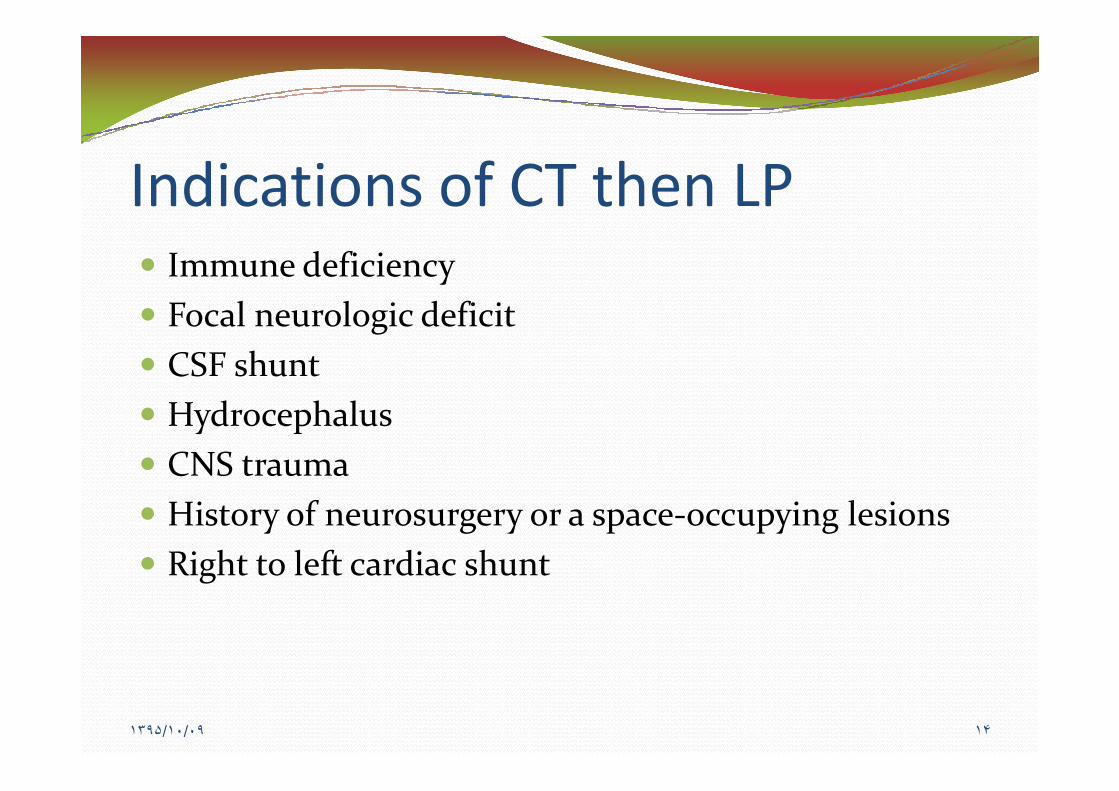
Indications of CT then LP� Immune deficiency� Focal neurologic deficit� CSF shunt� Hydrocephalus� CNS trauma� History of neurosurgery or a space-occupying lesions� Right to left cardiac shunt
����/��/�� ��
������ ��������� ���� �� �� �������������������ن������ن��������
����/��/�� ��

Elevated ICP findings on head CT� Midline shift� Effacement of the basilar cisterns� Effacement of the sulci
����/��/�� ��

��� ��� ����� ���� �� ن���� ����������ن����������� �ن��� ������
����/��/�� ��

�� �� LP� An experienced assistant who can position, restrain,
and comfort the patient� Lateral decubitus or seated position with the neck and
legs flexedlegs flexed� Sick neonates in a seated position� Shoulders and hips Be straight
����/��/�� ��

����� �ن���� ن���� ������CSF������ن � ������ ������ ����
����/��/�� ��
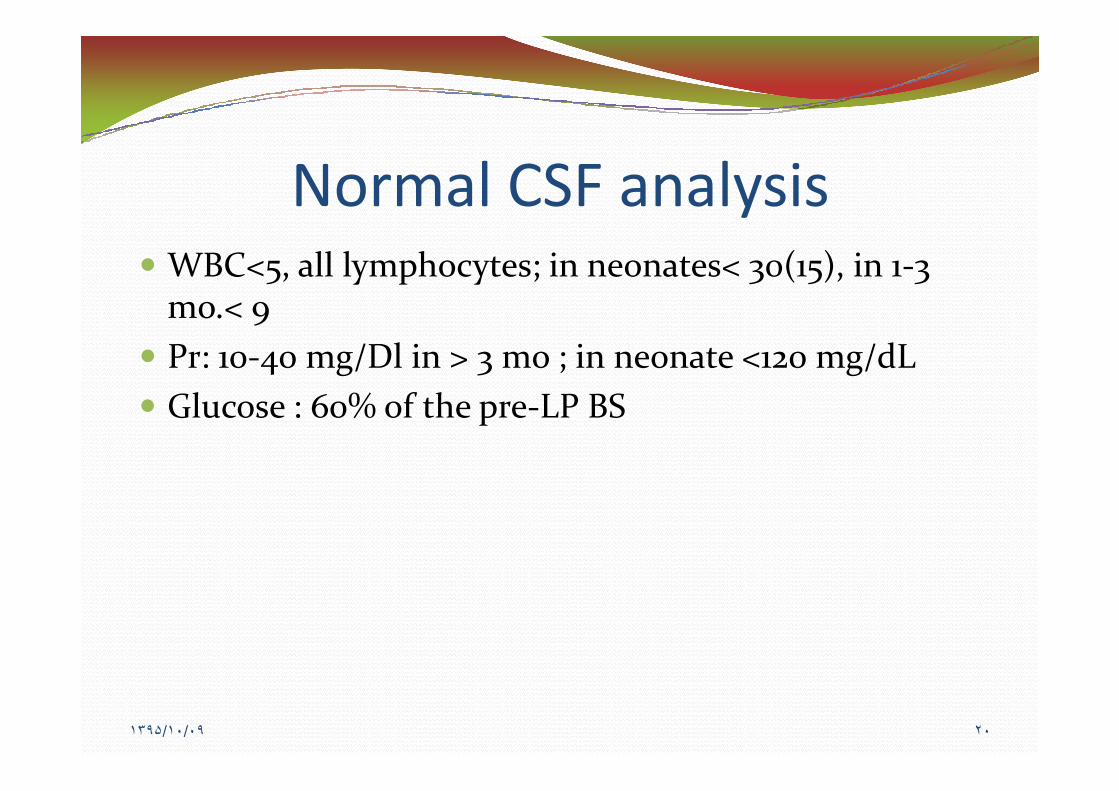
Normal CSF analysis� WBC<�, all lymphocytes; in neonates< ��(��), in �-�
mo.< �� Pr: ��-�� mg/Dl in > � mo ; in neonate <��� mg/dL
Gl � % f h LP BS� Glucose : ��% of the pre-LP BS
����/��/�� ��

�CSF�ن����� �������� ������ ���ن��������������
����/��/�� ��

CSF findings in typical bacterial meningitis� WBC >���� with a predominance of PMN� Glucose <�� mg/dL� Protein between ��� and ��� mg/dL
� Early in the course, few or no WBCs and/or NL Proteinand Glucose may be present
� Pleocytosis with a lymphocyte predominance may bepresent during the early stage
����/��/�� ��

CSF findings in typical viral meningitis� WBC of <��� with a mononuclear predominance� Normal CSF glucose� CSF protein <��� mg/dL� Negative CSF Gram stain� Improvement in symptoms following LP
� Normal WBC counts can be seen in enteroviral andrarely HSV
� Neutrophilic pleocytosis may be present in early stages(�-�� hr) of the initial LP.
����/��/�� ��

��� ����� ����� ������ �ن�� ���� ���� ��ن��� ��� �� �� �� ���� ���LP�� ���� �����
��������
����/��/�� ��

CSF analysis after appropriatetherapy� Pleocytosis with a predominance of neutrophils,
elevated protein, and a reduced glucose usually persistfor several days
� No change in �� to �� hours in most cases in chemical� No change in �� to �� hours in most cases in chemicaland cellular findings
� CSF culture can be negative� After conclusion of therapy WBC and protein generally
not returned completely to normal and glucose mayremained depressed
����/��/�� ��

����� ن��ن������������ ���CSF�ن������� ����� �� �ن���� ن���� ������
����/��/�� ��

Traumatic LP� Gram stain, culture, and glucose not be influenced� protein is increased by approximately � mg/dL for
every �,��� red blood cells/mm�
WBC RBC / i h b f l� � WBC per ���� RBCs/mm� in the absence of clot� It is prudent to rely on the bacteriologic results rather
than interpret the CSF leukocyte and protein
����/��/�� ��

����� ��� ��� �������� ������ ����� ����
����/��/�� ��

Antibiotic therapy in bacterialmeningitis� Penicillin-sensitive S. pneumoniae: third-generation
cephalosporin or penicillin for ��-�� days� Penicillin and the third-generation cephalosporin
resistant S pneumoniae: vancomycinresistant S. pneumoniae: vancomycin� N. meningitidis: penicillin for �-� days� H. influenzae type b: third-generation cephalosporin
for �-�� days� Culture negative: ceftriaxone or cefotaxime for �-��
days ??
����/��/�� ��

����� ����� � �� ��� ن�� �� �ن���� ���� ����� �� ن�����������������
����/��/�� ��

Response in bacterial meningitis� Neurologic signs in ��-�� hours:
� neurologic complications eg, subdural empyema,cerebral vascular thrombosis, ventricular dilation, brainabscess
� neuroimaging� Fever in � days:
� Inadequate treatment, nosocomial infection,suppurative complication (pericarditis, pneumonia,arthritis, subdural empyema), Drug fever (a diagnosis ofexclusion)
� Neuroimaging, repeat LP, …
����/��/�� ��

����� �� ������������������� �� � ن����� �� ����� ������������ ����� ���� ��
����/��/�� ��

Meningococcal chemoprophylaxis� All household, daycare center, and nursery school
contacts, healthcare workers who have direct exposureto oral secretions (mouth-to-mouth resuscitation,suctioning intubation)suctioning, intubation)
� No Schoolroom classmates and hospital contacts� Rifampin �� mg/kg/dose every �� hr (maximum dose
of ��� mg) for � days� Ciprofloxacin ��� mg in age> �� year� Third generation cephalosporin
����/��/�� ��

������� ������ �� ��������������� ����
����/��/�� ��
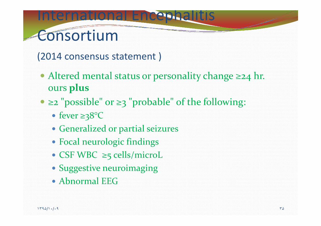
International EncephalitisConsortium(���� consensus statement )
� Altered mental status or personality change ≥�� hr.ours plus
� ≥� "possible" or ≥� "probable" of the following:f �°C� fever ≥��°C
� Generalized or partial seizures� Focal neurologic findings� CSF WBC ≥� cells/microL� Suggestive neuroimaging� Abnormal EEG
����/��/�� ��

����������ن���� �����ن��������CSFن���� ���ن�
����/��/�� ��

CSF analysis of encephalitis� Lymphocytic pleocytosis (��%); neutrophil
predominance in �st ��-�� hr� Mild RBCs in HSV , or other necrotizing
encephalitidesencephalitides� Moderately elevated protein ( <��� mg/dL).� Normal Glucose . Moderate reduction in HSV and
mumps
����/��/�� ��

���� ���� ���� ���� ��PCR�������� �� ���� �� �������������������� ��� ��
����/��/�� ��

Discontinuation of acyclovirin HSV-PCR negative patients� Low probability of HSV encephalitis(<� percent):
� Normal neuroimaging� <� cells/mm� in CSF� Normal mental status� Negative CSF HSV PCR after �� hours
� High probability of HSV encephalitis (� percent) each ofthe following:� Suggestive neuroimaging findings� CSF pleocytosis� Positive EEG findings� Seizures
����/��/�� ��