cerebral complications in infective endocarditis - … · cerebral complications in infective...
TRANSCRIPT

Cerebral complications in
infective endocarditis
!Ulrika Snygg-Martin
Department of Infectious Diseases, Institute of Biomedicine
Sahlgrenska Academy
University of Gothenburg
Sweden
2009

© 2009 Ulrika Snygg-Martin [email protected] Print: Intellecta Infolog AB, Västra Frölunda 2009 ISBN: 978-91-628-7795-8 URL: http://hdl.handle.net/2077/20044

Till Peter, Julius, Elvira och Nicholas
“Du blir aldrig färdig, och det är som det skall”
Tomas Tranströmer

Abstract
Infective endocarditis (IE) is a life-threatening disease. Cerebral emboliza-
tion complicates the course in 10-40% of IE episodes. Aims of study were to
investigate the frequency of cerebrovascular complications (CVC) in left-
sided IE and the influence of protective and risk factors with focus on anti-
platelet and anticoagulant therapy.
CVC rate was examined by repeated magnetic resonance imaging of the
brain and by assaying levels of brain damage markers in cerebrospinal fluid
in 60 IE patients in paper I. The overall CVC frequency was 65%, with 35%
of the patients experiencing neurological symptoms and 30% characterized
as having clinically silent CVC. The risk of neurological deterioration during
cardiac surgery after established cerebral embolism was low.
In paper II the relationship between symptomatic CVC and established use
of antiplatelet therapy was evaluated in 684 definite left-sided IE episodes.
Antiplatelet agents were used by 23% of the patients. These patients were
older and more often had a history of congestive heart failure. In 25% of all
episodes a CVC was seen. There was no statistically significant difference in
CVC rate between patients with and without previously established anti-
platelet therapy (24% vs. 25%, n.s.). Twelve-month mortality was signifi-
cantly higher for patients on previously established antiplatelet therapy in the
univariable analysis (34% vs. 24%, OR 1.6, 95% CI 1.1-2.4), but after ad-
justment for covariables the use of antiplatelet therapy was no longer a risk
factor.
The association between ongoing warfarin therapy and CVC incidence in
native valve endocarditis (NVE) was analyzed in paper III. Out of 587 NVE
episodes 8% were seen in patients using warfarin on admission. Patients on
warfarin suffered from CVC significantly less frequently than patients not on
warfarin (6% vs. 26%, 0.2 95% CI 0.06-0.6). In a multivariable model S.
aureus etiology (adjusted OR [aOR] 6.3, 95% CI 3.8-10.4) and vegetation
length (aOR 1.04, 95% CI 1.01-1.07) were associated with higher CVC fre-
quency. Warfarin use (aOR 0.26, 95% CI 0.07-0.94), history of congestive
heart failure (aOR 0.22, 95% CI 0.1-0.52) and previous IE episode (aOR 0.1,
95% CI 0.01-0.79) conferred a lower risk of CVC. Cerebral hemorrhagic
complications were few.
Keywords: Infective endocarditis (IE), cerebral embolism, cerebrovascular
complications, antiplatelet therapy, anticoagulation, warfarin, vegetation,
Staphylococcus aureus, mortality

List of papers
This thesis is based on the following papers, referred to in the text by their Roman numerals.
I Snygg-Martin, U., Gustafsson, L., Rosengren, L., Alsiö, Å.,
Ackerholm, P., Andersson, R., Olaison, L. (2008) Cerebrovascular complications in patients with left-sided infective endocarditis are common: a prospective study using magnetic resonance imaging and neurochemical brain damage markers. Clin Infect Dis, 47(1): 23-30.
II Snygg-Martin, U., Rasmussen, R.V., Hassager, C., Bruun, N.E., Andersson, R., Olaison, L. (2009) The relationship between cerebrovascular complications and established use of antiplate-let therapy: A cohort study of the effects in left-sided infective endocarditis. Submitted 2009.
III Snygg-Martin, U., Rasmussen, R.V., Hassager, C., Bruun, N.E., Andersson, R., Olaison, L. (2009) The influence of warfarin treatment on cerebrovascular complications in left-sided native valve infective endocarditis. Submitted 2009.

Contents 1 Introduction ..................................................................................................9
1.1 Infective endocarditis 9 1.2 Historical aspects 11 1.3 Epidemiology 12 1.4 Gender aspects 12 1.5 Embolic events 13 1.6 Neurological complications 13 1.7 Antiplatelet therapy 15 1.8 Anticoagulant therapy 16 1.9 Neurochemical markers of brain damage 18
2 Aims of the study .......................................................................................20
3 Patients and methods..................................................................................21 3.1 Patients 21 3.2 Study design 22 3.3 Methods 23 3.4 Definitions 25 3.5 Statistics 26
4 Results and discussion................................................................................27 4.1 Demography 27 4.2 Microbiology 30 4.3 Cerebrovascular complications 32 4.4 Factors related to the incidence of cerebrovascular complications 36 4.5 Echocardiography 39 4.6 Silent cerebral embolism 41 4.7 Mortality 44
5 General considerations ...............................................................................46
6 Conclusions ................................................................................................51
7 Svensk sammanfattning..............................................................................52
8 Acknowledgments......................................................................................53!
9 References ..................................................................................................54!

Abbreviations
aOR Adjusted odds ratio ASA Acetylsalicylic acid CHF Congestive heart failure CI Confidence interval CNS Central nervous system CoNS Coagulase-negative staphylococci CRP C-reactive protein CSF Cerebrospinal fluid CT Computed tomography CVC Cerebrovascular complication(s) GFAP Glial fibrillary acidic protein HACEK Haemophilus spp, Aggregatibacter actinomycetemcomitans,
Cardiobacterium hominis, Eikenella corrodens, Kingella spp
IE Infective endocarditis INR International normalized ratio IQR Interquartile range IVDU Intravenous drug use MRI Magnetic resonance imaging MRSA Methicillin-resistant Staphylococcus aureus
MSSA Methicillin-sensitive Staphylococcus aureus NFL Neurofilament protein - light chain NVE Native valve endocarditis OR Odds ratio PVE Prosthetic valve endocarditis ROC Reciever operating characteristic SD Standard deviation TEE Transesophageal echocardiography TIA Transient ischemic attack(s) TTE Transthoracic echocardiography

11
1 Introduction
1.1 Infective endocarditis Infective endocarditis (IE) is a serious disease, universally fatal if unrevealed and untreated. IE can be defined as an endovascular microbial infection of cardiac structures and intracardiac foreign bodies. The commonly accepted pathogenetic theory is that IE results from a complex interaction between initially hemodynamically derived mechanical damage to the endocardial lining, preferably on the valvular cusps, and transient bacteremia with mi-croorganism adhesion and escape from host defense mechanisms (1, 2). Di-rect contact between blood and subendothelial components results in platelet and fibrin deposition and the formation of a coagulum. Circulating microor-ganisms adhere to the coagulum and promote further enlargement of this infected structure, referred to as the vegetation (3, 4). These irregular excres-cences on the cardiac valves or endocardium, composed of platelets, fibrin, microorganisms, and inflammatory cells, are characteristic but not patho-gnomonic of IE. The detection of vegetations by echocardiography consti-tutes one of the two major clinical criteria for IE, according to the Duke and modified Duke criteria (5, 6) (table 1). Continuous release of bacteria from the vegetations gives rise to the persistent bacteremia, the other cornerstone of IE. Microorganisms adherent to cardiac valves have been shown to avoid host defense mechanisms inside the vegetation through impaired infiltration of phagocytes into the vegetation (i.e. localized agranulocytosis) (7, 8), reduced phagocytic ability (4) and protection from the humoral immune response (9). Local extension of the infection, tissue damage and embolic spread of in-fected material from valvular vegetations to distant organs give rise to the main complications of IE, congestive heart failure and embolic events. Causative microorganisms in IE are predominantly gram-positive bacteria, although a wide variety of pathogens can be found including gram-negative bacteria, fungi and rickettsiae. Viridans group streptococci and S. aureus are each found in approximately one third of IE episodes, enterococci in 10-15%, and in 5-15% of episodes blood cultures are negative.
9

12
Table 1. Definition of infective endocarditis according to the modified Duke Criteria
* Excludes positive cultures for coagulase-negative staphylococci and organisms that do not cause IE
Definite infective endocarditis Pathologic criteria
• Microorganisms demonstrated by results of cultures or histological examination of a vegetation, a vegetation that has embolized, or an intracardiac abscess specimen.
• Pathologic lesions; vegetation, or intracardiac abscess confirmed by histologic exami-nation showing active endocarditis.
Clinical criteria
• 2 major criteria • 1 major criterion and 3 minor criteria • 5 minor criteria
Possible infective endocarditis • 1 major criterion and 1 minor criterion • 3 minor criteria
Rejected • Firm alternate diagnosis explaining evidence of infective endocarditis. • Resolution of infective endocarditis syndrome with antibiotic therapy for ! 4 d. • No pathologic evidence of infective endocarditis at surgery or autopsy, with antibiotic
therapy for ! 4 d. • Does not meet criteria for possible infective endocarditis.
_______________________________________________________________
Major criteria Blood culture findings positive for infective endocarditis
• Typical microorganisms consistent with IE from 2 separate blood cultures: Viridans streptococci, Streptococcus bovis, HACEK group, or Staphylococcus aureus.
Community-acquired enterococci, in the absence of a primary focus. • Microorganisms consistent with IE from persistently positive blood cultures
(at least 2 positive cultures of blood samples drawn >12 h apart; or 3 or most of > 4 separate cultures of blood (with first and last sample drawn > 1 h apart).
• Single positive blood culture for Coxiella burnetii or antiphase I IgG antibody titer > 1:800).
Evidence of endocardial involvement (echocardiographic findings positive for IE) • Oscillating intracardiac mass on valve or supporting structures, in the path of regurgi-
tant jets, or on implanted material in the absence of an alternative anatomic explanation. • Abscess. • New partial dehiscence of prosthetic valve. • New valvular regurgitation.
Minor criteria • Predisposition: predisposing heart condition, or intravenous drug use. • Fever, temperature " 38°C. • Vascular phenomena: major arterial emboli, septic pulmonary infarcts, mycotic aneu-
rysm, intracranial hemorrhage, conjunctival hemorrhages, Janeway´s lesions. • Immunologic phenomena: glomerulonephritis, Osler´s nodes, Roth´s spots, rheumatoid
factor. • Microbiological evidence: positive blood culture but does not meet a major criterion*.
10
13
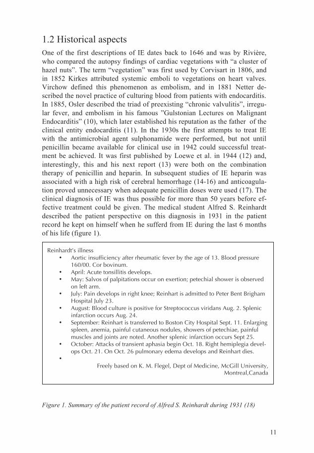
1.2 Historical aspects One of the first descriptions of IE dates back to 1646 and was by Rivière, who compared the autopsy findings of cardiac vegetations with “a cluster of hazel nuts”. The term “vegetation” was first used by Corvisart in 1806, and in 1852 Kirkes attributed systemic emboli to vegetations on heart valves. Virchow defined this phenomenon as embolism, and in 1881 Netter de-scribed the novel practice of culturing blood from patients with endocarditis. In 1885, Osler described the triad of preexisting “chronic valvulitis”, irregu-lar fever, and embolism in his famous ”Gulstonian Lectures on Malignant Endocarditis” (10), which later established his reputation as the father of the clinical entity endocarditis (11). In the 1930s the first attempts to treat IE with the antimicrobial agent sulphonamide were performed, but not until penicillin became available for clinical use in 1942 could successful treat-ment be achieved. It was first published by Loewe et al. in 1944 (12) and, interestingly, this and his next report (13) were both on the combination therapy of penicillin and heparin. In subsequent studies of IE heparin was associated with a high risk of cerebral hemorrhage (14-16) and anticoagula-tion proved unnecessary when adequate penicillin doses were used (17). The clinical diagnosis of IE was thus possible for more than 50 years before ef-fective treatment could be given. The medical student Alfred S. Reinhardt described the patient perspective on this diagnosis in 1931 in the patient record he kept on himself when he sufferd from IE during the last 6 months of his life (figure 1).
Figure 1. Summary of the patient record of Alfred S. Reinhardt during 1931 (18)
Reinhardt’s illness • Aortic insufficiency after rheumatic fever by the age of 13. Blood pressure
160/00. Cor bovinum. • April: Acute tonsillitis develops. • May: Salvos of palpitations occur on exertion; petechial shower is observed
on left arm. • July: Pain develops in right knee; Reinhart is admitted to Peter Bent Brigham
Hospital July 23. • August: Blood culture is positive for Streptococcus viridans Aug. 2. Splenic
infarction occurs Aug. 24. • September: Reinhart is transferred to Boston City Hospital Sept. 11. Enlarging
spleen, anemia, painful cutaneous nodules, showers of petechiae, painful muscles and joints are noted. Another splenic infarction occurs Sept 25.
• October: Attacks of transient aphasia begin Oct. 18. Right hemiplegia devel-ops Oct. 21. On Oct. 26 pulmonary edema develops and Reinhart dies.
• Freely based on K. M. Flegel, Dept of Medicine, McGill University,
Montreal,Canada
11

14
1.3 Epidemiology The annual incidence of IE in Sweden is estimated to be 5.9 cases per 100,000 inhabitants (19), as compared with estimated incidences of 1.9 to 9.6 cases per 100,000 individuals found in other studies (20-27). The inci-dence of IE is thought to have remained stable over the last three decades (20, 28) but international data indicate that the epidemiology of IE is chang-ing. In recent studies, IE patients are found to be older, have more prosthetic valve (PVE) or device associated IE episodes, and more often undergo car-diac surgery during the period of antibiotic treatment (19, 20, 29-33). A shift toward a higher frequency of S. aureus IE has both been reported (34, 35) and also questioned (28). These diverging data mirror the influence of refer-ral bias as well as differences in living conditions, genetic factors and bacte-rial resistance among different IE populations studied (34, 36, 37). In-hospital mortality in patients with IE reported to the National Swedish Endocarditis Registry 1995-2007 was 12% (pers comm L. Olaison). Similar in-hospital mortality rates of 10–18% have been found in other studies (21, 26, 38-42). Mortality rate was reduced after the introduction of thoracic sur-gery in the 1960s and 70s while reports on mortality in IE during the last two to three decades yield conflicting results (28, 29, 39, 43). This might reflect the contemporary epidemiological changes towards older ages, more comor-bid conditions, device-related episodes and more virulent microorganisms (25).
1.4 Gender aspects In most published studies IE occurs more frequently in men than in women (22, 29, 35, 44), while female dominance was found in a prospective cohort study in Gothenburg 1984-88 (19). Results from some earlier studies of IE also show more even sex distribution (5, 45-47) while in Osler’s original report there was male predominance (10). Several studies indicate that women diagnosed with IE are older than men (19, 35) while data on age is not reported in most publications. In most studies cerebral complications are reported in similar frequencies in both men and women with IE (47-52), while frequencies are not presented as separate sex data in other studies (53, 54). In one study, Roder et al. found a trend towards more neurological manifestations in women with S. aureus IE than in men with S. aureus IE (55), and S. aureus etiology has been found more often in women than in men with IE (34, 46). Lower surgery rates and/or higher in-hospital mortality have been reported among women as compared with men (50, 56-58) but interpretation was confounded by differences in age and comorbid condi-tions in some studies.
12
15
It is not known whether the observed demographic and clinical differences between women and men with IE reflect varying rates of comorbid condi-tions or inherent physiological differences, but may partly be explained by diagnostic, referral, or treatment bias (19). Other potential explanations have been extrapolated from animal models, suggesting that estrogen is protective against endothelial damage (59). In addition, human studies have shown that women are less likely than men to develop sepsis after traumatic hemor-rhagic shock (60). Women also tend to develop heart disease later in life than men (61, 62).
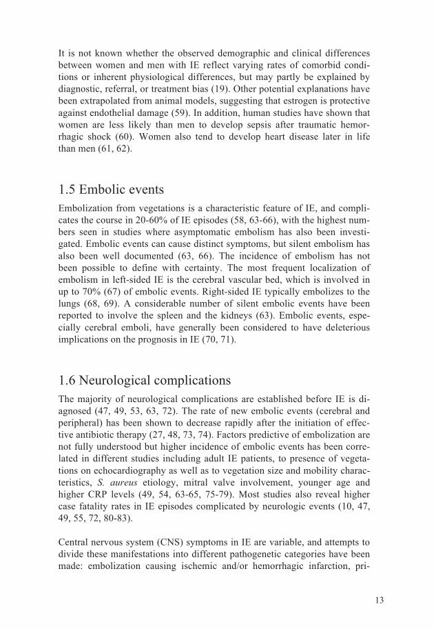
1.5 Embolic events Embolization from vegetations is a characteristic feature of IE, and compli-cates the course in 20-60% of IE episodes (58, 63-66), with the highest num-bers seen in studies where asymptomatic embolism has also been investi-gated. Embolic events can cause distinct symptoms, but silent embolism has also been well documented (63, 66). The incidence of embolism has not been possible to define with certainty. The most frequent localization of embolism in left-sided IE is the cerebral vascular bed, which is involved in up to 70% (67) of embolic events. Right-sided IE typically embolizes to the lungs (68, 69). A considerable number of silent embolic events have been reported to involve the spleen and the kidneys (63). Embolic events, espe-cially cerebral emboli, have generally been considered to have deleterious implications on the prognosis in IE (70, 71).
1.6 Neurological complications The majority of neurological complications are established before IE is di-agnosed (47, 49, 53, 63, 72). The rate of new embolic events (cerebral and peripheral) has been shown to decrease rapidly after the initiation of effec-tive antibiotic therapy (27, 48, 73, 74). Factors predictive of embolization are not fully understood but higher incidence of embolic events has been corre-lated in different studies including adult IE patients, to presence of vegeta-tions on echocardiography as well as to vegetation size and mobility charac-teristics, S. aureus etiology, mitral valve involvement, younger age and higher CRP levels (49, 54, 63-65, 75-79). Most studies also reveal higher case fatality rates in IE episodes complicated by neurologic events (10, 47, 49, 55, 72, 80-83).
Central nervous system (CNS) symptoms in IE are variable, and attempts to divide these manifestations into different pathogenetic categories have been made: embolization causing ischemic and/or hemorrhagic infarction, pri-
13

16
mary intracerebral bleeding, subarachnoid bleeding, mycotic aneurysm, in-fection of the brain or meninges and toxic or immune-mediated injury, en-cephalopathy and psychiatric manifestations (47, 84-87). It is well docu-mented that IE patients with cerebral involvement often exhibit more than one type of lesion and/or neurological sign (47, 88-91) although they may also be asymptomatic (56, 66, 72). In a carefully described material of 17 patients dying from IE in the 1930s, the fundamental pathological change in the brain was diffuse embolic meningoencephalitis, from which various clinical manifestations arose (92). Pruitt et al. postulated that cerebral infarc-tion, micro- and macroabscess formation, septic vasculitis and mycotic aneu-rysms represent a continuum, and that the advent of computed tomography (CT) and magnetic resonance imaging (MRI) has made the clinical distinc-tion between syndromes less clear (93). Encephalopathy with impaired con-sciousness or delirium and meningism have also been argued to be of septic embolic origin (77, 94). Lerner concluded that infected emboli account for all neurological complications in IE (mycotic aneurysm, intracerebral bleed-ing, meningitis/meningoencephalitis, and brain abscess) additional to caus-ing pure ischemic lesions (95). Roder et al. arrived at the opinion that the attempt to prove the precise nature of a neurological complication is inher-ently connected with difficulties (55) . Ischemic infarction is the most common neurological complication in IE occurring in 9–28% of all IE episodes (47-49, 54, 72, 78). Large emboli preferably lodge in the middle cerebral artery territory, resulting in hemipa-retic symptoms of varying degree. The clinical syndromes seen with micro-embolic punctuate cerebral infarctions are variable, often referred to as an altered level of consciousness or embolic encephalopathy. About half of IE episodes with cerebral emboli are described as having concomitant periph-eral emboli (47). Intracranial hemorrhage occurs in 2-7% of patients with IE (47, 49, 54, 72, 82, 91, 96) and three different mechanisms are thought to prevail, namely rupture of mycotic (infectious) aneurysm, pyogenic arteritis and hemorrhagic transformation of initially bland infarction. Anticoagulant therapy has been associated with a higher risk of hemorrhagic complications in IE (47, 88, 96, 97). The incidence of meningitis in IE varies from 3-16% in different studies and is, when it occurs, an early feature of the complex clinical manifestations of IE. Meningitis is most frequently seen with S. aureus as the causative patho-gen, but also in IE caused by other pathogens. The detected rate of meningi-tis in different studies depends to a large extent on the frequency of lumbar punctures performed in the specific study setting. More than one third of patients with meningitis experience additional neurological complications (98). The availability of better and non-invasive brain imaging methods have reduced the proportion of IE patients examined by lumbar puncture and
14

17
cerebrospinal fluid (CSF) analysis, since isolated meningism is seldom the only initial neurological symptom. This is illustrated by two studies by Pruitt et al. made at different time periods, the first with IE patients from 1964 to 1973 when 85% of the patients with neurological symptoms underwent lum-bar puncture, the second with patients from 1988 to 1992, when the corre-sponding figure was 43%. In the first study, the incidence of CSF anomalies indicative of meningitis was 16% of all IE cases, in the second it was 4% (47, 93). CSF culture is positive only in a minority of patients with IE asso-ciated meningitis (47, 49, 55, 82). Aseptic meningitis, often with moderately increased numbers of white blood cells in the CSF is thought to prevail, al-though this concept has been questioned (92, 98). It is widely accepted that mortality in patients with IE is reduced in a consid-erable subset of patients by early surgical intervention (80, 99-102). The safety of cardiac surgery in IE patients suffering from cerebral complications is an important question since cardiopulmonary bypass is suspected to ag-gravate preexisting neurological deficits and to carry an increased risk of secondary cerebral hemorrhage due to heparinization. In the absence of a hemorrhagic cerebral manifestation, valve replacement has been considered resonably safe when performed at least 72 h after the cerebral event, while patients with a recent hemorrhagic cerebral complication are anticipated to have an unacceptably high risk of intracranial bleeding during early cardiac surgery (103). In a study by Ruttmann et al. (52) the suspected risk of secon-dary hemorrhage in IE patients undergoing cardiac surgery despite preopera-tively established cerebral embolism was very low, and the authors recom-mended early surgical procedure for IE when indicated also after stroke.
1.7 Antiplatelet therapy
The pharmacological effects of salicylic acid have been known since ancient times. Hippocrates, the father of modern medicine, made the first recorded descriptions of the therapeutic benefits of extract of willow bark in the fifth century B.C. Salicylate-rich willow bark extract was used for its analgesic and anti-rheumatic effects. By the end of the nineteenth century German chemists had managed to synthesize acetylsalicylic acid (ASA) by acetyla-tion of salicylic acid, and this drug, subsequently named aspirin, soon be-came the most sold medication worldwide. In 1908 ASA was introduced in the Swedish Pharmacopeia and became a formal part of Swedish pharmacol-ogical history. The analgesic, antipyretic and anti-inflammatory effects of ASA were known long before its mechanisms of action through inhibition of cyclooxy-genase in the prostaglandin synthesis was elucidated by Vane and coworkers
15

18
in the late 1960s (104). The antiplatelet era of ASA began with the observa-tion that patients with regular ASA use had fewer heart attacks than ex-pected, and later the use of ASA to reduce the rate of stroke and myocardial infarctions was approved. This effect is mediated by irreversible inhibition of cyclooxygenase and termination of production of tromboxan in platelets, and requires lower doses than used to produce the analgesic and anti-inflammatory effects. ASA reduces the relative risk of stroke, myocardial infarction and vascular death by 25% in high risk patients and has wide-spread use as primary and secondary prophylaxis (105). The potential role of antiplatelet agents as adjunctive therapy in IE has pro-voked considerable interest, and a complex pattern of mechanisms involving platelets in the evolution of IE has been outlined (106-108). Advantageous effects of these agents have been shown under experimental conditions in several studies mainly concerning S. aureus IE (109-113). Other studies have failed to demonstrate such positive effects (114, 115). The results of prospective human treatment studies (116, 117) and recently published co-hort studies (51, 53, 118, 119) are contradictory. Occasional reports of anti-platelet drug use in neonatal IE (120) and bovine IE (121) have opened up for further hypothesizing, but international treatment guidelines on IE do not generally include comments concerning the use of antiplatelet agents. Newer antiplatelet drugs have been developed, of which clopidogrel has been shown to be significantly more effective than ASA in reducing the risk of stroke, myocardial infarction, or vascular death in patients with athero-sclerotic vascular disease (122). Clopidogrel blocks activation of platelets by another mechanism and has fewer upper gastrointestinal side effects than ASA. Bleeding, a common complication with antiplatelet drugs irrespective of mechanism of action, has also proven to be less frequent with clopidogrel.
1.8 Anticoagulant therapy
Heparin
In 1916 McLean first observed that an extract of dog liver contained a sub-stance that retarded the coagulation of blood in vitro. This substance was named “heparin” by Howell in 1918 and was tried in humans in 1935 (123). Its subsequent clinical use in the prevention and treatment of venous throm-boembolism was introduced early in Sweden (124). In 1941, McLean evalu-ated the use of heparin in IE patients (125) and found that heparin treatment did not lead to improvments and resulted in a high proportion of fatal cere-bral hemorrhages. Contrary to this were the results of combination therapy with heparin and penicillin found to be succesful by Loewe et al. (12). In still other studies the addition of anticoagulant therapy proved unnecessary
16

19
when adequate penicillin doses were used (45). Most authors (85, 126) have since discouraged the routine use of anticoagulant therapy in patients with IE. Heparin is a naturally occurring glycosaminoglycan, the main function of which is to inhibit the coagulation of blood by activating antitrombin III. Subsequent inactivation of the coagulation cascade is seen mainly through inhibition of thrombin (factor II) and factor X. Low molecular weight hepa-rin is composed of depolymerized heparin salts with lower molecular weight, longer biological half-life and more predictable effects on the coagulation system. Both unfractionated and low molecular weight heparins are contra-indicated during septic endocarditis according to Swedish pharmaceutical recommendations (www.fass.se), owing to the risk of cerebral bleeding dur-ing IE.
Coumarin
In the early 1920s a previously unknown disease in cattle was recognized in the United States and Canada. The animals died of uncontrollable bleeding from minor injuries or internal hemorrhage. The cause of this condition proved to be ingestion of spoiled sweet clover (Melilotus officinalis) that functioned as a potent anticoagulant. The affected cattle had prolonged clot-ting time, owing to profound prothrombin deficiency. Coumarin was, at this time, a known component of sweet clover, causing the combination of a sweet smell and a bitter taste, and it was commercially used to scent cheap tobacco, artificial vanilla and perfumes. Not until 1939, when Karl Link and associates isolated the hemorrhagic agent from sweet clover, the association between coumarin and sweet clover disease was revealed (127). Coumarin itself , like other natural coumarins, is atoxic, but in moldy hay it is oxidized to hydroxycoumarin and linked to another coumarin molecule, thus forming a dicoumarol with potent anticoagulant effects. This compound was subse-quently synthesized and the clinical use of dicoumarol began in 1941. As early as 1948 a randomized study sponsored by the American Heart Associa-tion concluded the beneficial effects of dicoumarol in myocardial infarction (128). Dicoumarol was, however, a weak and unpredictable drug and another more potent coumarin, given the name warfarin, was patented 10 years later. The initial use of warfarin was as a rat poison but the substance soon proved more reliable than dicoumarol in the clinical setting and was commercially introduced in 1954. Warfarin is a synthetic anticoagulant substance and works through competi-tive inhibition of vitamin K reductase. This enzyme recycles oxidated vita-min K after vitamin K has participated in the carboxylation of several blood coagulation proteins, mainly prothrombin (factor II), factors VII, IX and X, as well as other proteins involved in the blood clotting system. The decar-boxylated coagulation factors produced in the presence of warfarin are bio-
17

20
logically inactive and successively replace the active clotting factors in the circulatory system, explaining the slow onset of the anticoagulant effect of warfarin. Warfarin and related drugs are referred to as vitamin K antagonists, and their effects can be reversed with vitamin K or by transfusion of coagu-lation factors. The inter- and intraindividual therapeutic sensitivity to war-farin is variable and the intensity of the anticoagulant effect must be fol-lowed with standardized measurements of the prothrombin time (interna-tional normalized ratio, INR).
1.9 Neurochemical markers of brain damage Measurement of concentrations of organ-specific proteins in body fluids is well established in various medical fields, e.g. analysis of serum levels of troponin and creatine kinase in patients with suspected myocardial infarction or alanine aminotransferase to evaluate liver cell damage. Specific proteins that are found in high concentrations in CNS but in negligible concentrations in other organs can be used to assess various types of brain injury. The neu-rochemical marker proteins are specific for different CNS cell types and/or for distinct cellular components. Of the two brain damage markers used in this thesis (Paper I) the light chain of the neurofilament protein (NFL) is an established marker of neuronal/axonal damage, and glial fibrillary protein (GFAP) is a marker of astroglial cell injury. Ischemic infarctions and other types of acute or chronic brain diseases provoke brain cell destruction and leakage of marker proteins into CSF. NFL and GFAP are analyzed in clinical praxis at the Sahlgrenska University Hospital. The neurofilament is the main structural component of the neuronal cy-toskeleton. It is abundant in axons relative to nerve cell somas and in large myelinated axons (129). The neurofilament is composed of a triplet protein of which the light subunit (NFL) is the essential component. It is used as a marker of neurodegeneration, particularly of axonal damage. Following acute parenchymatous CNS damage, the levels gradually increase, with a maximum after a few weeks and normalization over a period of months. The late increase is related to the release of NFL from injured axons subsequent to damage of neuronal soma. Measurements of CSF-NFL as a marker of axonal damage show increased values after cerebral infarction (130-132) and in various other conditions with neuronal damage (131, 133-135).
Glial fibrillary acidic protein is a structural protein found almost exclusively in glial cells of the CNS. In astrocytes, GFAP builds up the intermediate filament, which is the main cytoskeleton structure (136). Increased levels of GFAP in CSF have been demonstrated in two principally different situa-tions: (i) associated to astrogliosis and (ii) after acute CNS injuries with dis-
18

21
integration of glial cells. Astrogliosis is a reactive proliferation, and hyper-trophy of astrocytes with increased intracellular amounts and extracellular release of GFAP to CSF. Astrogliosis is an unspecific reaction seen follow-ing brain injury, in inflammatory and certain chronic conditions, but also during ageing (137). Modest increases in CSF-GFAP levels are seen in mul-tiple sclerosis (133) and normal pressure hydrocephalus (138). After acute parenchymatous CNS damage, an increase in CSF-GFAP can be detected from days 1-2, with concentrations returning to normal within less than three weeks (139). The levels reflect the extent of the injury and are high after large cerebral infarctions (139, 140), in patients with herpes encephalitis (134) and after subarachnoid hemorrhage (141).
19

22
2 Aims of the study
1. To study the incidence of symptomatic and silent cerebral complications during episodes of left-sided infective endocarditis using sensitive diag-nostic methods (Paper I).
2. To study the influence of protective factors and risk factors for cere-
brovascular complications in left-sided infective endocarditis (Paper I-III).
3. To study the relationship between previously established daily antiplate-
let therapy and cerebrovascular complications and mortality in left-sided definite infective endocarditis (Paper II).
4. To study the association between ongoing oral anticoagulant therapy and cerebrovascular complications in patients with left-sided native valve endocarditis (Paper III).
20

23
3 Patients and methods
3.1 Patients Adult patients with left-sided possible or definite IE according to the Duke modified criteria (6) at three tertiary care centers in Sweden and Denmark were included. Altogether, 866 episodes of IE in 837 patients were consecu-tively enrolled in the study from January 1, 1996 to January 31, 2008 in Gothenburg (papers I–III) and from October 1, 2002 to January 31, 2008 in Copenhagen (papers II–III). Patients with isolated right-sided IE were ex-cluded from the analysis owing to the inherently low risk of cerebral embo-lism in such episodes (68). Thus the remaining 797 episodes of left-sided IE in 771 patients were the total study cohort as illustrated in figure 2. In paper I, only patients from Sweden were included, and the inclusion period was in two intervals between June 1, 1998 and January 31, 2005. During the final year of that study, patients from the Department of Infectious Diseases at Skaraborg Hospital, Skövde, Sweden were also included. The Department of Infectious Diseases, Sahlgrenska University Hospital, Gothenburg, Sweden, serves the Gothenburg area, with about 600 000 in-habitants, as both first-line and tertiary care center for IE patients. In addi-tion, the Departments of Infectious Diseases and Cardiothoracic Surgery at the Sahlgrenska University Hospital serve as tertiary referral center for IE patients from other hospitals in western Sweden (Västra Götaland regionen) with about 1.5 million inhabitants. The Department of Cardiology, Gentofte University Hospital, Copenhagen, Denmark and the Department of Cardiol-ogy, Copenhagen University Hospital (Rigshospitalet), Copenhagen, Den-mark provide care for patients with IE in the eastern part of Denmark with a catchment area of 2.4 million inhabitants. These centers are the only ones in the area receiving IE patients, and are thus responsible for both basic and highly specialized care. Patients with diagnosed IE are transferred to these centers and there are cardiothoracic surgery departments in both hospitals.
21

24
Figure 2. Distribution of patients with infective endocarditis included in the studies.
IE: infective endocarditis, NVE: native valve endocarditis, PVE: prosthetic valve
endocarditis.
* Referred to in text as ”total cohort”.
** Inclusion period: June 1998-April 2001, September 2002-January 2005. Two
patients from the Department of Infectious Diseases at Skaraborg Hospital, Skövde,
Sweden included in 2004.
.
3.2. Study design The studies were carried out as observational cohort studies. In paper I, pa-tients with high clinical suspicion of IE were enrolled irrespective of neuro-logical symptoms. Symptomatic and silent (asymptomatic) CVC during the
22

25
present IE episode was the primary outcome variable. Patients underwent repeated physical and neurological examinations during hospitalization. Magnetic resonance imaging (MRI) of the brain was performed during the first 10 days of antibiotic treatment and a follow-up MRI after 2-3 months. Lumbar puncture with analysis of CSF was done during the first and fourth weeks of treatment if no contraindications were found and the patient con-sented. The study was approved by the ethics committee of the University of Gothenburg (L 077-98). In papers II–III consecutive patients with IE treated in the participating cen-ters were eligible for study entry. Patients enrolled prospectively in the study were followed according to local protocols including uniform treatment regimens, clinical evaluation procedures and collections of specimens in-cluding blood cultures. Study participants were treated in regular care at the Cardiology Department (Denmark) or the Infectious Diseases Department (Sweden) and consultations with other specialists including thoracic sur-geons were performed regularly. Demographic and clinical characteristics including age, sex, presence of comorbidities, ongoing medication, cardiac rhythm, presence of prosthetic valve, or pacemaker/intravascular device and duration of symptoms prior to admission were recorded. Blood cultures, biochemical blood tests, radiological, electrocardiographic and echocardio-graphic examinations as well as antibiotic treatment, and frequency, timing and type of surgical treatment were recorded. Primary outcome variables were cerebrovascular complications (CVC) during IE, and in-hospital and 12-month mortality.
3.3 Methods
Magnetic resonance imaging (Paper I)
The MRI protocol contained T1, T2, and PD-weighted images before and after the administration of a gadolinium contrast medium. MR angiography was not routinely performed. Diffusion-weighted MRI was not available when the study began and is therefor not included in the protocol. If contra-indications for MRI were present, computed tomography (CT) of the brain with and without contrast was performed instead. MRI findings were evalu-ated directly by the supervising neuroradiologist and later reevaluated by one of two experienced neuroradiologists blinded to the clinical and neurological status of the patients. Identification of acute or subacute ischemic, hemor-rhagic or infectious lesions as well as changes between the two subsequent neuroradiological investigations in each patient were considered in the diag-nosis of a cerebral complication. Each patient served as his/her own control,
23

26
and findings with a temporally consistent association were considered re-lated to the present IE episode.
Cerebrospinal fluid analyses (Paper I)
The CSF analyses included bacterial culture, cell counts, and determination of glucose and albumin levels. Polymorphnuclear leukocytes >5 x 10⁶ cells/mL CSF were considered diagnostic of meningitis (49, 54), while iso-lated elevation of monomorphnuclear leukocytes was not. CSF concentrations of the neurochemical brain damage marker NFL (neuro-filament protein light chain) were analyzed using a sandwich ELISA as pre-viously described (131). The sensitivity of the assay was 125 ng/L and the standard curve ranged from 125 to 16 000 ng/L. Values are age dependent and were regarded as normal if the level of CSF-NFL was <250 ng/L for patients aged <59 years, <380 ng/L for patients aged 60-69 years and <750 ng/L for patients aged >70 years. GFAP (glial fibrillary acidic protein) was measured with a previously de-scribed ELISA procedure (137). The standard curve ranged from 32-16 000 ng/L and the sensitivity of the assay was 16 ng/L. Values are age dependent and were regarded as normal if the level of CSF-GFAP was <750 ng/L for patients aged 20-59 years, <1250 ng/L for patients aged >60 years. CSF-NFL and CSF-GFAP reference levels were based on measurments from 141 neurologically healthy individuals aged 18-83 years.
Echocardiography
Transthoracic and transoesophageal echocardiography were performed using standard techniques in all patients included in papers I-III with the exception of 17 cases where only TTE was performed. Seven of these were classified as possible IE due to lack of conclusive findings on TTE, four were definite through autopsy findings, one through peroperative findings, four through detection of vegetations on TTE, and one classified as definite based on five minor criteria.
Evidence of endocardial involvement detected using echocardiography was classified according to the modified Duke criteria (6). Vegetations were clas-sified by location and maximum size. Mobility characteristics were consid-ered as an insufficiently standardized variable in the absence of systematic reevaluation of the echocardiographic examinations and were therefore not included in any of the analyses. Paravalvular engagement of the infection was defined as the presence of intracardiac abscesses and/or pseodoaneu-rysm formation or new periprosthetic regurgitation in PVE.
24

27
3.4 Definitions
Case definition
A prospectively identified episode of left-sided IE satisfying the modified Duke criteria (6) of possible or definite IE constituted a defined case. Possi-ble IE episodes were included if treatment was given in accordance with IE treatment guidelines or, in cases of in-hospital death, intended to be given. In cases included prior to the year 2000 the modified criteria were applied ret-rospectively. Childhood cases were not included (<15 years). Left-sided IE cases included episodes with combined right and left-sided engagement of the infection.
Microbial etiology
The causative agents were divided into seven different categories of micro-bial etiology: Staphylococcus aureus including methicillin-sensitive (MSSA) and methicillin-resistant (MRSA) strains; viridans group streptococci includ-ing Streptococcus bovis; coagulase-negative staphylococci; Enterococci spp; other streptococci including groups A, B, C, G and pneumococci, miscella-neous microorganisms including HACEK (Haemophilus spp, Aggregatibac-
ter [former Actinobacillus] actinomycetemcomitans, Cardiobacterium
hominis, Eikenella corrodens, Kingella spp), Candida spp and other bacte-ria; and episodes with negative blood cultures.
Cerebrovascular complications
Cerebrovascular complications (CVC) included ischemic and hemorrhagic infarctions, intracerebral and subarachnoid hemorrhages, cerebral mycotic aneurysms, transient ischemic attacks (TIA), and cerebral infections includ-ing brain abscesses and culture-positive and culture-negative meningitis. A TIA was defined as a focal neurological sign or symptom of sudden occur-rence and resolution within 24 h. Cerebral events occurring in the prediagnostic period and during treatment of the studied IE episode were the focus of this thesis, while later neurologi-cal complications were not systematically registered. Diagnosis of CVC was based on repeated physical and neurological examinations, neuroimaging recordings and/or lumbar puncture. Cerebral CT or MRI was performed in all episodes with neurological symptoms except six cases in the total study cohort. In five cases presenting with meningism, only lumbar puncture with CSF analysis was performed and in one case of early in-hospital death owing to cardiac failure, a clinical diagnosis of ischemic stroke was made. In pa-tients without neurological symptoms no routine cerebral CT or MRI was performed.
25
28
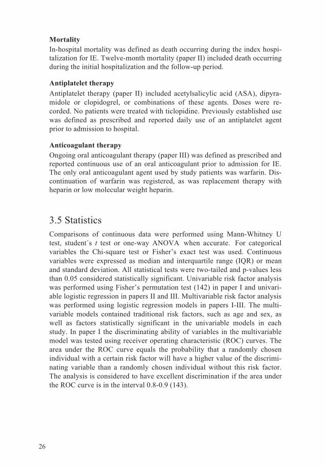
Mortality
In-hospital mortality was defined as death occurring during the index hospi-talization for IE. Twelve-month mortality (paper II) included death occurring during the initial hospitalization and the follow-up period.
Antiplatelet therapy
Antiplatelet therapy (paper II) included acetylsalicylic acid (ASA), dipyra-midole or clopidogrel, or combinations of these agents. Doses were re-corded. No patients were treated with ticlopidine. Previously established use was defined as prescribed and reported daily use of an antiplatelet agent prior to admission to hospital.
Anticoagulant therapy
Ongoing oral anticoagulant therapy (paper III) was defined as prescribed and reported continuous use of an oral anticoagulant prior to admission for IE. The only oral anticoagulant agent used by study patients was warfarin. Dis-continuation of warfarin was registered, as was replacement therapy with heparin or low molecular weight heparin.
3.5 Statistics Comparisons of continuous data were performed using Mann-Whitney U
test, student´s t test or one-way ANOVA when accurate. For categorical variables the Chi-square test or Fisher’s exact test was used. Continuous variables were expressed as median and interquartile range (IQR) or mean and standard deviation. All statistical tests were two-tailed and p-values less than 0.05 considered statistically significant. Univariable risk factor analysis was performed using Fisher’s permutation test (142) in paper I and univari-able logistic regression in papers II and III. Multivariable risk factor analysis was performed using logistic regression models in papers I-III. The multi-variable models contained traditional risk factors, such as age and sex, as well as factors statistically significant in the univariable models in each study. In paper I the discriminating ability of variables in the multivariable model was tested using receiver operating characteristic (ROC) curves. The area under the ROC curve equals the probability that a randomly chosen individual with a certain risk factor will have a higher value of the discrimi-nating variable than a randomly chosen individual without this risk factor. The analysis is considered to have excellent discrimination if the area under the ROC curve is in the interval 0.8-0.9 (143).
26

29
4 Results and discussion
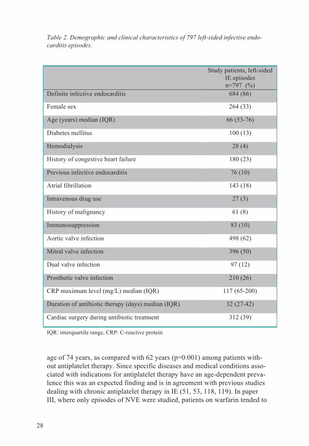
4.1 Demography Baseline characteristics of the 797 episodes of left-sided IE in the total study cohort are shown in table 2. Infective endocarditis fulfilling definite criteria was seen in 86% of the episodes. The prevalence of intravenous drug use (IVDU) was generally low among study patients but, as expected, a higher rate of IVDU was reported among patients with S. aureus IE compared to patients with IE caused by other pathogens (7% vs. 2%, p=0.007). Prosthetic valve endocarditis was seen in 26% of the patients and a history of conges-tive heart failure in 23%. Other underlying conditions such as diabetes (22% vs. 10%, p<0.001), immunosuppression (15% vs. 9%, p=0.026) and hemo-dialysis (9% vs. 2%, p<0.001) were more prevalent in patients with S.
aureus IE. Cardiac surgery during antibiotic treatment was performed in 39% of the patients in the total cohort.
Age
Mean and median age of patients in the total cohort was 63.5 (SD 15.9) and 66 (IQR 53-76) years, with a range of 15-99 years. A similar age span was seen in the National Swedish Endocarditis Registry (57, 144) and in other modern IE cohorts in the industrialized world (24, 31, 118, 145), while the median age in patients in a large worldwide multicenter study performed by the International Collaboration on Endocarditis (ICE) was slightly lower (42). These aged populations contrast the IE cohorts studied in the mid-1900s (45, 146) and in developing countries today (147, 148) with mean ages of 40 years or less. The numbers of patients divided by 10-year age intervals are shown in figure 3. The number of IE episodes increased in each 10-year age group up to 80 years of age, but no incidence rates were calcu-lated owing to lack of denominator data. However, in a study from Gothen-burg 1984-1988 also including retrospectively identified cases (19) median age was still higher, 70 years, and the calculated incidence continued to in-crease beyond 80 years of age.
Median age (67 years) among patients included in paper I was in accordance with the median age of the total cohort despite the different inclusion proce-dure in that part of the study. Among definite IE cases (paper II), the sub-group of patients on previously established antiplatelet therapy had a median
27
30
Table 2. Demographic and clinical characteristics of 797 left-sided infective endo-
carditis episodes.
Study patients, left-sided
IE episodes n=797 (%)
Definite infective endocarditis 684 (86)
Female sex 264 (33)
Age (years) median (IQR) 66 (53-76)
Diabetes mellitus 100 (13)
Hemodialysis 28 (4)
History of congestive heart failure 180 (23)
Previous infective endocarditis 76 (10)
Atrial fibrillation 143 (18)
Intravenous drug use 27 (3)
History of malignancy 61 (8)
Immunosuppression 83 (10)
Aortic valve infection 498 (62)
Mitral valve infection 396 (50)
Dual valve infection 97 (12)
Prosthetic valve infection 210 (26)
CRP maximum level (mg/L) median (IQR) 117 (65-200)
Duration of antibiotic therapy (days) median (IQR) 32 (27-42)
Cardiac surgery during antibiotic treatment 312 (39)
IQR: interquartile range, CRP: C-reactive protein age of 74 years, as compared with 62 years (p=0.001) among patients with-out antiplatelet therapy. Since specific diseases and medical conditions asso-ciated with indications for antiplatelet therapy have an age-dependent preva-lence this was an expected finding and is in agreement with previous studies dealing with chronic antiplatelet therapy in IE (51, 53, 118, 119). In paper III, where only episodes of NVE were studied, patients on warfarin tended to
28

31
be older than patients not on warfarin, but the differences did not reach sta-tistical significance for median ages (69 vs. 64 years, p=0.058). Patients with PVE were significantly older than patients with NVE (71 vs. 64 years, p<0.001)
Figure 3. Number of patients with infective endocarditis divided by 10-year age
intervals. Total number of episodes 797.
Sex differences
In the total cohort of 797 IE episodes, 33% (264) were seen in women, Women were, on average, older than men (68 vs. 65 years, p=0.02). The proportion of definite IE did not differ significantly between the sexes. Mi-tral valve involvement was more common in women than in men (54 vs. 46%, p=0.029) but the presence and length of vegetations did not differ be-tween the sexes. Contrary to a study concerning sex differences in IE by Aksoy et al. (50) there was no statistically significant difference between the frequency of comorbid conditions between women and men. A higher pro-portion of IE episodes in women were caused by S. aureus as compared with the proportion in men (29% vs. 20%, p=0.003).
In the total cohort, CVC were seen in 26% of the female episodes as com-pared with 20% of the male episodes (p=0.06). When definite episodes were studied separately (paper II), the higher CVC frequency in women was sta-tistically significant (30% vs. 22%, p=0.039). However, when the patients were stratified for microbial etiology, the CVC frequency did not differ be-tween the sexes. This is contrasted by the findings in a study of 260 S.
aureus IE by Roder et al. in which women tended to have more neurological
29

32
complications than men (55). Among episodes of definite IE, cardiac surgery during hospital stay was performed in 40% of the women vs. 44% of the men (n.s.). There was no statistically significant difference in in-hospital mortality between women and men (17% vs. 13%, n.s.), but 12-month mor-tality was higher in women (31% vs. 24%, p=0.031), similar to the findings of Thuny et al. (58).
4.2 Microbiology The most commonly found pathogens in the total cohort of 797 patients were viridans group streptococci (including S. bovis) seen in 29% of the episodes (figure 4). S. aureus was the etiological agent in 23% of all cases and in two episodes MRSA strains were identified. In paper II, where definite IE cases were studied separately, the S. aureus etiological fraction was 25% (168/684). This is consistent with the 28% S. aureus IE among 1213 definite IE cases from European countries prospectively included from 2000-2005 in the International Collaboration on Endocarditis (ICE) study (42). In paper I, the proportion of S. aureus IE among study patients was 25%.
Figure 4. Microbiological etiology of 797 left-sided infective endocarditis episodes.
23%
Staphylococcus
aureus
29%
Viridans group
streptococci 14%
Enterococci
9%
Coagualse-
negative
staphylococci
6%
Other streptococci
12%
Negative blood
culture
7%
Miscellaneous
30

33
Negative blood cultures were seen in 12% of all episodes, and 58% of the blood culture negative episodes were classified as possible. This is compati-ble with an analysis of culture negative IE reported in the National Swedish Endocarditis registry from 1995-2004, where the proportion of culture nega-tive episodes was 12%, and 75% were classified as possible IE (57). In a prospective epidemiological study from Gothenburg and Borås, Sweden (1984-1996), the proportion of culture negative IE was higher, 20%, and only one fifth of the culture negative IE cases were classified as definite IE (149). The combined secondary and tertiary referral center characteristics of the including sites in our study probably conferred a selection bias affecting the proportion of blood culture negative IE episodes.
Patients with enterococcal IE were significantly older (median 72 years) than patients with IE of other etiologies (median 60-65 years, p=0.01) except for coagulase-negative staphylococci (median 68 years), where the difference was not significant (figure 5). Enterococcal IE has been more prevalent in studies specifically concerning IE in older patients (41, 150). Prosthetic valve endocarditis (n=210) were caused by coagulase-negative staphylococci in 17% of the cases, in another 17% of the cases by viridans group strepto-cocci, and in 19% each by S. aureus and enterococci. Of coagulase-negative staphylococcal IE 51% were PVE as compared with 24% PVE episodes with other pathogens (p<0.001).
Figure 5. Median age (interquartile range) among 797 IE episodes divided by
pathogen.
31

34
4.3 Cerebrovascular complications
Symptomatic cerebrovascular complications
Cerebrovascular complications with concomitant neurological symptoms were diagnosed in 22% (177) of the IE episodes in the total cohort. The inci-dence of CVC in papers II and III was 25% in each. This higher figure is explained by the fact that CVC were diagnosed more often during definite IE episodes (paper II) as compared with possible episodes (25% vs. 7%, p<0.001), and that significantly more CVC were detected in episodes of NVE as compared with PVE (24% vs. 16%, p=0.008). Median ages did not differ significantly between patients with and without CVC. The CVCs were divided into three major groups; ischemic (including both established infarc-tions and TIA), hemorrhagic, and infectious complications depending on the type of sign or lesion presented. The proportion of episodes where two or more types of cerebral lesions were detected was 4% in the total cohort. More than one neurological symptom was displayed by 15% of the 60 pa-tients in paper I, although one of the symptoms dominated in each patient’s initial presentation. Neurological symptoms were present on admission for IE in three quarters of the patients with symptomatic CVC (paper I: 76%, paper II: 73%, paper III: 74%). Totally 17% of all patients experienced a CVC during admission (half recurrent CVC, half first time CVC) in paper I compared to 8% in papers II-III. The frequency of patients who underwent cardiac surgery after hosiptal admission for IE did not differ between pa-tients with and without CVC in the total cohort (42% vs. 38%, n.s.). Neurological symptoms were displayed by a significantly higher proportion of the patients in paper I (35%) than in papers II-III (25%, p=0.003). The high proportion of patients found to have symptomatic cerebral complica-tions in paper I could be explained by the additional clinical diagnostic ef-forts in the study with repeatedly performed standardized neurological ex-aminations of the patients included, apart from the radiological and neuro-chemical investigations also undertaken. A higher diagnostic sensitivity of this procedure could be assumed as compared with the routine physical and neurological examinations performed in patients in the other parts of the study.
In paper I patients were not included consecutively owing to late transfer of a considerable proportion of IE patients from other hospitals, the possible inclusion period of the study thereby being passed. Some patients were also reluctant to participate in a study involving additional investigations in the early period of this often unexpected and serious disease. The non-consecutive inclusion of IE patients might have impacted on the number of
32

35
patients with neurological symptoms included, but no selection of neurologi-cally symptomatic IE patients was done during the study period.
Ischemic infarctions and TIA
Ischemic infarction was the most common type of CVC, seen in 15% (120/797) of the episodes in the total study cohort. In paper I patients with neurological symptoms had ischemic lesions in 18 episodes, corresponding to 30% of the 60 patients included. Infarctions among the neurologically symptomatic patients were verified using MRI and release of brain damage markers in 11 cases, only by MRI findings in six cases and by isolated eleva-tion of NFL and GFAP in one case. There were multiple ischemic lesions on MRI in 10 patients, ranging from three to more than 10 in each patient. The sizes varied from punctuate to 3 cm, and in one patient there was a more specific pattern of septic embolism with early stages of abscess formation. The lesions were distributed in various parts of the brain including the corti-cal, subcortical, cerebellar and thalamic regions. In paper III, where 587 NVE episodes were studied separately, 96 cerebral ischemic infarctions verified by CT or MRI were found out of 144 episodes with symptomatic CVC (67%) (table 3). The size and localizations of these infarctions were not recorded systematically in the total study cohort, but all patients were neurologically symptomatic and the majority displayed focal or multifocal neurological symptoms.
Table 3. Types of cerebral lesions in 144 native valve endocarditis episodes with
cerebrovascular complications (paper II).
Number of NVE epi-sodes with CVC
n=144
Proportion of all NVE episodes n=587
Ischemic infarction 96 (67%) 16% Transient ischemic attack 20 (14%) 3% Hemorrhagic lesion 14 (10%) 2% Ruptured mycotic aneu-rysm
2 (1%) 0.3%
Cerebral infection 38 (26%) 6% More than one type of cerebral lesion
26 (18%) 4%
Among the 282 Swedish patients with NVE included in paper III, 16% suf-fered a symptomatic ischemic infarction verified by CT or MRI. Twenty percent of these 44 patients died during index hospitalization, 18% had ma-jor sequelae (hemiparesis, aphasia) and 25% minor sequelae (minor weak-ness, dysphasia, cognitive impairment) at hospital discharge. Studies on neu-
33

36
rological recovery in IE patients are scarce but our figures can be compared with the findings of Ruttmann et al. (52) in a study of 214 surgically treated IE patients. Thirty percent of the patients in that study had cerebral compli-cations prior to cardiac surgery. Overall 54% of the patients with preopera-tive cerebral lesions achieved full neurological recovery (in-hospital mortal-ity 17%), but a worse prognosis was seen in patients with large cerebral in-farctions and patients with multiple types of neurological complications. Stroke in IE patients was concluded to have a favorable prognosis as com-pared with stroke resulting from other causes.
A TIA was diagnosed in 3% of the 587 NVE episodes in paper III and in one out of 60 IE episodes (2%) in paper I. Similar incidences of TIA were found by Heiro et al. (5%) at a Finnish teaching hospital with IE patients included from 1980-1996 (49), and in a study by Thuny et al. (6%) among 496 pro-spectively studied definite left-sided IE episodes 1990-2005 at two French referral centers (72). Since a TIA, by definition, only gives transient neuro-logical signs, a risk for underdiagnosis accompanies this condition, but the specificity in the diagnosis of TIA could also be questioned.
Cerebral hemorrhage
Cerebral bleeding was detected in 14 out of the 587 NVE episodes (2%) and in two additional patients with PVE, the frequency of cererbral hemorrhage in PVE thereby being 1% (2/210). Accordingly, in 16 of the 797 episodes in the total cohort (2%) some degree of cerebral bleeding was found, and 75% of these complications had already occurred before admission to hospital. The cerebral hemorrhage was characterized as primary intracerebral in seven cases, caused by a ruptured mycotic aneurysm in two cases, complicating a primary infarction in six cases, and accompanying a brain abscess in one case. Patients suffering from cerebral hemorrhagic complications in IE were younger than patients with other types of CVC (median 54 vs. 65 years, p=0.024). The causative pathogen was S. aureus in 56% (9/16) of the IE cases with a hemorrhagic complication as compared with 49% (59/120) S.
aureus IE among patients with ischemic infarctions (n.s.). In-hospital mor-tality among patients with intracerebral bleeding complications was 38% (6/16), not significantly different from the in-hospital mortality of 25% (41/161) seen in patients with other types of CVC. In paper I the incidence of symptomatic cerebral bleeding was 2% (one patient) while minor hemor-rhagic components not influencing the management of the patients were found in another five patients. Mycotic aneurysms were detected in three patients in the total cohort, two of whom presented with a subarachnoid hemorrhage on admission. In the third patient, MRI detected an aneurysm without signs of bleeding that healed during antibiotic therapy. Blood cultures were negative in two cases and
34
37
grew Cardiobacterium hominis in the third case. None of the patients with mycotic aneurysms died during admission. One of the patients, with a rup-tured mycotic aneurysm on admission, underwent cardiac surgery on treat-ment day 71 owing to progressive aortic insufficiency. Two intracerebral mycotic aneurysms had been coiled prior to the cardiac operation. An addi-tional patient presented with a ruptured mycotic aneurysm four months after the completion of antibiotic therapy for aortic NVE caused by Salmonella
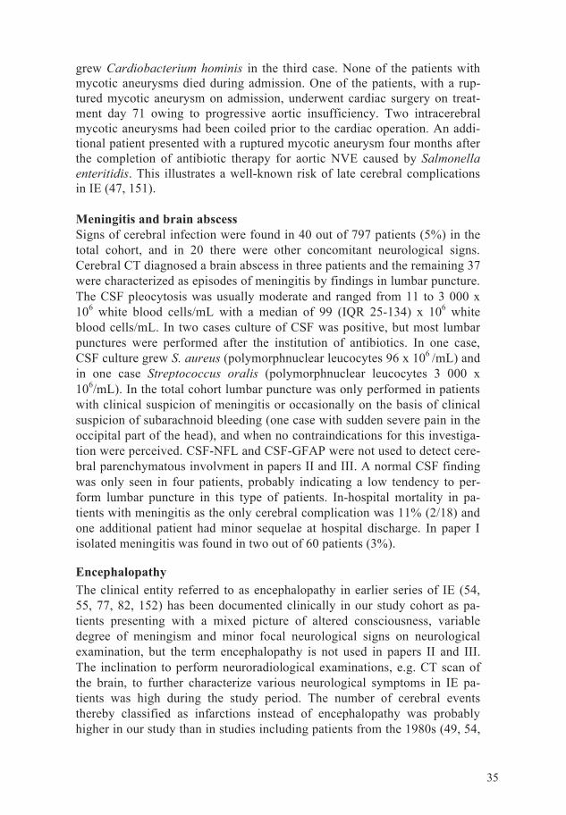
enteritidis. This illustrates a well-known risk of late cerebral complications in IE (47, 151). Meningitis and brain abscess
Signs of cerebral infection were found in 40 out of 797 patients (5%) in the total cohort, and in 20 there were other concomitant neurological signs. Cerebral CT diagnosed a brain abscess in three patients and the remaining 37 were characterized as episodes of meningitis by findings in lumbar puncture. The CSF pleocytosis was usually moderate and ranged from 11 to 3 000 x 106 white blood cells/mL with a median of 99 (IQR 25-134) x 106 white blood cells/mL. In two cases culture of CSF was positive, but most lumbar punctures were performed after the institution of antibiotics. In one case, CSF culture grew S. aureus (polymorphnuclear leucocytes 96 x 106 /mL) and in one case Streptococcus oralis (polymorphnuclear leucocytes 3 000 x 106/mL). In the total cohort lumbar puncture was only performed in patients with clinical suspicion of meningitis or occasionally on the basis of clinical suspicion of subarachnoid bleeding (one case with sudden severe pain in the occipital part of the head), and when no contraindications for this investiga-tion were perceived. CSF-NFL and CSF-GFAP were not used to detect cere-bral parenchymatous involvment in papers II and III. A normal CSF finding was only seen in four patients, probably indicating a low tendency to per-form lumbar puncture in this type of patients. In-hospital mortality in pa-tients with meningitis as the only cerebral complication was 11% (2/18) and one additional patient had minor sequelae at hospital discharge. In paper I isolated meningitis was found in two out of 60 patients (3%).
Encephalopathy
The clinical entity referred to as encephalopathy in earlier series of IE (54, 55, 77, 82, 152) has been documented clinically in our study cohort as pa-tients presenting with a mixed picture of altered consciousness, variable degree of meningism and minor focal neurological signs on neurological examination, but the term encephalopathy is not used in papers II and III. The inclination to perform neuroradiological examinations, e.g. CT scan of the brain, to further characterize various neurological symptoms in IE pa-tients was high during the study period. The number of cerebral events thereby classified as infarctions instead of encephalopathy was probably higher in our study than in studies including patients from the 1980s (49, 54,
35

38
77) when CT examinations assumedly were less common. In still earlier studies, such as the study by Pruitt et al. published in 1978 (47), the high in-hospital mortality rates in IE patients with neurological complications (58%) and the considerable proportion of post-mortem examinations executed in these patients (>90%) facilitated more precise diagnosis. The detected inci-dence of major cerebral infarctions was 17% in that study, and an additional 11% of the patients were found to have microembolic ischemic lesions. Pa-tients with microembolic lesions presented with altered levels of conscious-ness and fluctuating neurological signs, in accord with the clinical descrip-tion of encephalopathy. In paper I a more thorough characterization of patients with encephalopathic neurological manifestations was possible, and all these patients presented with confusion and/or altered consciousness and minor focal signs. Alto-gether six out of 60 patients (10%) were considered to be encephalopathic, and the median age of these patients was 78 years as compared with 54 years in patients presenting with other neurological signs (n.s.). In all six patients with the clinical picture of encephalopthy there were multiple ischemic le-sions demonstrated on MRI, and increased levels of GFAP and NFL were detected in CSF, illustrating the embolic nature of these lesions and the in-volvement of parenchymatous CNS damage.
4.4 Factors related to the incidence of cerebrovascular complications
Antiplatelet therapy (paper II)
In paper II the aim was to study the relationship between previously estab-lished antiplatelet therapy and the incidence of cerebrovascular complica-tions in left-sided definite IE episodes. Antiplatelet therapy was established prior to IE diagnosis in 23% of the 684 IE episodes in patients included in this part of the study. No difference in the incidence of CVC during IE was detected between patients with and without previously established antiplate-let therapy (24% vs. 25%, OR 0.9, 95% CI 0.6-1.4). To adjust for possible confounding variables a multiple logistic regression model was used, and variables that were significantly associated with both occurrence of CVC and to the use of antiplatelet therapy were included. After adjustment for congestive heart failure, vegetation length, pathogen and CRP level no sig-nificant relation between antiplatelet therapy and CVC incidence was found either (aOR 0.8, 95% CI 0.48-1.5). The possibility of a reduced embolic risk owing to use of antiplatelet therapy during the prediagnostic development of IE has been debated (51, 53, 153,
36

39
154) while in a prospective study, the initiation of antiplatelet therapy after IE diagnosis was found not to reduce the embolic risk (117). A study on specific influence of antiplatelet therapy on mortality rate (118) and of ASA on the rate of acute valvular replacement surgery in S. aureus IE (119) has been published recently. Only one small previous study has focused on the role of antiplatelet therapy in relation to cerebral complications (155).
Anticoagulant therapy in native valve endocarditis (paper III)
In paper III, including episodes of NVE, 8% (48/587) of the patients were on oral anticoagulants (warfarin) upon admission. There was, as expected, a significantly higher prevalence of atrial flutter among patients on warfarin as compared with patients not on warfarin (37% vs. 12%, p<0.001). Surgical rates did not differ between patients with and without warfarin on admission for IE. Symptomatic CVC were significantly less frequent in NVE patients on war-farin than in patients not on this treatment, i.e. in 6% vs. 26% (OR 0.2, 95% CI 0.06-0.6, P=0.006). In the warfarin group all CVC were established on admission, while 7% of the patients without warfarin experienced a first CVC during antibiotic treatment. The reduction in CVC rate consisted of a lower number of non-hemorrhagic events, while the incidence of hemor-rhagic complications did not differ significantly between patients with and without warfarin therapy (figure 6). After adjustment for age, sex, S. aureus etiology, history of congestive heart failure or previous IE episode, vegeta-tion length and CRP level, a significantly lower incidence of CVC prevailed among patients on warfarin (aOR 0.26, 95% CI 0.07-0.94, p=0.04).
Figure 6. Cerebrovascular complications in 587 left-sided native valve endocarditis
episodes in patients with and without warfarin therapy
37

40
The aim in paper III was to do a specific analysis of the relation between ongoing therapy with vitamin K antagonists and the incidence of CVC in IE affecting native valves. This design was chosen in order to avoid confound-ing from inherent differences in (i) CVC rate between NVE and PVE, (ii) PVE affecting different types of valve prostheses (mechanical, bioprosthetic, homograft) and (iii) effects on CVC rate between oral anticoagulants and heparin analogues. Anticoagulant therapy and prosthetic valve endocarditis
In the total cohort of 797 patients there were 210 PVE not separately ana-lyzed in papers I-III. As earlier mentioned, the CVC incidence was lower among PVE cases than in NVE (16% vs. 24%, p=0.008). The incidence tended to be lower in bioprosthetic PVE (13%, 13/98) than in mechanical PVE (18%, 20/112) but this difference was not statistically significant. War-farin use on admission for PVE was frequent (67%), and, as expected, it was more frequent in PVE on mechanical valve prostheses compared to PVE on bioprostheses (90%, vs. 41%, p<0.001). CVC incidence in PVE did not dif-fer between patients with and without warfarin therapy (17% [24/141 vs. 13% [9/69], n.s.), and this was similar in both mechanical and biological PVE. These findings evoke the hypothesis that embolic events in PVE and NVE have different predictors and favor separate analysis of the disease entities. Some recent studies have found a trend towards fewer CVC in PVE compared to NVE (49, 55, 65) or a lower rate of systemic major embolism in PVE (156), while the incidence of cerebral complications in NVE and PVE did not differ in other studies (48, 54, 72).
Microbial etiology and CVC
Patients with S. aureus IE suffered from cerebral complications significantly more frequently than patients with IE caused by other pathogens. The CVC incidence in S. aureus IE was 49% (89/182) in the total cohort, which corre-sponded to 52% among both definite IE episodes (paper II) and NVE epi-sodes (paper III). In table 4 unadjusted OR for CVC divided by pathogen in definite IE episodes are shown (paper II). Enterococcal IE carried the lowest CVC risk, significantly different from the risk seen in culture-negative IE (OR 2.8, 95% CI 1.1–7.0) and S. aureus IE (OR 8.5, 95% CI 4.4-16.8). In both papers I and III, S. aureus etiology remained a significant risk factor for symptomatic CVC in the multivariable models, with an adjusted odds ratio of 6.1 (95% CI 1.5-24.3) and 6.3 (95% CI 3.8-10.4), respectively.
IE episodes complicated with meningitis had a different etiological pattern, since this manifestation was seen more often in IE caused by non-viridans
38

41
Table 4. Odds ratio for cerebrovascular complications divided by pathogen in 684
definite infective endocarditis episodes.
Pathogen % CVC OR 95% CI for
OR
Enterococci (n=108)
11% (n=12) reference -
Coagulase-negative staphylococci (n=59)
14% (n=8) 1.3 0.5 - 3.3
Viridans streptococci (n=214)
15% (33) 1.5 0.7 – 2.9
Other streptococci (n=47)
19% (n=9) 1.9 0.7 – 4.9
Miscellaneous (n=49)
20% (n=10) 2.1 0.8 – 5.1
Culture negative (n=39)
26% (n=10) 2.8* 1.1 – 7.0
S. aureus
(n=168) 52% (n=87) 8.5* 4.4 – 16.8
* p<0.05.
streptococci (15%, 7/47) and in S. aureus IE (14%, 24/168) compared to episodes of other etiologies (1%, 6/469, p<0.001). Cerebral hemorrhage occurred in 5% of culture-negative IE (2/39) and 5% of S. aureus IE (9/168), but only the latter was significantly different from the incidence of cerebral hemorrhage seen in IE of other etiologies (1%, 5/477, p=0.003) (paper II). If the analysis of CVC frequency was performed on the total cohort of pa-tients including possible IE cases, the proportion of patients with culture-negative IE suffering any CVC (including hemorrhage) was lower, since definite IE diagnosis in culture negative IE relies on signs of embolism and other minor criteria.
4.5 Echocardiography
Incidence of vegetations
Vegetations were established in 75% of the 760 episodes in the total cohort that were possible to evaluate for the presence of vegetation. A symptomatic CVC was diagnosed in 25% of the episodes with vegetation on echocardi-ography as compared with in 12% of episodes without detected vegetations, p<0.001. Among episodes of NVE (paper III), a statistically significant dif-
39

42
ference in CVC rate between episodes with and without vegetations was detected as well (27%, vs. 14%, p=0.003). In paper II, where definite IE episodes were studied separately, the trend was similar but the difference was no longer statistically significant (26% vs. 18%, p=0.06). This discrep-ancy could be explained by the construction of the modified Duke criteria, since an embolic event is considered a minor criterion for IE. A suspected IE episode without an echocardiographic major criterion is frequently classified as definite, owing to the presence of embolism together with e.g. positive blood cultures, a predisposing condition and fever. This implies a higher incidence of CVC among IE episodes without an echocardiographically de-tected vegetation, when only definite episodes are studied. In paper I, a vege-tation was observed in 70% (42/60) of the patients. A symptomatic CVC occurred in 45% of the patients with a vegetation, as compared with in 11% of patients without a vegetation, p=0.02.
Size of vegetation
IE episodes complicated with symptomatic CVC had significantly longer vegetations, median 10 mm (IQR 6-16) vs. 8 mm (IQR 5-12), p<0.001, when the total cohort was analyzed. This difference was statistically significant among definite IE episodes in paper II (median 10 mm [IQR 6-16] vs. 8 mm [IQR 5-12], p<0.001) and among NVE cases studied in paper III (median 11 mm [IQR 6-16] vs. 9 mm [IQR 5-13], p=0.006). Vegetation length differed in all types of CVC as compared with episodes without CVC except for TIA, where median vegetation lengths were similar in cases with and without the complication. Between IE cases with cerebral hemorrhage and cases without any CVC the difference in vegetation length did not reach statistical signifi-cance, which was probably an effect of the limited number of patients with this complication (data not shown). In paper III the vegetation length in NVE episodes was found to be an inde-pendent predictive factor for CVC using a multivariable model. After ad-justment for age, sex, S.aureus etiology, history of congestive heart failure or previous IE, and CRP level, vegetation length remained significantly associ-ated with a higher incidence of CVC with an adjusted OR of 1.04 (95% CI 1.01-1.07, p=0.005). In paper I there was also a significant association between vegetation length and incidence of both symptomatic and total cerebral embolism (including silent emboli). Median vegetation length in episodes with symptomatic CVC as compared with episodes without was 12 mm (IQR 8-16) vs. 8 mm (IQR 6-11), p=0.001. A similar correlation was seen for the incidence of total CVC (10 mm |IQR 8-15] vs. 5 mm [IQR 4-10), p<0.001). In multivariable logistic regression analysis the only variable correlated to an increased risk of both clinically symptomatic CVC (adjusted OR 1.17 [95% CI 1.05-1.30],
40

43
p=0.005) and total CVC (adjusted OR 1.23 [95% CI 1.08-1.39], p=0.001) was the length of vegetation. As mentioned above, S aureus etiology also conferred a higher risk for clinical cerebral embolism but not total cerebral embolism. In the multivariable analysis, the area under the ROC curve was calculated for the discriminating variables (weighted sum of vegetation length and S. aureus etiology, and only vegetation length) and found to be 0.811 and 0.782, respectively.
Additional aspects on echocardiographic findings
In 37 patients the echocardiographic examinations were inconclusive regard-ing the presence of vegetations, e.g. in cases with severe regurgitation and perforated heart valves, intracardiac abscesses or concomitant ruptured chor-dae tendinae. These episodes were regarded as “missing values” in the analysis of the relationship between the presence of vegetation and the inci-dence of CVC. Five of the episodes were classified as possible, 22 were definite according to surgical findings, six were definite attributable to the presence of an intracardic abscess, and the remaining four patients were classified as definite without the echocardiographic criterion fulfilled. In another 65 cases the echocardiographic examinations (all TEE) defined the presence but not the size of vegetation. All these cases were classified as definite, and 31 underwent cardiac surgery. The remaining 34 episodes were seen in patients with varying types of predisposing cardiac valve anomalies. Paravalvular involvement was found in 112 (14%) of the IE episodes, in 63% of these together with a vegetation. Paravalvular complications were significantly more frequent in PVE than NVE (p<0.001).
Multivariable modeling
After multivariable analysis (paper III) additional variables found to corre-late to CVC incidence among NVE patients were history of congestive heart failure (aOR 0.22; 95% CI 0.1-0.52) and history of previous IE episode (aOR 0.1; 95% CI 0.01-0.79), both associated with a lower incidence of CVC.
4.6 Silent cerebral embolism In paper I the incidence of silent CVC was found to be 30% (18/60) when MRI of the brain and CSF analysis were used to detect lesions (figure 7). The MRI lesions in patients with silent cerebral embolism were all small ischemic infarctions with a maximum size of 1 cm, and the findings were multiple in 40% of the patients. Out of the 27 patients with no IE related pathological MRI findings, 14 had other types of lesions, mainly minor old infarctions or white matter lesions.
41

44
Figure 7. Incidence and distribution of cerebrovascular complications among 60 IE
patients (paper I). Circles represent the patients with cerebrovascular complications
detected by each method.
Only one of the patients in the study had a history of previous stroke, and no clinical or silent signs of CVC correlated to the current IE episode were de-tected in this patient. In one patient, previously operated on for falx men-ingeoma, minor residual meningeomal tissue and gliosis were found on MRI in combination with a new ischemic infarction in the cerebellum. In another patient a Chiari malformation not previously known was found, and this patient also had signs of silent CVC verified by both MRI and CSF findings. In yet another patient with clinically obvious cerebral embolism also verified with MRI and CSF findings, a previously diagnosed hypophyseal adenoma was seen. None of the patients in the study had a history of or signs attribut-able to other neurological diseases known to be associated with elevations in brain damage markers or specific MRI lesion (131, 133, 138). Elevated levels of NFL (figure 8) and/or GFAP (figure 9) in CSF as markers of parenchymatous brain damage was seen in 24 patients, 12 of whom had no concomitant neurological signs. In all symptomatic patients both CSF-NFL and CSF-GFAP were elevated above the reference levels, and elevated levels of polymorphnuclear cells were found in seven patients as well. Among the 12 patients with silent cerebral embolism detected on the basis of CSF findings, eight were detected from elevations in brain damage markers only (CSF-NFL and CSF-GFAP elevated in five, only CSF-NFL in three). There were no statistically significant differences between CSF-NFL or CSF-GFAP levels in patients with symptomatic and silent CVC, respec-
42

45
tively, but for CSF-NFL the highest values were seen in symptomatic pa-tients. For CSF-GFAP the highest value was detected in a patient with silent CVC and no ischemic lesion identified on MRI.
0
2000
4000
6000
8000
30 40 50 60 70 80 90
No CVC Silent CVC Symptomatic CVC Isolated meningitis
16800 11500 12700 13710
CSF - NFL(ng/L)
Age (years)
Figure 8. CSF-NFL levels in infective endocarditis patients according to presence and type of cerebrovascular complication (CVC). Reference levels: < 59 years: < 250 ng/L, 60-69 years: < 380 ng/L, > 70 years: < 750 ng/L
0
2000
4000
6000
8000
30 40 50 60 70 80 90
No CVC Symptomatic CVC Silent CVC Isolated meningiAge (years)
CSF - GFAP(ng/L)
19240
42150
Figure 9. CSF-GFAP levels in infective endocarditis patients according to presence and type of cerebrovascular complication (CVC). Reference levels: 21-59 years: < 750 ng/L, > 60 years: < 1250 ng/L
43
tis

46
The kinetics of NFL and GFAP elevations in CSF after acute CNS damage differ considerably, with the highest CSF-GFAP elevations detected after 24-48 hours and normalization within less than three weeks. For CSF-NFL there is a gradual increase peaking after a few weeks and normalizing months after the acute insult. This is consistent with the isolated CSF-NFL increment found in three neurologically asymptomatic patients, where the assumption is that the expected concomitant GFAP elevation had already passed when the sample was taken. In the neurologically symptomatic pa-tients the first LP was probably performed closer in time to the embolic event as compared with in the patients with silent CVC. No LP was performed in 21 patients for reasons of concomitant anticoagula-tion (9), other contraindications (4), early surgery before LP was possible (3), refusal (3), and technical problems (2). CSF findings were normal in 13 patients.
4.7 Mortality In-hospital mortality was 13% in the total cohort, 14% among definite cases (paper II) and 12% among NVE cases (paper III). Infective endocarditis patients with cerebral manifestation showed significantly higher in-hospital mortality than patients without such complications, 27% vs. 9%, p<0.001 (total cohort). Of all patients who died during hospitalization (104) for IE, 45% had a CVC prior to succumbing. The different in-hospital mortality between patients experiencing neurological complications were statistically significant when infarctions (30% vs. 9%, p<0.001) and cerebral hemorrhage (38% vs. 9%, p=0.006) were analyzed separately. For episodes complicated with meningitis the picture was different, and in patients with meningitis as the sole neurological manifestation, no significantly different in-hospital mortality was seen compared to patients without any CVC (11% vs. 9%, n.s.). For patients with isolated TIA there was a trend toward higher mortal-ity (18% vs. 9%, n.s.). In paper I in-hospital mortality was only 2%. In-hospital mortality was significantly correlated to age and increased for each 10-year interval from 50 to 70 years of age (<50 years: 5%, 51-60 years: 11%, 60-69 years: 14%, >70 years: 17%, p=0.002). Higher in-hospital mortality was seen for patients with CVC in all age groups, but for patients <50 years this trend was not significant (10% vs. 4%, n.s.). In-hospital mor-tality has been shown to correlate to age and CVC rate in other studies (39, 41, 42, 52, 150, 157).
44

47
Long-term outcome
Cerebral complications influenced long-term outcome. Among the 684 epi-sodes of definite IE a survival analysis using Kaplan-Meier procedure showed a significantly lower survival rate among patients with CVC during the follow-up period (log rank test, p=0.004) (figure 10). Follow up varied from 114 days to 4473 days (12.2 years) with a median of 3.6 years. After 10 years the survival rate was 35% among patients who experienced a CVC during the initial IE episode and 50% in patients without CVC. The analysis after 10 years was based on 25 survivors of 589 alive at hospital discharge. The detrimental long-term impact of neurological complications has been shown in other studies (40, 72, 78) but the low 10-year survival rates in all patients also reflects the high median ages in IE patients in the present series.
Figure 10. Kaplan-Meier analysis of long-term survival among IE patients with and
without a cerebrovascular complication during the initial IE episode. CVC: cerebrovascular complication, Cum survival: cumulative survival rate.
45

48
5 General considerations
Incidence of symptomatic cerebral manifestations
The symptomatic cerebral complication rate of 22% found in this large co-hort of adult IE patients, with ischemic stroke as the dominant lesion, is in accord with recent studies focusing on cerebral complications (48, 49, 72). The higher incidence of symptomatic CVC found in paper I might reflect more sensitive clinical examinations in that study as well as a higher aware-ness of neurological symptoms in IE patients during the study period, but inclusion bias could not be ruled out. Unchanged frequencies of cerebral complications have been asserted since the preantibiotic era of IE, but fig-ures are not comparable without further evaluation of the demographic char-acteristics of patients included in the different studies. The evolving epide-miology and varying definitions of IE and of cerebral complications, chang-ing diagnostic procedures, different study designs and the influence of selec-tion bias have to be considered in these comparisons. In the preantibiotic era the proportion of IE episodes caused by streptococci, especially the viridans group streptococci but also beta-hemolytic strepto-cocci and pneumococci, was much higher, and the natural history of the dis-ease could be observed in all patients, since no therapeutic measures were possible. The rate of cerebral complications in IE episodes caused by viri-dans group streptococci was around 40-50% in studies prior to the mid-1940s (92, 146), which is comparable with the CVC rate found in S. aureus IE in our cohort, but much higher than the rates of CVC seen in viridans group streptococcal IE today. The enhanced possibilities for early diagnose of IE and for effective treatment have probably decreased the rate of CVC in streptococcal IE. Regarding S. aureus IE, the development is less clear since the relative and probably also the absolute incidence of this disease has increased since the introduction of penicillin (34, 158) parallel to enhanced detection of IE and improved initial management of septic patients. Cerebral complications in S.
aureus IE are high in modern studies (34, 55, 159) and were also described as high in early IE patient series (46). The improved diagnostic methods might have given a paradoxical increase of cerebral events in S. aureus IE,
46
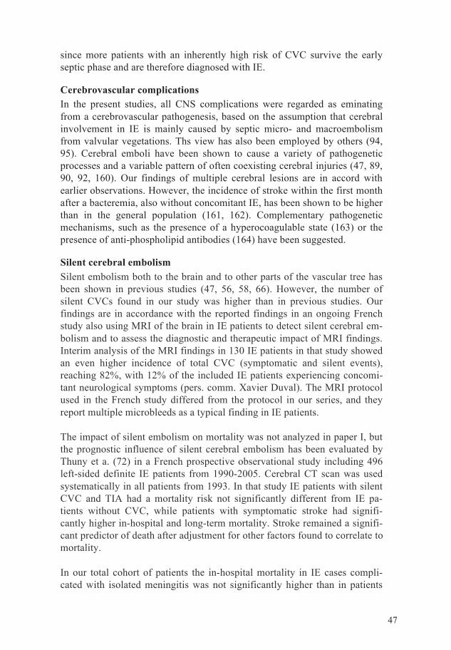
49
since more patients with an inherently high risk of CVC survive the early septic phase and are therefore diagnosed with IE.
Cerebrovascular complications
In the present studies, all CNS complications were regarded as eminating from a cerebrovascular pathogenesis, based on the assumption that cerebral involvement in IE is mainly caused by septic micro- and macroembolism from valvular vegetations. Ths view has also been employed by others (94, 95). Cerebral emboli have been shown to cause a variety of pathogenetic processes and a variable pattern of often coexisting cerebral injuries (47, 89, 90, 92, 160). Our findings of multiple cerebral lesions are in accord with earlier observations. However, the incidence of stroke within the first month after a bacteremia, also without concomitant IE, has been shown to be higher than in the general population (161, 162). Complementary pathogenetic mechanisms, such as the presence of a hyperocoagulable state (163) or the presence of anti-phospholipid antibodies (164) have been suggested.
Silent cerebral embolism
Silent embolism both to the brain and to other parts of the vascular tree has been shown in previous studies (47, 56, 58, 66). However, the number of silent CVCs found in our study was higher than in previous studies. Our findings are in accordance with the reported findings in an ongoing French study also using MRI of the brain in IE patients to detect silent cerebral em-bolism and to assess the diagnostic and therapeutic impact of MRI findings. Interim analysis of the MRI findings in 130 IE patients in that study showed an even higher incidence of total CVC (symptomatic and silent events), reaching 82%, with 12% of the included IE patients experiencing concomi-tant neurological symptoms (pers. comm. Xavier Duval). The MRI protocol used in the French study differed from the protocol in our series, and they report multiple microbleeds as a typical finding in IE patients. The impact of silent embolism on mortality was not analyzed in paper I, but the prognostic influence of silent cerebral embolism has been evaluated by Thuny et a. (72) in a French prospective observational study including 496 left-sided definite IE patients from 1990-2005. Cerebral CT scan was used systematically in all patients from 1993. In that study IE patients with silent CVC and TIA had a mortality risk not significantly different from IE pa-tients without CVC, while patients with symptomatic stroke had signifi-cantly higher in-hospital and long-term mortality. Stroke remained a signifi-cant predictor of death after adjustment for other factors found to correlate to mortality. In our total cohort of patients the in-hospital mortality in IE cases compli-cated with isolated meningitis was not significantly higher than in patients
47
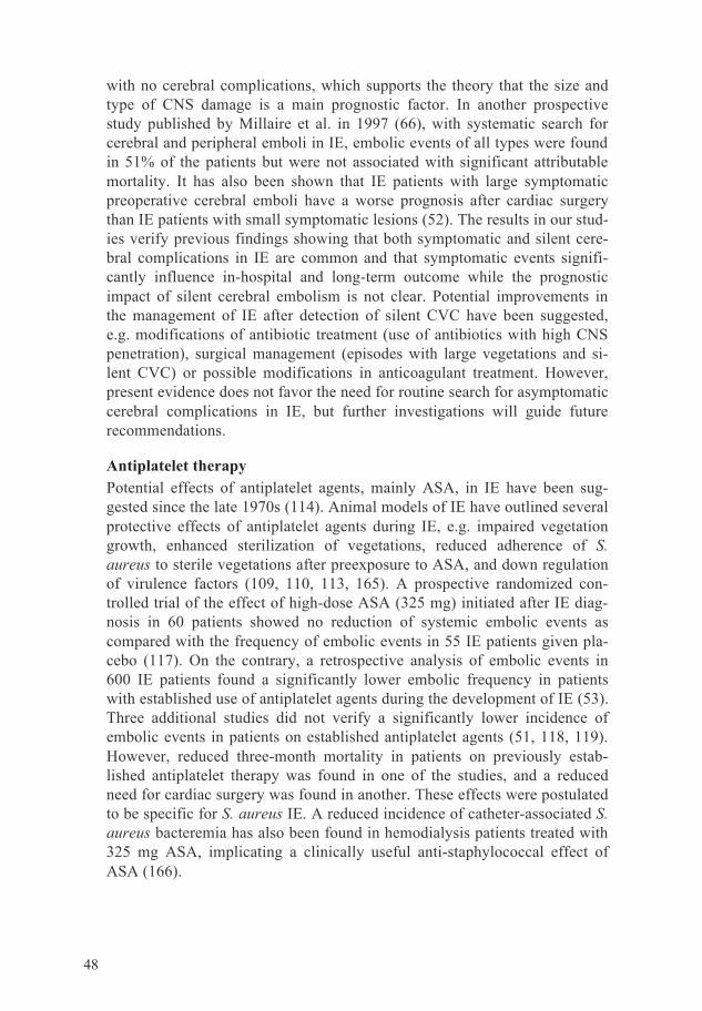
50
with no cerebral complications, which supports the theory that the size and type of CNS damage is a main prognostic factor. In another prospective study published by Millaire et al. in 1997 (66), with systematic search for cerebral and peripheral emboli in IE, embolic events of all types were found in 51% of the patients but were not associated with significant attributable mortality. It has also been shown that IE patients with large symptomatic preoperative cerebral emboli have a worse prognosis after cardiac surgery than IE patients with small symptomatic lesions (52). The results in our stud-ies verify previous findings showing that both symptomatic and silent cere-bral complications in IE are common and that symptomatic events signifi-cantly influence in-hospital and long-term outcome while the prognostic impact of silent cerebral embolism is not clear. Potential improvements in the management of IE after detection of silent CVC have been suggested, e.g. modifications of antibiotic treatment (use of antibiotics with high CNS penetration), surgical management (episodes with large vegetations and si-lent CVC) or possible modifications in anticoagulant treatment. However, present evidence does not favor the need for routine search for asymptomatic cerebral complications in IE, but further investigations will guide future recommendations.
Antiplatelet therapy
Potential effects of antiplatelet agents, mainly ASA, in IE have been sug-gested since the late 1970s (114). Animal models of IE have outlined several protective effects of antiplatelet agents during IE, e.g. impaired vegetation growth, enhanced sterilization of vegetations, reduced adherence of S.
aureus to sterile vegetations after preexposure to ASA, and down regulation of virulence factors (109, 110, 113, 165). A prospective randomized con-trolled trial of the effect of high-dose ASA (325 mg) initiated after IE diag-nosis in 60 patients showed no reduction of systemic embolic events as compared with the frequency of embolic events in 55 IE patients given pla-cebo (117). On the contrary, a retrospective analysis of embolic events in 600 IE patients found a significantly lower embolic frequency in patients with established use of antiplatelet agents during the development of IE (53). Three additional studies did not verify a significantly lower incidence of embolic events in patients on established antiplatelet agents (51, 118, 119). However, reduced three-month mortality in patients on previously estab-lished antiplatelet therapy was found in one of the studies, and a reduced need for cardiac surgery was found in another. These effects were postulated to be specific for S. aureus IE. A reduced incidence of catheter-associated S.
aureus bacteremia has also been found in hemodialysis patients treated with 325 mg ASA, implicating a clinically useful anti-staphylococcal effect of ASA (166).
48

51
In the analysis of CVC incidence in paper II no correlation was found be-tween prior established use of antiplatelet therapy and the frequency of CVC, and unadjusted 12-month mortality was significantly higher in patients on antiplatelet agents. However, when adjusted for other variables signifi-cantly associated with 12-month mortality, no significant relationship re-mained between 12-month mortality and antiplatelet therapy. Our findings thus do not support the hypothesis that antiplatelet therapy influences em-bolic tendency in IE patients, and no statistically significant conclusions could be drawn regarding the influence of antiplatelet therapy on mortality. However, important variables with previously shown major impact on mor-tality in IE, e.g. development of new or worsening congestive heart failure during IE, were not retained in our model. Additionally, the study was not designed to evaluate other possible benefits of antiplatelet therapy in IE. Despite the large number of patients in our study, the number of S. aureus IE episodes might have been too small to evaluate specific interactions between S. aureus and antiplatelet agents.
Hemorrhagic cerebral complications
Hemorrhagic complications were few in the present series, reaching 2% symptomatic bleeding events both in the total cohort of IE patients and in the patients systematically investigated using MRI and CSF analysis in paper I. No increase was detected among patients on antiplatelet therapy or oral anti-coagulants. The number of detected ruptured cerebral mycotic (infectious) aneurysms was less than 0.5% in the total cohort of IE patients and only one patient with an unruptured aneurysm was found. Similar low rates of rup-tured mycotic aneurysms have been found in other recent studies (49, 54, 72), but data supporting the occurrence of a considerably higher number of unruptured and clinically silent aneurysms that heal during antibiotic therapy have also been presented (47, 167, 168). The detected number of silent aneu-rysms will probably increase in the future following the use of further en-hanced neuroradiological imaging, such as e.g. MR angiography. The low but prolonged risk of aneurysm rupture after antibiotic therapy has been completed, as seen in one of the patients in our study, is well recognized in previous studies (76, 151).
Oral anticoagulant therapy
The proportion of NVE patients on warfarin who suffered a CVC was sig-nificantly lower than the proportion of patients not on warfarin who suffered a CVC (6% vs. 26%, p=0.006). This was an unexpected finding since the only previous study separately addressing the issue of anticoagulation in NVE patients (96) found cerebrovascular accidents significantly more fre-quently among anticoagulated patients as compared with non-anticoagulated patients. The CVC proportion in that restrospective study remained signifi-cantly higher among anticoagulated NVE patients when cerebral hemor-
49

52
rhagic complications were analyzed separately, but not when ischemic cere-bral complications (including CVC of unclear origin but not TIA) were ana-lyzed. Among 269 NVE patients in that study, including patients from 1970-1987, cerebral hemorrhagic complications were seen in 5% and cerebral ischemic infarctions in 7% of the patients. The number of ischemic events was relatively low as compared with most other series, which implies very restricted diagnostic criteria for ischemic cerebrovascular accidents or possi-ble selection bias. Other previous studies addressing the relationship between anticoagulant therapy and cerebral embolic complications in IE patients have included very few anticoagulated patients (47) or have mainly focused on patients with PVE (97, 169-171). These studies have included patients over an ex-tended period of time, and Leport et al. (169) found better management of anticoagulant treatment among patients in the later part of the study. It is not known whether improved general management of anticoagulant therapy in IE patients has since been achieved. One recently published study primarily addressing the effects of antiplatelet therapy in IE (118) detected a signifi-cantly higher incidence of hemorrhagic stroke among patients who were on warfarin prior to IE diagnosis, although no more detailed information was given. An increasing frequency of anticoagulant associated intracerebral hemorrhage was reported during the 1990s in patients with anticoagulant therapy for any indication (172).
The lower CVC rate among NVE patients on warfarin therapy in our study remained significant after adjustment for microbiological etiology and vege-tation length, indicating that warfarin treatment may diminish the liability for embolization independently from vegetation length. Fibrin formation plays important roles during the development of IE, both in the attachment of bac-teria to the surface of the vegetations and in the formation of a protective layer against host defense mechanisms within the vegetation. A disturbance in fibrin formation could therefore possibly influence both the development of IE and the frequency of embolism. Only a few experimental studies with diverging results were published in this area in the 1970s (7), while the ques-tion has not been addressed in later studies and merits further attention in future studies.
50

6 Conclusions
• Cerebrovascular complications were seen in 65% of patients with
left-sided infective endocarditis when sensitive diagnostic methods
were used, but almost half of these were clinically silent.
• Prior established antiplatelet therapy gave no protection against
cerebral complications in infective endocarditis. After adjustment
for covariables antiplatelet therapy had no significant impact on
mortality.
• Ongoing oral anticoagulation with warfarin on admission for native
valve endocarditis was associated with lower risk of cerebrovascular
complications.
• Staphylococcus aureus etiology and vegetation length as detected
using echocardiography were each correlated to a higher risk of
cerebrovascular complications in infective endocarditis.
• Cerebral hemorrhagic complications were few. Antiplatelet therapy,
warfarin therapy and prosthetic valve endocarditis conferred no
higher frequency of these complications.
51

54
7 Svensk sammanfattning Infektiös hjärtklaffsinfektion är en relativt ovanlig men livshotande sjukdom. I Sverige diagnosticeras cirka 550 fall årligen. Risk för hjärtklaffsinfektion är högre hos individer med skador på hjärtklaffarna eller inopererade hjärt-klaffsproteser. Personer med kvarliggande intravenösa infarter, t ex hemodi-alyspatienter, har också förhöjd risk liksom individer med intravenöst miss-bruk.
Vid hjärtklaffsinfektion finns det bakterier i blodet, som fått fäste på en hjärtklaff och gett upphov till infekterade koagel där, s.k. vegetationer. Hjärtklaffen kan skadas och börja läcka, vilket kan medföra hjärtsvikt. Akut hjärtkirurgi behövs i c:a 30% av alla fall. En annan vanlig och allvarlig komplikation vid hjärtklaffsinfektion är s.k. embolisering, då små infektera-de fragment från vegetationerna lossnar och förs med blodet till olika organ med åtföljande skador. Detta sker i 20-60% av episoderna och hjärnan drab-bas mest frekvent, vilket medför olika typer av neurologiska symtom och skador. Kliniskt tysta (asymtomatiska) embolier förekommer också. Skador-na i hjärnan är av varierande typ. Vanligast är hjärninfarkt orsakad av för-hindrad blodförsörjning till en del av hjärnan, men infektion i hjärnan eller hjärnhinnorna och hjärnblödning förekommer också. Olika typer av skador kan uppstå parallellt.
Syftet med avhandlingens studier var att undersöka frekvensen symtomgi-vande och tysta embolier till hjärnan samt att kartlägga faktorer associerade med ökad och minskad emboliseringsrisk. I delstudie I undersöktes 60 pati-enter med magnetresonanstomografi (MR) och provtagning av ryggmärgs-vätska med analys av markörer för hjärnskada. Andelen patienter som drab-bades av symtomgivande neurologiska komplikationer var 35% och därut-över påvisades tyst embolisering till hjärnan hos ytterligare 30%.
I delstudie II undersöktes om mediciner som minskar blodets levringsförmå-ga via hämning av blodplättarnas funktion (vanligen acetylsalicylsyra, ASA) minskar risken för symtomgivande embolisering till hjärnan. Andelen pati-enter som drabbades av neurologiska komplikationer var likartad hos patien-ter med och utan blodplättshämmande medicinering.
I delstudie III påvisades en minskad andel symtomgivande embolier till hjärnan hos patienter som stod på blodförtunnande medicinering i form av warfarin, ett potent antikoagulerande läkemedel som verkar via minskad produktion av ett flertal koagulationsfaktorer. Hjärtklaffsinfektion orsakad av Staphylococcus aureus samt större uppmätt vegetationslängd medförde större risk för neurologiska komplikationer.
52

55
8 Acknowledgments I wish to express my sincere thanks to all those who have supported me and helped me to complete this work, and especially to: Lars Olaison, my supervisor, for generously sharing his great knowledge of infective endocarditis, for clear analyses, fruitful discussions and friendli-ness, and for patience during all these years, Rune Andersson, my co-supervisor, for invaluable scientific guidance and unfailing support, for considerate and constructive ways to bring things for-ward, and for recruiting me to this clinical field, Lars Hagberg, head of the Department of Infectious Diseases, for providing a generous research environment, and for encouragement and problem solv-ing whenever needed, Rune Wejstål and all other collegues and friends at the Department of Infec-tious Diseases, for creating a stimulating working atmosphere every day, and for contributing to this work, Harriet Hogevik, for introducing me to this area of research and for support in many aspects during my carrier, and Inger Johansson, my room mate, for good times and friendship, Lars Rosengren, for contributing to the original idea of this thesis and for help over the years, and Lars Gustafsson, for guiding me in the shadows of MRI and for trustworthy answers, My Danish co-authors Niels Eske Bruun, Rasmus Vedby Rasmussen, and
Salmir Nasic and Pär Johansson, for excellent assistance in areas beyond my knowledge, and Linda Schenk, for linguistic accuracy and encouragement, Yvonne Ballard, for skilful universal assistance and delightful humor, My parents, Benkt Göran and Anna-Clara Snygg, for neverending love, Peter, Julius, Elvira, and Nicholas, my outstanding family and co-warriors in the field of life.
53
Christian Hassager for great ideas, enthusiasm and clear scientific criticism,

56
9 References
1. Sullam PM, Drake TA, Sande MA. Pathogenesis of endocarditis.
Am J Med. 1985;78(6B):110-5. 2. Thiene G, Basso C. Pathology and pathogenesis of infective
endocarditis in native heart valves. Cardiovasc Pathol. 2006;15(5):256-63.
3. Moreillon P, Que YA, Bayer AS. Pathogenesis of streptococcal and staphylococcal endocarditis. Infect Dis Clin North Am. 2002;16(2):297-318.
4. Durack DT. Experimental bacterial endocarditis. IV. Structure and evolution of very early lesions. J Pathol. 1975;115(2):81-9.
5. Durack DT, Lukes AS, Bright DK. New criteria for diagnosis of infective endocarditis: utilization of specific echocardiographic findings. Duke Endocarditis Service. Am J Med. 1994;96(3):200-9.
6. Li JS, Sexton DJ, Mick N, Nettles R, Fowler VG, Jr., Ryan T, et
al. Proposed modifications to the Duke criteria for the diagnosis of infective endocarditis. Clin Infect Dis. 2000;30(4):633-8.
7. Thompson J, Meddens MJ, Thorig L, van Furth R. The role of bacterial adherence in the pathogenesis of infective endocarditis. Infection. 1982;10(3):196-8.
8. Livornese LL. Pathogenesis of infective endocarditis. In: Kaye D, editor. Infective endocarditis1992. p. 19-36.
9. McCormick JK, Tripp TJ, Dunny GM, Schlievert PM. Formation of vegetations during infective endocarditis excludes binding of bacterial-specific host antibodies to Enterococcus faecalis. J Infect Dis. 2002;185(7):994-7.
10. Osler W. Gulstonian lectures on malignant endocarditis. Lancet. 1885;1:415-18, 59-64, 505-8.
11. Pruitt RD. William Osler and his Gulstonian Lectures on malignant endocarditis. Mayo Clin Proc. 1982;57(1):4-9.
12. Loewe L, Rosenblatt p, Greene H. Combined penicillin and heparin therapy of subacute bacterial endocarditis: report of seven consecutive successfully treated patients. Jama. 1944;124:144-9.
13. Loewe L. The combined use of anti-infectives and anticoagulants in the treatment of subacute bacterial endocarditis. Bull N Y Acad Med. 1945;21(2):59-86.
54

57
14. Katz LN, Elek SR. Combined heparin and chemotherapy in subacute bacterial endocarditis. JAMA. 1944;124:149.
15. Thill A. The use of anticoagulants in bacterial endocarditis. Am j med sci. 1947;213:300.
16. Priest W, Smith J, McGee C. The effect of anticoagulants on the penicillin therapy and the pathologic lesion of subacute bacterial endocarditis. N Engl J Med. 1946;235:699-706.
17. Chritsies R. Penicillin in bacterial endocarditis. Lancet. 1945. 18. Flegel KM. Our medical past. Subacute bacterial endocarditis
observed: the illness of Alfred S. Reinhart. Cmaj. 2002;167(12):1379-83.
19. Hogevik H, Olaison L, Andersson R, Lindberg J, Alestig K. Epidemiologic aspects of infective endocarditis in an urban population. A 5-year prospective study. Medicine (Baltimore). 1995;74(6):324-39.
20. Hoen B, Alla F, Selton-Suty C, Beguinot I, Bouvet A, Briancon
S, et al. Changing profile of infective endocarditis: results of a 1-year survey in France. Jama. 2002;288(1):75-81.
21. Lee CH, Tsai WC, Liu PY, Tsai LM, Ho MT, Chen JH, et al. Epidemiologic features of infective endocarditis in Taiwanese adults involving native valves. Am J Cardiol. 2007;100(8):1282-5.
22. van der Meer JT, Thompson J, Valkenburg HA, Michel MF. Epidemiology of bacterial endocarditis in The Netherlands. I. Patient characteristics. Arch Intern Med. 1992;152(9):1863-8.
23. Delahaye F, Goulet V, Lacassin F, Ecochard R, Selton-Suty C,
Hoen B, et al. Characteristics of infective endocarditis in France in 1991. A 1-year survey. Eur Heart J. 1995;16(3):394-401.
24. Walpot J, Blok W, van Zwienen J, Klazen C, Amsel B. Incidence and complication rate of infective endocarditis in the Dutch region of Walcheren: a 3-year retrospective study. Acta Cardiol. 2006;61(2):175-81.
25. Moreillon P, Que YA. Infective endocarditis. Lancet. 2004;363(9403):139-49.
26. Cecchi E, Forno D, Imazio M, Migliardi A, Gnavi R, Dal Conte
I, et al. New trends in the epidemiological and clinical features of infective endocarditis: results of a multicenter prospective study. Ital Heart J. 2004;5(4):249-56.
27. Alestig K, Hogevik H, Olaison L. Infective endocarditis: a diagnostic and therapeutic challenge for the new millennium. Scand J Infect Dis. 2000;32(4):343-56.
28. Tleyjeh IM, Steckelberg JM, Murad HS, Anavekar NS,
Ghomrawi HM, Mirzoyev Z, et al. Temporal trends in infective endocarditis: a population-based study in Olmsted County, Minnesota. Jama. 2005;293(24):3022-8.
55

58
29. Heiro M, Helenius H, Makila S, Hohenthal U, Savunen T,
Engblom E, et al. Infective endocarditis in a Finnish teaching hospital: a study on 326 episodes treated during 1980-2004. Heart. 2006;92(10):1457-62.
30. Tleyjeh IM, Abdel-Latif A, Rahbi H, Scott CG, Bailey KR,
Steckelberg JM, et al. A systematic review of population-based studies of infective endocarditis. Chest. 2007;132(3):1025-35.
31. Hill EE, Herijgers P, Claus P, Vanderschueren S, Herregods
MC, Peetermans WE. Infective endocarditis: changing epidemiology and predictors of 6-month mortality: a prospective cohort study. Eur Heart J. 2007;28(2):196-203.
32. Ferreiros E, Nacinovich F, Casabe JH, Modenesi JC,
Swieszkowski S, Cortes C, et al. Epidemiologic, clinical, and microbiologic profile of infective endocarditis in Argentina: a national survey. The Endocarditis Infecciosa en la Republica Argentina-2 (EIRA-2) Study. American heart journal. 2006;151(2):545-52.
33. Martin-Davila P, Fortun J, Navas E, Cobo J, Jimenez-Mena M,
Moya JL, et al. Nosocomial endocarditis in a tertiary hospital: an increasing trend in native valve cases. Chest. 2005;128(2):772-9.
34. Fowler VG, Jr., Miro JM, Hoen B, Cabell CH, Abrutyn E,
Rubinstein E, et al. Staphylococcus aureus endocarditis: a consequence of medical progress. Jama. 2005;293(24):3012-21.
35. Ekdahl C. Infective endocarditis - aspects of pathophysiology, epidemiology, management and prognosis. Linköping, Sweden: Linköping University; 2008.
36. Steckelberg JM, Melton LJ, 3rd, Ilstrup DM, Rouse MS, Wilson
WR. Influence of referral bias on the apparent clinical spectrum of infective endocarditis. Am J Med. 1990;88(6):582-8.
37. Chang FY, MacDonald BB, Peacock JE, Jr., Musher DM,
Triplett P, Mylotte JM, et al. A prospective multicenter study of Staphylococcus aureus bacteremia: incidence of endocarditis, risk factors for mortality, and clinical impact of methicillin resistance. Medicine (Baltimore). 2003;82(5):322-32.
38. Olaison L, Hogevik H, Myken P, Oden A, Alestig K. Early surgery in infective endocarditis. Qjm. 1996;89(4):267-78.
39. Delahaye F, Alla F, Beguinot I, Bruneval P, Doco-Lecompte T,
Lacassin F, et al. In-hospital mortality of infective endocarditis: prognostic factors and evolution over an 8-year period. Scand J Infect Dis. 2007;39(10):849-57.
40. Heiro M, Helenius H, Hurme S, Savunen T, Engblom E,
Nikoskelainen J, et al. Short-term and one-year outcome of infective endocarditis in adult patients treated in a Finnish teaching hospital during 1980-2004. BMC Infect Dis. 2007;7(78):78.
56

59
41. Durante-Mangoni E, Bradley S, Selton-Suty C, Tripodi MF,
Barsic B, Bouza E, et al. Current features of infective endocarditis in elderly patients: results of the International Collaboration on Endocarditis Prospective Cohort Study. Arch Intern Med. 2008;168(19):2095-103.
42. Murdoch DR, Corey GR, Hoen B, Miro JM, Fowler VG, Jr.,
Bayer AS, et al. Clinical presentation, etiology, and outcome of infective endocarditis in the 21st century: the International Collaboration on Endocarditis-Prospective Cohort Study. Arch Intern Med. 2009;169(5):463-73.
43. Netzer RO, Altwegg SC, Zollinger E, Tauber M, Carrel T, Seiler
C. Infective endocarditis: determinants of long term outcome. Heart. 2002;88(1):61-6.
44. Mylonakis E, Calderwood SB. Infective endocarditis in adults. N Engl J Med. 2001;345(18):1318-30.
45. Cates JE, Christie RV. Subacute bacterial endocarditis: a review of 442 patients in 14 centres appointed by the Penicillin Trials Committee of the Medical Research Council. Q J Med. 1951;20:93-130.
46. Morgan Lm, Bland EF. Bacterial Endocarditis in the antibiotic era with special reference to the later. Circulation. 1956;19:753-65.
47. Pruitt AA, Rubin RH, Karchmer AW, Duncan GW. Neurologic complications of bacterial endocarditis. Medicine (Baltimore). 1978;57(4):329-43.
48. Dickerman SA, Abrutyn E, Barsic B, Bouza E, Cecchi E,
Moreno A, et al. The relationship between the initiation of antimicrobial therapy and the incidence of stroke in infective endocarditis: an analysis from the ICE Prospective Cohort Study (ICE-PCS). American heart journal. 2007;154(6):1086-94.
49. Heiro M, Nikoskelainen J, Engblom E, Kotilainen E, Marttila R,
Kotilainen P. Neurologic manifestations of infective endocarditis: a 17-year experience in a teaching hospital in Finland. Arch Intern Med. 2000;160(18):2781-7.
50. Aksoy O, Meyer LT, Cabell CH, Kourany WM, Pappas PA,
Sexton DJ. Gender differences in infective endocarditis: pre- and co-morbid conditions lead to different management and outcomes in female patients. Scand J Infect Dis. 2007;39(2):101-7.
51. Chan KL, Tam J, Dumesnil JG, Cujec B, Sanfilippo AJ, Jue J,
et al. Effect of long-term aspirin use on embolic events in infective endocarditis. Clin Infect Dis. 2008;46(1):37-41.
52. Ruttmann E, Willeit J, Ulmer H, Chevtchik O, Hofer D, Poewe
W, et al. Neurological outcome of septic cardioembolic stroke after infective endocarditis. Stroke. 2006;37(8):2094-9.
53. Anavekar NS, Tleyjeh IM, Anavekar NS, Mirzoyev Z,
Steckelberg JM, Haddad C, et al. Impact of prior antiplatelet
57

60
therapy on risk of embolism in infective endocarditis. Clin Infect Dis. 2007;44(9):1180-6.
54. Corral I, Martin-Davila P, Fortun J, Navas E, Centella T, Moya
JL, et al. Trends in neurological complications of endocarditis. J Neurol. 2007;27:27.
55. Roder BL, Wandall DA, Espersen F, Frimodt-Moller N, Skinhoj
P, Rosdahl VT. Neurologic manifestations in Staphylococcus aureus endocarditis: a review of 260 bacteremic cases in nondrug addicts. Am J Med. 1997;102(4):379-86.
56. Ting W, Silverman N, Levitsky S. Valve replacement in patients with endocarditis and cerebral septic emboli. Ann Thorac Surg. 1991;51(1):18-21; discussion 2.
57. Werner M, Andersson R, Olaison L, Hogevik H. A 10-year survey of blood culture negative endocarditis in Sweden: aminoglycoside therapy is important for survival. Scand J Infect Dis. 2008;40(4):279-85.
58. Thuny F, Di Salvo G, Belliard O, Avierinos JF, Pergola V,
Rosenberg V, et al. Risk of embolism and death in infective endocarditis: prognostic value of echocardiography: a prospective multicenter study. Circulation. 2005;112(1):69-75.
59. Bakir S, Mori T, Durand J, Chen YF, Thompson JA, Oparil S. Estrogen-induced vasoprotection is estrogen receptor dependent: evidence from the balloon-injured rat carotid artery model. Circulation. 2000;101(20):2342-4.
60. Oberholzer A, Keel M, Zellweger R, Steckholzer U, Trentz O,
Ertel W. Incidence of septic complications and multiple organ failure in severely injured patients is sex specific. J Trauma. 2000;48(5):932-7.
61. Schenck-Gustafsson K. Are the symptoms of myocardial infarction different in men and women, if so, will there be any consequences? Scand Cardiovasc J. 2006;40(6):325-6.
62. Stramba-Badiale M, Fox KM, Priori SG, Collins P, Daly C,
Graham I, et al. Cardiovascular diseases in women: a statement from the policy conference of the European Society of Cardiology. Eur Heart J. 2006;27(8):994-1005.
63. Di Salvo G, Habib G, Pergola V, Avierinos JF, Philip E, Casalta
JP, et al. Echocardiography predicts embolic events in infective endocarditis. J Am Coll Cardiol. 2001;37(4):1069-76.
64. Hill EE, Herijgers P, Claus P, Vanderschueren S, Peetermans
WE, Herregods MC. Clinical and echocardiographic risk factors for embolism and mortality in infective endocarditis. Eur J Clin Microbiol Infect Dis. 2008;16:16.
65. Durante Mangoni E, Adinolfi LE, Tripodi MF, Andreana A,
Gambardella M, Ragone E, et al. Risk factors for "major" embolic
58

61
events in hospitalized patients with infective endocarditis. American heart journal. 2003;146(2):311-6.
66. Millaire A, Leroy O, Gaday V, de Groote P, Beuscart C,
Goullard L, et al. Incidence and prognosis of embolic events and metastatic infections in infective endocarditis. Eur Heart J. 1997;18(4):677-84.
67. Piper C, Wiemer M, Schulte HD, Horstkotte D. Stroke is not a contraindication for urgent valve replacement in acute infective endocarditis. J Heart Valve Dis. 2001;10(6):703-11.
68. Julander I, Arneborn P, Back E, Hoglund C, Svanbom M. Intravenous drug addiction--staphylococcal septicemia--pulmonary embolism: a triad pathognomonic for tricuspid valve endocarditis? Scand J Infect Dis. 1983;15(3):257-65.
69. Heydari AA, Safari H, Sarvghad MR. Isolated tricuspid valve endocarditis. Int J Infect Dis. 2008;3:3.
70. Werner GS, Schulz R, Fuchs JB, Andreas S, Prange H,
Ruschewski W, et al. Infective endocarditis in the elderly in the era of transesophageal echocardiography: clinical features and prognosis compared with younger patients. Am J Med. 1996;100(1):90-7.
71. Chu VH, Cabell CH, Benjamin DK, Jr., Kuniholm EF, Fowler
VG, Jr., Engemann J, et al. Early predictors of in-hospital death in infective endocarditis. Circulation. 2004;109(14):1745-9.
72. Thuny F, Avierinos JF, Tribouilloy C, Giorgi R, Casalta JP,
Milandre L, et al. Impact of cerebrovascular complications on mortality and neurologic outcome during infective endocarditis: a prospective multicentre study. Eur Heart J. 2007;28(9):1155-61.
73. Steckelberg JM, Murphy JG, Ballard D, Bailey K, Tajik AJ,
Taliercio CP, et al. Emboli in infective endocarditis: the prognostic value of echocardiography. Ann Intern Med. 1991;114(8):635-40.
74. Paschalis C, Pugsley W, John R, Harrison MJ. Rate of cerebral embolic events in relation to antibiotic and anticoagulant therapy in patients with bacterial endocarditis. Eur Neurol. 1990;30(2):87-9.
75. Macarie C, Iliuta L, Savulescu C, Moldovan H, Gherghiceanu
DP, Vasile R, et al. Echocardiographic predictors of embolic events in infective endocarditis. Kardiol Pol. 2004;60(6):535-40.
76. Salgado AV, Furlan AJ, Keys TF, Nichols TR, Beck GJ. Neurologic complications of endocarditis: a 12-year experience. Neurology. 1989;39(2 Pt 1):173-8.
77. Kanter MC, Hart RG. Neurologic complications of infective endocarditis. Neurology. 1991;41(7):1015-20.
78. Anderson DJ, Goldstein LB, Wilkinson WE, Corey GR, Cabell
CH, Sanders LL, et al. Stroke location, characterization, severity, and outcome in mitral vs aortic valve endocarditis. Neurology. 2003;61(10):1341-6.
59

62
79. Tischler MD, Vaitkus PT. The ability of vegetation size on echocardiography to predict clinical complications: a meta-analysis. J Am Soc Echocardiogr. 1997;10(5):562-8.
80. Remadi JP, Habib G, Nadji G, Brahim A, Thuny F, Casalta JP,
et al. Predictors of death and impact of surgery in Staphylococcus aureus infective endocarditis. Ann Thorac Surg. 2007;83(4):1295-302.
81. Chao TH, Li YH, Tsai WC, Tsai LM, Lin LJ, Chen JH, et al. Prognostic determinants of infective endocarditis in the 1990s. J Formos Med Assoc. 1999;98(7):474-9.
82. Jones HR, Jr., Siekert RG, Geraci JE. Neurologic manifestations of bacterial endocarditis. Ann Intern Med. 1969;71(1):21-8.
83. Fernandez Guerrero ML, Gonzalez Lopez JJ, Goyenechea A,
Fraile J, de Gorgolas M. Endocarditis caused by Staphylococcus aureus: A reappraisal of the epidemiologic, clinical, and pathologic manifestations with analysis of factors determining outcome. Medicine (Baltimore). 2009;88(1):1-22.
84. Tunkel AR, Kaye D. Neurologic complications of infective endocarditis. Neurol Clin. 1993;11(2):419-40.
85. Ziment I. Nervous system complications in bacterial endocarditis. Am J Med. 1969;47(4):593-607.
86. Antel JJ, Rome HP, Geraci JE, Sayre GP. Toxic-organic psychosis as a presenting feature in bacterial endocarditis. Proc Staff Meet Mayo Clin. 1955;30(3):45-50.
87. Bademosi O, Falase AO, Jaiyesimi F, Bademosi A. Neuropsychiatric manifestations of infective endocarditis: a study of 95 patients at Ibadan, Nigeria. J Neurol Neurosurg Psychiatry. 1976;39(4):325-9.
88. Bitsch A, Nau R, Hilgers RA, Verheggen R, Werner G, Prange
HW. Focal neurologic deficits in infective endocarditis and other septic diseases. Acta Neurol Scand. 1996;94(4):279-86.
89. Kim SJ, Lee JY, Kim TH, Kim SC, Choi YH, Pai H, et al. Imaging of the neurological complications of infective endocarditis. Neuroradiology. 1998;40(2):109-13.
90. Bakshi R, Wright PD, Kinkel PR, Bates VE, Mechtler LL,
Kamran S, et al. Cranial magnetic resonance imaging findings in bacterial endocarditis: the neuroimaging spectrum of septic brain embolization demonstrated in twelve patients. J Neuroimaging. 1999;9(2):78-84.
91. Hart RG, Foster JW, Luther MF, Kanter MC. Stroke in infective endocarditis. Stroke. 1990;21(5):695-700.
92. Toone CE. Cerebral manifestations of bacterial endocarditis. Ann Intern Med. 1941;14:1551-74.
60

63
93. Pruitt AA. Neurological complications of infective endocarditis: a review of an evolving disease and its management issues in the 1990s. Infect Dis Clin Pract. 1996;5:101-13.
94. Bush LM, Johnson CC. Clinical syndrome and diagnosis. In: Kaye D, editor. Infective endocarditis. New York: Raven Press Ltd; 1992. p. 108-11.
95. Lerner PI. Neurologic complications of infective endocarditis. Med Clin North Am. 1985;69(2):385-98.
96. Delahaye JP, Poncet P, Malquarti V, Beaune J, Gare JP, Mann
JM. Cerebrovascular accidents in infective endocarditis: role of anticoagulation. Eur Heart J. 1990;11(12):1074-8.
97. Tornos P, Almirante B, Mirabet S, Permanyer G, Pahissa A,
Soler-Soler J. Infective endocarditis due to Staphylococcus aureus: deleterious effect of anticoagulant therapy. Arch Intern Med. 1999;159(5):473-5.
98. Angstwurm K, Halle E, Wetzel K, Schultze J, Schielke E, Weber
JR. Isolated bacterial meningitis as the key syndrome of infective endocarditis. Infection. 2004;32(1):47-50.
99. Olaison L, Pettersson G. Current best practices and guidelines. Indications for surgical intervention in infective endocarditis. Cardiol Clin. 2003;21(2):235-51, vii.
100. Aksoy O, Sexton DJ, Wang A, Pappas PA, Kourany W, Chu V,
et al. Early surgery in patients with infective endocarditis: a propensity score analysis. Clin Infect Dis. 2007;44(3):364-72.
101. Thuny F, Beurtheret S, Mancini J, Gariboldi V, Casalta JP,
Riberi A, et al. The timing of surgery influences mortality and morbidity in adults with severe complicated infective endocarditis: a propensity analysis. Eur Heart J. 2009;26:26.
102. Bannay A, Hoen B, Duval X, Obadia JF, Selton-Suty C, Le
Moing V, et al. The impact of valve surgery on short- and long-term mortality in left-sided infective endocarditis: do differences in methodological approaches explain previous conflicting results? Eur Heart J. 2009;9:9.
103. Westling K, Aufwerber E, Ekdahl C, Friman G, Gardlund B,
Julander I, et al. Swedish guidelines for diagnosis and treatment of infective endocarditis. Scand J Infect Dis. 2007;39(11-12):929-46.
104. Vane JR. Inhibition of prostaglandin synthesis as a mechanism of action for aspirin-like drugs. Nat New Biol. 1971;231(25):232-5.
105. Collaborative meta-analysis of randomised trials of antiplatelet therapy for prevention of death, myocardial infarction, and stroke in high risk patients. Bmj. 2002;324(7329):71-86.
106. Sullam PM, Frank U, Yeaman MR, Tauber MG, Bayer AS,
Chambers HF. Effect of thrombocytopenia on the early course of streptococcal endocarditis. J Infect Dis. 1993;168(4):910-4.
61

64
107. Dankert J, Krijgsveld J, van Der Werff J, Joldersma W, Zaat
SA. Platelet microbicidal activity is an important defense factor against viridans streptococcal endocarditis. J Infect Dis. 2001;184(5):597-605.
108. Bayer AS, Sullam PM, Ramos M, Li C, Cheung AL, Yeaman
MR. Staphylococcus aureus induces platelet aggregation via a fibrinogen-dependent mechanism which is independent of principal platelet glycoprotein IIb/IIIa fibrinogen-binding domains. Infect Immun. 1995;63(9):3634-41.
109. Nicolau DP, Freeman CD, Nightingale CH, Quintiliani R, Coe
CJ, Maderazo EG, et al. Reduction of bacterial titers by low-dose aspirin in experimental aortic valve endocarditis. Infect Immun. 1993;61(4):1593-5.
110. Nicolau DP, Marangos MN, Nightingale CH, Quintiliani R. Influence of aspirin on development and treatment of experimental Staphylococcus aureus endocarditis. Antimicrob Agents Chemother. 1995;39(8):1748-51.
111. Nicolau DP, Tessier PR, Nightingale CH. Beneficial effect of combination antiplatelet therapy on the development of experimental Staphylococcus aureus endocarditis. Int J Antimicrob Agents. 1999;11(2):159-61.
112. Nicolau DP, Tessier PR, Nightingale CH, Quintiliani R. Influence of adjunctive ticlopidine on the treatment of experimental Staphylococcus aureus endocarditis. Int J Antimicrob Agents. 1998;9(4):227-9.
113. Kupferwasser LI, Yeaman MR, Shapiro SM, Nast CC, Sullam
PM, Filler SG, et al. Acetylsalicylic acid reduces vegetation bacterial density, hematogenous bacterial dissemination, and frequency of embolic events in experimental Staphylococcus aureus endocarditis through antiplatelet and antibacterial effects. Circulation. 1999;99(21):2791-7.
114. Levison ME, Carrizosa J, Tanphaichitra D, Schick PK, Rubin
W. Effect of aspirin on thrombogenesis and on production of experimental aortic valvular Streptococcus viridans endocarditis in rabbits. Blood. 1977;49(4):645-50.
115. Johnson CM, Helgeson SC. Platelet adherence to cardiac and noncardiac endothelial cells in culture: lack of a prostacyclin effect. J Lab Clin Med. 1988;112(3):372-9.
116. Taha TH, Durrant S, Crick J, Bowcock S, Bradshaw A, Oakley
CM. Hemostatic studies in patients with infective endocarditis: a report on nine consecutive cases with evidence of coagulopathy. Heart Vessels. 1991;6(2):102-6.
117. Chan KL, Dumesnil JG, Cujec B, Sanfilippo AJ, Jue J, Turek
MA, et al. A randomized trial of aspirin on the risk of embolic
62

65
events in patients with infective endocarditis. J Am Coll Cardiol. 2003;42(5):775-80.
118. Pepin J, Tremblay V, Bechard D, Rodier F, Walker C, Dufresne
D, et al. Chronic antiplatelet therapy and mortality among patients with infective endocarditis. Clin Microbiol Infect. 2009;22:22.
119. Eisen DP, Corey GR, McBryde ES, Fowler VG, Jr., Miro JM,
Cabell CH, et al. Reduced valve replacement surgery and complication rate in Staphylococcus aureus endocarditis patients receiving acetyl-salicylic acid. J Infect. 2009;20:20.
120. Adler A, Litmanovitz I, Bauer S, Dolfin T. Aspirin treatment for neonatal infectious endocarditis. Pediatr Cardiol. 2004;25(5):562-4.
121. Tyler JW, George L, Bartram PA. Endocarditis in a cow. J Am Vet Med Assoc. 1991;198(8):1410-2.
122. A randomised, blinded, trial of clopidogrel versus aspirin in patients at risk of ischaemic events (CAPRIE). CAPRIE Steering Committee. Lancet. 1996;348(9038):1329-39.
123. Hedenius P, Wilander O. The influence of intravenous injections of heparin in man in the time of coagulation. Acta Med Scand. 1936;88.
124. Crafoord C. Preliminary report on postoperative treatment with heparin as a prevention of thrombosis. Acta Chir Scand. 1937;79:407-26.
125. McLean J, Meyer B, Griffith J. Heparin in subacute bacterial endocarditis: report of a case and critical review of litterature. Jama. 1941;117:1879.
126. Finland M. Current status of therapy in bacterial endocarditis. J Am Med Assoc. 1958;166(4):364-73.
127. Link KP. The anticoagulant from spoiled sweet clover hay. Harvey lectures. 1943-44;34:162-207.
128. Wright IS, Marple CD, Beck DF. Report of the Committee for the Evaluation of Anticoagulants in the Treatment of Coronary Thrombosis with Myocardial Infarction; a progress report on the statistical analysis of the first 800 cases studied by this committee. American heart journal. 1948;36(6):801-15.
129. Lee MK, Cleveland DW. Neuronal intermediate filaments. Annu Rev Neurosci. 1996;19:187-217.
130. Norgren N, Rosengren L, Stigbrand T. Elevated neurofilament levels in neurological diseases. Brain Res. 2003;987(1):25-31.
131. Rosengren LE, Karlsson JE, Karlsson JO, Persson LI, Wikkelso
C. Patients with amyotrophic lateral sclerosis and other neurodegenerative diseases have increased levels of neurofilament protein in CSF. J Neurochem. 1996;67(5):2013-8.
132. Rosen H, Karlsson JE, Rosengren L. CSF levels of neurofilament is a valuable predictor of long-term outcome after cardiac arrest. J Neurol Sci. 2004;221(1-2):19-24.
63

66
133. Malmestrom C, Haghighi S, Rosengren L, Andersen O, Lycke J. Neurofilament light protein and glial fibrillary acidic protein as biological markers in MS. Neurology. 2003;61(12):1720-5.
134. Studahl M, Rosengren L, Gunther G, Hagberg L. Difference in pathogenesis between herpes simplex virus type 1 encephalitis and tick-borne encephalitis demonstrated by means of cerebrospinal fluid markers of glial and neuronal destruction. J Neurol. 2000;247(8):636-42.
135. Nylen K, Csajbok LZ, Ost M, Rashid A, Karlsson JE, Blennow
K, et al. CSF -neurofilament correlates with outcome after aneurysmal subarachnoid hemorrhage. Neurosci Lett. 2006;404(1-2):132-6.
136. Eng LF, Ghirnikar RS, Lee YL. Glial fibrillary acidic protein: GFAP-thirty-one years (1969-2000). Neurochem Res. 2000;25(9-10):1439-51.
137. Rosengren LE, Wikkelso C, Hagberg L. A sensitive ELISA for glial fibrillary acidic protein: application in CSF of adults. J Neurosci Methods. 1994;51(2):197-204.
138. Tullberg M, Rosengren L, Blomsterwall E, Karlsson JE,
Wikkelso C. CSF neurofilament and glial fibrillary acidic protein in normal pressure hydrocephalus. Neurology. 1998;50(4):1122-7.
139. Aurell A, Rosengren LE, Karlsson B, Olsson JE, Zbornikova V,
Haglid KG. Determination of S-100 and glial fibrillary acidic protein concentrations in cerebrospinal fluid after brain infarction. Stroke. 1991;22(10):1254-8.
140. Herrmann M, Vos P, Wunderlich MT, de Bruijn CH, Lamers
KJ. Release of glial tissue-specific proteins after acute stroke: A comparative analysis of serum concentrations of protein S-100B and glial fibrillary acidic protein. Stroke. 2000;31(11):2670-7.
141. Nylen K, Csajbok LZ, Ost M, Rashid A, Blennow K, Nellgard B,
et al. Serum glial fibrillary acidic protein is related to focal brain injury and outcome after aneurysmal subarachnoid hemorrhage. Stroke. 2007;38(5):1489-94.
142. Oden A, Wedel H. Arguments for Fisher's Permutation Test. Ann Statist. 1975;3:518-20.
143. Hosmer D, Lemeshow S. Applied Logistic Regression. 2nd ed: John Wiley & Sons; 2000.
144. Olaison L, Schadewitz K. Enterococcal endocarditis in Sweden, 1995-1999: can shorter therapy with aminoglycosides be used? Clin Infect Dis. 2002;34(2):159-66.
145. Olaison L, Hogevik H. Comparison of the von Reyn and Duke criteria for the diagnosis of infective endocarditis: a critical analysis of 161 episodes. Scand J Infect Dis. 1996;28(4):399-406.
64

67
146. Correll HL, Lubitz JM, Lindert MC. Bacterial endocarditis: clinicopathologic studies of untreated, treated and cured patients. Ann Intern Med. 1951;35(1):45-58.
147. Garg N, Kandpal B, Tewari S, Kapoor A, Goel P, Sinha N. Characteristics of infective endocarditis in a developing country-clinical profile and outcome in 192 Indian patients, 1992-2001. Int J Cardiol. 2005;98(2):253-60.
148. Tariq M, Alam M, Munir G, Khan MA, Smego RA, Jr. Infective endocarditis: a five-year experience at a tertiary care hospital in Pakistan. Int J Infect Dis. 2004;8(3):163-70.
149. Werner M, Andersson R, Olaison L, Hogevik H. A clinical study of culture-negative endocarditis. Medicine (Baltimore). 2003;82(4):263-73.
150. Terpenning MS, Buggy BP, Kauffman CA. Infective endocarditis: clinical features in young and elderly patients. Am J Med. 1987;83(4):626-34.
151. Hart RG, Kagan-Hallet K, Joerns SE. Mechanisms of intracranial hemorrhage in infective endocarditis. Stroke. 1987;18(6):1048-56.
152. Heiro M. Infective endocarditis in a Finish teaching hospital. 25 years of experience of adult patients (thesis) [Thesis]. Turku: University of Turku; 2007.
153. Shapiro S, Kupferwasser LI. Echocardiography predicts embolic events in infective endocarditis. J Am Coll Cardiol. 2001;37(4):1077-9.
154. Eisen DP, Bayer AS. Aspirin use in infective endocarditis. Clin Infect Dis. 2008;46(9):1481-2.
155. Taha TH, Durrant SS, Mazeika PK, Nihoyannopoulos P, Oakley
CM. Aspirin to prevent growth of vegetations and cerebral emboli in infective endocarditis. J Intern Med. 1992;231(5):543-6.
156. Romano G, Carozza A, Della Corte A, De Santo LS, Amarelli C,
Torella M, et al. Native versus primary prosthetic valve endocarditis: comparison of clinical features and long-term outcome in 353 patients. J Heart Valve Dis. 2004;13(2):200-8; discussion 8-9.
157. Remadi JP, Nadji G, Goissen T, Zomvuama NA, Sorel C,
Tribouilloy C. Infective endocarditis in elderly patients: clinical characteristics and outcome. Eur J Cardiothorac Surg. 2009;35(1):123-9.
158. Cabell CH, Jollis JG, Peterson GE, Corey GR, Anderson DJ,
Sexton DJ, et al. Changing patient characteristics and the effect on mortality in endocarditis. Arch Intern Med. 2002;162(1):90-4.
159. Nadji G, Remadi JP, Coviaux F, Mirode AA, Brahim A,
Enriquez-Sarano M, et al. Comparison of clinical and morphological characteristics of Staphylococcus aureus endocarditis with endocarditis caused by other pathogens. Heart. 2005;91(7):932-7.
65

68
160. Weeks SG, Silva C, Auer RN, Doig CJ, Gill MJ, Power C. Encephalopathy with staphylococcal endocarditis: multiple neuropathological findings. Can J Neurol Sci. 2001;28(3):260-4.
161. Syrjanen J. Central nervous system complications in patients with bacteremia. Scand J Infect Dis. 1989;21(3):285-96.
162. Lindsberg PJ, Grau AJ. Inflammation and infections as risk factors for ischemic stroke. Stroke. 2003;34(10):2518-32.
163. Ileri M, Alper A, Senen K, Durmaz T, Atak R, Hisar I, et al. Effect of infective endocarditis on blood coagulation and platelet activation and comparison of patients with to those without embolic events. Am J Cardiol. 2003;91(6):689-92.
164. Kupferwasser LI, Hafner G, Mohr Kahaly S, Erbel R, Meyer J,
Darius H. The presence of infection-related antiphospholipid antibodies in infective endocarditis determines a major risk factor for embolic events. J Am Coll Cardiol. 1999;33(5):1365-71.
165. Kupferwasser LI, Yeaman MR, Nast CC, Kupferwasser D,
Xiong YQ, Palma M, et al. Salicylic acid attenuates virulence in endovascular infections by targeting global regulatory pathways in Staphylococcus aureus. J Clin Invest. 2003;112(2):222-33.
166. Sedlacek M, Gemery JM, Cheung AL, Bayer AS, Remillard BD. Aspirin treatment is associated with a significantly decreased risk of Staphylococcus aureus bacteremia in hemodialysis patients with tunneled catheters. Am J Kidney Dis. 2007;49(3):401-8.
167. Salgado AV, Furlan AJ, Keys TF. Mycotic aneurysm, subarachnoid hemorrhage, and indications for cerebral angiography in infective endocarditis. Stroke. 1987;18(6):1057-60.
168. Peters PJ, Harrison T, Lennox JL. A dangerous dilemma: management of infectious intracranial aneurysms complicating endocarditis. Lancet Infect Dis. 2006;6(11):742-8.
169. Leport C, Vilde JL, Bricaire F, Cohen A, Pangon B, Gaudebout
C, et al. Fifty cases of late prosthetic valve endocarditis: improvement in prognosis over a 15 year period. Br Heart J. 1987;58(1):66-71.
170. Wilson WR, Geraci JE, Danielson GK, Thompson RL, Spittell
JA, Jr., Washington JR, 2nd, et al. Anticoagulant therapy and central nervous system complications in patients with prosthetic valve endocarditis. Circulation. 1978;57(5):1004-7.
171. Karchmer AW, Dismukes WE, Buckley MJ, Austen WG. Late prosthetic valve endocarditis: clinical features influencing therapy. Am J Med. 1978;64(2):199-206.
172. Flaherty ML, Kissela B, Woo D, Kleindorfer D, Alwell K, Sekar
P, et al. The increasing incidence of anticoagulant-associated intracerebral hemorrhage. Neurology. 2007;68(2):116-21.
66