cerebral vasculitis associated with cocaine abuse rk ... · cranial nerves were intact. motor exam...
TRANSCRIPT

RK Fredericks, DS Lefkowitz, VR Challa and BT TroostCerebral vasculitis associated with cocaine abuse
1524-4628 Copyright © 1991 American Heart Association. All rights reserved. Print ISSN: 0039-2499. Online ISSN:
Stroke is published by the American Heart Association. 7272 Greenville Avenue, Dallas, TX 72514doi: 10.1161/01.STR.22.11.1437
1991, 22:1437-1439Stroke
http://stroke.ahajournals.org/content/22/11/1437located on the World Wide Web at:
The online version of this article, along with updated information and services, is
http://www.lww.com/reprintsReprints: Information about reprints can be found online at
[email protected]. E-mail: Kluwer Health, 351 West Camden Street, Baltimore, MD 21202-2436. Phone: 410-528-4050. Fax: Permissions: Permissions & Rights Desk, Lippincott Williams & Wilkins, a division of Wolters
http://stroke.ahajournals.org//subscriptions/Subscriptions: Information about subscribing to Stroke is online at
by guest on May 3, 2012http://stroke.ahajournals.org/Downloaded from

1437
Case Reports
Cerebral Vasculitis AssociatedWith Cocaine Abuse
Ruth K. Fredericks, MD; David S. Lefkowitz, MD;
Venkata R. Challa, MD; and B. Todd Troost, MD
Background: Earlier reports of cocaine-associated cerebral vasculitis have been basedprimarily on angiographic findings without pathological verification.
Case Description: We present a case of acute encephalopathy following intravenous andintranasal administration of cocaine. Brain biopsy revealed vascular changes involvingprimarily small arteries. Findings included lymphocytic infiltration, endothelial thickening,and deposition of proteinaceous amorphous material within and around vessel walls.
Conclusions: These abnormalities are consistent with pathological features of arteritis previouslyreported in association with amphetamine and multiple-drug abuse. Vasospasm-induced changesare an alternative explanation for the vascular picture seen in this case. The patient made modestimprovement with high-dose intravenous steroids. (Stroke 1991;22:1437-1439)
Cerebral vasculitis has been attributed to co-caine abuse in two reports on the basis ofangiography12 and on pathological grounds
in two other patients.3 Cerebral vasculitis has beendemonstrated pathologically in two additional caseswith multiple drug abuse, including cocaine andmethamphetamines.4-5 We present a case of histolog-ically verified cerebral vasculitis temporally related tococaine use and not confounded by concurrent abuseof recreational drugs other than ethanol.
Case ReportA 24-year-old right-handed female presented to an
outlying emergency room for evaluation of inappro-priate behavior. She had been found attempting tolight a fire on a kitchen floor shortly after usingintravenous and intranasal cocaine. She was cur-rently using 4-6 ounces of ethanol daily and hadused marijuana in the remote past. She was dis-charged from the emergency department with adiagnosis of panic disorder. She continued to exhibitbizarre behavior and was committed to a psychiatricfacility, where she was noted to be dysphasic, re-sponding inappropriately, ataxic, and hypertonic inall extremities. Three days after the onset of symp-toms, she was transferred to our institution forfurther evaluation. She had no history of a recent
From the Departments of Neurology (R.K.F., D.S.L., B.T.T.)and Pathology (V.R.C.), Bowman Gray School of Medicine ofWake Forest University, Winston-Salem, N.C.
Address for reprints: David Lefkowitz, MD, Department of Neu-rology, Bowman Gray School of Medicine of Wake Forest University,300 South Hawthorne Road, Winston-Salem, NC 27103.
Received February 19, 1990; accepted June 5, 1991.
intercurrent illness, hypertension, diabetes, or previ-ous vascular disease.
On physical examination, her blood pressure was110/80 mm Hg and pulse was 72 and regular. She wasarousable, but her speech was slurred and her an-swers inappropriate. She was globally confused andcombative. Right-left disassociation was present.Cranial nerves were intact. Motor exam was difficultto assess, but there were no asymmetries of strength,and tone was increased throughout. Appendicularataxia was noted. Deep tendon reflexes were 3+/4 inthe arms and 4+/4 in the legs with ankle and patellarclonus bilaterally. There were bilateral extensor pla-nar responses. Gait could not be evaluated.
Normal laboratory tests included complete bloodcell count; prothrombin time; partial thromboplastintime; electrolytes; erythrocyte sedimentation rate;human immunodeficiency virus titers; toxoplasmosis,other viruses, rubella, cytomegalovirus, herpes(TORCH) titers; antinuclear antibody; latex andsheep cell agglutination tests for rheumatoid factor;immune complexes; hepatitis screen; and blood cul-tures. Urine drug screen performed at the psychiatricfacility the day after onset of symptoms was positiveonly for cocaine. Lumbar puncture revealed clear,colorless fluid under normal pressure with 10 mono-nuclear cells/mm3, normal glucose, and a protein of185 mg/dl. Cerebrospinal fluid cultures and gramstain were negative. Chest roentgenogram andechocardiogram were normal. Electroencephalogra-phy revealed diffuse delta activity. Computed cranialtomography was normal. T2-weighted magnetic reso-nance imaging revealed multiple foci of increasedsignal in the deep white matter and basal ganglia
by guest on May 3, 2012http://stroke.ahajournals.org/Downloaded from
1438 Stroke Vol 22, No 11 November 1991
• % ' '
*4 i
•
t *
\ m/- :'•% •
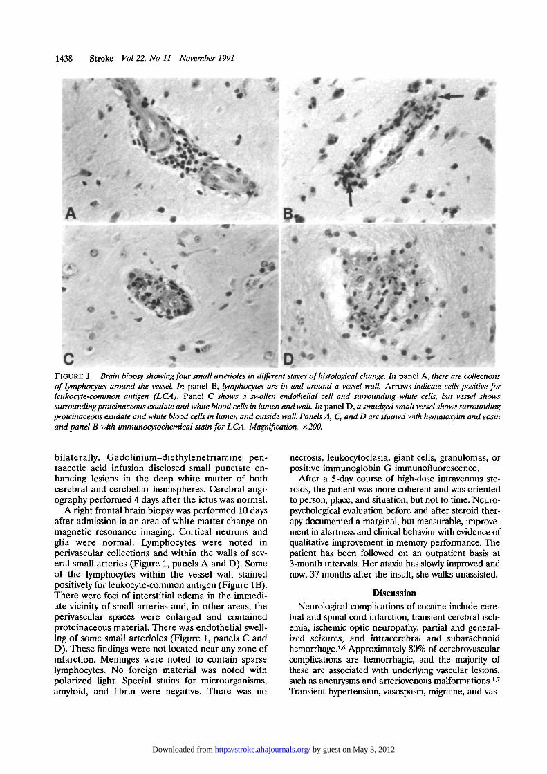
D . • . tFIGURE 1. Brain biopsy showing four small arterioles in different stages of histological change. In panel A, there are collectionsof lymphocytes around the vessel. In panel B, lymphocytes are in and around a vessel wall. Arrows indicate cells positive forleukocyte-common antigen (LCA). Panel C shows a swollen endothelial cell and surrounding white cells, but vessel showssurrounding proteinaceous exudate and white blood cells in lumen and wall. In panel D, a smudged small vessel shows surroundingproteinaceous exudate and white blood cells in lumen and outside wall. Panels A, C, and D are stained with hematoxylin and eosinand panel B with immunocytochemical stain for LCA. Magnification, x.200.
bilaterally. Gadolinium-diethylenetriamine pen-taacetic acid infusion disclosed small punctate en-hancing lesions in the deep white matter of bothcerebral and cerebellar hemispheres. Cerebral angi-ography performed 4 days after the ictus was normal.
A right frontal brain biopsy was performed 10 daysafter admission in an area of white matter change onmagnetic resonance imaging. Cortical neurons andglia were normal. Lymphocytes were noted inperivascular collections and within the walls of sev-eral small arteries (Figure 1, panels A and D). Someof the lymphocytes within the vessel wall stainedpositively for leukocyte-common antigen (Figure IB).There were foci of interstitial edema in the immedi-ate vicinity of small arteries and, in other areas, theperivascular spaces were enlarged and containedproteinaceous material. There was endothelial swell-ing of some small arterioles (Figure 1, panels C andD). These findings were not located near any zone ofinfarction. Meninges were noted to contain sparselymphocytes. No foreign material was noted withpolarized light. Special stains for microorganisms,amyloid, and fibrin were negative. There was no
necrosis, leukocytoclasia, giant cells, granulomas, orpositive immunoglobin G immunofluorescence.
After a 5-day course of high-dose intravenous ste-roids, the patient was more coherent and was orientedto person, place, and situation, but not to time. Neuro-psychological evaluation before and after steroid ther-apy documented a marginal, but measurable, improve-ment in alertness and clinical behavior with evidence ofqualitative improvement in memory performance. Thepatient has been followed on an outpatient basis at3-month intervals. Her ataxia has slowly improved andnow, 37 months after the insult, she walks unassisted.
DiscussionNeurological complications of cocaine include cere-
bral and spinal cord infarction, transient cerebral isch-emia, ischemic optic neuropathy, partial and general-ized seizures, and intracerebral and subarachnoidhemorrhage.1-6 Approximately 80% of cerebrovascularcomplications are hemorrhagic, and the majority ofthese are associated with underlying vascular lesions,such as aneurysms and arteriovenous malformations.17
Transient hypertension, vasospasm, migraine, and vas-
by guest on May 3, 2012http://stroke.ahajournals.org/Downloaded from

Fredericks et al Cocaine-Induced Vasculitis 1439
culitis have also been suggested as mechanisms ofcocaine-induced cerebrovascular disease.1-8
Drug-associated cerebral vasculitis has been bestdocumented with amphetamine abuse. A multiorgansystem necrotizing vasculitis resembling polyarteritisnodosa has been reported in patients using amphet-amine alone or with other drugs.9 Intravenousmethamphetamine has been shown to produce tran-sient decreased vessel diameter in rhesus monkeyswithin 1 hour of administration.10 Arteriographicchanges consistent with vasculitis have been reportedwith other recreational drugs, including phenylpro-panolamine,11-13 ephedrine,14 pseudoephedrine,15
and heroin.16
Previous reports of vasculitis associated with drugabuse have been flawed by a lack of pathologicalverification and the frequent history of multiple-druguse. Two previous cases of cerebral vasculitis as-cribed to cocaine abuse, one with intracerebral hem-orrhage1 and the other with ischemic infarction,2
were diagnosed exclusively on the basis of the non-specific angiographic finding of segmental constric-tion or beading. There are two reports of pathologi-cally proven cerebral vasculitis in patients withconcurrent abuse of intravenous cocaine, heroin, andamphetamines, in whom vasculitis was attributed tothe amphetamines.45 Only two published cases withbiopsies verified that small-vessel vasculitis existed inpatients with cocaine abuse who had no history ofconcomitant drug use.3
Our patient presented with multifocal ischemiashortly after using intravenous and inhaled cocaine.She had no history of concurrent use of heroin oramphetamines, nor were these agents present on adrug screen performed within 24 hours of the event.The pathological changes in the small arteries andarterioles in her brain biopsy are similar to thosepreviously reported by Citron et al9 and Kessler et al4
in patients with multidrug-induced vasculitis, byRumbaugh et al10 in experimental animals givenmethamphetamine, and by Krendel et al3 in patientswith cocaine abuse. The sparing of capillaries andveins, amorphous eosinophilic deposits, and intimalthickening seen in our patient have also been de-scribed previously in patients with drug abuse.45
We speculate that the vascular changes described inthe brain biopsy may be secondary to hypersensitivityangiitis or angiospasm. Despite the absence of fibrinoidnecrosis, leukocytoclastic infiltration, and immunofluo-rescent material, the morphological changes may re-flect the stage of the disease during which the biopsywas obtained. Hypersensitivity angiitis has been notedin endomyocardial biopsies from cocaine abusers.17 It isalso conceivable that these findings represent a re-sponse to vascular spasm. The literature on cocaine-induced myocardial ischemia and infarction empha-sizes the pharmacological effects of cocaine oncoronary artery smooth muscle.1819 Lange et al18 havedemonstrated a-adrenergic-mediated coronary arteryvasoconstriction in patients receiving pharmacologicaldoses of intranasal cocaine. However, no such evidence
exists to support the occurrence of cerebrovascularspasm due to cocaine. Intraluminal thrombosis hasbeen found in some cases with cocaine-related myocar-dial infarction.17 In vitro studies indicate that cocaineincreases thromboxane synthesis and platelet reactivityto arachidonic acid.1 The presence of lesions at variousstages makes direct toxicity a less plausible explanationfor these findings.
In conclusion, this case supports the existence of anonnecrotizing, nonleukocytoclastic small-vessel ar-teritis associated with lone cocaine use. One shouldconsider a diagnosis of cerebral vasculitis in thecocaine abuser with signs of focal or diffuse centralnervous system dysfunction. The incidence and etiol-ogy of this disorder remain speculative. The role ofsteroid therapy warrants further investigation.
References1. Klonoff DC, Andrews BT, Obana WG: Stroke associated with
cocaine use. Arch Neurol 1989;46:989-9932. Kaye BR, Fainstat M: Cerebral vasculitis associated with
cocaine abuse. JAMA 1987;258:2104-21063. Krendel DA, Ditter SM, Frankel MR, Ross WK: Biopsy-
proven cerebral vasculitis associated with cocaine abuse. Neu-rology 1990;40:1092-1094
4. Kessler JT, Jortner BS, Adapon BD: Cerebral vasculitis in adrug abuser. / Clin Psychol 1978;39:559-564
5. Bostwick DG: Amphetamine induced cerebral vasculitis. HumPathol 1981;12:1031-1033
6. Mody CK, Miller BL, Mclntyre HB, Cobb SK, Goldberg MA:Neurologic complications of cocaine abuse. Neurology 1988;38:1189-1193
7. Wojak JC, Flamm ES: Intracranial hemorrhage and cocaineuse. Stroke 1987;18:712-715
8. Levine SR, Washington JM, Jefferson MF, Kieran SN, MoenM, Feit H, Welch KMA: "Crack" cocaine-associated stroke.Neurology 1987;37:1849-1853
9. Citron BP, Halpern M, McCarron M, Lundberg GD, McCormickR, Pincus IJ, Tatter D, Haverback BJ: Necrotizing angiitisassociated with drug abuse. N EnglJ Med 1970;283:1003-1011
10. Rumbaugh CL, Bergeron RT, Scanlan RL, Teal JS, SegallHD, Fang HCH, McCormick R: Cerebral vascular changessecondary to amphetamine abuse in the experimental animal.Radiology 1971;101:345-351
11. Stoessl AJ, Young GB, Feasby TE: Intracerebral hemorrhageand angiographic beading following ingestion of catecholamin-ergics. Stroke 1985;16:734-736
12. Fallis RJ, Fisher M: Cerebral vasculitis and hemorrhage associ-ated with phenylpropanolamine. Neurology 1985;35:405-407
13. Kase CS, Foster TE, Reed JE, Spatz EL, Girgis GN: Intra-cerebral hemorrhage and phenylpropanolamine. Neurology1987;37:399-404
14. Wooten MR, Khangure MS, Murphy MJ: Intracerebral hem-orrhage and vasculitis related to ephedrine abuse. Ann Neurol1983; 13:337-340
15. Loizou LA, Hamilton JG, Tsementzis SA: Intracranial hem-orrhage in association with pseudoephedrine overdosage.J Neurol Neurosurg Psychiatry 1982;45:471-472
16. King J, Richards M, Tress B: Cerebral arteritis associated withheroin abuse. Med J Aust 1978;2:444-445
17. Levine SR, Welch KMA: Cocaine and stroke. Stroke 1988;19:779-783
18. Lange RA, Cigarroa RG, Yancy CW, Willard JE, Popma JJ, SillsMN, McBride W, Kim AS, Hillis LD: Cocaine-induced coronary-artery vasoconstriction. N Engl J Med 1989;321:1557-1562
19. Isner JM, Chokshi SK: Cocaine and vasospasm. N Engl J Med1989;321:1604-1606
KEY WORDS • cocaine • vasculitis
by guest on May 3, 2012http://stroke.ahajournals.org/Downloaded from