cerebrovascular diseases-aneurysm, vascular malformation
TRANSCRIPT

New 2021
ACR Appropriateness Criteria® 1 CVD-Aneurysm, Vascular Malformation, and SAH
American College of Radiology ACR Appropriateness Criteria®
Cerebrovascular Diseases-Aneurysm, Vascular Malformation, and Subarachnoid Hemorrhage
Variant 1: Known acute subarachnoid hemorrhage (SAH) on CT. Next imaging study.
Procedure Appropriateness Category Relative Radiation Level
Arteriography cervicocerebral Usually Appropriate ☢☢☢
CTA head with IV contrast Usually Appropriate ☢☢☢
MRA head without IV contrast May Be Appropriate O
US duplex Doppler carotid Usually Not Appropriate O
US duplex Doppler transcranial Usually Not Appropriate O
MRA head with IV contrast Usually Not Appropriate O
MRA head without and with IV contrast Usually Not Appropriate O
MRA neck with IV contrast Usually Not Appropriate O
MRA neck without and with IV contrast Usually Not Appropriate O
MRA neck without IV contrast Usually Not Appropriate O
MRI head perfusion with IV contrast Usually Not Appropriate O
MRI head with IV contrast Usually Not Appropriate O
MRI head without and with IV contrast Usually Not Appropriate O
MRI head without IV contrast Usually Not Appropriate O
MRV head with IV contrast Usually Not Appropriate O
MRV head without and with IV contrast Usually Not Appropriate O
MRV head without IV contrast Usually Not Appropriate O
CT head perfusion with IV contrast Usually Not Appropriate ☢☢☢
CT head with IV contrast Usually Not Appropriate ☢☢☢
CT head without and with IV contrast Usually Not Appropriate ☢☢☢
CT head without IV contrast Usually Not Appropriate ☢☢☢
CTA neck with IV contrast Usually Not Appropriate ☢☢☢
CTV head with IV contrast Usually Not Appropriate ☢☢☢

ACR Appropriateness Criteria® 2 CVD-Aneurysm, Vascular Malformation, and SAH
Variant 2: Suspected cerebral vasospasm. Initial imaging.
Procedure Appropriateness Category Relative Radiation Level
Arteriography cervicocerebral Usually Appropriate ☢☢☢
CTA head with IV contrast Usually Appropriate ☢☢☢
US duplex Doppler transcranial May Be Appropriate O
MRI head perfusion with IV contrast May Be Appropriate O
MRI head without IV contrast May Be Appropriate O
CT head perfusion with IV contrast May Be Appropriate ☢☢☢
CT head without IV contrast May Be Appropriate ☢☢☢
US duplex Doppler carotid Usually Not Appropriate O
MRA head with IV contrast Usually Not Appropriate O
MRA head without and with IV contrast Usually Not Appropriate O
MRA head without IV contrast Usually Not Appropriate O
MRA neck with IV contrast Usually Not Appropriate O
MRA neck without and with IV contrast Usually Not Appropriate O
MRA neck without IV contrast Usually Not Appropriate O
MRI head with IV contrast Usually Not Appropriate O
MRI head without and with IV contrast Usually Not Appropriate O
MRV head with IV contrast Usually Not Appropriate O
MRV head without and with IV contrast Usually Not Appropriate O
MRV head without IV contrast Usually Not Appropriate O
CT head with IV contrast Usually Not Appropriate ☢☢☢
CT head without and with IV contrast Usually Not Appropriate ☢☢☢
CTA neck with IV contrast Usually Not Appropriate ☢☢☢
CTV head with IV contrast Usually Not Appropriate ☢☢☢

ACR Appropriateness Criteria® 3 CVD-Aneurysm, Vascular Malformation, and SAH
Variant 3: Known cerebral aneurysm; untreated. Surveillance monitoring.
Procedure Appropriateness Category Relative Radiation Level
MRA head without IV contrast Usually Appropriate O
CTA head with IV contrast Usually Appropriate ☢☢☢
Arteriography cervicocerebral May Be Appropriate ☢☢☢
MRA head with IV contrast May Be Appropriate (Disagreement) O
MRA head without and with IV contrast May Be Appropriate O
US duplex Doppler carotid Usually Not Appropriate O
US duplex Doppler transcranial Usually Not Appropriate O
MRA neck with IV contrast Usually Not Appropriate O
MRA neck without and with IV contrast Usually Not Appropriate O
MRA neck without IV contrast Usually Not Appropriate O
MRI head perfusion with IV contrast Usually Not Appropriate O
MRI head with IV contrast Usually Not Appropriate O
MRI head without and with IV contrast Usually Not Appropriate O
MRI head without IV contrast Usually Not Appropriate O
MRV head with IV contrast Usually Not Appropriate O
MRV head without and with IV contrast Usually Not Appropriate O
MRV head without IV contrast Usually Not Appropriate O
CT head perfusion with IV contrast Usually Not Appropriate ☢☢☢
CT head with IV contrast Usually Not Appropriate ☢☢☢
CT head without and with IV contrast Usually Not Appropriate ☢☢☢
CT head without IV contrast Usually Not Appropriate ☢☢☢
CTA neck with IV contrast Usually Not Appropriate ☢☢☢
CTV head with IV contrast Usually Not Appropriate ☢☢☢

ACR Appropriateness Criteria® 4 CVD-Aneurysm, Vascular Malformation, and SAH
Variant 4: Known cerebral aneurysm; previously treated. Surveillance monitoring.
Procedure Appropriateness Category Relative Radiation Level
Arteriography cervicocerebral Usually Appropriate ☢☢☢
MRA head without and with IV contrast Usually Appropriate O
MRA head without IV contrast Usually Appropriate O
CTA head with IV contrast Usually Appropriate ☢☢☢
MRA head with IV contrast May Be Appropriate (Disagreement) O
US duplex Doppler carotid Usually Not Appropriate O
US duplex Doppler transcranial Usually Not Appropriate O
MRA neck with IV contrast Usually Not Appropriate O
MRA neck without and with IV contrast Usually Not Appropriate O
MRA neck without IV contrast Usually Not Appropriate O
MRI head perfusion with IV contrast Usually Not Appropriate O
MRI head with IV contrast Usually Not Appropriate O
MRI head without and with IV contrast Usually Not Appropriate O
MRI head without IV contrast Usually Not Appropriate O
MRV head with IV contrast Usually Not Appropriate O
MRV head without and with IV contrast Usually Not Appropriate O
MRV head without IV contrast Usually Not Appropriate O
CT head perfusion with IV contrast Usually Not Appropriate ☢☢☢
CT head with IV contrast Usually Not Appropriate ☢☢☢
CT head without and with IV contrast Usually Not Appropriate ☢☢☢
CT head without IV contrast Usually Not Appropriate ☢☢☢
CTA neck with IV contrast Usually Not Appropriate ☢☢☢
CTV head with IV contrast Usually Not Appropriate ☢☢☢

ACR Appropriateness Criteria® 5 CVD-Aneurysm, Vascular Malformation, and SAH
Variant 5: High-risk cerebral aneurysm screening.
Procedure Appropriateness Category Relative Radiation Level
MRA head without IV contrast Usually Appropriate O
CTA head with IV contrast Usually Appropriate ☢☢☢
US duplex Doppler carotid Usually Not Appropriate O
US duplex Doppler transcranial Usually Not Appropriate O
Arteriography cervicocerebral Usually Not Appropriate ☢☢☢
MRA head with IV contrast Usually Not Appropriate O
MRA head without and with IV contrast Usually Not Appropriate O
MRA neck with IV contrast Usually Not Appropriate O
MRA neck without and with IV contrast Usually Not Appropriate O
MRA neck without IV contrast Usually Not Appropriate O
MRI head perfusion with IV contrast Usually Not Appropriate O
MRI head with IV contrast Usually Not Appropriate O
MRI head without and with IV contrast Usually Not Appropriate O
MRI head without IV contrast Usually Not Appropriate O
MRV head with IV contrast Usually Not Appropriate O
MRV head without and with IV contrast Usually Not Appropriate O
MRV head without IV contrast Usually Not Appropriate O
CT head perfusion with IV contrast Usually Not Appropriate ☢☢☢
CT head with IV contrast Usually Not Appropriate ☢☢☢
CT head without and with IV contrast Usually Not Appropriate ☢☢☢
CT head without IV contrast Usually Not Appropriate ☢☢☢
CTA neck with IV contrast Usually Not Appropriate ☢☢☢
CTV head with IV contrast Usually Not Appropriate ☢☢☢
ACR Appropriateness Criteria® 6 CVD-Aneurysm, Vascular Malformation, and SAH
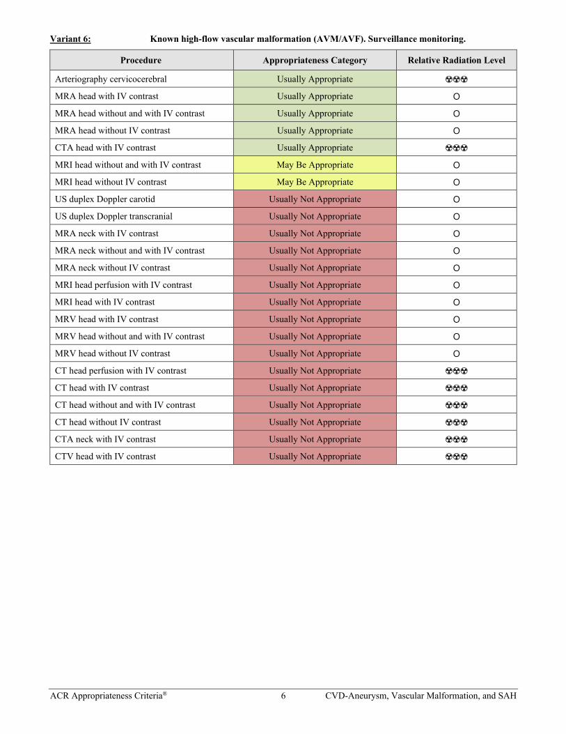
Variant 6: Known high-flow vascular malformation (AVM/AVF). Surveillance monitoring.
Procedure Appropriateness Category Relative Radiation Level
Arteriography cervicocerebral Usually Appropriate ☢☢☢
MRA head with IV contrast Usually Appropriate O
MRA head without and with IV contrast Usually Appropriate O
MRA head without IV contrast Usually Appropriate O
CTA head with IV contrast Usually Appropriate ☢☢☢
MRI head without and with IV contrast May Be Appropriate O
MRI head without IV contrast May Be Appropriate O
US duplex Doppler carotid Usually Not Appropriate O
US duplex Doppler transcranial Usually Not Appropriate O
MRA neck with IV contrast Usually Not Appropriate O
MRA neck without and with IV contrast Usually Not Appropriate O
MRA neck without IV contrast Usually Not Appropriate O
MRI head perfusion with IV contrast Usually Not Appropriate O
MRI head with IV contrast Usually Not Appropriate O
MRV head with IV contrast Usually Not Appropriate O
MRV head without and with IV contrast Usually Not Appropriate O
MRV head without IV contrast Usually Not Appropriate O
CT head perfusion with IV contrast Usually Not Appropriate ☢☢☢
CT head with IV contrast Usually Not Appropriate ☢☢☢
CT head without and with IV contrast Usually Not Appropriate ☢☢☢
CT head without IV contrast Usually Not Appropriate ☢☢☢
CTA neck with IV contrast Usually Not Appropriate ☢☢☢
CTV head with IV contrast Usually Not Appropriate ☢☢☢

ACR Appropriateness Criteria® 7 CVD-Aneurysm, Vascular Malformation, and SAH
Variant 7: Suspected central nervous system (CNS) vasculitis. Initial imaging.
Procedure Appropriateness Category Relative Radiation Level
MRA head without IV contrast Usually Appropriate O
MRI head without and with IV contrast Usually Appropriate O
MRI head without IV contrast Usually Appropriate O
Arteriography cervicocerebral May Be Appropriate ☢☢☢
CTA head with IV contrast May Be Appropriate ☢☢☢
US duplex Doppler carotid Usually Not Appropriate O
US duplex Doppler transcranial Usually Not Appropriate O
MRA head with IV contrast Usually Not Appropriate O
MRA head without and with IV contrast Usually Not Appropriate O
MRA neck with IV contrast Usually Not Appropriate O
MRA neck without and with IV contrast Usually Not Appropriate O
MRA neck without IV contrast Usually Not Appropriate O
MRI head perfusion with IV contrast Usually Not Appropriate O
MRI head with IV contrast Usually Not Appropriate O
MRV head with IV contrast Usually Not Appropriate O
MRV head without and with IV contrast Usually Not Appropriate O
MRV head without IV contrast Usually Not Appropriate O
CT head perfusion with IV contrast Usually Not Appropriate ☢☢☢
CT head with IV contrast Usually Not Appropriate ☢☢☢
CT head without and with IV contrast Usually Not Appropriate ☢☢☢
CT head without IV contrast Usually Not Appropriate ☢☢☢
CTA neck with IV contrast Usually Not Appropriate ☢☢☢
CTV head with IV contrast Usually Not Appropriate ☢☢☢

ACR Appropriateness Criteria® 8 CVD-Aneurysm, Vascular Malformation, and SAH
CEREBROVASCULAR DISEASES-ANEURYSM, VASCULAR MALFORMATION, AND SUBARACHNOID HEMORRHAGE
Expert Panel on Neurological Imaging: Luke N. Ledbetter, MDa; Judah Burns, MDb; Robert Y. Shih, MDc; Amna A. Ajam, MD, MBBSd; Michael D. Brown, MD, MSce; Santanu Chakraborty, MBBS, MScf; Melissa A. Davis, MD, MBAg; Andrew F. Ducruet, MDh; Christopher H. Hunt, MDi; Mary E. Lacy, MDj; Ryan K. Lee, MD, MBAk; Jeffrey S. Pannell, MDl; Jeffrey M. Pollock, MDm; William J. Powers, MDn; Gavin Setzen, MDo; Matthew D. Shaines, MDp; Pallavi S. Utukuri, MDq; Lily L. Wang, MBBS, MPHr; Amanda S. Corey, MD.s
Summary of Literature Review
Introduction/Background Cerebrovascular diseases encompass broad and varied clinical presentations and disease processes. This topic will focus on clinical presentations based on aneurysms, vascular malformations, subarachnoid hemorrhage (SAH), and related cerebrovascular abnormalities, such as vasospasm and central nervous system (CNS) vasculitis. For discussion regarding the presentation of SAH and appropriate imaging, please see the ACR Appropriateness Criteria® topic on “Headache” [1]. For potential SAH in the setting of head trauma, please see the ACR Appropriateness Criteria® topic on “Head Trauma” [2]. The subset of cerebrovascular diseases and presentations are also broad and varied; therefore, the introduction and background of each variant will be discussed individually.
For discussion of cerebrovascular diseases related to stroke, stroke-related conditions, or intraparenchymal hemorrhage, please see the ACR Appropriateness Criteria® topic on “Cerebrovascular Disease-Stroke and Stroke-Related Conditions” that will be made available on the ACR website when completed.
Special Imaging Considerations For the purposes of distinguishing between CT and CT angiography (CTA), ACR Appropriateness Criteria topics use the definition in the ACR–NASCI–SIR–SPR Practice Parameter for the Performance and Interpretation of Body Computed Tomography Angiography (CTA) [3]:
“CTA uses a thin-section CT acquisition that is timed to coincide with peak arterial or venous enhancement. The resultant volumetric dataset is interpreted using primary transverse reconstructions as well as multiplanar reformations and 3-D renderings.”
All elements are essential: 1) timing, 2) reconstructions/reformats, and 3) 3-D renderings. Standard CTs with contrast also include timing issues and reconstructions/reformats. Only in CTA, however, is 3-D rendering a required element. This corresponds to the definitions that the CMS has applied to the Current Procedural Terminology codes.
Initial Imaging Definition Initial imaging is defined as imaging at the beginning of the care episode for the medical condition defined by the variant. More than one procedure can be considered usually appropriate in the initial imaging evaluation when:
• There are procedures that are equivalent alternatives (ie, only one procedure will be ordered to provide the clinical information to effectively manage the patient’s care)
OR
aUniversity of California Los Angeles, Los Angeles, California. bPanel Chair, Montefiore Medical Center, Bronx, New York. cPanel Vice-Chair, Uniformed Services University, Bethesda, Maryland. dOhio State University, Columbus, Ohio. eMichigan State University, East Lansing, Michigan; American College of Emergency Physicians. fOttawa Hospital Research Institute and the Department of Radiology, The University of Ottawa, Ottawa, Ontario, Canada; Canadian Association of Radiologists. gEmory University, Atlanta, Georgia. hBarrow Neurological Institute, Phoenix, Arizona; Neurosurgery expert. iMayo Clinic, Rochester, Minnesota. jUniversity of New Mexico, Albuquerque, New Mexico; American College of Physicians. kEinstein Healthcare Network, Philadelphia, Pennsylvania. lUniversity of California San Diego Medical Center, San Diego, California. mOregon Health & Science University, Portland, Oregon. nUniversity of North Carolina School of Medicine, Chapel Hill, North Carolina; American Academy of Neurology. oAlbany ENT & Allergy Services, PC, Albany, New York; American Academy of Otolaryngology-Head and Neck Surgery. pAlbert Einstein College of Medicine Montefiore Medical Center, Bronx, New York, Internal medicine physician. qColumbia University Medical Center, New York, New York. rUniversity of Cincinnati Medical Center, Cincinnati, Ohio. sSpecialty Chair, Atlanta VA Health Care System and Emory University, Atlanta, Georgia. The American College of Radiology seeks and encourages collaboration with other organizations on the development of the ACR Appropriateness Criteria through representation of such organizations on expert panels. Participation on the expert panel does not necessarily imply endorsement of the final document by individual contributors or their respective organization. Reprint requests to: [email protected]
ACR Appropriateness Criteria® 9 CVD-Aneurysm, Vascular Malformation, and SAH
• There are complementary procedures (ie, more than one procedure is ordered as a set or simultaneously where each procedure provides unique clinical information to effectively manage the patient’s care).
Discussion of Procedures by Variant Variant 1: Known acute subarachnoid hemorrhage (SAH) on CT. Next imaging study. Recommendations for imaging in the setting of suspected SAH with the clinical presentation of sudden, severe headache, or “worst headache of life” are guided by the ACR Appropriateness Criteria® topic on “Headache” [1].
This variant will focus on imaging examinations used to determine the source of SAH after initial detection. SAH, involving the basal cisterns, requires rapid triage and workup because a ruptured cerebral aneurysm is responsible for 70% of all nontraumatic SAHs [4,5]. The overall incidence of aneurysmal SAH in the United States is between 9.7 and 14.5 cases per 100,000 population and may be underestimated due to the high risk of death prior to hospital admission [6-9]. Aneurysmal SAH results in significant morbidity and mortality with a quarter of aneurysmal subarachnoid patients dying after presentation; therefore, early diagnosis and repair is crucial to prevent rebleeding [6]. Less common causes of SAH, often presenting as isolated convexity SAH, such as tumors, stroke transformation, cerebral amyloid angiopathy, or reversible cerebral vasoconstriction syndrome, are not considered here as their imaging and diagnosis often follows the initial, commonly emergent, imaging revaluation for common vascular lesions. Follow-up imaging for delayed complications of SAH, such as hydrocephalus, should be directed by local protocols and clinical symptoms. The delayed complication of vasospasm after SAH is discussed in Variant 2 of this topic.
Arteriography Cervicocerebral Catheter-directed angiography of the cerebral vasculature demonstrates high spatial resolution, large field of view, and dynamic acquisition that leads to high diagnostic value in the evaluation of cerebrovascular diseases resulting in SAH. Sensitivity and specificity are both >98% for catheter cerebral angiography when compared with surgical findings, including small aneurysms <3 mm [10]. Catheter cerebral angiography also identified vascular abnormalities in up to 13% of patients with SAH and negative CTA imaging [11]. Although catheter cerebral angiography has been reported to be negative in 2% to 24% of patients with aneurysmal SAH, 3-D rotational angiography has been shown to identify an aneurysm on 25% of previously angiogram, both 2-D and 3-D, negative patients [12]. Angiography is an invasive procedure with a small complication risk related to intravascular instrumentation.
CT Head Perfusion There is no relevant literature to support the use of CT head perfusion in the evaluation of known acute SAH.
CT Head There is no relevant literature to support the use of CT head in the evaluation of known (previously diagnosed by imaging or lumbar puncture) acute SAH. Recommendations for imaging in the setting of suspected SAH with the clinical presentation of sudden, severe headache or “worst headache of life” are guided by the ACR Appropriateness Criteria® topic on “Headache” [1].
CTA Head CTA head is a fast, noninvasive study to evaluate patients with acute SAH. CTA head has been shown to have >90% sensitivity and specificity in the evaluation for aneurysms [4,10,13-18] responsible for SAHs. However, CTA head sensitivity for detecting aneurysm decreases for aneurysms <3 mm in size [4,10,13,15,17,19], in the setting of diffuse SAH [20], and for aneurysms occurring adjacent to an osseous structure [19]. CTA head may be sufficient to rule out a vascular cause of SAH when the location of hemorrhage is isolated to the perimesencephalic region with follow-up catheter-directed angiography indicated in CTA negative diffuse or peripheral SAHs [20].
CTA Neck There is no relevant literature to support the use of CTA neck in the initial imaging evaluation of known acute SAH. CTA neck may be useful for potential treatment planning, but preference will be individual or site specific.
CTV Head There is no relevant literature to support the use of CT venography (CTV) head in the evaluation of known acute SAH.
ACR Appropriateness Criteria® 10 CVD-Aneurysm, Vascular Malformation, and SAH
MRA Head MR angiography (MRA) head for the evaluation of intracranial aneurysm demonstrated a pooled sensitivity of 95% and specificity of 89% in one meta-analysis [21]. Diagnostic accuracy is increased, including for aneurysms >5 mm in size and at 3T scanner strength [21,22]. The decrease in specificity, when compared with CTA, is reported to have false-positive cases related to normal vascular variants of infundibular origin of vessels and vessel loops [23]. Limitations of MRA head include required safety screening and relatively long acquisition time in urgent clinical scenarios.
MRA Neck There is no relevant literature to support the use of MRA neck in the evaluation of known acute SAH. MRA neck may be useful for potential treatment planning, but preference will be individual or site specific.
MRI Head Perfusion There is no relevant literature to support the use of MRI head perfusion in the evaluation of known acute SAH.
MRI Head Although there is no relevant literature to support the use of MRI head in the evaluation for a vascular source of known acute SAH, several studies evaluated the use of MRI head in predicting clinical outcomes. Patients with acute poor-grade SAH and diffusion-weighted imaging positive findings on MRI head had a less favorable long-term outcome when compared with patients without diffusion-weighted imaging positive findings [24,25].
MRV Head There is no relevant literature to support the use of MR venography (MRV) head in the evaluation of known acute SAH.
US Duplex Doppler Carotid There is no relevant literature to support the use of carotid ultrasound (US) duplex Doppler in the evaluation of known acute SAH.
US Duplex Doppler Transcranial There is no relevant literature to support the use of US transcranial with duplex Doppler (TCD) in the evaluation of known acute SAH.
Variant 2: Suspected cerebral vasospasm. Initial imaging. Vasospasm in the cerebral arteries occurs in approximately 30% of patients with SAH and frequently occurs 7 to 10 days after hemorrhage with spontaneous resolution by day 21 [6]. Vasospasm is associated with delayed cerebral ischemia defined by delayed development of neurologic deficits after SAH not related to aneurysm treatment or other neurologic complications, such as hydrocephalus, cerebral edema, or metabolic derangements [26]. Morbidity and mortality in SAH increases between 10% and 20% after onset of clinical symptoms of delayed cerebral ischemia [27], and the symptoms are frequently nonreversible [28,29]. Imaging findings of vasospasm and guidance of treatment does not appear to improve clinical outcome after the onset of clinical symptoms [28].
Despite the association of moderate to severe vasospasm and poor clinical outcome [30], only 50% patients with large-vessel vasospasm develop clinical ischemic neurologic symptoms [6], and delayed ischemia can occur in the absence of imaging findings of vasospasm [26]. However, given the clinical implications of delayed cerebral ischemia, early screening and detection of vasospasm remains recommended [6].
Arteriography Cervicocerebral Conventional catheter-directed cerebrovascular arteriography is the reference standard for characterization of intracranial vasospasm. However, only approximately 50% of radiographic large-vessel vasospasm develops delayed cerebral ischemia, and given the invasive nature and potential rare neurologic complications, other less invasive screening methods are often performed before catheter angiogram [28]. In a large, international multicenter randomized trial, the presence of angiographic vasospasm was strongly associated (odds ratio of 9.3) with the development of cerebral infarction. In the same study, a small number of patients (3%) developed infarction without evidence of vasospasm on angiogram [31]. An additional consideration to the angiographic evaluation of vasospasm is the potential for intra-arterial treatment of vessel narrowing. However, intra-arterial treatment of vasospasm lacks high-quality evidence of improvement of outcomes at this time [6].

ACR Appropriateness Criteria® 11 CVD-Aneurysm, Vascular Malformation, and SAH
CT Head Perfusion CT head perfusion is a useful tool in the evaluation of vasospasm. CT perfusion data provide information regarding the intraparenchymal small-vessel perfusion as opposed to CTA and TCD evaluation of large- and medium-sized vessels. CT perfusion has been studied in 3 separate clinical scenarios: early (0–3 days after SAH) prediction of future of delayed cerebral ischemia, late detection of vasospasm, and late detection of ischemic injury. Early use of CT head perfusion within the first 3 days after SAH with qualitative perfusion abnormalities were associated with later development of vasospasm [26]. A more recent retrospective study of CT perfusion within 24 hours of aneurysmal SAH and demonstrating perfusion abnormalities did not correlate with the development of delayed cerebral ischemia [32]. For the later use of CT perfusion after SAH, a meta-analysis demonstrated a sensitivity of 74% and specificity of 93% in detecting vasospasm [33], and retrospective studies showed sensitivities of 84% to 93% and specificities of 57% to 73% in detected delayed cerebral ischemia [29,34]. However, using CT head perfusion to guide treatment decision in the setting of neurologic symptoms of delayed cerebral ischemia did not improve outcomes when compared with treating all patients without imaging guidance [28].
CT Head SAH on CT head can be graded by the Fisher or modified Fisher scale. The higher the Fisher grade of SAH, the higher the patient risk for vasospasm [35]. Although CT head may be useful to provide a Fisher grade and risk for vasospasm, the examination does not directly give information regarding the presence or absence of vasospasm. Anatomic changes of completed infarct related to delayed cerebral ischemia can also be identified on CT head.
CTA Head CTA head can provide a less invasive evaluation of the intracranial cerebral vasculature compared with catheter-directed angiography. In a meta-analysis, CTA head detected vasospasm with a sensitivity and specificity of 80% and 93%, respectively [33]. CTA head is highly correlated to conventional angiography for larger proximal intracranial vessels with decreasing correlation in the smaller more distal arteries [36].
CTA Neck There is no relevant literature to support the use of CTA neck in the evaluation of suspected cerebral vasospasm.
CTV Head There is no relevant literature to support the use of CTV head in the evaluation of suspected cerebral vasospasm.
MRA Head There is no relevant literature to support the use of MRA head in the evaluation of suspected cerebral vasospasm.
MRA head evaluation of the intracranial arteries in the setting of suspected vasospasm is limited by background of hemorrhage and hemodynamic flow alterations with poor correlation to digital subtraction angiography (DSA) findings [37].
MRA Neck There is no relevant literature to support the use of MRA neck in the evaluation of suspected cerebral vasospasm.
MRI Head Perfusion Given the continued difficulty in identifying patients at risk for and in preventing vasospasm and delayed cerebral ischemia, advanced MRI head perfusion studies are now being performed. MRI head perfusion with decreased intravoxel incoherent motion microvascular perfusion has been associated with vasospasm [38], and elevated blood-brain barrier permeability (Ktrans) was associated with patients who went on to develop delayed cerebral ischemia [39]. Despite these early positive studies, no large or prospective studies have been performed to support the widespread use of MRI head perfusion in the evaluation of suspected vasospasm.
MRI Head MRI head offers evaluation of consequences of delayed cerebral ischemia including completed infarction. However, there is no relevant literature to support the use of MRI head in the evaluation of suspected cerebral vasospasm.
MRV Head There is no relevant literature to support the use of MRV head in the evaluation of suspected cerebral vasospasm.
US Duplex Doppler Carotid There is no relevant literature to support the use of carotid US duplex Doppler in the evaluation of suspected cerebral vasospasm.

ACR Appropriateness Criteria® 12 CVD-Aneurysm, Vascular Malformation, and SAH
US Duplex Doppler Transcranial TCD is a quick and noninvasive modality to evaluate for increased arterial velocities in the setting of vasospasm. Given the ability to perform the examination at the bedside, daily TCD is frequently used in the screening for vasospasm in at-risk populations. Vasospasm identified on TCD predicts delayed cerebral ischemia with 90% sensitivity, 92% negative predictive value, 71% specificity, and 57% positive predictive value [40]. Although screening for vasospasm with TCD has high sensitivity and negative predictive value, prolonged TCD screening past day 10 post-SAH does not appear to increase detection of delayed cerebral ischemia [41]. In addition, there is no current high-quality literature relating detection of vasospasm on TCD to improved patient outcomes [40].
Variant 3: Known cerebral aneurysm; untreated. Surveillance monitoring. Cerebral aneurysms are often incidentally discovered on intracranial vascular imaging. Definitive algorithmic guidelines for management and follow-up of incidentally found cerebral aneurysms are lacking [42]. Between 4% and 18% of aneurysms demonstrate growth on imaging follow-up [43,44], with a 12-fold higher risk of rupture in growing aneurysms [44]. Although aneurysm growth is associated with size >7 mm, smaller aneurysms can grow and rupture [44]. Given the evidence of potential for growth and rupture of untreated and unruptured aneurysms, vascular imaging surveillance is recommended.
Arteriography Cervicocerebral Cervicocerebral arteriography remains the reference standard imaging examination for the evaluation of cerebral aneurysms with high spatial resolution, high signal-to-noise ratio, and dynamic image acquisition. However, given the invasive nature and potential complications of cervicocerebral arteriography, it is not ideal for routine patient surveillance.
CT Head Perfusion There is no relevant literature to support the use of CT head perfusion in the surveillance of a known, untreated cerebral aneurysm.
CT Head There is no relevant literature to support the use of CT head in the surveillance of a known, untreated cerebral aneurysm.
CTA Head CTA head is a fast and noninvasive study to evaluate the intracranial vasculature. CTA head has been shown to be >90% sensitive and specific in the evaluation for aneurysms [4,10,13-18]. However, CTA head sensitivity for detecting an aneurysm decreases for aneurysms <3 mm in size [4,10,13,15,17,19] and for aneurysms occurring adjacent to an osseous structure [19].
CTA Neck There is no relevant literature to support the use of CTA neck in the surveillance of a known, untreated cerebral aneurysm.
CTV Head There is no relevant literature to support the use of CTV head in the surveillance of a known, untreated cerebral aneurysm.
MRA Head MRA head is an ideal candidate for imaging surveillance of known, untreated aneurysms because of its noninvasive nature and ability to obtain diagnostic information without intravenous (IV) contrast. The evaluation of intracranial aneurysm with MRA head demonstrated a pooled sensitivity of 95% and specificity of 89% in one meta-analysis [21]. Diagnostic accuracy is increased, including for aneurysms <5 mm in size, at 3T scanner strength [21,22]. Vessel loops and infundibular origins of vessels can lead to false-positives for aneurysm on MRA [23]. Contrast-enhanced MRA head may increase visualized detail of large aneurysms with complex flow dynamics or thrombosis [45]. However, there is no significant difference in diagnostic performance between time-of-flight MRA and contrast-enhanced MRA on the aforementioned meta-analysis of MRA examinations in the diagnosis of aneurysms [21].
MRA Neck There is no relevant literature to support the use of MRA neck in the surveillance of a known, untreated cerebral aneurysm.

ACR Appropriateness Criteria® 13 CVD-Aneurysm, Vascular Malformation, and SAH
MRI Head Perfusion There is no relevant literature to support the use of MRI head perfusion in the surveillance of a known, untreated cerebral aneurysm.
MRI Head There is no relevant literature to support the use of MRI head in the surveillance of a known, untreated cerebral aneurysm.
MRV Head There is no relevant literature to support the use of MRV head in the surveillance of a known, untreated cerebral aneurysm.
US Duplex Doppler Carotid There is no relevant literature to support the use of carotid US duplex Doppler in the surveillance of a known, untreated cerebral aneurysm.
US Duplex Doppler Transcranial There is no relevant literature to support the use of TCD in the surveillance of a known, untreated cerebral aneurysm.
Variant 4: Known cerebral aneurysm; previously treated. Surveillance monitoring. Treatment of cerebral aneurysms is common to reduce the risk of aneurysm rupture or rebleeding. Endovascular treatment is now the first-line therapy in most cases, whereas aneurysms not amenable to endovascular repair require surgical clipping or observation. Follow-up imaging after treatment is often performed to assess for potential refilling of aneurysms and detect formation of new aneurysms. Aneurysm remnants after surgical clipping are identified in up to 11% of patients [46] and more frequently after endovascular repair [47,48]. Recurrence of treated aneurysm is most common within 6 months of treatment but can occur in a more delayed manner [49]. Development of de novo aneurysm occurs in 1% to 8% of patients with treated aneurysms [50-52].
Imaging evaluation is focused on not only the treated aneurysm but also the integrity of the parent vessel and formation of new aneurysms. Intracranial aneurysms are treated with several different devices, including surgical clips, detachable coils, stents, and flow diverters, and each device will result in unique appearances as well as challenges, depending on the imaging modality used. Specific knowledge of the technique utilized in prior treatment is helpful in choosing a particular follow-up modality for each patient.
Arteriography Cervicocerebral Cervicocerebral arteriography remains the reference standard imaging examination for the evaluation of treated cerebral aneurysms with high spatial resolution, high signal-to-noise ratio, and dynamic image acquisition. Aneurysm and parent vessel appearance is better visualized, as indwelling occlusion device artifacts are less apparent on cervicocerebral arteriography than on MRI or CT. Drawbacks for surveillance include invasiveness and small risk of vascular complication.
CT Head Perfusion There is no relevant literature to support the use of CT head perfusion in the surveillance of known, treated cerebral aneurysm.
CT Head There is no relevant literature to support the use of CT head in the surveillance of known, treated cerebral aneurysm.
CTA Head CTA head is useful for surveillance imaging of treated cerebral aneurysms because of its noninvasive nature. However, CTA is limited by large metallic streak artifacts encountered with metallic coils, stents, and devices. Although artifact from metal cannot be removed, several metal artifact reduction techniques are available to improve evaluation of treated aneurysms and the parent vessels [53-57].
CTA Neck There is no relevant literature to support the use of CTA neck in the surveillance of known, treated cerebral aneurysm.
CTV Head There is no relevant literature to support the use of CTV head in the surveillance of known, treated cerebral aneurysm.

ACR Appropriateness Criteria® 14 CVD-Aneurysm, Vascular Malformation, and SAH
MRA Head MRA head is a noninvasive examination commonly used for treated aneurysm surveillance. This examination can be obtained without IV contrast using time-of-flight imaging, with IV contrast to improve flow-related artifacts occasionally encountered in aneurysms, or a combination of both. In the setting of coiled aneurysms, a meta-analysis found similar performance of both noncontrast and contrast-enhanced examinations with sensitivities of 86% for both time-of-flight and contrast-enhanced MRA, as well as specificities of 84% and 89%, respectively [58]. MRA head was also compared directly with catheter-directed angiography and found to result in substantial agreement (kappa 0.73) regarding treatment recommendations between the 2 examinations [59]. Treatment with stents or flow diverters results in challenges in MRA intraluminal evaluation of the stent. Contrast-enhanced MRA outperforms time-of-flight MRA in the evaluation of a treated aneurysm and parent vessel patency with indwelling stent; however, intraluminal detail is limited with both techniques [60]. Newer endovascular devices demonstrate magnetic susceptibility and Faraday cage effects, which limits MRA head utility in assessing for aneurysm thrombosis or parent vessel patency when compared with conventional arteriography [61-63].
MRA Neck There is no relevant literature to support the use of MRA neck in the surveillance of known, treated cerebral aneurysm.
MRI Head Perfusion There is no relevant literature to support the use of MRI head perfusion in the surveillance of known, treated cerebral aneurysm.
MRI Head There is no relevant literature to support the use of MRI head in the surveillance of known, treated cerebral aneurysm.
MRV Head There is no relevant literature to support the use of MRV head in the surveillance of known, treated cerebral aneurysm.
US Duplex Doppler Carotid There is no relevant literature to support the use of carotid US duplex Doppler in the surveillance of known, treated cerebral aneurysm.
US Duplex Doppler Transcranial There is no relevant literature to support the use of TCD in the surveillance of known, treated cerebral aneurysm.
Variant 5: High-risk cerebral aneurysm screening. Certain populations are at high risk of developing a cerebral aneurysm. Given the high morbidity and mortality associated with aneurysm rupture, screening high-risk patients may be beneficial. The incidence of aneurysm in the general population is near 1.8% [64]. The most studied high-risk population is patients with autosomal dominant polycystic kidney disease (ADPKD). Patients with ADPKD have an increased prevalence of aneurysms between 10% and 11.5% [65], with up to 21% of patients with ADPKD and a first-degree relative with history of aneurysm [66]. Aneurysmal SAH occurs at a younger age, and risk of de novo aneurysm formation is higher in ADPKD patients when compared with the general population [67]. Given the relationship of ADPKD and cerebral aneurysms, screening has been shown to be cost effective in several studies [65,68]. The American Heart Association guidelines also recommend offering screening to patients with ≥2 family members with intracranial aneurysms or SAH. A higher risk of aneurysm occurrence in such families is found in those with a history of hypertension, smoking, and female sex [42]. Other conditions with increased risk of cerebral aneurysm are moyamoya [69], aortic dissection [70], bicuspid aortic valve [71], aortic aneurysm [72], and coarctation of the aorta [73]. Arteriography Cervicocerebral Although cerebral arteriography is the reference standard for known or suspected aneurysm, the invasive nature and potential complications are not suited for screening in a high-risk population. There is no relevant literature to support the use of cerebral arteriography in this population.
CT Head Perfusion There is no relevant literature to support the use of CT head perfusion in the screening of patients at high risk for cerebral aneurysm.

ACR Appropriateness Criteria® 15 CVD-Aneurysm, Vascular Malformation, and SAH
CT Head There is no relevant literature to support the use of CT head in the screening of patients at high risk for cerebral aneurysm.
CTA Head CTA head is a fast, noninvasive study to evaluate the intracranial vasculature. CTA head has been shown to be >90% sensitive and specific in the evaluation for aneurysms [4,10,13-18]. However, CTA head sensitivity for detecting an aneurysm decreases for both aneurysms <3 mm in size [4,10,13,15,17,19] and aneurysms occurring adjacent to an osseous structure [19].
CTA Neck There is no relevant literature to support the use of CTA neck in the screening of patients at high risk for cerebral aneurysm.
CTV Head There is no relevant literature to support the use of CTV head in the screening of patients at high risk for cerebral aneurysm.
MRA Head MRA head is an ideal candidate for screening high-risk populations for cerebral aneurysm due to its noninvasive nature and ability to obtain diagnostic information without IV contrast. The evaluation of intracranial aneurysm with MRA head demonstrated a pooled sensitivity of 95% and specificity of 89% in one meta-analysis, in which 45% of the 67 missed aneurysms were <3 mm in size, and another 45% were between 3 and 5 mm in size, 6% were between 5 and 10 mm in size, and 4% >10 mm in size [21]. Diagnostic accuracy is increased, including for aneurysms <5 mm in size, at 3T scanner strength [21,22]. Vessel loops and infundibular origins of vessels can lead to false-positives for aneurysm on MRA [23]. Contrast-enhanced MRA head has no relevant literature to support its use in the screening of patients at high risk for cerebral aneurysm.
MRA Neck There is no relevant literature to support the use of MRA neck in the screening of patients at high risk for cerebral aneurysm.
MRI Head Perfusion There is no relevant literature to support the use of MRI head perfusion in the screening of patients at high risk for cerebral aneurysm.
MRI Head There is no relevant literature to support the use of MRI head in the screening of patients at high risk for cerebral aneurysm.
MRV Head There is no relevant literature to support the use of MRV head in the screening of patients at high risk for cerebral aneurysm.
US Duplex Doppler Carotid There is no relevant literature to support the use of carotid US duplex Doppler in the screening of patients at high risk for cerebral aneurysm.
US Duplex Doppler Transcranial There is no relevant literature to support the use of TCD in the screening of patients at high risk for cerebral aneurysm.
Variant 6: Known high-flow vascular malformation (AVM/AVF). Surveillance monitoring. Intracranial high-flow vascular malformations include arteriovenous malformations (AVMs) and arteriovenous fistulas (AVFs). Both lesions are defined by an abnormal connection between the relatively high-pressure arterial system and the low-pressure venous system resulting in a high-flow shunting of blood.
AVMs are direct connections of artery to vein via abnormal dilated vascular channels without normal intermediary capillary bed. The abnormal dilated vascular channels are known as the nidus [74]. Although the true incidence of brain AVM is unknown, asymptomatic prevalence on MRI is estimated at 0.05% [74,75]. Between 10% and 20% of patients with hereditary hemorrhagic telangiectasia will have at least one AVM during their lifetime [74,76].

ACR Appropriateness Criteria® 16 CVD-Aneurysm, Vascular Malformation, and SAH
Symptomatic brain AVMs present most commonly with hemorrhage or epilepsy [74]. The annual rupture risk of a brain AVM is 1.3% for previously unruptured AVM and up to 4.8% for previously ruptured lesions [74,77]. Imaging findings associated with higher hemorrhage risk include intranidal aneurysm, deep venous drainage, deep location, or venous outflow obstruction [74,78]. Treatment for AVMs include surgical resection, endovascular embolization, stereotactic radiosurgery, or medical management. The ARUBA (A Randomised trial of Unruptured Brain Arteriovenous Malformations) trial concluded medical management alone was superior to medical management with interventional therapy for the prevention of death or stroke in patients with unruptured brain AVMs [79]. However, the trial did not establish the benefit of interventional treatment of unruptured AVMs, which remains a debated issue. The optimal methods for surveillance of untreated AVMs is not well established in the literature. However, treated lesions usually require long-term follow-up, specifically lesions treated with radiosurgery or embolization.
Intracranial dural AVF (dAVF) is an abnormal shunt between a dural artery and venous sinus or cortical vein. dAVFs demonstrate similar high-flow vascular shunting but lack the central nidus associated with AVM. Signs and symptoms of dAVF depend on the location, with posterior dural venous sinus lesion frequently presenting with pulsatile tinnitus or cavernous sinus lesions presenting with pain, proptosis, chemosis, and ophthalmoplegia [80]. Complications of high-grade dAVF include hemorrhage or nonhemorrhagic neurologic defects and are associated with retrograde cortical venous drainage [80-83]. Treatment via endovascular or microsurgical approach is usually indicated in high-grade dAVF with cortical venous drainage or symptomatic lesions. Observation can be utilized in lower-grade lesions with less risk of hemorrhagic or neurologic complications [80,83]. This variant covers the surveillance of both treated and untreated high-flow vascular malformations.
Arteriography Cervicocerebral Cervicocerebral angiography remains the reference standard for imaging of cerebrovascular disease, including AVM and dAVF. Angiography demonstrates high spatial and temporal resolution of critical importance in the characterization of the intranidal aneurysm in AVM as well as potentially small arterial feeding vessels and venous drainage characteristics in both AVM and dAVF. Arteriography is critical in planning treatment in all high-flow intracranial vascular malformations. Specifically, an arteriogram of an AVM provides high-resolution imaging of the nidus; however, 2-D angiographic images may overestimate lesion volumes when compared with MRA or CTA [84]. The addition of 3-D rotational cerebral arteriography results in more precise AVM nidus volume measurement when compared with CT and MRI [85].
CT Head Perfusion There is no relevant literature to support the use of CT head perfusion in the surveillance of high-flow intracranial vascular malformations.
CT Head Although larger AVMs can be visualized on CT head because of hyperattenuating prominent vascular structures [83] and the osseous landmarks can be useful in radiation therapy treatment planning [85], there is no relevant literature to support the use of CT head in the surveillance of high-flow intracranial vascular malformations.
CTA Head Sensitivity of CTA head was shown to be 90% for the overall detection of AVMs, 100% for AVMs >3 cm, and 88% for associated flow-related aneurysms when compared with DSA [86]. For high-flow AVFs, CTA head demonstrated a sensitivity of 86% and specificity of 100% in patients with pulsatile tinnitus [87]. Indirect signs of cortical venous drainage, indicating higher-risk lesion for future complication, on CTA exhibited sensitivities between 96% for cortical venous dilatation and 62% for identification of a medullary or pial vein. Drawbacks to CTA for surveillance monitoring include the lack of temporal resolution to directly determine flow dynamics of complex vascular lesions.
CTA Neck There is no relevant literature to support the use of CTA neck in the surveillance of high-flow intracranial vascular malformations. CTA neck may be useful for potential treatment planning, but preference will be individual or site specific.
CTV Head There is no relevant literature to support the use of CTV head in the surveillance of high-flow intracranial vascular malformations.
ACR Appropriateness Criteria® 17 CVD-Aneurysm, Vascular Malformation, and SAH
MRA Head MRA head is frequently used in surveillance of known high-flow vascular malformations. In the setting of AVM, time-of-flight and contrast-enhanced MRA offer good diagnostic accuracy but lack temporal resolution for hemodynamics and information regarding the small angioarchitecture [88]. Multiple 4-D MRA techniques are available to provide temporal resolution with trade-off in spatial resolution. Although 4-D MRA demonstrates good agreement with DSA [89-91], MRA has limited sensitivity for small nidus (<1 cm) or complete resolution after treatment [88,92].
For AVF, time-of-flight MRA demonstrates excellent intermodality agreement with DSA regarding the location of the fistula site and good agreement regarding the arterial feeding vessels and venous drainage [93]. Time-of-flight and contrast-enhanced MRA demonstrated slightly lower negative predictive values in the evaluation of signs of cortical venous reflux when compared with CTA [94]. There is good to excellent correlation of 4-D MRA techniques to DSA demonstrated in multiple studies [95-97].
MRA Neck There is no relevant literature to support the use of MRA neck in the surveillance of high-flow intracranial vascular malformations. MRA neck may be useful for potential treatment planning, but preference will be individual or site specific.
MRI Head Perfusion MRI head perfusion demonstrates variable perfusion abnormalities in the evaluation of hemodynamic physiology of AVM [83]. Perfusion examinations, including arterial spin-labeled perfusion imaging, may have a role in the evaluation of improved perfusion from obliteration of AVMs after radiation therapy [98].
MRI Head High-flow intracranial vascular malformations can be identified on MRI head because of dilated vessels. In a study evaluating MRI and AVM, T2-weighted images demonstrated overall sensitivity of 89% and 100% for lesions >3 cm as well as low (29%) sensitivity for AVM-associated aneurysms [86]. MRI can also provide important information regarding the associated brain parenchyma including ischemia on diffusion-weighted imaging or gliosis on T2 and Fluid-attenuated inversion-recovery imaging [83].
MRV Head There is no relevant literature to support the use of MRV head in the surveillance of high-flow intracranial vascular malformations.
US Duplex Doppler Carotid There is no relevant literature to support the use of carotid US duplex Doppler in the surveillance of high-flow intracranial vascular malformations.
US Duplex Doppler Transcranial There is no relevant literature to support the use of TCD in the surveillance of high-flow intracranial vascular malformations.
Variant 7: Suspected central nervous system (CNS) vasculitis. Initial imaging. CNS vasculitis refers to inflammation and destruction of the blood vessels of the brain, spinal cord, or meninges [99]. This variant will focus on primary CNS vasculitis, defined by vasculitis only involving the CNS as well as intracranial findings systemic vasculitis secondarily involving the CNS. For evaluation of systemic vasculitis outside of the CNS, please see the ACR Appropriateness Criteria® topic on “Noncerebral Vasculitis” [100]. Processes that can result in secondary vasculitis involvement of the CNS include, but are not limited to, autoimmune and autoinflammatory etiologies, such as polyarteritis nodosa, microscopic polyangiitis, granulomatosis with polyangiitis, rheumatoid arthritis, or systemic lupus erythematosus, as well as infectious causes, such as varicella zoster virus, hepatitis C virus, human deficiency virus, cytomegalovirus, and cysticercosis [99]. Secondary CNS vasculitis is frequently a late manifestation of the disease and frequently the systemic process is already known at the time of CNS involvement.
Primary CNS vasculitis is a rare disorder with 2.4 cases per 1 million person-years [99,101]. Primary CNS vasculitis typically presents with headache, followed by encephalopathy and behavioral changes. Focal neurological deficit occurs in 20% to 30% of patients. Seizures and intracranial hemorrhage may also occur. The diagnosis of primary CNS vasculitis is challenging because of its nonspecific and varied symptoms. Diagnostic criteria for CNS vasculitis
ACR Appropriateness Criteria® 18 CVD-Aneurysm, Vascular Malformation, and SAH
proposed by Calabrese and Mallek in 1988 required diagnosis via histopathology or characteristic findings on DSA [102,103]. Although angiographic diagnosis continues to be accepted by some authors [104], other authors have proposed diagnostic criteria that do not accept diagnosis based on angiography and require histology from biopsy or autopsy [105]. With a strong clinical suspicion, brain imaging is important for supporting the diagnostic process and directing biopsy [99,106]. Imaging examinations with CNS vasculitis demonstrate numerous nonspecific findings, such as infarcts, white matter injury, mass lesions, meningeal enhancement, or hemorrhage. Characteristic vessel imaging findings, though not always present on histologically proven cases, include multifocal stenosis and dilatation of the intracranial vasculature as well as characteristic pattern of vessel wall inflammation [107]. Many of the imaging features overlap with other cerebrovascular diseases, such as reversible cerebral vasoconstriction syndrome or atherosclerotic disease.
Arteriography Cervicocerebral Cerebral arteriography has long been the standard in imaging diagnosis of CNS vasculitis due to its submillimeter resolution. However, cerebral angiography has low specificity for vasculitis given significant overlap of findings with other cerebrovascular diseases, such as atherosclerosis or reversible cerebral vasoconstriction syndrome, and limited sensitivity as the degree of vascular involvement can be below angiography resolution [99,107].
CT Head Perfusion There is no relevant literature to support the use of CT head perfusion in the initial imaging for suspected CNS vasculitis.
CT Head There is no relevant literature to support the use of CT head in the initial imaging for suspected CNS vasculitis.
CTA Head CTA head can characterize intracranial vessel luminal characteristics with limited resolution and evaluation of the distal small arteries. Findings of CNS vasculitis on CTA include multifocal vessel wall narrowing and dilatation with considerable overlap with other nonvasculitis cerebral vascular diseases and sensitivity is limited to resolution [99].
CTA Neck There is no relevant literature to support the use of CTA neck in the initial imaging for suspected CNS vasculitis. For the evaluation of systemic vasculitis, please see the ACR Appropriateness Criteria® topic on “Noncerebral Vasculitis” [100].
CTV Head There is no relevant literature to support the use of CTV head in the initial imaging for suspected CNS vasculitis.
MRA Head MRA head offers a noninvasive and radiation free examination of the intracranial vessels. As stated above in the arteriography and CTA head sections, specificity of vascular luminal imaging is limited by considerable overlap with other cerebrovascular disease, such as atherosclerosis and reversible cerebral vasoconstriction syndrome, and sensitivity is limited to resolution as vasculitis can involve small distal arteries below native resolution of MRA [99]. In a recent retrospective comparison of time-of-flight MRA to DSA, time-of-flight MRA was abnormal in 81% of patients with angiographic findings of vasculitis and normal in 100% of patients with a normal angiogram. Although postcontrast imaging is utilized in vessel wall imaging MRI brain protocols and MRA is typically included in the imaging protocol, no relevant literature supports the use of postcontrast MRA in the initial imaging for suspected CNS vasculitis.
MRA Neck There is no relevant literature to support the use of MRA neck in the initial imaging for suspected CNS vasculitis. For the evaluation of systemic vasculitis, please see the ACR Appropriateness Criteria® topic on “Noncerebral Vasculitis” [100].
MRI Head Perfusion There is no relevant literature to support the use of MRI head perfusion in the initial imaging for suspected CNS vasculitis.

ACR Appropriateness Criteria® 19 CVD-Aneurysm, Vascular Malformation, and SAH
MRI Head MRI head is a useful examination in the evaluation of CNS vasculitis given its superior soft-tissue characteristics of the brain parenchyma and vessel walls. Multiple infarcts of variable ages are identified on MRI in up to 50% of patients with CNS vasculitis [99,101]. Other findings of primary CNS vasculitis include mass lesions, meningeal enhancement, and hemorrhage in 5%, 8%, and 9% of cases, respectively [99,101]. Progressive confluent white matter lesions, cortical and subcortical T2 lesions, multiple microhemorrhages, large single or multiple enhancing mass lesions, and enhancing small vessels/perivascular spaces are also seen [105]. Although parenchymal abnormalities on MRI have considerable overlap with other CNS diseases, sensitivity of a normal MRI for CNS vasculitis approaches 100% [99,101].
Recent advances in MRI intracranial vessel wall imaging shows promise in helping to differentiate CNS vasculitis from other cerebrovascular diseases as inflammatory changes of the vessel wall differ between conditions, whereas luminal stenoses and dilations can overlap [99,107-109]. In a recent retrospective study, the addition of contrast-enhanced MRI vessel wall imaging to luminal imaging (DSA, CTA, or MRA) increased radiological diagnostic accuracy to 89% when compared with 36% in luminal imaging alone in differentiating among nonocclusive cerebrovascular diseases. In this study the reference standard was the clinical diagnosis, so it remains to be determined whether these findings have any clinical value [107].
MRV Head There is no relevant literature to support the use of MRV head in the initial imaging for suspected CNS vasculitis.
US Duplex Doppler Carotid There is no relevant literature to support the use of carotid US duplex Doppler in the initial imaging for suspected CNS vasculitis.
US Duplex Doppler Transcranial There is no relevant literature to support the use of TCD in the initial imaging for suspected CNS vasculitis.
Summary of Recommendations • Variant 1: Arteriography cervicocerebral or CTA head with IV contrast is usually appropriate as a next imaging
study for patients with known acute SAH on CT. These procedures are equivalent alternatives (ie, only one initial procedure will be ordered to provide the clinical information to effectively manage the patient’s care).
• Variant 2: Arteriography cervicocerebral or CTA head with IV contrast is usually appropriate for the initial imaging of patients with suspected cerebral vasospasm. These procedures are equivalent alternatives (ie, only one initial procedure will be ordered to provide the clinical information to effectively manage the patient’s care).
• Variant 3: MRA head without IV contrast or CTA head with IV contrast is usually appropriate for the surveillance monitoring of patients with a known, untreated cerebral aneurysm. These procedures are equivalent alternatives (ie, only one initial procedure will be ordered to provide the clinical information to effectively manage the patient’s care). The panel did not agree on recommending MRA head with IV contrast for this clinical scenario. There is insufficient medical literature to conclude whether or not these patients would benefit from MRA head with IV contrast. This procedure in this patient population is controversial but may be appropriate.
• Variant 4: Arteriography cervicocerebral or MRA head without and with IV contrast or MRA head without IV contrast or CTA head with IV contrast is usually appropriate for the surveillance monitoring of patients with known, treated cerebral aneurysm. These procedures are equivalent alternatives (ie, only one initial procedure will be ordered to provide the clinical information to effectively manage the patient’s care). The panel did not agree on recommending MRA head with IV contrast for this clinical scenario. There is insufficient medical literature to conclude whether or not these patients would benefit from MRA head with IV contrast. This procedure in this patient population is controversial but may be appropriate.
• Variant 5: MRA head without IV contrast or CTA head with IV contrast is usually appropriate for screening patients with high risk of cerebral aneurysm. These procedures are equivalent alternatives (ie, only one initial procedure will be ordered to provide the clinical information to effectively manage the patient’s care).
• Variant 6: Arteriography cervicocerebral or MRA head with IV contrast or MRA head without and with IV contrast or CTA head with IV contrast or MRA head without IV contrast is usually appropriate for surveillance

ACR Appropriateness Criteria® 20 CVD-Aneurysm, Vascular Malformation, and SAH
monitoring for patients with known high-flow vascular malformation (AVM/AVF). Arteriography cervicocerebral can be complementary to MRA head with IV contrast or MRA head without and with IV contrast or CTA head with IV contrast or MRA head without IV. MRA head with IV contrast or MRA head without and with IV contrast or CTA head with IV contrast or MRA head without IV are equivalent alternatives (ie, only one initial procedure will be ordered to provide the clinical information to effectively manage the patient’s care).
• Variant 7: MRA head without IV contrast or MRI head without and with IV contrast or MRI head without IV contrast is usually appropriate for the initial imaging of patients with suspected CNS vasculitis. These procedures can be complementary (ie, both can be performed simultaneously).
Supporting Documents The evidence table, literature search, and appendix for this topic are available at https://acsearch.acr.org/list. The appendix includes the strength of evidence assessment and the final rating round tabulations for each recommendation.
For additional information on the Appropriateness Criteria methodology and other supporting documents go to www.acr.org/ac.
Appropriateness Category Names and Definitions
Appropriateness Category Name Appropriateness Rating Appropriateness Category Definition
Usually Appropriate 7, 8, or 9 The imaging procedure or treatment is indicated in the specified clinical scenarios at a favorable risk-benefit ratio for patients.
May Be Appropriate 4, 5, or 6
The imaging procedure or treatment may be indicated in the specified clinical scenarios as an alternative to imaging procedures or treatments with a more favorable risk-benefit ratio, or the risk-benefit ratio for patients is equivocal.
May Be Appropriate (Disagreement) 5
The individual ratings are too dispersed from the panel median. The different label provides transparency regarding the panel’s recommendation. “May be appropriate” is the rating category and a rating of 5 is assigned.
Usually Not Appropriate 1, 2, or 3
The imaging procedure or treatment is unlikely to be indicated in the specified clinical scenarios, or the risk-benefit ratio for patients is likely to be unfavorable.
Relative Radiation Level Information Potential adverse health effects associated with radiation exposure are an important factor to consider when selecting the appropriate imaging procedure. Because there is a wide range of radiation exposures associated with different diagnostic procedures, a relative radiation level (RRL) indication has been included for each imaging examination. The RRLs are based on effective dose, which is a radiation dose quantity that is used to estimate population total radiation risk associated with an imaging procedure. Patients in the pediatric age group are at inherently higher risk from exposure, because of both organ sensitivity and longer life expectancy (relevant to the long latency that appears to accompany radiation exposure). For these reasons, the RRL dose estimate ranges for pediatric examinations are lower as compared with those specified for adults (see Table below). Additional information regarding radiation dose assessment for imaging examinations can be found in the ACR Appropriateness Criteria® Radiation Dose Assessment Introduction document [110].

ACR Appropriateness Criteria® 21 CVD-Aneurysm, Vascular Malformation, and SAH
Relative Radiation Level Designations
Relative Radiation Level* Adult Effective Dose Estimate Range
Pediatric Effective Dose Estimate Range
O 0 mSv 0 mSv
☢ <0.1 mSv <0.03 mSv
☢☢ 0.1-1 mSv 0.03-0.3 mSv
☢☢☢ 1-10 mSv 0.3-3 mSv
☢☢☢☢ 10-30 mSv 3-10 mSv
☢☢☢☢☢ 30-100 mSv 10-30 mSv *RRL assignments for some of the examinations cannot be made, because the actual patient doses in these procedures vary as a function of a number of factors (eg, region of the body exposed to ionizing radiation, the imaging guidance that is used). The RRLs for these examinations are designated as “Varies.”
References
1. Whitehead MT, Cardenas AM, Corey AS, et al. ACR Appropriateness Criteria® Headache. J Am Coll Radiol 2019;16:S364-S77.
2. American College of Radiology. ACR Appropriateness Criteria®: Head Trauma. Available at: https://acsearch.acr.org/docs/69481/Narrative/. Accessed March 26, 2021.
3. American College of Radiology. ACR–NASCI–SIR–SPR Practice Parameter for the Performance and Interpretation of Body Computed Tomography Angiography (CTA). Available at: https://www.acr.org/-/media/ACR/Files/Practice-Parameters/body-cta.pdf. Accessed March 26, 2021.
4. Westerlaan HE, van Dijk JM, Jansen-van der Weide MC, et al. Intracranial aneurysms in patients with subarachnoid hemorrhage: CT angiography as a primary examination tool for diagnosis--systematic review and meta-analysis. Radiology 2011;258:134-45.
5. Khurram A, Kleinig T, Leyden J. Clinical associations and causes of convexity subarachnoid hemorrhage. Stroke 2014;45:1151-3.
6. Connolly ES, Jr., Rabinstein AA, Carhuapoma JR, et al. Guidelines for the management of aneurysmal subarachnoid hemorrhage: a guideline for healthcare professionals from the American Heart Association/american Stroke Association. Stroke 2012;43:1711-37.
7. Labovitz DL, Halim AX, Brent B, Boden-Albala B, Hauser WA, Sacco RL. Subarachnoid hemorrhage incidence among Whites, Blacks and Caribbean Hispanics: the Northern Manhattan Study. Neuroepidemiology 2006;26:147-50.
8. Schievink WI, Wijdicks EF, Parisi JE, Piepgras DG, Whisnant JP. Sudden death from aneurysmal subarachnoid hemorrhage. Neurology 1995;45:871-4.
9. Shea AM, Reed SD, Curtis LH, Alexander MJ, Villani JJ, Schulman KA. Characteristics of nontraumatic subarachnoid hemorrhage in the United States in 2003. Neurosurgery 2007;61:1131-7; discussion 37-8.
10. Wang H, Li W, He H, Luo L, Chen C, Guo Y. 320-detector row CT angiography for detection and evaluation of intracranial aneurysms: comparison with conventional digital subtraction angiography. Clin Radiol 2013;68:e15-20.
11. Heit JJ, Pastena GT, Nogueira RG, et al. Cerebral Angiography for Evaluation of Patients with CT Angiogram-Negative Subarachnoid Hemorrhage: An 11-Year Experience. AJNR Am J Neuroradiol 2016;37:297-304.
12. Bechan RS, van Rooij WJ, Peluso JP, Sluzewski M. Yield of Repeat 3D Angiography in Patients with Aneurysmal-Type Subarachnoid Hemorrhage. AJNR Am J Neuroradiol 2016;37:2299-303.
13. Donmez H, Serifov E, Kahriman G, Mavili E, Durak AC, Menku A. Comparison of 16-row multislice CT angiography with conventional angiography for detection and evaluation of intracranial aneurysms. Eur J Radiol 2011;80:455-61.
14. Guo W, He XY, Li XF, et al. Meta-analysis of diagnostic significance of sixty-four-row multi-section computed tomography angiography and three-dimensional digital subtraction angiography in patients with cerebral artery aneurysm. J Neurol Sci 2014;346:197-203.
15. McKinney AM, Palmer CS, Truwit CL, Karagulle A, Teksam M. Detection of aneurysms by 64-section multidetector CT angiography in patients acutely suspected of having an intracranial aneurysm and
ACR Appropriateness Criteria® 22 CVD-Aneurysm, Vascular Malformation, and SAH
comparison with digital subtraction and 3D rotational angiography. AJNR Am J Neuroradiol 2008;29:594-602.
16. Prestigiacomo CJ, Sabit A, He W, Jethwa P, Gandhi C, Russin J. Three dimensional CT angiography versus digital subtraction angiography in the detection of intracranial aneurysms in subarachnoid hemorrhage. J Neurointerv Surg 2010;2:385-9.
17. Xing W, Chen W, Sheng J, et al. Sixty-four-row multislice computed tomographic angiography in the diagnosis and characterization of intracranial aneurysms: comparison with 3D rotational angiography. World Neurosurg 2011;76:105-13.
18. Zhao B, Lin F, Wu J, et al. A Multicenter Analysis of Computed Tomography Angiography Alone Versus Digital Subtraction Angiography for the Surgical Treatment of Poor-Grade Aneurysmal Subarachnoid Hemorrhage. World Neurosurg 2016;91:106-11.
19. Philipp LR, McCracken DJ, McCracken CE, et al. Comparison Between CTA and Digital Subtraction Angiography in the Diagnosis of Ruptured Aneurysms. Neurosurgery 2017;80:769-77.
20. Agid R, Andersson T, Almqvist H, et al. Negative CT angiography findings in patients with spontaneous subarachnoid hemorrhage: When is digital subtraction angiography still needed? AJNR Am J Neuroradiol 2010;31:696-705.
21. Sailer AM, Wagemans BA, Nelemans PJ, de Graaf R, van Zwam WH. Diagnosing intracranial aneurysms with MR angiography: systematic review and meta-analysis. Stroke 2014;45:119-26.
22. Li MH, Li YD, Gu BX, et al. Accurate diagnosis of small cerebral aneurysms </=5 mm in diameter with 3.0-T MR angiography. Radiology 2014;271:553-60.
23. Cho YD, Lee JY, Kwon BJ, Kang HS, Han MH. False-positive diagnosis of cerebral aneurysms using MR angiography: location, anatomic cause, and added value of source image data. Clin Radiol 2011;66:726-31.
24. Sato K, Shimizu H, Fujimura M, Inoue T, Matsumoto Y, Tominaga T. Acute-stage diffusion-weighted magnetic resonance imaging for predicting outcome of poor-grade aneurysmal subarachnoid hemorrhage. J Cereb Blood Flow Metab 2010;30:1110-20.
25. Wartenberg KE, Sheth SJ, Michael Schmidt J, et al. Acute ischemic injury on diffusion-weighted magnetic resonance imaging after poor grade subarachnoid hemorrhage. Neurocrit Care 2011;14:407-15.
26. Washington CW, Zipfel GJ, Participants in the International Multi-disciplinary Consensus Conference on the Critical Care Management of Subarachnoid H. Detection and monitoring of vasospasm and delayed cerebral ischemia: a review and assessment of the literature. Neurocrit Care 2011;15:312-7.
27. Marshall SA, Kathuria S, Nyquist P, Gandhi D. Noninvasive imaging techniques in the diagnosis and management of aneurysmal subarachnoid hemorrhage. Neurosurg Clin N Am 2010;21:305-23.
28. Rawal S, Barnett C, John-Baptiste A, Thein HH, Krings T, Rinkel GJ. Effectiveness of diagnostic strategies in suspected delayed cerebral ischemia: a decision analysis. Stroke 2015;46:77-83.
29. Westermaier T, Pham M, Stetter C, et al. Value of transcranial Doppler, perfusion-CT and neurological evaluation to forecast secondary ischemia after aneurysmal SAH. Neurocrit Care 2014;20:406-12.
30. Ibrahim GM, Morgan BR, Macdonald RL. Patient phenotypes associated with outcomes after aneurysmal subarachnoid hemorrhage: a principal component analysis. Stroke 2014;45:670-6.
31. Crowley RW, Medel R, Dumont AS, et al. Angiographic vasospasm is strongly correlated with cerebral infarction after subarachnoid hemorrhage. Stroke 2011;42:919-23.
32. Takahashi Y, Sasahara A, Yamazaki K, Inazuka M, Kasuya H. Disturbance of CT perfusion within 24 h after onset is associated with WFNS grade but not development of DCI in patients with aneurysmal SAH. Acta Neurochir (Wien) 2017;159:2319-24.
33. Greenberg ED, Gold R, Reichman M, et al. Diagnostic accuracy of CT angiography and CT perfusion for cerebral vasospasm: a meta-analysis. AJNR Am J Neuroradiol 2010;31:1853-60.
34. Killeen RP, Gupta A, Delaney H, et al. Appropriate use of CT perfusion following aneurysmal subarachnoid hemorrhage: a Bayesian analysis approach. AJNR Am J Neuroradiol 2014;35:459-65.
35. Phan K, Moore JM, Griessenauer CJ, et al. Ultra-Early Angiographic Vasospasm After Aneurysmal Subarachnoid Hemorrhage: A Systematic Review and Meta-Analysis. World Neurosurg 2017;102:632-38 e1.
36. Ionita CC, Graffagnino C, Alexander MJ, Zaidat OO. The value of CT angiography and transcranial doppler sonography in triaging suspected cerebral vasospasm in SAH prior to endovascular therapy. Neurocrit Care 2008;9:8-12.
ACR Appropriateness Criteria® 23 CVD-Aneurysm, Vascular Malformation, and SAH
37. Hattingen E, Blasel S, Dumesnil R, Vatter H, Zanella FE, Weidauer S. MR angiography in patients with subarachnoid hemorrhage: adequate to evaluate vasospasm-induced vascular narrowing? Neurosurg Rev 2010;33:431-9.
38. Heit JJ, Wintermark M, Martin BW, et al. Reduced Intravoxel Incoherent Motion Microvascular Perfusion Predicts Delayed Cerebral Ischemia and Vasospasm After Aneurysm Rupture. Stroke 2018;49:741-45.
39. Russin JJ, Montagne A, D'Amore F, et al. Permeability imaging as a predictor of delayed cerebral ischemia after aneurysmal subarachnoid hemorrhage. J Cereb Blood Flow Metab 2018;38:973-79.
40. Kumar G, Shahripour RB, Harrigan MR. Vasospasm on transcranial Doppler is predictive of delayed cerebral ischemia in aneurysmal subarachnoid hemorrhage: a systematic review and meta-analysis. J Neurosurg 2016;124:1257-64.
41. Miller CM, Palestrant D, Schievink WI, Alexander MJ. Prolonged transcranial Doppler monitoring after aneurysmal subarachnoid hemorrhage fails to adequately predict ischemic risk. Neurocrit Care 2011;15:387-92.
42. Thompson BG, Brown RD, Jr., Amin-Hanjani S, et al. Guidelines for the Management of Patients With Unruptured Intracranial Aneurysms: A Guideline for Healthcare Professionals From the American Heart Association/American Stroke Association. Stroke 2015;46:2368-400.
43. Malhotra A, Wu X, Forman HP, et al. Management of Unruptured Intracranial Aneurysms in Older Adults: A Cost-effectiveness Analysis. Radiology 2019;291:411-17.
44. Villablanca JP, Duckwiler GR, Jahan R, et al. Natural history of asymptomatic unruptured cerebral aneurysms evaluated at CT angiography: growth and rupture incidence and correlation with epidemiologic risk factors. Radiology 2013;269:258-65.
45. Li J, Shen B, Ma C, et al. 3D contrast enhancement-MR angiography for imaging of unruptured cerebral aneurysms: a hospital-based prevalence study. PLoS One 2014;9:e114157.
46. Golitz P, Struffert T, Ganslandt O, Lang S, Knossalla F, Doerfler A. Contrast-enhanced angiographic computed tomography for detection of aneurysm remnants after clipping: a comparison with digital subtraction angiography in 112 clipped aneurysms. Neurosurgery 2014;74:606-13; discussion 13-4.
47. Jamali S, Fahed R, Gentric JC, et al. Inter- and Intrarater Agreement on the Outcome of Endovascular Treatment of Aneurysms Using MRA. AJNR Am J Neuroradiol 2016;37:879-84.
48. Schaafsma JD, Velthuis BK, Majoie CB, et al. Intracranial aneurysms treated with coil placement: test characteristics of follow-up MR angiography--multicenter study. Radiology 2010;256:209-18.
49. Mortimer AM, Marsh H, Klimczak K, et al. Is long-term follow-up of adequately coil-occluded ruptured cerebral aneurysms always necessary? A single-center study of recurrences after endovascular treatment. J Neurointerv Surg 2015;7:373-9.
50. Vourla E, Filis A, Cornelius JF, et al. Natural History of De Novo Aneurysm Formation in Patients with Treated Aneurysmatic Subarachnoid Hemorrhage: A Ten-Year Follow-Up. World Neurosurg 2019;122:e291-e95.
51. Wang JY, Smith R, Ye X, et al. Serial Imaging Surveillance for Patients With a History of Intracranial Aneurysm: Risk of De Novo Aneurysm Formation. Neurosurgery 2015;77:32-42; discussion 42-3.
52. Zali A, Khoshnood RJ, Zarghi A. De novo aneurysms in long-term follow-up computed tomographic angiography of patients with clipped intracranial aneurysms. World Neurosurg 2014;82:722-5.
53. Bier G, Bongers MN, Hempel JM, et al. Follow-up CT and CT angiography after intracranial aneurysm clipping and coiling-improved image quality by iterative metal artifact reduction. Neuroradiology 2017;59:649-54.
54. Jia Y, Zhang J, Fan J, et al. Gemstone spectral imaging reduced artefacts from metal coils or clips after treatment of cerebral aneurysms: a retrospective study of 35 patients. Br J Radiol 2015;88:20150222.
55. Katsura M, Sato J, Akahane M, et al. Single-energy metal artifact reduction technique for reducing metallic coil artifacts on post-interventional cerebral CT and CT angiography. Neuroradiology 2018;60:1141-50.
56. Lv F, Li Q, Liao J, et al. Detection and Characterization of Intracranial Aneurysms with Dual-Energy Subtraction CTA: Comparison with DSA. Acta Neurochir Suppl 2011;110:239-45.
57. Mocanu I, Van Wettere M, Absil J, Bruneau M, Lubicz B, Sadeghi N. Value of dual-energy CT angiography in patients with treated intracranial aneurysms. Neuroradiology 2018;60:1287-95.
58. van Amerongen MJ, Boogaarts HD, de Vries J, et al. MRA versus DSA for follow-up of coiled intracranial aneurysms: a meta-analysis. AJNR Am J Neuroradiol 2014;35:1655-61.
ACR Appropriateness Criteria® 24 CVD-Aneurysm, Vascular Malformation, and SAH
59. Schaafsma JD, Velthuis BK, van den Berg R, et al. Coil-treated aneurysms: decision making regarding additional treatment based on findings of MR angiography and intraarterial DSA. Radiology 2012;265:858-63.
60. Attali J, Benaissa A, Soize S, Kadziolka K, Portefaix C, Pierot L. Follow-up of intracranial aneurysms treated by flow diverter: comparison of three-dimensional time-of-flight MR angiography (3D-TOF-MRA) and contrast-enhanced MR angiography (CE-MRA) sequences with digital subtraction angiography as the gold standard. J Neurointerv Surg 2016;8:81-6.
61. Mine B, Tancredi I, Aljishi A, et al. Follow-up of intracranial aneurysms treated by a WEB flow disrupter: a comparative study of DSA and contrast-enhanced MR angiography. J Neurointerv Surg 2016;8:615-20.
62. Nawka MT, Sedlacik J, Frolich A, Bester M, Fiehler J, Buhk JH. Multiparametric MRI of intracranial aneurysms treated with the Woven EndoBridge (WEB): a case of Faraday's cage? J Neurointerv Surg 2018;10:988-94.
63. Timsit C, Soize S, Benaissa A, Portefaix C, Gauvrit JY, Pierot L. Contrast-Enhanced and Time-of-Flight MRA at 3T Compared with DSA for the Follow-Up of Intracranial Aneurysms Treated with the WEB Device. AJNR Am J Neuroradiol 2016;37:1684-9.
64. Agarwal N, Gala NB, Choudhry OJ, et al. Prevalence of asymptomatic incidental aneurysms: a review of 2,685 computed tomographic angiograms. World Neurosurg 2014;82:1086-90.
65. Malhotra A, Wu X, Matouk CC, Forman HP, Gandhi D, Sanelli P. MR Angiography Screening and Surveillance for Intracranial Aneurysms in Autosomal Dominant Polycystic Kidney Disease: A Cost-effectiveness Analysis. Radiology 2019;291:400-08.
66. Bor AS, Rinkel GJ, van Norden J, Wermer MJ. Long-term, serial screening for intracranial aneurysms in individuals with a family history of aneurysmal subarachnoid haemorrhage: a cohort study. Lancet Neurol 2014;13:385-92.
67. Nurmonen HJ, Huttunen T, Huttunen J, et al. Polycystic kidney disease among 4,436 intracranial aneurysm patients from a defined population. Neurology 2017;89:1852-59.
68. Flahault A, Trystram D, Nataf F, et al. Screening for intracranial aneurysms in autosomal dominant polycystic kidney disease is cost-effective. Kidney Int 2018;93:716-26.
69. Kim JH, Kwon TH, Kim JH, Chong K, Yoon W. Intracranial Aneurysms in Adult Moyamoya Disease. World Neurosurg 2018;109:e175-e82.
70. Jung WS, Kim JH, Ahn SJ, et al. Prevalence of Intracranial Aneurysms in Patients with Aortic Dissection. AJNR Am J Neuroradiol 2017;38:2089-93.
71. Egbe AC, Padang R, Brown RD, et al. Prevalence and predictors of intracranial aneurysms in patients with bicuspid aortic valve. Heart 2017;103:1508-14.
72. Rouchaud A, Brandt MD, Rydberg AM, et al. Prevalence of Intracranial Aneurysms in Patients with Aortic Aneurysms. AJNR Am J Neuroradiol 2016;37:1664-8.
73. Curtis SL, Bradley M, Wilde P, et al. Results of screening for intracranial aneurysms in patients with coarctation of the aorta. AJNR Am J Neuroradiol 2012;33:1182-6.
74. Derdeyn CP, Zipfel GJ, Albuquerque FC, et al. Management of Brain Arteriovenous Malformations: A Scientific Statement for Healthcare Professionals From the American Heart Association/American Stroke Association. Stroke 2017;48:e200-e24.
75. Morris Z, Whiteley WN, Longstreth WT, Jr., et al. Incidental findings on brain magnetic resonance imaging: systematic review and meta-analysis. BMJ 2009;339:b3016.
76. Nishida T, Faughnan ME, Krings T, et al. Brain arteriovenous malformations associated with hereditary hemorrhagic telangiectasia: gene-phenotype correlations. Am J Med Genet A 2012;158A:2829-34.
77. Kim H, Al-Shahi Salman R, McCulloch CE, Stapf C, Young WL, Coinvestigators M. Untreated brain arteriovenous malformation: patient-level meta-analysis of hemorrhage predictors. Neurology 2014;83:590-7.
78. Gross BA, Du R. Natural history of cerebral arteriovenous malformations: a meta-analysis. J Neurosurg 2013;118:437-43.
79. Mohr JP, Parides MK, Stapf C, et al. Medical management with or without interventional therapy for unruptured brain arteriovenous malformations (ARUBA): a multicentre, non-blinded, randomised trial. Lancet 2014;383:614-21.
80. Gandhi D, Chen J, Pearl M, Huang J, Gemmete JJ, Kathuria S. Intracranial dural arteriovenous fistulas: classification, imaging findings, and treatment. AJNR Am J Neuroradiol 2012;33:1007-13.

ACR Appropriateness Criteria® 25 CVD-Aneurysm, Vascular Malformation, and SAH
81. Borden JA, Wu JK, Shucart WA. A proposed classification for spinal and cranial dural arteriovenous fistulous malformations and implications for treatment. J Neurosurg 1995;82:166-79.
82. Cognard C, Gobin YP, Pierot L, et al. Cerebral dural arteriovenous fistulas: clinical and angiographic correlation with a revised classification of venous drainage. Radiology 1995;194:671-80.
83. Mossa-Basha M, Chen J, Gandhi D. Imaging of cerebral arteriovenous malformations and dural arteriovenous fistulas. Neurosurg Clin N Am 2012;23:27-42.
84. Huang YJ, Hsu SW, Lee TF, Ho JT, Chen WF. Consistency between Targets Delineated by Angiography, Computed Tomography, and Magnetic Resonance Imaging in Stereotactic Radiosurgery for Arteriovenous Malformation. Stereotact Funct Neurosurg 2017;95:236-42.
85. Veeravagu A, Hansasuta A, Jiang B, Karim AS, Gibbs IC, Chang SD. Volumetric analysis of intracranial arteriovenous malformations contoured for CyberKnife radiosurgery with 3-dimensional rotational angiography vs computed tomography/magnetic resonance imaging. Neurosurgery 2013;73:262-70.
86. Gross BA, Frerichs KU, Du R. Sensitivity of CT angiography, T2-weighted MRI, and magnetic resonance angiography in detecting cerebral arteriovenous malformations and associated aneurysms. J Clin Neurosci 2012;19:1093-5.
87. Narvid J, Do HM, Blevins NH, Fischbein NJ. CT angiography as a screening tool for dural arteriovenous fistula in patients with pulsatile tinnitus: feasibility and test characteristics. AJNR Am J Neuroradiol 2011;32:446-53.
88. Soize S, Bouquigny F, Kadziolka K, Portefaix C, Pierot L. Value of 4D MR angiography at 3T compared with DSA for the follow-up of treated brain arteriovenous malformation. AJNR Am J Neuroradiol 2014;35:1903-9.
89. Hadizadeh DR, Kukuk GM, Steck DT, et al. Noninvasive evaluation of cerebral arteriovenous malformations by 4D-MRA for preoperative planning and postoperative follow-up in 56 patients: comparison with DSA and intraoperative findings. AJNR Am J Neuroradiol 2012;33:1095-101.
90. Oleaga L, Dalal SS, Weigele JB, et al. The role of time-resolved 3D contrast-enhanced MR angiography in the assessment and grading of cerebral arteriovenous malformations. Eur J Radiol 2010;74:e117-21.
91. Raoult H, Bannier E, Robert B, Barillot C, Schmitt P, Gauvrit JY. Time-resolved spin-labeled MR angiography for the depiction of cerebral arteriovenous malformations: a comparison of techniques. Radiology 2014;271:524-33.
92. Buis DR, Bot JC, Barkhof F, et al. The predictive value of 3D time-of-flight MR angiography in assessment of brain arteriovenous malformation obliteration after radiosurgery. AJNR Am J Neuroradiol 2012;33:232-8.
93. Azuma M, Hirai T, Shigematsu Y, et al. Evaluation of Intracranial Dural Arteriovenous Fistulas: Comparison of Unenhanced 3T 3D Time-of-flight MR Angiography with Digital Subtraction Angiography. Magn Reson Med Sci 2015;14:285-93.
94. Lin YH, Wang YF, Liu HM, Lee CW, Chen YF, Hsieh HJ. Diagnostic accuracy of CTA and MRI/MRA in the evaluation of the cortical venous reflux in the intracranial dural arteriovenous fistula DAVF. Neuroradiology 2018;60:7-15.
95. Edjlali M, Roca P, Rabrait C, et al. MR selective flow-tracking cartography: a postprocessing procedure applied to four-dimensional flow MR imaging for complete characterization of cranial dural arteriovenous fistulas. Radiology 2014;270:261-8.
96. Iryo Y, Hirai T, Kai Y, et al. Intracranial dural arteriovenous fistulas: evaluation with 3-T four-dimensional MR angiography using arterial spin labeling. Radiology 2014;271:193-9.
97. Nishimura S, Hirai T, Sasao A, et al. Evaluation of dural arteriovenous fistulas with 4D contrast-enhanced MR angiography at 3T. AJNR Am J Neuroradiol 2010;31:80-5.
98. Amponsah K, Ellis TL, Chan MD, et al. Retrospective analysis of imaging techniques for treatment planning and monitoring of obliteration for gamma knife treatment of cerebral arteriovenous malformation. Neurosurgery 2012;71:893-9.
99. Hajj-Ali RA, Calabrese LH. Diagnosis and classification of central nervous system vasculitis. J Autoimmun 2014;48-49:149-52.
100. American College of Radiology. ACR Appropriateness Criteria®: Noncerebral Vasculitis. Available at: https://acsearch.acr.org/docs/3158180/Narrative/. Accessed March 26, 2021.
101. Salvarani C, Brown RD, Jr., Calamia KT, et al. Primary central nervous system vasculitis: analysis of 101 patients. Ann Neurol 2007;62:442-51.

ACR Appropriateness Criteria® 26 CVD-Aneurysm, Vascular Malformation, and SAH
102. Calabrese LH, Mallek JA. Primary angiitis of the central nervous system. Report of 8 new cases, review of the literature, and proposal for diagnostic criteria. Medicine (Baltimore) 1988;67:20-39.
103. de Boysson H, Boulouis G, Parienti JJ, et al. Concordance of Time-of-Flight MRA and Digital Subtraction Angiography in Adult Primary Central Nervous System Vasculitis. AJNR Am J Neuroradiol 2017;38:1917-22.
104. de Boysson H, Zuber M, Naggara O, et al. Primary angiitis of the central nervous system: description of the first fifty-two adults enrolled in the French cohort of patients with primary vasculitis of the central nervous system. Arthritis Rheumatol 2014;66:1315-26.
105. Powers WJ. Primary angiitis of the central nervous system: diagnostic criteria. Neurol Clin 2015;33:515-26.
106. Lie JT. Classification and histopathologic spectrum of central nervous system vasculitis. Neurol Clin 1997;15:805-19.
107. Mossa-Basha M, Shibata DK, Hallam DK, et al. Added Value of Vessel Wall Magnetic Resonance Imaging for Differentiation of Nonocclusive Intracranial Vasculopathies. Stroke 2017;48:3026-33.
108. Obusez EC, Hui F, Hajj-Ali RA, et al. High-resolution MRI vessel wall imaging: spatial and temporal patterns of reversible cerebral vasoconstriction syndrome and central nervous system vasculitis. AJNR Am J Neuroradiol 2014;35:1527-32.
109. Swartz RH, Bhuta SS, Farb RI, et al. Intracranial arterial wall imaging using high-resolution 3-tesla contrast-enhanced MRI. Neurology 2009;72:627-34.
110. American College of Radiology. ACR Appropriateness Criteria® Radiation Dose Assessment Introduction. Available at: https://www.acr.org/-/media/ACR/Files/Appropriateness-Criteria/RadiationDoseAssessmentIntro.pdf. Accessed March 26, 2021.
The ACR Committee on Appropriateness Criteria and its expert panels have developed criteria for determining appropriate imaging examinations for diagnosis and treatment of specified medical condition(s). These criteria are intended to guide radiologists, radiation oncologists and referring physicians in making decisions regarding radiologic imaging and treatment. Generally, the complexity and severity of a patient’s clinical condition should dictate the selection of appropriate imaging procedures or treatments. Only those examinations generally used for evaluation of the patient’s condition are ranked. Other imaging studies necessary to evaluate other co-existent diseases or other medical consequences of this condition are not considered in this document. The availability of equipment or personnel may influence the selection of appropriate imaging procedures or treatments. Imaging techniques classified as investigational by the FDA have not been considered in developing these criteria; however, study of new equipment and applications should be encouraged. The ultimate decision regarding the appropriateness of any specific radiologic examination or treatment must be made by the referring physician and radiologist in light of all the circumstances presented in an individual examination.