cesarean scar pregnancies and their managementfile.lookus.net/tajev/sunumlar/mutlu ercan -cesarean...
TRANSCRIPT

Cesarean Scar Pregnancies andTheir Management
Assoc. Prof. C. Mutlu ERCAN University of Health Sciences, Gulhane Medical Faculty, Ankara
Department of Obstetrics and Gynecology, Division of Reproductive Endocrinology and Infertility

Agenda
▪ Definition
▪ Prevalence
▪ Risk Factors
▪ Pathogenesis
▪ Complications
▪ Diagnosis and differential diagnosis
▪ Management and follow-up
▪ Prevention
▪ Concluding remarks

Cesarean scar defect = uterine scar defect = uterine
diverticulum = niche = isthmocele = pouch = sacculation
Wedge-shapeanechoic area
• no clear criteria for diagnosing a cesarean scar defect.
• May be described as thinning of the myometrium, an anechoic area with a depth of at least 1 mm, and
indentation of the myometrium of at least 2-mm depth at the site of the cesarean scar
Voet LF., BJOG. 2014

Longutidinal SectionNon-pregnant
Well-healed lower segment C/S scar
Longutidinal Section of a retrovertedNon-pregnant uterus
Highly deficient lower segment C/S scar

Schematic presentation of niche measurement
(1) niche depth (in the sagittal plane)
(2) residual myometrium (RM), from the serosal surface of the uterus to the apex of the niche
(3) adjacent myometrial thickness (AMT)
LF van der Voet, BJOG; 2013

Definition of CSP
• A relatively new type of ectopic pregnancy
– related to the increasing number of cesarean deliveries
– the advances in imaging technology
• GS implanted in a previous cesarean scar and is
surrounded by myometrium and connective tissue.
– Terminology; scar pregnancy, cesarean scar pregnancy, cesarean ectopic
pregnancy.
Larsen JV., S Afr Med J., 1978Seow KM., Ultrasound Obstet Gynecol;2004
Prevalence
• 6% of all ectopic pregnancies in women with at least 1 previous low
uterine segment incision
• The incidence does not appear to correlate with the number of CD
• The estimated incidence of CSP is
– 1 in 1688 pregnancies
– 1 in 3000 in the general obstetric population
– 1 in 2000 of all CDGonzalez N., J Min Inv Gynecol; 2017Parker VL., Arch Gynecol Obstet; 2016

Risk FactorsThe myometrial Defect
• Previous C/S
• Previous D&C
• Adenomyosis,
• IVF
• IVF associated heterotopic CSP
• Metroplasty
• Myomectomy
• Manual removal of the placenta

Pathogenesis
Roeder HA., reprod Sci., 2012Yang OZ., J Ultrasound Med., 2014
Migration of the embryo through either a wedge defect in the lower uterine segment or a microscopic fistula within the scar

Pathogenesis
• impaired healing of the cesarean incision
• Factors predisposing to poor wound healing;
– inadequate closure of the uterine incision,
– postoperative infections,
– impaired health conditions; diabetes or collagen disturbances
– decreased blood flow to the affected tissue predisposes incomplete or
delayed healing.
– Clinically, a short interval between the cesarean pregnancy and a
subsequent pregnancy

Uterine Closure Technique ???
• Whether the technique of uterine closure during cesarean
sections (i.e., either single or double uterine closure)
correlates with the occurrence of CSP remains unclear
Rotas MA., Obstet Gynecol. 2006

low- to moderate-quality evidence that single- and double-layer closure of the uterine incisionfollowing CD are associated with similar incidences of uterine scar defects detected by ultrasound after CD, and uterine dehiscence and rupture in subsequent pregnancy

Types of CSP
• Type I, endogenic type: Progression to the cervicoisthmic
space or uterine cavity
– could result in a viable pregnancy but with a high risk of bleeding at
the placental site.
• Type II, exogenic type: Deep invasion of a scar defect with
progression toward the bladder and abdominal cavity.
– could be complicated with uterine rupture and bleeding early in
pregnancy.
Vial Y., Ultrasound Obstet Gynecol; 2000

Type I, Endogenic
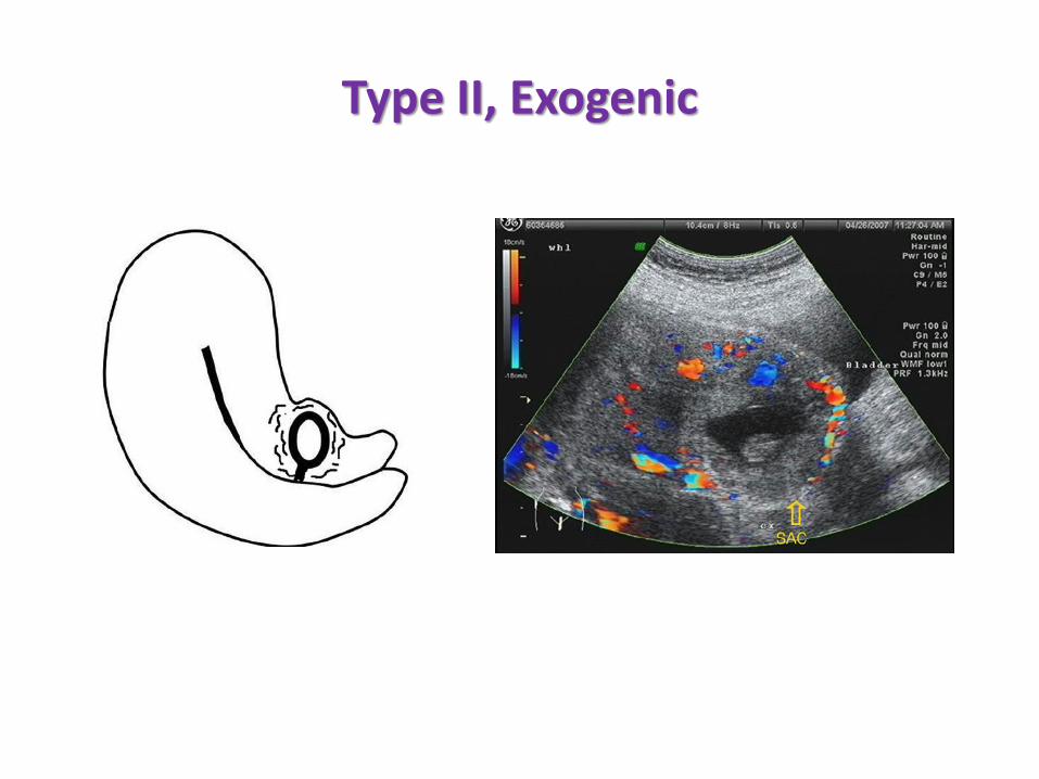
Type II, Exogenic

• CSP implanted “on the scar” had a substantially better outcome
then CSP implanted “in the niche.”
• Myometrial thickness <2 mm in the first-trimester TVUS is associated
with morbidly adherent placenta at delivery.


• In CSP, the gestation sac is completely surrounded by myometrium and the
fibrous tissue of the scar, quite separate from the endometrial cavity
Clark SL, Obstet Gynecol; 1985
Absent retroplacental clear space
Transvaginal scan showing low implantation of gestational sac embedded eccentrically in lower uterine segment and implanted in location of prior CS
Rich vascular pattern in area between CS and placenta

Complications
• Placental adhesion abnormalities
– Placenta accreta X4-5
• Invasion of the bladder by the growing placenta
• Uterine rupture with catastrophic haemorrhage
• Risk of hysterectomy causing serious maternal morbidity
• Increased maternal morbidity and mortality
• Preterm labour
• Loss of future fertility
Ash A., BJOG; 2007Miller DA., Am J Obstet Gynecol; 1997
CSP & Placenta accreta
• CSP and placenta accreta share a common histology.
– deep invasion of the myometrium that could reach the uterine serosa.
– Trophoblastic implantations in CSP and placenta accreta are
histologically undistinguishable
• Placenta accreta could be a progression of CSP.
• The endometrium at the cesarean scar site differs from the
rest of the endometrium
– fewer leukocytes and less vascularization
Timor-Tritsch IE et al. Cesarean scar pregnancy and early placenta accreta share common histology. Ultrasound Obstet Gynecol. 2014.
Timor-Tritsch IE et al. Cesarean scar pregnancy is a precursor of morbidly adherent placenta. Ultrasound Obstet Gynecol. 2014

Diagnosis
• Due to the serious consequences of CSP, early diagnosis and
management are paramount
– 1/3 of incidentally diagnosed CSP are asymptomatic
– usually non-specific symptoms
– The most common clinical finding is vaginal bleeding
– 24.6% of cases low abdominal pain ± vaginal bleeding
– GA at the time of diagnosis ranged from 5 to 16 weeks with a mean of
7 ± 2.5 weeks
Riaz RM., Abdom Imaging; 2015Zhang Y., J Obstet Gynaecol Res; 2013

Ultrasound
• The first-line diagnostic tool
– visualization of an empty uterine cavity;
– Detection of the placenta and/or gestational sac embedded in a hysterotomy
scar;
– triangular gestational sac filling the niche of the scar;
– thin (1–3 mm) or absent myometrial layer between the gestational sac and the
bladder;
– closed cervix and empty endocervical canal;
– Presence of an embryonic/fetal pole and/or yolk sac with or without heart
activity;
– presence of a prominent, and at times rich, vascular pattern at or around the
chorionic sac and placenta.
Cali G., Ultrasound Obstet Gynecol; 2018

A midline sagittal transvaginal image demonstrating a gestation sac implanted at the previous caesarean scar with an empty uterine cavity.
Maymon R, Hum Reprod 2004

US image of a sagittal section of uterus showing protrusion of the GS with fetus anteriorly through the scar, with empty uterine cavity at fundus.
Ash A., BJOG; 2007

In sagittal view of uterus, a straight line is drawn connecting internal cervical os and uterine fundus through endometrium (endometrial line; long yellow line). Gestational sac is identified and superior–inferior (S–I) diameter perpendicular to endometrial line is traced (short yellow line).

The Crossover Sign

The Crossover Sign
• Cesarean scar pregnancy (CSP) is the precursor of
Morbidly Adherent Placenta
– Pl. accreta
– Pl. percreta
– Pl. increta
COS 1
COS 2+
Cali G, Forlani F, Timor-Tritsch IE, Palacios-Jaraquemada J, Minneci G, D'Antonio F. Natural history of Cesarean scar pregnancy on prenatal ultrasound: the crossover sign. Ultrasound Obstet Gynecol. 2017 Jul;50(1):100-104.

Sliding Sac Sign
• a positive ‘‘sliding sac sign’’ suggests an aborted IU pregnancy
where the sac slides with a slight pressure of the endovaginal
probe to the cervix, suggesting no intimate attachment
between the sac and the uterus
Timor-Tritsch IE., Am J Obstet Gynecol; 2012

Heterotopic CSP
Ouyang Y., Reprod Biol Endocrinol; 2015

Smith A, Ash A, Maxwell D. Sonographic diagnosis of cesarean scar pregnancy at 16 weeks. J Clin Ultrasound. 2007 May;35(4):212-5.
• a case of a CSP diagnosed at 16 weeks• The patient subsequently suffered a ruptured uterus, who was preserved at surgery

Doppler US
• Additional diagnostic information can be obtained by colour
flow Doppler
– distinct circular peritrophoblastic perfusion surrounding the gestation
sac
– with pulsed Doppler functions consistent with normal early pregnancy
• a prominent high-velocity (peak velocity > 20 cm/second),
• low impedance (pulsatility index < 1) flow velocity waveforms
– an aborted IU pregnancy has no vascular flow bordering
Jurkovic D, Obstet Gynecol; 1991

Doppler US
The presence of a prominent, and at times rich, vascular pattern at or around the chorionic sac and placenta.
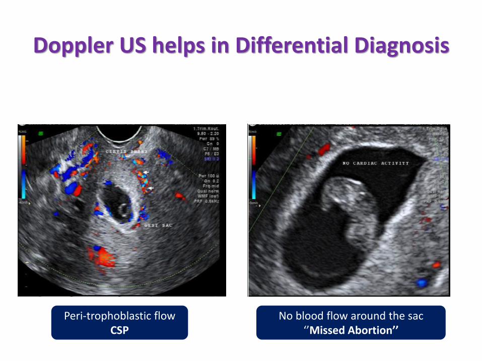
Doppler US helps in Differential Diagnosis
Peri-trophoblastic flowCSP
No blood flow around the sac ‘’Missed Abortion’’

MRI
• could be helpful when TV US + power Doppler US is
inconclusive
• T2-weighed sagittal section is best to identify a cesarean scar
defect, the trophoblastic layer, and the myometrium
separately
• does not detect placental invasion to the cesarean scar and
its extension
Peng KW, Clin Radiol; 2015

Sagittal T2 MRI:
• implantation of GS in the anterior LUS with bulging of the anterior contour and
thinning of the myometrium between GS and bladder (long arrows).
• Empty endometrial and cervical canals.
• CS scar is shown in the anterior lower abdominal wall (short arrows).
Elnashar A. Cesarean scar Pregnancy; 2015

Treatment of CSP
• No consensus yet
• Early treatment will provide the best results,
• most are combined treatments
– expectant management,
– medical management,
– local treatment,
– surgical approach

Treatment should be individualized, based on
• Medical Center capacity– Availability
– Expertise of the clinicians
• Patient- Fertility status– Age
– Number of children
– Number of previous C/S
– Clinical symptoms at presentation
– Severity of symptoms
• CSP– Gestational age
– Viability
– Evidence of myometrial deficiency
– Level of HCG
– Thickness of coveringmyometrium

Treatment Objectives
• should be to perform feticide prior to rupture,
• to remove the gestation sac and to retain patient’s future
fertility.
Ash A., BJOG; 2007

• Expectant management may be a reasonable option for CSP with no
detectable embryonic/fetal heart activity, as 70% of the included cases did not
experience any major maternal complication and had an uncomplicated
miscarriage.

Proposed algorithm for management of Cesarean scar pregnancy
Cali G., Ultrasound Obstet Gynecol; 2018

Conservative medical treatment
• Systemic administration of MTX
– No reason to doubt its efficacy on CSP
– in those with β-hCG levels < 5000 miu/ml
– at a dose of 50 mg/m2
• appropriate for a woman who is pain free and
haemodynamically stable with an unruptured CSP of <8 GW
and a myometrial thickness < 2 mm between the CSP and the
bladder
Chuang J., BLOG; 2003

Local injection of embryocides
– MTX
– potassium chloride
– hyperosmolar glucose
– crystalline trichosanthi
• 16-gauge double-lumen oocyte-retrieval IVF needles may be
used to ensure better aspiration of the trophoblastic tissue via
one lumen and injection of MTX through the other
Hwu YM., BJOG; 2005

Combined medical treatment
• Local injection of 8 mEq KCI (2 mEq/ml) followed by 60mg of MTX injected into the GS,
• Direct injection of 3 ml of 50% glucose + oral MTX (2.5mg three times a day for 5 days),
• Multi-dose systemic MTX (1mg/kg) with alternate day folinic acid rescue,
• Failed systemic MTX followed by successful local MTX,
Donnez J., Br J Obstet Gynaecol; 1997Roberts H., Aust N Z J ObstetGynaecol; 1998
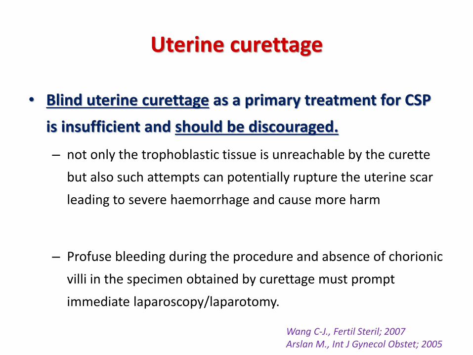
Uterine curettage
• Blind uterine curettage as a primary treatment for CSP
is insufficient and should be discouraged.
– not only the trophoblastic tissue is unreachable by the curette
but also such attempts can potentially rupture the uterine scar
leading to severe haemorrhage and cause more harm
– Profuse bleeding during the procedure and absence of chorionic
villi in the specimen obtained by curettage must prompt
immediate laparoscopy/laparotomy.
Wang C-J., Fertil Steril; 2007Arslan M., Int J Gynecol Obstet; 2005

MTX and Suction Curettage
• Similar success rates compared with MTX treatment
alone
• Can be preferred when the serum β-hCG <50 IU/L
and US revealed the absence of blood flow velocity
Wang JH., Fertil Steril; 2009

Uterine Artery Embolization
• Adjuvant treatment of CS
– an efficient treatment for bleeding prevention before curettage
– embolization particles are a gelatin sponge or polyvinyl alcohol
• Concomitant use of UAE increases
– the success rate of the primary treatment of CSP
– it might be associated with decreased ovarian reserve, IUGR,
premature delivery, placental abruption, or placenta accreta.
Lian F., Cardiovasc Intervent Radiol; 2012Zhuang Y., Am J Obstet Gynecol; 2009

Hysteroscopic evacuation
• offers an important alterative treatment
– with a short operative time (mean 36.7 ± 20.8 minutes),
– less blood loss (mean 50.0 ± 0.0 ml),
– short postoperative stay (mean 1.1 ± 0.9 days)
– rapid return of the pregnancy test to negative (<4 weeks)
• The fertility is conserved after the surgery.
Wang C-J, Chao A-S, Yuen L-T, Wang C-W, Soong Y-K, Lee C-L. Endoscopic management of cesarean scar pregnancy. Fertil Steril 2006; 85:494–7.

Primary open surgical treatment
• L/T followed by wedge resection of the lesion (hysterotomy) should be
considered in women who do not respond to conservative medical and/or
surgical treatments, present too late or if facilities and expertise for operative
endoscopy are not available
• mandatory when uterine rupture is confirmed or strongly suspected
• has the advantage of complete removal of the CSP and simultaneous repair of
the scar, followed by a quick return of the β-hCG to normal level within 1–2
weeks.
• a larger surgical wound, longer hospital stay and longer recovery time, with a
possible higher risk of a future placenta praevia accreta
Fylstra DL., Am J Obstet Gynecol; 2002

Hysterectomy
• In case of failed all other treatment modalities
‘’CSP is a potentially serious condition despite advances in many of the diagnostic
techniques and therapeutic measures.’’
Gonzalez N., JMIG; 2017
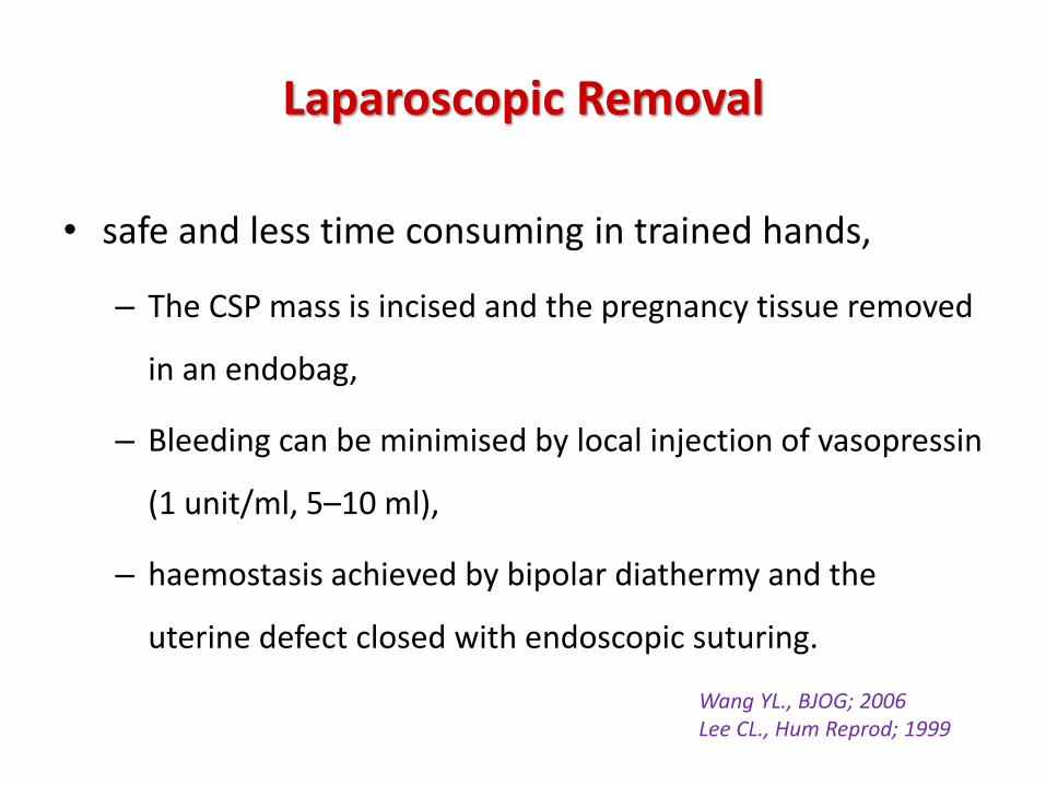
Laparoscopic Removal
• safe and less time consuming in trained hands,
– The CSP mass is incised and the pregnancy tissue removed
in an endobag,
– Bleeding can be minimised by local injection of vasopressin
(1 unit/ml, 5–10 ml),
– haemostasis achieved by bipolar diathermy and the
uterine defect closed with endoscopic suturing.
Wang YL., BJOG; 2006Lee CL., Hum Reprod; 1999
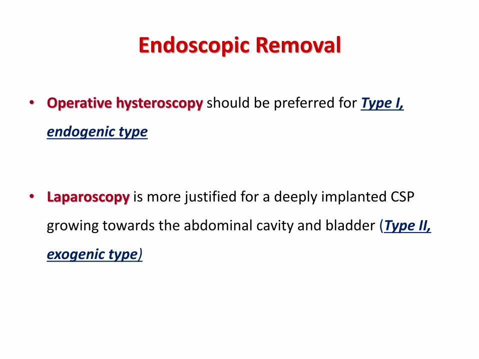
Endoscopic Removal
• Operative hysteroscopy should be preferred for Type I,
endogenic type
• Laparoscopy is more justified for a deeply implanted CSP
growing towards the abdominal cavity and bladder (Type II,
exogenic type)
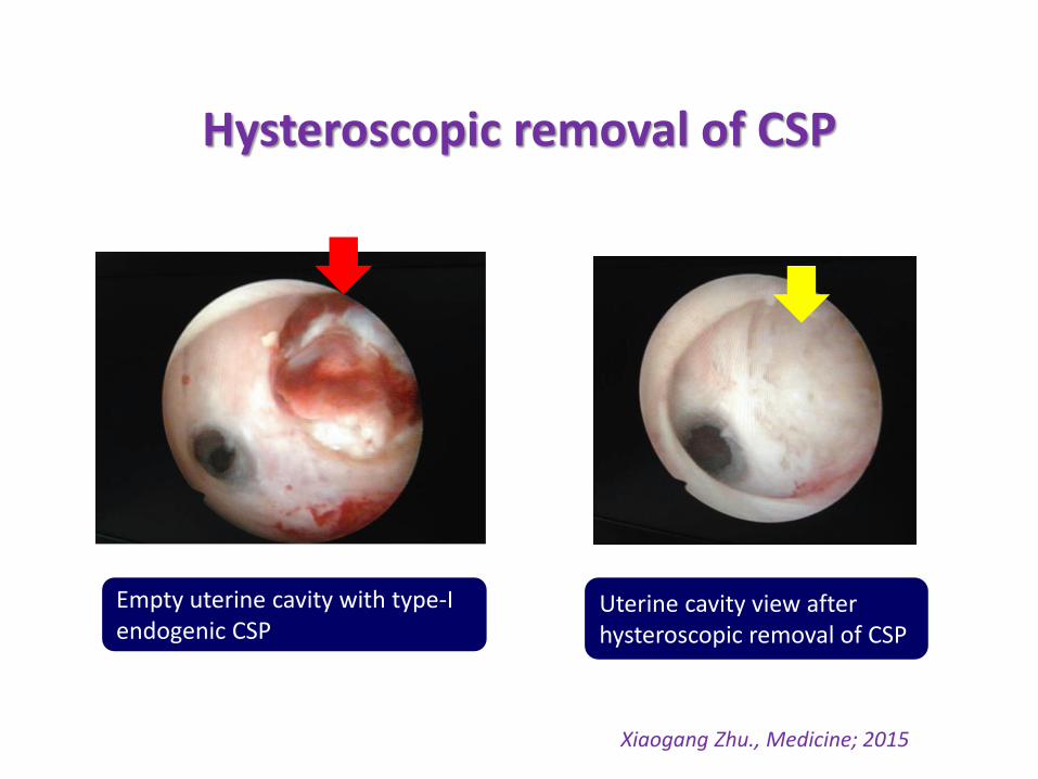
Hysteroscopic removal of CSP
Empty uterine cavity with type-I endogenic CSP
Uterine cavity view afterhysteroscopic removal of CSP
Xiaogang Zhu., Medicine; 2015

Randomized trials of treatment of CSP
Gonzalez N., JMIG; 2017

• HIFU combined with suction curettage under hysteroscopic
guidance is safe and effective in treating patients with CSP at
gestational ages <8 weeks.

• For patients who are searching for definitive symptom release
and future children bearing plans,
– HIFU treatment is a better therapeutic option due to its significant
lower adverse effects and better quality of life improvement than UAE
treatment.

Concluding Remarks
• CSP remains rare; however, it may lead to severe hemorrhage
• Because early diagnosis and treatment are important for the best
outcome, every pregnant woman with history of a cesarean
delivery should be screened early in the first trimester of
pregnancy to rule out this life-threatening complication
• Diagnosis can be achieved with ultrasound and Doppler imaging
Concluding Remarks
• Treatment should be individualized
– Termination of pregnancy in the first trimester is strongly
recommended
– D&C should be avoided because it can lead to profuse bleeding and in
many instances laparotomy and loss of the uterus

A Conservative Approach for CSP
• Multidose MTX therapy with or without intra-amniotic and/or intrafetal
injection of local KCI when FHR is present.
• The dose of MTX is 1 mg/kg body weight intramuscularly on days 1, 3, 5, and 7
and oral leucovorin (0.1 mg/kg) on days 2, 4, 6, and 8.
• Hysteroscopic removal of the gestation for type I CSP or laparoscopic excision for
type II CSP is performed when the hCG levels do not adequately decline or when
the patient becomes symptomatic.
• Hysterectomy is life saving in profusely bleeding patients with scar dehiscence

Thank you…