ch04
TRANSCRIPT

Clinical Immunology & SerologyA Laboratory Perspective, Third Edition
Copyright © 2010 F.A. Davis CompanyCopyright © 2010 F.A. Davis Company
Antibody Structure and Function
Chapter Four
Clinical Immunology & SerologyA Laboratory Perspective, Third Edition
Copyright © 2010 F.A. Davis Company
Antibody Structure and Function Immunoglobulins are glycoproteins found in
the serum portion of the blood. These immunoglobulins are designated IgG,
IgM, IgA, IgD, and IgE. Immunoglobulins are considered to be part of
the humoral branch of the immune response.
Clinical Immunology & SerologyA Laboratory Perspective, Third Edition
Copyright © 2010 F.A. Davis Company
Antibody Structure and Function All immunoglobulin molecules are made up of
a basic four-chain polypeptide unit consisting of two large chains called heavy, or H, chains and two smaller chains called light, or L, chains.
These chains are held together by noncovalent forces and disulfide interchain bridges.
Clinical Immunology & SerologyA Laboratory Perspective, Third Edition
Copyright © 2010 F.A. Davis Company
Antibody Structure and Function When subjected to electrophoresis at pH 8.6,
immunoglobulins appear primarily in the gamma (γ) band.
See Figure 4-1.

Clinical Immunology & SerologyA Laboratory Perspective, Third Edition
Copyright © 2010 F.A. Davis Company
Antibody Structure and FunctionFigure 4-1

Clinical Immunology & SerologyA Laboratory Perspective, Third Edition
Copyright © 2010 F.A. Davis Company
Antibody Structure and Function The FC fragment is important in effector
functions of immunoglobulin molecules, which include opsonization and complement fixation.
The remaining two identical fragments were found to have antigen-binding capacity and were named Fab fragments (fragment antigen-binding).

Clinical Immunology & SerologyA Laboratory Perspective, Third Edition
Copyright © 2010 F.A. Davis Company
Antibody Structure and Function Such a molecule will be able to form a cross-
linked complex with soluble antigen, and the complex would precipitate; particulate antigen will agglutinate.
Each Fab fragment thus consists of one L chain and one-half of an H chain, held together by disulfide bonding.
Clinical Immunology & SerologyA Laboratory Perspective, Third Edition
Copyright © 2010 F.A. Davis Company
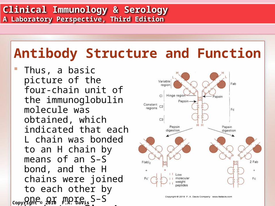
Antibody Structure and Function Thus, a basic picture of
the four-chain unit of the immunoglobulin molecule was obtained, which indicated that each L chain was bonded to an H chain by means of an S–S bond, and the H chains were joined to each other by one or more S–S bonds (see Fig. 4-2).
Clinical Immunology & SerologyA Laboratory Perspective, Third Edition
Copyright © 2010 F.A. Davis Company
Antibody Structure and Function Bence-Jones proteins, found in the urine of
patients with multiple myeloma, are in fact L chains being secreted by malignant plasma cells.
Analysis of several Bence-Jones proteins revealed that there were two main types of L chains, designated kappa (κ) and lambda (λ).
Clinical Immunology & SerologyA Laboratory Perspective, Third Edition
Copyright © 2010 F.A. Davis Company
Antibody Structure and Function The difference between the κ and λ chains lies
in the amino acid substitutions at a few locations along the chain.
Both κ and λ L chains are found in all five classes of immunoglobulins, but only one type is present in a given molecule.
Clinical Immunology & SerologyA Laboratory Perspective, Third Edition
Copyright © 2010 F.A. Davis Company
Antibody Structure and Function Constant regions of the H chain are unique to
each class and give each immunoglobulin type its name.
Hence, IgG has a γ H chain, IgM a μ chain, IgA an α chain, IgD a δ chain, and IgE an ε chain.
Clinical Immunology & SerologyA Laboratory Perspective, Third Edition
Copyright © 2010 F.A. Davis Company
Antibody Structure and Function Each of these represents an isotype, a unique
amino acid sequence that is common to all immunoglobulin molecules of a given class in a given species.
Minor variations of these sequences that are present in some individuals but not others are known as allotypes (see Fig. 4-3 in text).

Clinical Immunology & SerologyA Laboratory Perspective, Third Edition
Copyright © 2010 F.A. Davis Company
Antibody Structure and FunctionFigure 4-3

Clinical Immunology & SerologyA Laboratory Perspective, Third Edition
Copyright © 2010 F.A. Davis Company
Antibody Structure and Function The variable portions of each chain are unique
to a specific antibody molecule, and they constitute the idiotype of the molecule.
The aminoterminal ends of both L and H chains contain these variable regions, which are essential to the formation of the antigen-binding site.
Together they serve as the antigen recognition unit of the immunoglobulin.

Clinical Immunology & SerologyA Laboratory Perspective, Third Edition
Copyright © 2010 F.A. Davis Company
Antibody Structure and Function The basic four-chain structure of all
immunoglobulin molecules does not actually exist as a straight Y shape; instead it is folded into compact globular subunits, based on the formation of balloon-shaped loops at each of the domains.
Intrachain disulfide bonds stabilize these globular regions.

Clinical Immunology & SerologyA Laboratory Perspective, Third Edition
Copyright © 2010 F.A. Davis Company
Antibody Structure and Function Antigen is captured within the cleft by
binding to a small number of amino acids at strategic locations on each chain known as hypervariable regions.

Clinical Immunology & SerologyA Laboratory Perspective, Third Edition
Copyright © 2010 F.A. Davis Company
Antibody Structure and Function IgG is the predominant immunoglobulin in
humans, containing approximately 75–80 percent of the total serum immunoglobulins.
As seen in Table 4-1, IgG has the longest half-life of any immunoglobulin class, approximately 23–25 days, which may help to account for its predominance in serum.

Clinical Immunology & SerologyA Laboratory Perspective, Third Edition
Copyright © 2010 F.A. Davis Company
Antibody Structure and Function There are four major subclasses of IgG,
with the following distribution: IgG1, 67 percent; IgG2, 22 percent; IgG3, 7 percent; and IgG4, 4 percent.
These subclasses differ mainly in the number and position of the disulfide bridges between the γ chains, as seen in Figure 4-5 in text.

Clinical Immunology & SerologyA Laboratory Perspective, Third Edition
Copyright © 2010 F.A. Davis Company
Antibody Structure and Function All subclasses of IgG appear to be able to
cross the placenta, although IgG2 is the least efficient.
Macrophages, monocytes, and neutrophils have FC receptors on their surfaces that are specific for the FC region of IgG.
This enhances contact between antibody-coated antigen and phagocytic cells and generally increases the efficiency of phagocytosis.

Clinical Immunology & SerologyA Laboratory Perspective, Third Edition
Copyright © 2010 F.A. Davis Company
Antibody Structure and Function Major functions of IgG include the
following: (1) providing immunity for the newborn, because IgG can cross the placenta; (2) fixing complement; (3) coating antigen for enhanced phagocytosis (opsonization); (4) neutralizing toxins and viruses; and (5) participating in agglutination and precipitation reactions.

Clinical Immunology & SerologyA Laboratory Perspective, Third Edition
Copyright © 2010 F.A. Davis Company
Antibody Structure and Function Agglutination and precipitation reactions take
place in vitro. These reactions would make phagocytosis more efficient in vivo.
IgG is better at precipitation reactions than at agglutination, because precipitation involves small soluble particles, which are more easily brought together by the relatively small IgG molecule. Ig M is better for agglutination.
Clinical Immunology & SerologyA Laboratory Perspective, Third Edition
Copyright © 2010 F.A. Davis Company
Antibody Structure and Function IgM is known as a macroglobulin, and is a
pentamer aggregate providing 10 potential ag-binding sites.
It accounts for 5–10 percent of all serum immunoglobulins.
As seen in Table 4-1, the half-life of IgM is about 10 days—much shorter than that of IgG.

Clinical Immunology & SerologyA Laboratory Perspective, Third Edition
Copyright © 2010 F.A. Davis Company
Antibody Structure and Function IgM can exist as a monomer or as a
pentamer. The pentamer form is found in secretions,
while the monomer form occurs on the surface of naïve B cells.
When it forms a pentamer, the five monomeric units are held together by a J, or joining, chain, which is a glycoprotein with several cysteine residues.
Clinical Immunology & SerologyA Laboratory Perspective, Third Edition
Copyright © 2010 F.A. Davis Company
Antibody Structure and Function Because of its large size, IgM is found mainly
in the intravascular pool and not in other body fluids or tissues.
IgM cannot cross the placenta.
Clinical Immunology & SerologyA Laboratory Perspective, Third Edition
Copyright © 2010 F.A. Davis Company
Antibody Structure and Function IgM is known as the primary response
antibody, because it is the first to appear after antigenic stimulation and the first to appear in the maturing infant.
It is synthesized only as long as antigen remains present, because there are no memory cells for IgM.
Clinical Immunology & SerologyA Laboratory Perspective, Third Edition
Copyright © 2010 F.A. Davis Company
Antibody Structure and Function Figure 4-7 depicts the difference between the
primary response, which is predominantly IgM, and the secondary response, which is mainly IgG.
The primary response is characterized by a long lag phase, while the secondary response has a shortened lag period and a much more rapid increase in antibody titer.

Clinical Immunology & SerologyA Laboratory Perspective, Third Edition
Copyright © 2010 F.A. Davis Company
Antibody Structure and Function Figure 4-7

Clinical Immunology & SerologyA Laboratory Perspective, Third Edition
Copyright © 2010 F.A. Davis Company
Antibody Structure and Function The functions of IgM include (1)
complement fixation, (2) agglutination, (3) opsonization, and (4) toxin neutralization.
IgM is the most efficient of all immunoglobulins at triggering the classical complement pathway (see Chapter 6), because a single molecule can initiate the reaction as a result of its multiple binding sites.

Clinical Immunology & SerologyA Laboratory Perspective, Third Edition
Copyright © 2010 F.A. Davis Company
Antibody Structure and Function The larger number of binding sites also makes
IgM more efficient at agglutination reactions, especially with multivalent antigens.
Thus, IgM forms a potent defense against many bacterial diseases.
Clinical Immunology & SerologyA Laboratory Perspective, Third Edition
Copyright © 2010 F.A. Davis Company
Antibody Structure and Function Because IgM has a J chain, it can
occasionally acquire a secretory component like IgA does.
This protein allows it to cross epithelial cells and patrol mucous membranes.
IgM also serves as a surface receptor for antigen.
The presence of membrane IgM classifies lymphocytes as mature B cells.
Clinical Immunology & SerologyA Laboratory Perspective, Third Edition
Copyright © 2010 F.A. Davis Company
Antibody Structure and Function In the serum, IgA represents 10–15 percent of
all circulating immunoglobulin. Ig A1 may be susceptible to certain bacterial
proteases.
Clinical Immunology & SerologyA Laboratory Perspective, Third Edition
Copyright © 2010 F.A. Davis Company
Antibody Structure and Function Hence, IgA2 is the predominant form in
secretions at mucosal surfaces, while IgA1 is mainly found in serum.
IgA2 is found as a dimer along the respiratory, urogenital, and intestinal mucosa, and it also appears in milk, saliva, tears, and sweat.
Since mucosal surfaces are a major point of entry for pathogens, IgA2 serves to keep antigen from penetrating further into the body.

Clinical Immunology & SerologyA Laboratory Perspective, Third Edition
Copyright © 2010 F.A. Davis Company
Antibody Structure and Function The dimer consists of two monomers held
together by a J chain Secretory IgA is synthesized in plasma cells
found mainly in mucosal-associated lymphoid tissue, and it is released in dimeric form.
Then it picks up the secretory component at the surface of the epithelial cell; this complex exits into the mucosa-lined cavity.

Clinical Immunology & SerologyA Laboratory Perspective, Third Edition
Copyright © 2010 F.A. Davis Company
Antibody Structure and Function The SC precursor is
actually found on the surface of epithelial cells and serves as a specific receptor for IgA. SC facilitates transport of IgA.
Plasma cells that secrete IgA actually home to subepithelial tissue, where IgA can bind as soon as it is released from the plasma cells. (See Fig. 4-8.)

Clinical Immunology & SerologyA Laboratory Perspective, Third Edition
Copyright © 2010 F.A. Davis Company
Antibody Structure and Function SC also makes the dimer more resistant to
enzymatic digestion by masking sites that would be susceptible to protease cleavage.
The main function of secretory IgA is to patrol mucosal surfaces and act as a first line of defense, by neutralizing toxins produced by microorganisms and helping to prevent bacterial adherence to mucosal surfaces.
Clinical Immunology & SerologyA Laboratory Perspective, Third Edition
Copyright © 2010 F.A. Davis Company
Antibody Structure and Function Complexes of IgA and antigen are easily
trapped in mucus and then eliminated by the ciliated epithelial cells of the respiratory and intestinal tracts.
Clinical Immunology & SerologyA Laboratory Perspective, Third Edition
Copyright © 2010 F.A. Davis Company
Antibody Structure and Function In addition, neutrophils, monocytes, and
macrophages possess specific receptors for serum and secretory IgA.
Binding to these sites triggers a respiratory burst and degranulation of the cells involved.
Both forms of IgA can thus act as opsonins, or promoters of phagocytosis.
Clinical Immunology & SerologyA Laboratory Perspective, Third Edition
Copyright © 2010 F.A. Davis Company
Antibody Structure and Function IgD was not discovered until 1965, when it
was found in a patient with multiple myeloma (a plasma cell neoplasm).
It is extremely scarce in the serum, representing less than 0.001 percent of total immunoglobulins.
Clinical Immunology & SerologyA Laboratory Perspective, Third Edition
Copyright © 2010 F.A. Davis Company
Antibody Structure and Function Most of the IgD present is found on the
surface of immunocompetent but unstimulated (naïve) B lymphocytes.
It is the second type of immunoglobulin to appear (IgM being the first), and it may play a role in B-cell activation.
Clinical Immunology & SerologyA Laboratory Perspective, Third Edition
Copyright © 2010 F.A. Davis Company
Antibody Structure and Function B lymphocytes bearing only IgM receptors
appear incapable of an IgG response. Those with both IgM and IgD receptors are
capable of responding to T-cell help and switching to synthesis of IgG, IgA, or IgE.
IgD may thus play a role in regulating B-cell maturation and differentiation.

Clinical Immunology & SerologyA Laboratory Perspective, Third Edition
Copyright © 2010 F.A. Davis Company
Antibody Structure and Function In the secreted form in the serum, IgD does
not appear to serve a protective function, because it does not bind complement, it does not bind to neutrophils or macrophages, and it does not cross the placenta.
Clinical Immunology & SerologyA Laboratory Perspective, Third Edition
Copyright © 2010 F.A. Davis Company
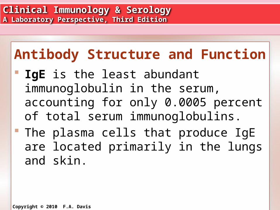
Antibody Structure and Function IgE is the least abundant immunoglobulin in
the serum, accounting for only 0.0005 percent of total serum immunoglobulins.
The plasma cells that produce IgE are located primarily in the lungs and skin.

Clinical Immunology & SerologyA Laboratory Perspective, Third Edition
Copyright © 2010 F.A. Davis Company
Antibody Structure and Function IgE does not participate in typical
immunoglobulin reactions such as complement fixation, agglutination, or opsonization.
In addition, it is incapable of crossing the placenta.

Clinical Immunology & SerologyA Laboratory Perspective, Third Edition
Copyright © 2010 F.A. Davis Company
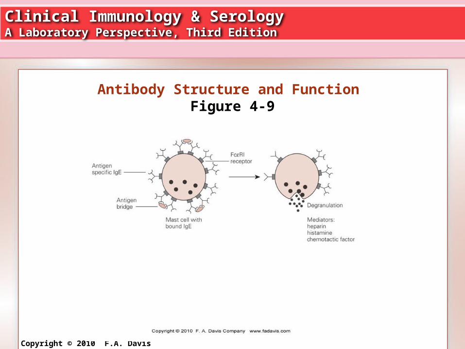
Antibody Structure and Function Instead, shortly after synthesis, it attaches to
basophils and tissue mast cells by means of specific surface proteins, termed high-affinity FC ε RI receptors, which are found exclusively on these cells.
The IgE molecule binds at the FC region, leaving the antigen-binding sites free to interact with specific antigen. (See Fig. 4-9.)
Clinical Immunology & SerologyA Laboratory Perspective, Third Edition
Copyright © 2010 F.A. Davis Company
Antibody Structure and Function Figure 4-9
Clinical Immunology & SerologyA Laboratory Perspective, Third Edition
Copyright © 2010 F.A. Davis Company
Antibody Structure and Function When two adjacent IgE molecules on a mast
cell bind specific antigen, a cascade of cellular events is initiated that results in degranulation of the mast cells with release of vasoactive amines such as histamine and heparin.
Release of these mediators induces what is known as a Type I immediate hypersensitivity, or an allergic reaction (see Chapter 13 for details).

Clinical Immunology & SerologyA Laboratory Perspective, Third Edition
Copyright © 2010 F.A. Davis Company
Antibody Structure and Function Typical reactions include hay fever, asthma,
vomiting and diarrhea, hives, and life-threatening anaphylactic shock.
IgE may thus serve a protective role by triggering an acute inflammatory reaction that recruits neutrophils and eosinophils to the area to help destroy invading antigens that have penetrated IgA defenses.

Clinical Immunology & SerologyA Laboratory Perspective, Third Edition
Copyright © 2010 F.A. Davis Company
Antibody Structure and Function Eosinophils, especially, play a major part in
the destruction of large antigens, such as parasitic worms, that cannot be easily phagocytized (see Chapter 20 for details).
Clinical Immunology & SerologyA Laboratory Perspective, Third Edition
Copyright © 2010 F.A. Davis Company
Antibody Structure and Function The key premise of clonal selection is that
individual lymphocytes are genetically preprogrammed to produce one type of immunoglobulin and that a specific antigen finds or selects those particular cells capable of responding to it, causing them to proliferate.
Clinical Immunology & SerologyA Laboratory Perspective, Third Edition
Copyright © 2010 F.A. Davis Company
Antibody Structure and Function The receptors are in fact the surface
immunoglobulins IgM and IgD, found on immune-competent but unstimulated B lymphocytes.
Repeated contact with antigen would continually increase a specific lymphocyte pool.

Clinical Immunology & SerologyA Laboratory Perspective, Third Edition
Copyright © 2010 F.A. Davis Company
Antibody Structure and Function In 1965, Dryer and Bennett proposed a
solution to the issue of the large number of genes required by the clonal selection theory.
They suggested that the constant and variable portions of immunoglobulin chains are actually coded for by separate genes. Gene rearrangements within the lymphocyte allow for antibody diversity.

Clinical Immunology & SerologyA Laboratory Perspective, Third Edition
Copyright © 2010 F.A. Davis Company
Antibody Structure and Function More than one gene controls synthesis of a
particular immunoglobulin, and through a random selection process these individual segments are joined (spliced) to commit that lymphocyte to making antibody of a single specificity. (See Figs. 4-10 and 4-11.)

Clinical Immunology & SerologyA Laboratory Perspective, Third Edition
Copyright © 2010 F.A. Davis Company
Antibody Structure and Function Figure 4-10

Clinical Immunology & SerologyA Laboratory Perspective, Third Edition
Copyright © 2010 F.A. Davis Company
Antibody Structure and Function The knowledge that B cells are genetically
preprogrammed to synthesize very specific antibody has been used in developing antibodies, known as monoclonal antibodies, for diagnostic testing and for therapeutic purposes (eg. Herceptin for Her2-neu + breast tumors).