ch0576/rhy biology of disease ch0576 aspects of apoptosis
TRANSCRIPT
CH0576/RHY
Biology of Disease CH0576
Aspects of Apoptosis
CH0576/RHY
Apoptosis
• Apoptosis, or ‘programmed cell death’ is a term used to describe how cells die under both pathological and physiological conditions.
• It is an active process, unlike necrosis.
• It is a genetically controlled process, unlike necrosis.
CH0576/RHY
Apoptosis:Physiological examples
• A number of clear situations exist, in which apoptosis is fundamentally important, in the development and survival of the organism.– Limb development in embryology– Potentially autoreactive thymocytes, or cells
bearing inappropriate receptors, fail to develop, they undergo apoptosis, being destroyed before populating the secondary lymphoid organs.
– Involution of the thymus with age.
CH0576/RHY
Apoptosis:Pathological examples
• In pathological states apoptosis can be induced by:-– Cytotoxic T-cells ***– Natural Killer cells (NK cells)– By the action of cell free cytokines such as
tumour necrosis factor (TNF)– Apoptosis plays an important role in a
range of autoimmune diseases, viral infections, and in some tumour cell death.
– Mutations in various elements of the apoptotic pathways have recently been linked with specific disease states
CH0576/RHY
Mechanisms of apoptosis
Activation of the Caspase System• Work with the nematode worm
C.elegans revealed that an enzyme activity, ced-3, was essential for apoptosis to occur.
• Various enzymes present in mammalian systems, which are homologous to ced-3, have been described.
• The first such enzyme was termed ICE.
CH0576/RHY
Caspase cascade
• Several such enzymes have now been found and described as ICE-like enzymes, more recently they are described as CASPASES.
• These are a series of cysteine protease enzymes which become activated within the cell undergoing apoptosis.
• The activated caspases can cause the cleavage of poly(ADP-ribose) polymerase, or PARP, a crucial enzyme in DNA repair.
CH0576/RHY
Mechanism of apoptosis:
• Hence DNA repair mechanisms become disabled, leading to the activation of a range of apoptotic endonucleases, which initiate the fragmentation of DNA.
• A wide range of cellular proteins are broken down by the activated caspase enzymes.
• The question is: How is the caspase enzyme cascade activated initially?
CH0576/RHY
Caspase Classes• Caspases are grouped into two classes
based on similarities in their primary structures.
• Class I Caspases possess long N-terminal prodomains.
• Classs II Caspases possess either short prodomains or lack prodomains.
• Activation of the Class II molecules (e.g. Caspases-3,6, and 7) seem to need proteolytic processing by class I caspases
CH0576/RHY
Caspase classes
• Class II caspases are the downstream caspases that mediate the proteolysis of a range of cellular proteins, in a cell destined to die.
• The activation of the class I caspases (upstream) is fundamental in cell death commitment.
• Recent work suggests how these class I caspases are activated.
CH0576/RHY
Molecule-Molecule Interactions
• Interaction between various components of the death complexes are crucial in apoptotic signalling.
• Three types of protein-protein interaction domains are present in apoptotic molecules.
• DDs - death domains.• DEDs - death-effector domains.• CARDs - caspase recruitment domains.
CH0576/RHY
Molecule-Molecule Interactions
• DDs are common in:-– the upstream components of the apoptotic
pathway, such as the death receptors (e.g. Fas, TNFR and DR3)
– molecules that are recruited to these death receptors (e.g. FADD, TRADD and RIP) - these molecules are cytoplasmic ‘adaptor’ molecules.
• DEDs and CARDs are responsible for recruiting class I caspases to specific death complexes through adaptor molecules.
CH0576/RHY
Molecule-Molecule Interactions
• The interaction between the death domains of the death receptors and those of the adaptor molecules require the initial activation of the death receptor by a suitable ligand.
• The binding of the ligand to the receptor causes a conformational change in the intracytoplasmic death domain and allows combination with a similar domain in the adaptor molecules.
CH0576/RHY
Activation of Caspase cascade:
• Activators or inducers of apoptosis interact with receptors present on the cell surface.
• These receptors are members of the TNF/NGF receptor family.
• The two key receptors are Fas, also known as APO-1 or CD95, and TNF receptor.
• Both these receptors possess an intracytoplasmic domain associated with them termed a ‘death domain’.
CH0576/RHY
Caspase activation• The binding of a suitable ligand to the
specific receptor on the cell surface causes a signal transduction to the death domain and the activation of a pro-caspase activity.
• Fas receptors are closely associated with a pro-caspase 8 activity, and TNF receptors associated with a pro-caspase 2 activity.
• This activation triggers a sequential cascade within the cell.
CH0576/RHY
Caspase activation: (2001)
• Activation Induced Cell Death. – Current Opinion in Immunology, 2001, 13:3:
356-362• Oligomerization of Fas by FasL induces
the recruitment of the adaptor molecule FADD by their mutual DDs.
• The opposite end of FADD contains DEDs which allows the recruitment of either pro-caspase 8 or FLIP (an inhibitory molecule)
CH0576/RHY
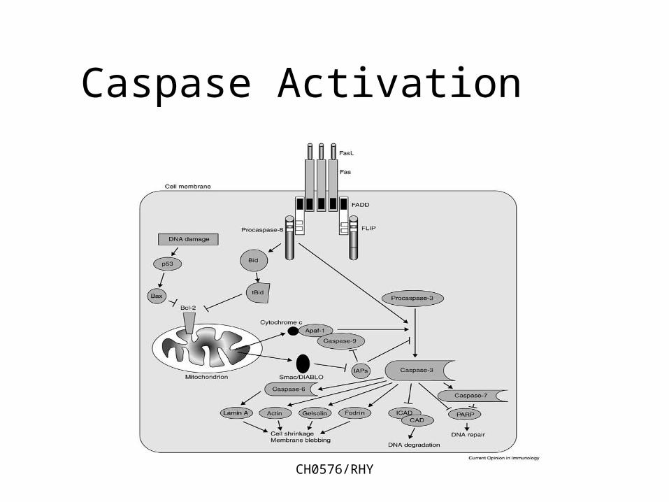
Caspase Activation
CH0576/RHY
Caspase activation
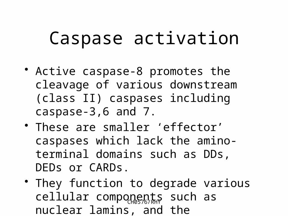
• Active caspase-8 promotes the cleavage of various downstream (class II) caspases including caspase-3,6 and 7.
• These are smaller ‘effector’ caspases which lack the amino-terminal domains such as DDs, DEDs or CARDs.
• They function to degrade various cellular components such as nuclear lamins, and the cytoskeletal proteins fodrin and gelsolin.
CH0576/RHY
Caspase activation
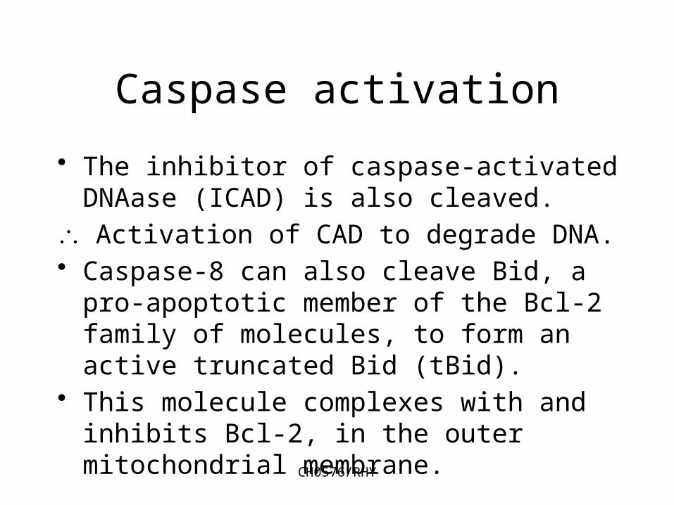
• The inhibitor of caspase-activated DNAase (ICAD) is also cleaved.
Activation of CAD to degrade DNA.• Caspase-8 can also cleave Bid, a pro-
apoptotic member of the Bcl-2 family of molecules, to form an active truncated Bid (tBid).
• This molecule complexes with and inhibits Bcl-2, in the outer mitochondrial membrane.
CH0576/RHY
Caspase activation
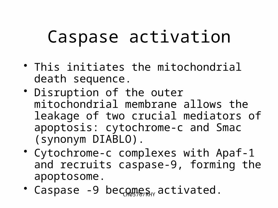
• This initiates the mitochondrial death sequence.
• Disruption of the outer mitochondrial membrane allows the leakage of two crucial mediators of apoptosis: cytochrome-c and Smac (synonym DIABLO).
• Cytochrome-c complexes with Apaf-1 and recruits caspase-9, forming the apoptosome.
• Caspase -9 becomes activated.
CH0576/RHY
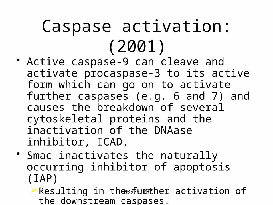
Caspase activation: (2001)• Active caspase-9 can cleave and activate
procaspase-3 to its active form which can go on to activate further caspases (e.g. 6 and 7) and causes the breakdown of several cytoskeletal proteins and the inactivation of the DNAase inhibitor, ICAD.
• Smac inactivates the naturally occurring inhibitor of apoptosis (IAP)Resulting in the further activation of the
downstream caspases.
CH0576/RHY
Killing by Cytotoxic T-cells:
• In addition to the activation of the caspase system through the death domains of Fas and TNFR, the process of granule mediated apoptosis takes part in both innate immunity, via NK cells, and also adaptive defence mechanisms, via CTL’s against:-
• Intracellular pathogens (e.g. viruses)• Tumours• Non-self cells.
CH0576/RHY
Granule mediated apoptosis
• This is a calcium dependent mechanism induced by the actions of a pore froming protein called PERFORIN and a family of granule associated proteases called GRANZYMES.
• The granules of cytotoxic T cells and their constituent proteins are synthesized about 24 - 48 hours after stimulation via the T cell receptor.

CH0576/RHY
Granule mediated apoptosis• Perforin and granzymes are stored
within cytoplasmic cytotoxic granules.• Following recognition of a specific
target by the CTL a tight junction is formed between the effector and target cell.
• When purified granules from CTLs are added to suitable targets in vitro the cells are destroyed by the creation of pores within the lipid bilayer.
CH0576/RHY
Granule mediated apoptosis
• The pores consist of polymers of perforin.• Perforin, in the presence of calcium ions,
spontaneously polymerises and forms a cylindrical structure.
• The outer surface of the cylinder is lipophilic, and the inner surface of the structure is hydrophilic.
• The inner diameter of the cylinder being about 16nms.
CH0576/RHY
Granule mediated apoptosis
• The structure inserts itself into the target membrane.
• The pore allows the passage of water and salts into the cell and causes a rapid destruction of the target by a lytic event.
• In vitro there is no evidence of the nuclear DNA fragmentation which is a feature characteristic of apoptosis.
CH0576/RHY
Granule mediated apoptosis
• It is suggested that this lytic method of killing is only seen at artificially high levels of perforin.
• It is probably not a true reflection of the role of perforin in apoptosis initiated by CTLs in vivo.
• It is more likely that sub-lytic levels of perforin are generated in vivo, serving as a means of entry of granzymes into the target cell.
CH0576/RHY
Granule mediated apoptosis
• Recent evidence suggests that there are a number of means by which granzymes enter the target cell in order to initiate apoptosis.
• Granzymes are not themselves directly responsible for DNA fragmentation, they are proteases and not nucleases.
• Granzymes A G have been identified, the major granzymes in terms of apoptosis are GrA and GrB.
CH0576/RHY
Granzymes:role in apoptosis
• Granzyme B in an aspase, i.e. it cleaves proteins after an aspartic acid residue.
• The pro-caspases are generally activated by cleavage at specific aspartic acid residues.
• GrB is capable of activating most of the caspases (3,6,7,8,9,and 10).
• Evidence suggests that in addition to activation of caspases, GrB can activate ‘death substrates’ directly.
CH0576/RHY
Granzymes:role in apoptosis
• In experiments performed using purified GrB the enzyme cleaved nuclear substrates including DNA-PK and NuMA.
• The cleavage sites were different to those acted upon by the caspases.
CH0576/RHY
Death Receptors
• The well known death receptors, Fas and TNFR1 trigger apoptosis by recognition of their specific ligands.
• A recent finding is that there are several homologues of Fas and TNFR1.
• Death Receptor (DR) 3,4,5 and 6 all function as receptors which signal apoptosis.
• Decoy Receptors (DcR) 1, 2 & 3 act as decoys– they compete with DRs for ligand binding.
CH0576/RHY
Death Ligands• Research using ‘gene knockout’ studies
in the murine system has allowed some of the links between death receptors and the cell’s apoptotic mechanism to be elucidated.
• Studies suggest that FasL is critical for the activation-induced, or instructive apoptosis of T -cells.– Individuals with mutations in the genes
which encode Fas or FasL accumulate enormous numbers of lymphocytes.
– There is massive, lethal lymphadenopathy.
CH0576/RHY
Death Ligands• The research and clinical findings
suggest that the main biological role for FasL is to initiate instructive apoptosis and deletion of peripheral lymphocytes.
• TNF or TNFR ‘knockout’ mice show an susceptibility to microbial infection and a inflammatory response, when challenged with bacterial endotoxins:– main biological role for TNF is the induction
of inflammatory response and stress response genes.
CH0576/RHY
Death Ligands
• Through bioinformatics database screening two groups of workers found another death ligand - Apo2 ligand or TRAIL– TNF-related apoptosis-inducing ligand.
• Apo2L’s closest sequence homologue is FasL
• Research indicates that in vitro both these ligands potentially induce apoptosis of tumour cells.
• Apo2L mRNA is expressed in many tissues.
CH0576/RHY
Death Ligands
• Transcript levels in T cells following their stimulation with mitogens such as PHA.
• Resting peripheral T cells are resistant to the induction of apoptosis by Apo2L, but IL-2 stimulated T cells acquire sensitivity to this ligand.
• this ligand plays a role in peripheral deletion of lymphocytes.
CH0576/RHY
Death Ligands
• Apo2L may also contribute to the instructive apoptosis of virally infected cells.
• T-cells from HIV infected patients are more susceptible to this ligand than are uninfected cells.
• Apo2L also seems to be involved in tumour cell killing mechanisms.
CH0576/RHY
Death Receptors• The receptors in the TNFR family all
possess several cysteine rich domains (CRD’s) in their NH2 terminal region.
• The family of receptors is broadly divided into two subgroups on the basis of their cytoplasmic amino acid sequences.
• The subgroups either possess or lack a so called ‘death domain’
• This death domain links the receptor to the caspase cascade.
CH0576/RHY
Death Receptors• Receptors bearing the ‘death domain’
(DDs) include:-– TNFR1– Fas– DR3 (also called Apo3 plus other synonyms)– DR4– DR5 (also called TRAIL-R2, TRICK2, KILLER)– DR6– The death domains of these molecules
either connect the receptor to the caspases, which induce apoptosis or to kinase cascades, capable of activating genes.
CH0576/RHY
Decoy Receptors
• The other subgroup of TNFR family, lacking a functional ‘death domain’ act as inhibitors as opposed to transducers of signalling.
• They are referred to as the Decoy Receptors.
• This subgroup includes DcR1 and DcR2 - both cell surface expressed molecules.
• It also includes DcR3 and osteoprotegerin (OPG) - both of which are secreted, soluble proteins.
CH0576/RHY
Decoy Receptors
• DcR3 acts as a decoy for FasL and has been shown to have a high expression in several tumours.
• It is thought to play a role in tumour immune evasion.
• DcR1 and DcR2 both act as decoys for Apo2L, inhibiting its activation of DR4 and DR5 - see explanatory diagram.
CH0576/RHY
Mutations in Apoptosis Genes
• A range of mutations in genes encoding different elements of various apoptosis pathways have been recently implicated in disease pathogenesis.
• It is envisaged that knowledge of the mutations of apoptosis genes will benefit the research into the clinical management of a range of disease states.
CH0576/RHY
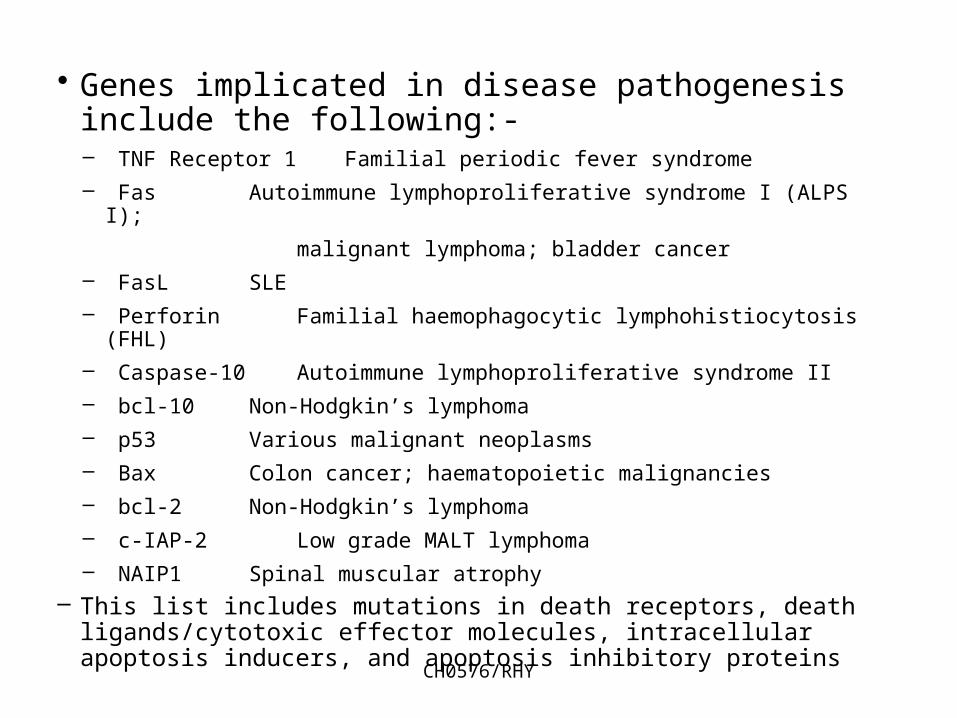
• Genes implicated in disease pathogenesis include the following:-– TNF Receptor 1 Familial periodic fever syndrome– Fas Autoimmune lymphoproliferative syndrome I (ALPS I);
malignant lymphoma; bladder cancer– FasL SLE– Perforin Familial haemophagocytic lymphohistiocytosis (FHL)– Caspase-10 Autoimmune lymphoproliferative syndrome II – bcl-10 Non-Hodgkin’s lymphoma– p53 Various malignant neoplasms– Bax Colon cancer; haematopoietic malignancies– bcl-2 Non-Hodgkin’s lymphoma– c-IAP-2 Low grade MALT lymphoma– NAIP1 Spinal muscular atrophy
– This list includes mutations in death receptors, death ligands/cytotoxic effector molecules, intracellular apoptosis inducers, and apoptosis inhibitory proteins