ch17-18 urinary system - sintich...
TRANSCRIPT
Ch17-18 Urinary System
Main Function: • Filter the blood
Other Functions: • maintain purity and consistency of internal fluids • eliminates nitrogenous wastes, toxins, and drugs from the body • regulates blood volume and chemical makeup to maintain
proper balance between water and salts and between acids and bases
• produces the enzyme renin to help regulate blood pressure • releases hormone erythropoietin to stimulate red blood cell
production • converts vitamin D into active form • manufactures urine
Every day, kidneys filter gallons of fluid from our bloodstream

Organs
• kidney, paired ureters, urinary bladder, and urethra
– Provide reservoirs or transportation channels

Location
• Along dorsal wall (T12 to L3 vertebrae level) in the retroperitoneal space
• Right kidney is slightly lower because of liver
• Layer of fat helps hold each kidney in place

Structure of Kidney
• Hilus – medial indentation where ureters, renal blood vessels, and nerves enter and exit
• Outer region – renal cortex (light colored)
• Inner region – renal medulla (dark colored)
• Triangular part of renal medulla – medullary pyramids
• Cavity leading from the hilus to the pyramids – renal pelvis

• Nephron: structural and functional unit of kidneys and responsible for the formation of urine
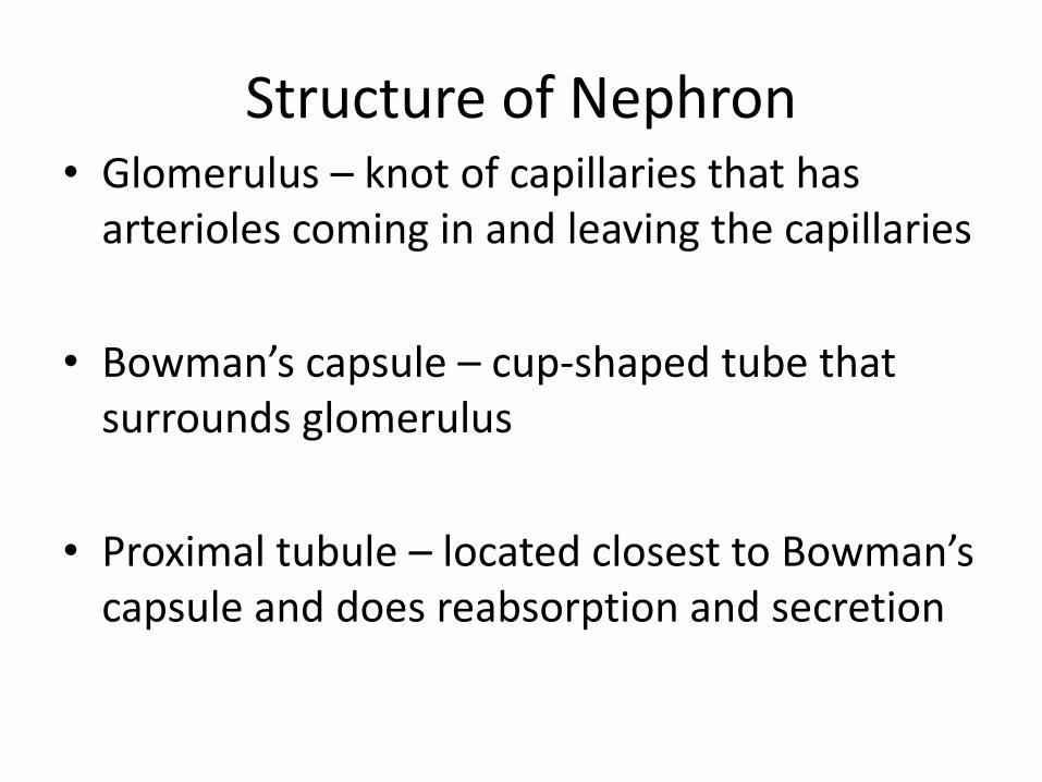
Structure of Nephron • Glomerulus – knot of capillaries that has
arterioles coming in and leaving the capillaries
• Bowman’s capsule – cup-shaped tube that surrounds glomerulus
• Proximal tubule – located closest to Bowman’s capsule and does reabsorption and secretion
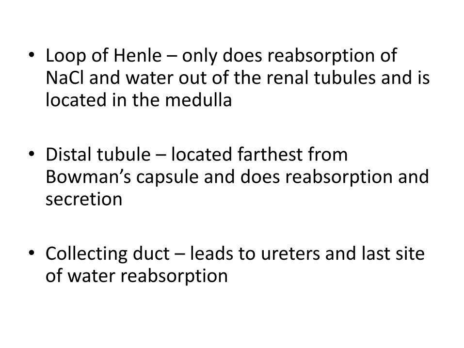
• Loop of Henle – only does reabsorption of NaCl and water out of the renal tubules and is located in the medulla
• Distal tubule – located farthest from Bowman’s capsule and does reabsorption and secretion
• Collecting duct – leads to ureters and last site of water reabsorption

Steps of Urine Formation
• Filtration – all materials small enough to fit through the membrane moves into the renal tubes (ALL IN)
• Reabsorption – good nutrients are moved back to the blood through passive and active transport (GOOD BACK OUT)
• Secretion – any remaining bad materials in the blood move into the renal tubes (BAD IN)
• Excretion – urine leaves the body

Characteristics of Urine • Clear to pale yellow
• Yellow = pigment urochrome that is produced when blood destroys hemoglobin
• Sterile
• Has an odor; drugs, vegetables, or diseases can alter the odor

• pH slightly acidic; can change with diet or body metabolism
• Water plus solutes
– Normal solutes: Na, K, urea, uric acid, creatinine, ammonia, bicarbonate ions, and other ions
– Abnormal solutes: RBC, WBC (pus), glucose, blood proteins, hemoglobin, and bile

Blood Composition
• Depends on: – diet
– metabolism
– urine output
• Our kidneys can keep our blood composition fairly constant by allowing different amounts of filtration and reabsorption despite a wide variation in our diets and cell activity.

3 Roles in Regulating Blood Composition
• Excretion of nitrogenous wastes
• Maintaining water and electrolyte balance
• Ensuring proper blood pH

Nitrogenous wastes
• Nitrogenous wastes – by-products of reactions that the body needs to get rid of
– Urea = less toxic form of ammonia and is formed in liver when proteins are broken down
– Uric acid = less common and even less toxic and released when nucleic acids are metabolized
– Creatinine = released when creatine metabolism takes place in muscles

Water and Electrolyte Balance
• Water intake needs to be greater than water loss
– Water is found in many locations in body: intracellular fluid, extracellular fluid, and fluid in plasma, cerebrospinal, and eyes
– It is important to maintain a balance of water in all areas of the body for cells to work properly
– Water % decreases as you age (45% water in old age; 75% water as a baby)

• Electrolytes = charged ions that conduct electrical signals
– Examples: Na+, K+, Ca2+, Cl-
• Solute changes cause osmosis to occur
– This can change blood volume and pressure as well as functioning of nerves and muscles
– Rule: Water follows Salt
• The more concentrated one side of a membrane, the more water will move to that area (high to low)

Blood pH
• Cell metabolism continually adds H+ to blood as a byproduct
– Disrupts acid-base balance
• CO2 forms carbonic acid (regulated by lungs with breathing to decrease pH of blood) and other acids such as lactic acid (all other molecules are regulated by kidneys)

• Blood buffers help “tie up” acids and bases in blood to regulate pH levels
– Release or bind H+ to increase or decrease pH
• Increase or decrease in respiratory rate depends on need for CO2 in blood
Hormone Regulation
• Blood pressure balance:
– Juxtaglomerular complex, smooth muscle cells in wall of capillaries near the glomerulus, detects a drop in blood pressure
– Renin (enzyme) is then secreted from the smooth muscle cells and moves to the liver
– Renin reacts with a protein from the liver to then trigger the release of aldosterone (glucocorticoid and mineralcorticoid) from the adrenal gland
– Aldosterone will act on the kidney and help to move Na+ and K+ from the renal tubes back into the blood
– This causes more water to move into the blood
– Meanwhile, blood vessels get a signal to constrict
– Blood pressure is returned to normal

Simply Put
• Some cells in your blood vessels in the kidneys detect a drop in blood pressure
• The cells release a chemical, which activates another chemical from the liver, which causes the adrenal gland to release its hormone
• The hormone constricts your blood vessels and has the kidneys keep more Na+ and K+ in your blood, which also puts water back in your blood
• Now, blood pressure can go back up

Hormone Regulation
• Water balance
– Antidiuretic hormone is released from the pituitary gland if blood water levels are low
– ADH attaches to the collecting duct and more water is moved back into the blood by adding more water channel proteins
– This keeps water in the body instead of being released with the urine

Disorders • Diabetes mellitus – disruption of the acid-base
balance in which ketones (acids) are not broken down in the body and leave through the urine
• Water intoxication – babies who drink too much water under the age of 3 months start to have swelling and eventually seizures
• Kidney disease – reduces glomerular filtration
• Incontinence – inability to voluntarily control the external sphincter
• Urinary retention – when bladder is unable to expel its urine
• Dehydration – water output exceeds water intake

Development • In young embryos, there are 3 different times
that kidneys form and then are degenerated
– Third formation, they become the functioning kidneys
• By 3rd month, kidneys are excreting urine through the placenta

• Voluntary control of sphincters follows nervous development
– By 18 months, bladder can be held for 2 hours – start of potty training
– Complete nighttime control by 4 years old
• Micturition (Voiding) = act of emptying the bladder by the control of 2 sphincters
• Old age = decrease function of nephrons and less efficient filtrate reabsorption

Video
• Organs
• Nephron
• Kidney stone removal: Laparoscopic Pyelolithotomy
• Urine Formation