ch17: pediatric endodontics - … · in more detail later in this chapter. ... cavity with little...
TRANSCRIPT
Treatment of pulpally inflamed primary and perma-nent teeth in children presents a unique challenge tothe dental clinician. Pulp diagnosis in the child isimprecise as clinical symptoms do not correlate wellwith histologic pulpal status. Age and behavior cancompromise the reliability of pain as an indicator ofthe extent of pulp inflammation. Furthermore, treat-ment goals are developmentally oriented and may berelatively short term by comparison to the long-termrestorative permanence of adult endodontics.Because of this latter fact, a major focus in pediatricpulp therapy is vital pulp treatment, that capitalizeson the healing potential of the noninflamed remain-ing portions of the pulp. With instances of irre-versibly inflamed and necrotic radicular pulps, con-ventional concepts of nonvital pulp treatment areindicated. However, they must be modified to accom-modate physiologic root resorption in primary teethand continued root development in young perma-nent teeth.
Lewis and Law succinctly stated the ultimate objec-tive of pediatric pulp therapy: “The successful treat-ment of the pulpally involved tooth is to retain thattooth in a healthy condition so it may fulfill its role as auseful component of the primary and young perma-nent dentition.”1 Premature loss of primary teeth fromdental caries and infection may result in the followingsequelae:
• Loss of arch length• Insufficient space for erupting permanent teeth• Ectopic eruption and impaction of premolars• Mesial tipping of molar teeth adjacent to primary
molar loss • Extrusion of opposing permanent teeth• Shift of the midline with a possibility of crossbite
occlusion• Development of certain abnormal tongue positions
It is for this reason that maximum attempts must bemade to preserve primary teeth in a healthy state untilnormal exfoliation occurs. A major contention in con-temporary research involving vital pulp treatment isthe definition of “healthy pulp status” ascribed to manyof the treatment outcomes. This issue will be addressedin more detail later in this chapter.
Vital pulp therapy is based on the premise that pulptissue has the capacity to heal. In addition to the bio-logic basis for the healing capacity of the pulp, differ-ences between primary and permanent teeth exist froma morphologic and histologic standpoint. These differ-ences must be addressed by the clinician to successful-ly treat pulpally inflamed teeth in children.
PULP MORPHOLOGY
Anatomic Differences Between Primary andPermanent Teeth
Anatomic differences between the pulp chambers androot canals of primary teeth and those of young perma-nent teeth have been described2 (Figure 17-1): (1) Pulpchamber anatomy in primary teeth approximates thesurface shape of the crown more closely than in perma-nent teeth. (2) The pulps of primary teeth are propor-tionately larger and the pulp horns extend closer to theouter surfaces of the cusps than in permanent teeth. (3)The pulp-protecting dentin thickness between the pulpchamber and the dentinoenamel junction is less than inpermanent teeth. These three factors increase the poten-tial for pulp exposure from mechanical preparation,dental caries, and trauma. (4) An increased number ofaccessory canals and foramina, as well as porosity in pul-pal floors of primary teeth, has been noted in compari-son with permanent teeth.3 This is thought to accountfor the consistent pulp necrosis response of furcationradiolucency in primary teeth versus periapical radiolu-cency in permanent teeth.4–6
Chapter 17
PEDIATRIC ENDODONTICSClifton O. Dummett Jr and Hugh M. Kopel

A comparison of root canals in primary teeth withthose of young permanent teeth reveals the followingcharacteristics: (1) the roots of primary teeth are pro-portionately longer and more slender; (2) primary rootcanals are more ribbon-like and have multiple pulp fil-aments within their more numerous accessory canals;(3) the roots of primary molars flare outward from thecervical part of the tooth to a greater degree than per-manent teeth and continue to flare apically to accom-modate the underlying succedaneous tooth follicle; (4)the roots of primary anterior teeth are narrowermesiodistally than permanent anterior tooth roots; and(5) in contrast to permanent teeth, the roots of primaryteeth undergo physiologic root resorption. These fac-tors make complete extirpation of pulp remnantsalmost impossible and increase the potential of rootperforation during canal instrumentation. As a result,the requirements of primary root canal filling materialsmust encompass germicidal action, good obturation,and resorptive capability.3
Histologic Considerations
Numerous descriptions of pulp histology exist thatidentify the various cell components of pulp tissue.7,8
Consistently, the pulp is primarily connective tissueand has considerable healing potential. Features that
862 Endodontics
distinguish pulp tissue from other connective tissueinclude the presence of odontoblasts, absence of hista-mine-releasing mast cells, tissue confinement in a hardcavity with little collateral circulation, and vascularaccess limited to the root apex.7,8 Pulp healing capabil-ity is affected by endogenous factors of coronal cellu-larity and apical vascularity. Both are increased in pri-mary and young permanent teeth.8 Pulps become morefibrous, less cellular, and less vascular with age.8
Exogenous factors affecting pulp healing include bacte-rial invasion and chemical/thermal insult. Currentresearch in pulp biology and restorative materialsstrongly substantiates the need for bacterial microleak-age control in maximizing pulp survival.9
Fox and Heeley concluded that, histologically, nostructural differences exist between primary pulp tissueand young permanent pulp tissue with the exception ofthe presence of a cap-like zone of reticular and collage-nous fibers in the primary coronal pulp.10 However,many clinicians have noted different pulp responsesbetween primary and young permanent teeth to trau-ma, bacterial invasion, irritation, and medication.Anatomic differences may contribute to these respons-es. Primary roots have an enlarged apical foramen, incontrast to the foramen of permanent roots, which isconstricted. The resultant reduced blood supply inmature permanent teeth favors a calcific response andhealing by “calcific scarring.”11 This hypothesis isexemplified in older pulps, in which more calcifiednodules and ground substance are found than in youngpulps. Primary teeth, with their abundant blood sup-ply, demonstrate a more typical inflammatoryresponse than that seen in mature permanent teeth.
The exaggerated inflammatory response in primaryteeth may account for increased internal and externalroot resorption from calcium hydroxide pulpotomies.The alkalinity of calcium hydroxide can produce severepulp inflammation and subsequent metaplasia withresultant internal primary root resorption. It has beenshown that the greater the inflammation, the moresevere the resorption (Figure 17-2). Although it is sus-pected that pulps of primary teeth have a differentfunction from those of permanent teeth, no supportingdata are available.
Some clinicians believe that primary teeth are lesssensitive to pain than permanent teeth, probablybecause of differences in the number and/or distribu-tion of neural elements. When comparing primary andpermanent teeth, Bernick found differences in the finaldistribution of pulp nerve fibers.12 In permanent teeth,these fibers terminate mainly among the odontoblastsand even beyond the predentin. In primary teeth, pulp
Figure 17-1 Comparative anatomy between primary (left) andpermanent (right) molars. Primary teeth are smaller in all dimen-sions; their enamel cap is thinner, with less tooth structure protect-ing the pulp. Primary pulp horns are higher, particularly mesial.The roots of primary molars are longer and more slender, are“pinched in” at the cervical part of the tooth, and flare more towardthe apex to accommodate permanent tooth buds. All of these fac-tors tend to increase the incidence of pulp involvement from cariesor complicate canal preparation and obturation. Reproduced withpermission from Finn SB.2

Pediatric Endodontics 863
nerve fibers pass to the odontoblastic area, where theyterminate as free nerve endings. Bernick postulatedthat if primary teeth were not so short-lived in the oralcavity, their nerve endings might terminate among theodontoblasts and in the predentin as in permanentteeth12 (Figure 17-3).
Rapp and associates concurred with Bernick’shypothesis and also stated that the density of the inner-vation of the primary tooth is not as great as that of thepermanent tooth and may be the reason why primaryteeth are less sensitive to operative procedures.13 Theyagree, however, that as the primary teeth resorb, there isa degeneration of the neural elements as with otherpulp cells. Neural tissue is the first to degenerate whenroot resorption begins, just as it is the last tissue tomature when the pulp develops.
Primary and permanent teeth also differ in their cel-lular responses to irritation, trauma, and medication. Ithas been shown, for example, that the incidence ofreparative dentin formation beneath carious lesions is
more extensive in primary than in permanentteeth.14–17 McDonald reported that the localization ofinfection and inflammation is poorer in the primarypulp than in the pulp of permanent teeth.18
MANAGEMENT OF DEEP CARIOUS LESIONSAND PULP INFLAMMATION IN PRIMARY
AND YOUNG PERMANENT TEETH
Pulp therapy for primary and young permanent teethhas historically been subject to change and controversy.Pulp medicaments, such as zinc oxide–eugenol (ZOE)cement, calcium hydroxide, and formocresol, have beenthe basis for much of this controversy. A better under-standing of the reactions of the pulp and dentin to thesemedicaments has developed over time, primarilythrough improvements in histologic techniques.Anderson and colleagues felt that the pulp and dentinshould be considered as one organ.19 Frankl determinedthat this pulpodentinal system reaction is proportionalto the intensity and duration of the offending agents ofcaries, trauma, medicaments, or restorative materials.20
A correct diagnosis of pulp conditions in primaryteeth is important for treatment planning. McDonaldand Avery have outlined several diagnostic aids in select-
Figure 17-2 Internal resorption triggered by inflammation. A,Advanced caries in a 5-year-old child. Note calcification (arrow) inthe first primary molar (contraindication for pulp therapy). B,Same patient 6 months later. Marked internal resorption, forecast inthe earlier radiograph, indicates advanced degenerative changes.Reproduced with permission from Law DB, Lewis TM, Davis JM.An atlas of pedodontics. Philadelphia: WB Saunders; 1969.
Figure 17-3 Section of pulp from a human primary molar. Notethat the majority of nerves terminate at the pulp-odontoblastic(PO) border. Only isolated nerve fiber penetrates the P-O border toterminate in the zone of Weil. D = dentin; N = nerve fiber;O = odontoblasts; Pr = predentin; PO = pulp-odontoblast border.Reproduced with permission from Bernick S.12
A
B

ing teeth for vital pulp therapy.3 Eidelman et al.21 andProphet and Miller22 have emphasized that no singlediagnostic means can be relied on for determining adiagnosis of pulp conditions. Rayner and Southam havestated that the inflammation response to the effects ofdentin caries in the deciduous pulp is more rapid than inthe permanent pulp.23 Yet Taylor concluded that in spiteof being inflamed and infected by the carious process,primary molars are still capable of marked defense reac-tions similar to those observed in permanent teeth.24
The goal in managing the deep carious lesion ispreservation of pulp vitality before arbitrarily institut-ing endodontic therapy. A suggested outline for deter-mining the pulpal status of cariously involved teeth inchildren involves the following:
1. Visual and tactile examination of carious dentin andassociated periodontium
2. Radiographic examination ofa. periradicular and furcation areasb. pulp canalsc. periodontal spaced. developing succedaneous teeth
3. History of spontaneous unprovoked pain4. Pain from percussion5. Pain from mastication6. Degree of mobility7. Palpation of surrounding soft tissues8. Size, appearance, and amount of hemorrhage associ-
ated with pulp exposures
Pediatric pulp therapy for primary and young per-manent teeth involves the following techniques:
1. Indirect pulp capping2. Direct pulp capping 3. Coronal pulpotomy4. Pulpectomy
The first three methods are vital techniques thatinvolve conservative management of portions ofinflamed pulp tissue with the preservation of theremaining vital pulp. The pulpectomy procedure is anonvital technique and involves the complete extirpa-tion of the irreversibly inflamed and/or necrotic pulpfollowed by canal obturation with a resorbable medica-ment in primary teeth and conventional root canal fill-ing in permanent teeth.
INDIRECT PULP CAPPING
Indirect pulp capping is defined as the application of amedicament over a thin layer of remaining carious
864 Endodontics
dentin, after deep excavation, with no exposure of thepulp. In 1961, Damele described the purpose of indi-rect pulp capping as the use of “reconstructed” dentinto prevent pulp exposure.25 The treatment objective isto avoid pulp exposure and the necessity of more inva-sive measures of pulp therapy by stimulating the pulpto generate reparative dentin beneath the cariouslesion. This results in the arrest of caries progressionand preservation of the vitality of the nonexposedpulp.26 This technique can be used as a one-sitting pro-cedure or the more classic two-sitting procedure. Thelatter involves re-entry after a 6 to 8-week interval toremove any remaining carious dentin and place thefinal restoration3,27 (Figure 17-4).
DiMaggio found, in a histologic evaluation of teethselected for indirect treatment, that 75% would havebeen pulp exposures if all of the caries had initiallybeen removed. Using clinical criteria, this same studyshowed a failure rate of only 1% for indirect pulp capscompared with 25% failure for direct caps.28 A histo-logic examination, however, raised these failure rates to12% and 33%, respectively. Trowbridge and Bergerstated that complete removal of softened dentin, withensuing pulp exposure, may contribute nothing ofdiagnostic value in estimating the extent of existingpulp disease.29 In fact, other studies have shown thatthe true picture of pulp disease cannot be assessed onthe basis of such diagnostic criteria as history of pain,response to temperature change, percussion, and elec-tric pulp testing.30,31
Figure 17-4 Indirect pulp-capping technique. A, Medicament,either zinc oxide–eugenol cement, calcium hydroxide, or both,against remaining caries. B, Lasting temporary restoration.Following repair, both materials are removed along with softenedcaries, and final restorations are placed.

Pediatric Endodontics 865
Historical Review
The concept of indirect pulp capping was firstdescribed by Pierre Fauchard as reported by JohnTomes in the mid-18th century, who recommendedthat all caries should not be removed in deep, sensitivecavities “for fear of exposing the nerve and making thecure worse than the disease.”32 John Tomes, in his mid-19th century textbook, stated, “It is better that a layerof discolored dentin should be allowed to remain forthe protection of the pulp rather than run the risk ofsacrificing the tooth.”32 Although neither of these den-tal pioneers referred to any specific medication for thesoftened dentin, they recognized the healing capacityof the pulp.
In 1891, W. D. Miller discussed various “antisep-tics” that should be used for sterilizing dentin.34 Incontrast to these early reports advocating conservativemanagement of deep lesions, G. V. Black felt that in theinterest of scientific dental practice, no decayed or soft-ened material should be left in a cavity preparation,whether or not the pulp was exposed.35
Rationale
Indirect pulp capping is based on the knowledge thatdecalcification of the dentin precedes bacterial invasionwithin the dentin.36–38 This technique is predicated onremoving the outer layers of the carious dentin, thatcontain the majority of the microorganisms, reducingthe continued demineralization of the deeper dentinlayers from bacterial toxins, and sealing the lesion toallow the pulp to generate reparative dentin. Fusayamaand colleagues demonstrated that in acute caries,dentin discoloration occurred far in advance of themicroorganisms, and as much as 2 mm of softened ordiscolored dentin was not infected.38 In a later study,Fusayama found that carious dentin actually consists oftwo distinct layers having different ultramicroscopicand chemical structures.39 The outer carious layer isirreversibly denatured, infected, and incapable of beingremineralized and should be removed. The inner cari-ous layer is reversibly denatured, not infected, andcapable of being remineralized and should be pre-served. The two layers can be differentiated clinically bya solution of basic fuchsin.39
Whitehead and colleagues compared deep excava-tions in primary and permanent teeth.40 After all soft-ened dentin had been removed from the cavity floor,they found that 51.5% of the permanent teeth werefree from all signs of organisms, and a further 34%had only 1 to 20 infected dentinal tubules in any onesection.40 Primary teeth, however, showed a muchhigher percentage of bacteria in the cavity floor after
all softened dentin was removed. These results werefurther supported by Shovelton, who found thatalthough the deepest demineralized layers of dentinwere generally free from infection, the possibility of afew dentinal tubules containing organisms did exist,especially in primary teeth.41 This finding was sup-ported by Seltzer and Bender.42 Thus, complete clini-cal removal of carious dentin does not necessarilyensure that all infected tubules have been eradicated.Conversely, the presence of softened dentin does notnecessarily indicate infection.
A number of investigators have provided evidencethat the pulp can readily cope with minute contami-nation. Reeves and Stanley43 and Shovelton44 showedthat when the carious lesion proximity to the pulp wasgreater than 0.8 mm (including reparative dentinwhen present), no significant disturbance occurredwithin the pulp of permanent teeth. Rayner andSoutham, in studying carious primary teeth, found themean depth of pulp inflammatory changes from bac-terial dentin penetration to be 0.6 mm in proximity tothe pulp, with some changes occurring within a 1.8 mm pulp proximity.23 Massler considered thatpulp reactions under deep carious lesions result frombacterial toxins rather than the bacteria themselves.45
Massler and Pawlak used the terms “affected” and“infected” to describe pulp reaction to deep cariousattack.46 This histologic study showed that the “affect-ed” pulp, beneath a deep carious lesion with a thinlayer of dentin between the pulp and the bacterialfront, was often inflamed and painful but containedno demonstrable bacteria. However, when significantnumbers of bacteria were found within the “infected”pulp, a microscopic exposure in the carious dentin wasseen. Canby and Bernier concluded that the deeperlayers of carious dentin tend to impede the bacterialinvasion of the pulp because of the acid nature of theaffected dentin.47
The results of these studies indicate the presence ofthree dentinal layers in a carious lesion: (1) a necrotic,soft, brown dentin outer layer, teeming with bacteriaand not painful to remove; (2) a firmer, discoloreddentin layer with fewer bacteria but painful to remove,suggesting the presence of viable odontoblastic exten-sions from the pulp; and (3) a hard, discolored dentindeep layer with a minimal amount of bacterial invasionthat is painful to instrumentation.
Response to Treatment
Sayegh found three distinct types of new dentin inresponse to indirect pulp capping: (1) cellular fibrillardentin at 2 months post-treatment, (2) presence of

globular dentin during the first 3 months, and (3)tubular dentin in a more uniformly mineralized pat-tern.17 In this study of 30 primary and permanentteeth, Sayegh concluded that new dentin forms fastestin teeth with the thinnest dentin remaining after cavitypreparation. He also found that the longer treatmenttimes enhanced dentin formation.17
Diagnosis of the type of caries influences the treat-ment planning for indirect pulp capping. In the activelesion, most of the caries-related organisms are found inthe outer layers of decay, whereas the deeper decalcifiedlayers are fairly free of bacteria. In the arrested lesion, thesurface layers are not always contaminated, especiallywhere the surface is hard and leathery. The deepest lay-ers are quite sclerotic and free of microorganisms.48
Deep carious dentin is even more resistant to decompo-sition by acids and proteolysis than is normal dentin.This was especially true in arrested caries.49,50
Procedures for Indirect Pulp Capping
Case selection based on clinical and radiographic assess-ment to substantiate the health of the pulp is critical forsuccess. Only those teeth free from irreversible signsand symptoms should be considered for indirect pulpcapping. The following measures should be employedfor those teeth appropriate for this technique.
Indications. The decision to undertake the indi-rect pulp capping procedure should be based on thefollowing findings:
1. Historya. Mild discomfort from chemical and thermal
stimulib. Absence of spontaneous pain
2. Clinical examinationa. Large carious lesionb. Absence of lymphadenopathyc. Normal appearance of adjacent gingivad. Normal color of tooth
3. Radiographic examinationa. Large carious lesion in close proximity to the pulpb. Normal lamina durac. Normal periodontal ligament spaced. No interradicular or periapical radiolucency
Contraindications. Findings that contraindicatethis procedure are listed below:
1. Historya. Sharp, penetrating pain that persists after
withdrawing stimulusb. Prolonged spontaneous pain, particularly at night
866 Endodontics
2. Clinical examinationa. Excessive tooth mobilityb. Parulis in the gingiva approximating the roots of
the toothc. Tooth discolorationd. Nonresponsiveness to pulp testing techniques
3. Radiographic examinationa. Large carious lesion with apparent pulp exposureb. Interrupted or broken lamina durac. Widened periodontal ligament spaced. Radiolucency at the root apices or furcation areas
If the indications are appropriate for indirect pulpcapping, such treatment may be performed as a two-appointment or a one-appointment procedure.
Two-Appointment Technique (First Sitting).
1. Administer local anesthesia and isolate with a rubber dam.
2. Establish cavity outline with a high-speed hand-piece.
3. Remove the majority of soft, necrotic, infecteddentin with a large round bur in a slow-speed hand-piece without exposing the pulp.
4. Remove peripheral carious dentin with sharp spoonexcavators. Irrigate the cavity and dry with cottonpellets.
5. Cover the remaining affected dentin with a hard-set-ting calcium hydroxide dressing.
6. Fill or base the remainder of the cavity with a rein-forced ZOE cement (IRM Dentsply-Caulk; Milford.)or a glass-ionomer cement to achieve a good seal.
7. Do not disturb this sealed cavity for 6 to 8 weeks. Itmay be necessary to use amalgam, composite resin,or a stainless steel crown as a final restoration tomaintain this seal.
Two-Appointment Technique (Second Sitting, 6 to8 Weeks Later). If the tooth has been asymptomatic,the surrounding soft tissues are free from swelling, andthe temporary filling is intact, the second step can beperformed:
1. Bitewing radiographs of the treated tooth should beassessed for the presence of reparative dentin.
2. Again use local anesthesia and rubber dam isolation.3. Carefully remove all temporary filling material,
especially the calcium hydroxide dressing over thedeep portions of the cavity floor.
4. The remaining affected carious dentin shouldappear dehydrated and “flaky” and should be easilyremoved. The area around the potential exposure

Pediatric Endodontics 867
should appear whitish and may be soft; this is “pre-dentin.” Do not disturb!
5. The cavity preparation should be irrigated and gen-tly dried.
6. Cover the entire floor with a hard-setting calciumhydroxide dressing.
7. A base should be placed with a reinforced ZOE orglass ionomer cement, and the tooth should receivea final restoration.
One-Appointment Technique. The value ofre-entry and re-excavation has been questioned bysome clinicians when viewed in light of numerousstudies reporting success rates of indirect pulp cappingwith calcium hydroxide ranging from 73 to 98% (Table17-1). On this basis, the need to uncover the residualdentin to remove dehydrated dentin and view the scle-rotic changes has been questioned. The second entrysubjects the pulp to potential risk of exposure owing tooverzealous re-excavation.7
Leung et al.51 and Fairbourn and colleagues52 havebeen able to show a significant decrease of bacteria indeep carious lesions after being covered with calciumhydroxide (Dycal, Dentsply-Caulk; Milford.) or a modi-fied ZOE (IRM) for periods ranging from 1 to 15months. These investigators suggested that re-entry to
remove the residual minimal carious dentin after cap-ping with calcium hydroxide may not be necessary if thefinal restoration maintains a seal and the tooth is asymp-tomatic.
After cavity preparation, if all carious dentin wasremoved except the portion that would expose the pulp,re-entry might be unnecessary.7 Conversely, if the clini-cian had to leave considerably more carious dentinowing to patient symptoms, re-entry would be advisedto confirm reparative dentin and pulp exposure status. Ifa pulp exposure occurs during re-entry, a more invasivevital pulp therapy technique such as direct pulp cappingor pulpotomy would be indicated. Tooth selection forone-appointment indirect pulp capping must be basedon clinical judgment and experience with many cases inaddition to the previously mentioned criteria.
Evaluation of Therapy. A histologic evaluation ofpulp reactions to indirect pulp capping has been report-ed in a varying number of samples. Law and Lewisreported irritational dentin formation, an active odonto-blastic layer, an intact zone of Weil, and a slightly hyper-active pulp with the presence of some inflammatorycells.53 Held-Wydler demonstrated irritational dentin in40 of 41 young molars in which the carious dentin wascovered with ZOE cement.54 The pulp tissue was eithercompletely normal or mildly inflamed over a period of
Table 17-1 Studies on Indirect Pulp Capping in Primary and Young Permanent Teeth
Study Agent Cases Observation Period % of Success
Sowden, 1956 Ca(OH)2 4,000 Up to 7 y “Very high”
Law and Lewis, 1961 Ca(OH)2 38 Up to 2 y 73.6
Hawes and DiMaggio, 1964 Ca(OH)2 475 Up to 4 y 97
Kerkhove et al., 1964 Ca(OH)2 41 12 mo 95 ZOE 35 12 mo 95
Held-Wydler, 1964 Ca(OH)2 41 35–630 d 88
King et al., 1965 Ca(OH)2 21 25–206 d 62 ZOE 22 88
Aponte, 1966 Ca(OH)2 30 6–46 mo 93
Jordan and Suzuki, 1971 Ca(OH)2 243 10–12 wk 98
Nordstrom et al., 1974 Ca(OH)2 64 94 d 84 SnFl 90
Magnusson, 1977 Ca(OH)2 55 85
Sawusch, 1982 Ca(OH)2 184 13–15 mo 97
Nirschl and Avery, 1983 Ca(OH)2 38 6 mo 94
Coll, 1988 Ca(OH)2 26 20–58 mo 92.3
Ca(OH)2 = calcium hydroxide; ZOE = zinc oxide–eugenol; SnFI = stannous fluoride.

34 to 630 days. In the histologic sections, four layerscould be demonstrated (Figure 17-5): (1) carious decal-cified dentin, (2) rhythmic layers of irregular reparativedentin, (3) regular tubular dentin, and (4) normal pulpwith a slight increase in fibrous elements.
Clinical studies have shown no significant differ-ences in the ultimate success of this technique regard-less of whether calcium hydroxide or ZOE cement isused over residual carious dentin.55–57 However,Torstenson et al. demonstrated slight to moderateinflammation when ZOE was used in deep unlinedcavities that were less than 0.5 mm to the pulp itself.58
Nordstrom and colleagues reported that cariousdentin, wiped with a 10% solution of stannous fluoridefor 5 minutes and covered with ZOE, can be remineral-ized.59 It was further stated that no particular differ-ence was found in failure rates of teeth treated with cal-cium hydroxide and those treated with stannous fluo-ride. As so many others have also concluded, the resultsfor primary and young permanent teeth do not differsignificantly (see Table 17-1).
King and associates,60 as well as Aponte et al.61 andParikh et al.,62 determined that the residual layer ofcarious dentin, left in the indirect pulp-capping tech-nique, can be sterilized with either ZOE cement or cal-cium hydroxide. However, it cannot be presumed thatall of the remaining infected or affected dentin
868 Endodontics
becomes remineralized. In contrast to ZOE, residualdentin will increase in mineral content when in contactwith calcium hydroxide.63,64
Sawusch evaluated calcium hydroxide liners forindirect pulp capping in primary and young perma-nent teeth. After periods ranging from 13 to 21 months,he concluded that Dycal was a highly effective agent.65
Nirschl and Avery reported greater than 90% successrates for both Dycal and LIFE (SybronEndo/KerrCorp.; Orange, Calif.) calcium hydroxide preparationswhen used as bases in both primary and permanentteeth for indirect pulp-capping therapy.66
Coll et al., in an evaluation of several modes of pulptherapy in primary incisors, stated that the success ratesof indirect pulp cappings in primary incisors did notdiffer from comparable molar rates.67 They showed a92.3% success rate for treated incisors after a mean fol-low-up time of 42 months.
The medicament choice for indirect pulp capping canbe based on the clinical history of the carious tooth inquestion. Some investigators recommend ZOE becauseof its sealing and obtundant properties, which reducepulp symptoms. Others recommend calcium hydroxidebecause of its ability to stimulate a more rapid formationof reparative dentin. Stanley believes that it makes nodifference which is used because neither is in direct con-tact with pulp tissue, and increased dentin thickness wasobserved to occur beneath deep lesions treated with bothagents.57 However, in case of an undetected microscopicpulp exposure during caries excavation, calcium hydrox-ide will better stimulate a dentinal bridge.57,68 Primoschet al. noted that the majority of US pediatric dentistryundergraduate programs used calcium hydroxide as theprincipal indirect pulp capping medicament in theirteaching protocols.69
Lado and Stanley demonstrated that light-cured cal-cium hydroxide compounds were equally effective ininhibiting growth of organisms commonly found atthe base of cavity preparations.70
A minimum indirect pulp post-treatment time peri-od of 6 to 8 weeks should be allowed to produce ade-quate remineralization of the cavity floor.7,17,71 Thisdesirable outcome is essentially dependent on themaintenance of a patent seal against microleakage bythe temporary and final restorations. In this regard, thenewer resin-reinforced glass ionomer cements anddentin bonding agents should be considered.
DIRECT PULP CAPPING
Direct pulp capping involves the placement of a bio-compatible agent on healthy pulp tissue that has beeninadvertently exposed from caries excavation or trau-
Figure 17-5 Photomicrograph of four layers of healing underindirect pulp capping of a permanent molar of a 141⁄2-year-oldchild. Zinc oxide–eugenol cement capping after excavation of thenecrotic dentin layer only. No pain 480 days later when extracted. 1= carious decalcified dentin; 2 = rhythmic layers of irregular irrita-tional dentin; 3 = regular tubular dentin; 4 = normal pulp withslight increase in fibrous elements. Reproduced with permissionfrom Held-Wydler E.54

Pediatric Endodontics 869
matic injury72 (Figure 17-6). The treatment objective isto seal the pulp against bacterial leakage, encourage thepulp to wall off the exposure site by initiating a dentinbridge, and maintain the vitality of the underlying pulptissue regions (Figure 17-7).
Case Selection
Success with direct pulp capping is dependent on thecoronal and radicular pulp being healthy and free frombacterial invasion.73,74 The clinician must rely on thephysical appearance of the exposed pulp tissue, radi-ographic assessment, and diagnostic tests to determinepulpal status.
Indications. Tooth selection for direct pulp cap-ping involves the same vital pulp therapy considerationsmentioned previously, to rule out signs of irreversiblepulp inflammation and degeneration. The classic indi-cation for direct pulp capping has been for “pinpoint”mechanical exposures that are surrounded with sounddentin.3,7,21–24 The exposed pulp tissue should be brightred in color and have a slight hemorrhage that is easilycontrolled with dry cotton pellets applied with minimalpressure. Frigoletto noted that small exposures and agood blood supply have the best healing potential.75
Although imprecise, the term “pinpoint” conveys theconcept of smallness to the exposed tissue, whichshould have the lowest possibility of bacterial access. Anempirical guideline has been to limit the technique toexposure diameters of less than 1 mm. Stanley has
Figure 17-6 Direct pulp-capping technique. A, Capping materialcovers pulp exposure and the floor of the cavity. B, Protective baseof zinc oxide–eugenol cement. C, Amalgam restoration.
Figure 17-7 Effect of calcium hydroxide and time on the healing of the capped pulp. A, Twenty-four hours after application of calciumhydroxide. B, After 2 or 3 weeks. C, After 4 or 5 weeks. D, After 8 weeks. Reproduced with permission from Vermeersch AG.107
A B
C D

determined, however, that the size of the exposure is lesssignificant than the quality of the capping technique inavoiding contamination and mechanical trauma to theexposure site and careful application of the medicamentto hemostatically controlled pulp tissue.74 Equallyimportant is the quality of the temporary or permanentrestoration to exclude microleakage.
Contraindications. Contraindications to directpulp-capping therapy include a history of (1) sponta-neous and nocturnal toothaches, (2) excessive toothmobility, (3) thickening of the periodontal ligament,(4) radiographic evidence of furcal or periradiculardegeneration, (5) uncontrollable hemorrhage at thetime of exposure, and (6) purulent or serous exudatefrom the exposure.
Clinical Success
The salient features of a clinically successful direct pulp-capping treatment (with or without bridging) are (1)maintenance of pulp vitality, (2) absence of sensitivity orpain, (3) minimal pulp inflammatory responses, and (4)absence of radiographic signs of dystrophic changes.
Permanent Teeth. Several investigators have pro-vided evidence that direct pulp capping cannot be suc-cessful in the presence of pulpal inflammation andidentify this condition as a contraindication to directpulp capping.2 Tronstad and Mjör capped inflamedpulps in monkey teeth with calcium hydroxide or ZOEand found no beneficial healing of the exposed pulpwhen calcium hydroxide was used.76 More recently,however, other investigators have shown in animal
870 Endodontics
studies that pulp healing can take place irrespective ofthe presence of overt inflammation.77,78 Cottonobserved that when there is minimal pulp inflamma-tion, a bridge may form against the capping material,but when inflammation is more severe, the bridge is aptto form at a distance from the exposure.79
Dentin bridge formation has been considered to bethe sine qua non for success in response to direct pulp-capping procedures.73,80–82 Weiss and Bjorvatn havedemonstrated, however, that a healthy pulp can existbeneath a direct pulp cap even in the absence of adentinal bridge.83 Kakehashi et al., in a germ-free ani-mal study, found pulp exposure healing with bridgingeven when left uncovered84 (Figure 17-8). Seltzer andBender42 and Langeland et al.85 have shown that adentin bridge is not as complete as it appears, whichcan ultimately lead to untoward pulp reactions. Coxand Subay found that 89% of bridges formed inresponse to calcium hydroxide direct pulp caps demon-strated tunnel defects, which allowed access ofmicroleakage products beneath the restoration into thepulp. They found recurrent pulp inflammationbeneath 41% of all bridges formed in the sample.86
It is generally considered that pulps inadvertentlyexposed and asymptomatic in the preoperative periodare more apt to survive when capped. The prognosis isfar less favorable if an attempt is made to cap aninflamed pulp infected from caries or trauma.87 Also,the wide-open apices and high vascularity of youngpermanent teeth enhance the successful outcome ofdirect capping techniques.
Figure 17-8 Role of bacteria in dentin repair following pulp exposure. A, Germ-free specimen, obtained 14 days after surgery, with foodand debris in occlusal exposure. Nuclear detail of the surviving pulp tissue (arrow) can be observed beneath the bridge consisting of dentinfragments united by a new matrix. B, Intentional exposure of a first molar in a control rat (with bacteria) 28 days postoperatively. Completepulp necrosis with apical abscess. A reproduced with permission from Kakehashi S et al.84 B reproduced with permission from Clark JW andStanley HR. Clinical dentistry. Hagerstown (MD): Harper & Row; 1976.
A B

Pediatric Endodontics 871
Primary Teeth. Kennedy and Kapala attributedthe high cellular content of pulp tissue to be responsi-ble for direct pulp-capping failures in primary teeth.88
Undifferentiated mesenchymal cells may give rise toodontoclastic cells in response to either the cariesprocess or the pulp-capping material, resulting ininternal resorption.
Because of the pulp cellular content, increasedinflammatory response, and increased incidence ofinternal resorption, some pediatric dentists feel that thedirect capping procedure is contraindicated in primaryteeth.27,89,90 Starkey and others feel that a high degreeof success with direct pulp capping in primary teethcan be achieved in carefully selected cases using specif-ic criteria and treatment methods.91–94
Treatment Considerations
Débridement. Kalins and Frisbee have shown thatnecrotic and infected dentin chips are invariablypushed into the exposed pulp during the last stages ofcaries removal.95 This debris can impede healing in thearea by causing further pulpal inflammation andencapsulation of the dentin chips. Therefore, it is pru-dent to remove peripheral masses of carious dentinbefore beginning the excavation where an exposuremay occur. When an exposure occurs, the area shouldbe appropriately irrigated with nonirritating solutionssuch as normal saline to keep the pulp moist.81
Hemorrhage and Clotting. Hemorrhage at theexposure site can be controlled with cotton pellet pres-sure. A blood clot must not be allowed to form after thecessation of hemorrhage from the exposure site as itwill impede pulpal healing.96 The capping materialmust directly contact pulp tissue to exert a reparativedentin bridge response. Hemolysis of erythrocytesresults in an excess of hemosiderin and inflammatorycellular infiltrate, which prolongs pulpal healing.74
Exposure Enlargement. There have been recom-mendations that the exposure site be enlarged by amodification of the direct capping technique known aspulp curettage or partial pulpotomy prior to the place-ment of the capping material.3,93,96,97 Enlarging thisopening into the pulp itself serves three purposes: (1) itremoves inflamed and/or infected tissue in the exposedarea; (2) it facilitates removal of carious and noncari-ous debris, particularly dentin chips; and (3) it ensuresintimate contact of the capping medicament withhealthy pulp tissue below the exposure site.
Cvek98 and Zilberman et al.99 have described highlyfavorable results with this partial pulpotomy techniquefor pulp-exposed, traumatized, anterior teeth and cari-ous molars. After a 24-month waiting period, Mejare
and Cvek were able to show a 93.5% success rate of par-tial pulpotomy in permanent posterior teeth with deepcarious lesions with exposed pulps.100 Fuks et al. foundsimilar partial pulpotomy success rates above 90% inpermanent incisors with fracture-exposed pulps.101
Bacterial Contamination. Watts and Paterson102
and Cox103 have both emphasized the fact that bacter-ial microleakage under various restorations causes pul-pal damage in deep lesions, not the toxic properties ofthe cavity liners and/or restorative materials. The suc-cess of pulp-capping procedures is dependent on pre-vention of microleakage by an adequate seal. Cox et al.have shown that pulp healing is more dependent on thecapacity of the capping material to prevent bacterialmicroleakage rather than the specific properties of thematerial itself.104
Medications and Materials. Many medicamentsand materials have been suggested to cover pulp expo-sures and initiate tissue healing and/or hard structurerepair. Calcium hydroxide, in one form or another, hasbeen singled out by a myriad of authors as the medica-ment of choice for pulp exposures.80,82,105,106
Antibiotics, calcitonin, collagen, corticosteroids, cyano-acrylate, formocresol, and resorbable tricalcium phos-phate ceramic have also been investigated, with varyingdegrees of success. These latter compounds, with theexception of formocresol, have not had sufficient clinicalimpact to be adopted as the material of choice in directpulp capping, especially in the pediatric age groups.
Calcium Hydroxide. Calcium hydroxide producescoagulation necrosis at the contact surface of the pulp.The underlying tissue then differentiates into odonto-blasts, which elaborate a matrix in about 4 weeks.107
This results in the formation of a reparative dentinbridge, caused by the irritating quality of the highlyalkaline calcium hydroxide, which has a pH of 11 to12.108 Stanley has identified that the dentin bridgingeffects of calcium hydroxide occur only when the agentis in direct contact with healthy pulp tissue.74 Tamburicet al. summarized the mineralizing effects of calciumhydroxide, which include cellular adenosine triphos-phate activation resulting from calcium and hydroxylion enhancement of alkalinity in the mineralizationprocess.109
Yoshiba et al. provided immunofluorescence evi-dence of the possible contribution of calcium hydrox-ide to odontoblastic differentiation. They foundincreased amounts of fibronectin, an extracellular gly-coprotein implicated in cell differentiation, amongmigrating fibroblasts and newly formed odontoblastsin areas of initial bridge calcification in response to cal-cium hydroxide. They noted that although calcium

hydroxide was not unique in initiating reparativedentinogenesis, it demonstrated the most rapid tubulardentin formation in comparison to calcium phosphateceramics and tricalcium phosphate.110
Calcium hydroxide has significant antibacterialaction, which has been identified as an additional ben-efit in capping procedures.111,112 Estrela et al. summa-rized the antibacterial properties of calcium hydroxide,which include hydrolyzing bacterial cell wall lipo-polysaccharides, neutralizing bacterial endotoxins, andreducing anaerobic organisms through carbon dioxideabsorption.113
There is some controversy as to the source of calci-um ions necessary for dentinal bridge repair at theexposure site. Sciaky and Pisanti114 and Attalla andNoujaim115 demonstrated that calcium ions from thecapping material were not involved in the bridge for-mation. Stark and his colleagues, however, believe thatcalcium ions from the capping medicament do enterinto bridge formation.116 Holland et al. provided addi-tional evidence to support this concept.117
Seltzer and Bender identified the osteogenic potentialof calcium hydroxide.42 It is capable of inducing calcif-ic metamorphosis, resulting in obliteration of the pulpchamber and root canals. This fact has raised concernamong clinicians.42 Lim and Kirk, in an extensive reviewof direct pulp capping literature, found little support forpulp obliteration and internal resorption being a majorcomplication of pulp capping.81 Although internalresorption has been documented following calciumhydroxide pulpotomies in primary teeth, it does notappear to be a problem in permanent teeth.
Jeppersen, in a long-term study using a creamy mixof calcium hydroxide placed on exposed pulps of pri-mary teeth, reported a 97.6% clinical success and 88.4%histologic success.93 Although calcium hydroxide pasteshave been shown to be effective in promoting dentinbridges, their higher pH, water solubility, and lack ofphysical barrier strength led manufacturers to introducemodified calcium hydroxide cements that set quicklyand hard for lining cavities and pulp capping.
Various studies have shown successful results of upto 80% with calcium hydroxide pulp capping ofinvolved primary teeth with or without coronalinflammation.65,94,96,118 These investigations supportthe use of hard-set calcium hydroxide cements in placeof calcium hydroxide pastes without causing patholog-ic sequelae, such as internal resorption, associated withpulp-capping failure. For example, the so-called“necrobiotic” and inflammatory zones are minimal,and dentin bridges seem to form directly under thesecommercial compounds instead of at a distance from
872 Endodontics
the paste forms.82,118,119 Antibacterial properties andphysical strength to support amalgam condensationhave been shown for the hard-set calcium hydroxidecements.51,103,120
After a clinical investigation of two formulas of ahard, self-setting calcium hydroxide compound(Dycal), Sawusch found calcium hydroxide liners to beeffective agents for direct and indirect pulp capping inboth primary and young permanent teeth.65 He alsofound that failures in this study tended to be associat-ed with failed restorations and microleakage. Fuks etal. observed an 81.5% success in young permanentfractured teeth with pinpoint exposures when calciumhydroxide was the capping material of choice.121
With the advent of visible light-curing restorativeresins, it was inevitable that, in the interest of efficien-cy and improving the hardness of a cavity lining mate-rial, light-cured calcium hydroxide pulp-capping prod-ucts were introduced. Stanley and Pameijer122 andSeale and Stanley,123 in histologic studies, found that acalcium hydroxide product (Prisma VLC Dycal, L. D.Caulk Co.), cured by visible light, maintained all of thecharacteristics of healing and bridge formation equiva-lent to the original self-curing Dycal. Lado, in an invitro study comparing the bacterial inhibition of thesenew light-cured products to the self-setting calciumhydroxide cements, also found no differences.112
Howerton and Cox reported the same results as Stanleyand Pameijer122 and Seale and Stanley123 usinglight-cured calcium hydroxide in monkeys.124
Alternative Agents to Calcium Hydroxide Suggested for Direct Pulp Capping in Primary
and Permanent Teeth
Zinc Oxide–Eugenol Cement. Glass and Zanderfound that ZOE, in direct contact with the pulp tissue,produced chronic inflammation, a lack of calcific bar-rier, and an end result of necrosis.80
Hembree and Andrews, in a literature review of ZOEused as a direct pulp-capping material, could find nopositive recommendations.125 Watts also found mild tomoderate inflammation and no calcific bridges in thespecimens under his study,126 and this was confirmedby Holland et al.127 Weiss and Bjorvatn, on the otherhand, noted negligible necrosis of the pulp in directcontact with ZOE but stated that any calcific bridgingof an exposure site was probably a layer of dentinalchips.83 They also found no apparent difference in thepulp reactions of primary and permanent teeth.
In spite of the reported lack of success with ZOEcement, Sveen reported 87% success with the cappingof primary teeth with ZOE in ideal situations of pulp
Pediatric Endodontics 873
exposure.128 He offered no histologic evidence, butTronstad and Mjör, comparing ZOE with calciumhydroxide, found ZOE more beneficial for inflamed,exposed pulps and felt that the production of a calcificbridge is not necessary if the pulp is free of inflamma-tion following treatment.76
Corticosteroids and Antibiotics. Corticosteroidsand/or antibiotics were suggested for direct pulp cap-ping in the pretreatment phase and also to be mixed inwith calcium hydroxide with the thought of reducingor preventing pulp inflammation. These agents includ-ed neomycin and hydrocortisone,129 Cleocin,130 corti-sone,131 Ledermix (calcium hydroxide plus pred-nisolone),132 penicillin,133 and Keflin (cephalothinsodium).134 Although many of these combinationsreduced pain for the most part, they were found only topreserve chronic inflammation and/or reduce repara-tive dentin. Also, Watts and Paterson cautioned thatanti-inflammatory compounds should not be used inpatients at risk from bacteremia.135 Gardner et al.found, however, that vancomycin, in combination withcalcium hydroxide, was somewhat more effective thancalcium hydroxide used alone and stimulated a moreregular reparative dentin bridge.136
Polycarboxylate Cements. These cements havealso been suggested as a direct capping material. Thematerial was shown to lack an antibacterial effect anddid not stimulate calcific bridging in the pulps of mon-key primary and permanent teeth.134 Negm et al.placed calcium hydroxide and zinc oxide into a 42%aqueous polyacrylic acid and used this combination fordirect pulp exposure in patients from 10 to 45 years ofage. This mixture showed faster dentin bridging overthe exposures in 88 to 91% of the patients when com-pared to Dycal as the control.137
Inert Materials. Inert materials such as isobutylcyanoacrylate138 and tricalcium phosphate ceramic139
have also been investigated as direct pulp-cappingmaterials. Although pulpal responses in the form ofreduced inflammation and unpredictable dentin bridg-ing were found, to date, none of these materials havebeen promoted to the dental profession as a viabletechnique. At Istanbul University, dentists capped 44pulps, half with tricalcium phosphate hydroxyapatiteand half with Dycal (calcium hydroxide). At 60 days,none of the hydroxyapatite-capped pulps exhibitedhard tissue bridging but instead had mild inflamma-tion. Nearly all of the Dycal-capped pulps, however,were dentin bridged, with little or no inflammation.140
Collagen Fibers. Because collagen fibers areknown to influence mineralization, Dick andCarmichael placed modified wet collagen sponges with
reduced antigenicity in pulp-exposed teeth of youngdogs.141 Although the material was found to be rela-tively less irritating than calcium hydroxide, and withminimal dentin bridging in 8 weeks, it was concludedthat collagen was not as effective in promoting a dentinbridge as was calcium hydroxide. Fuks et al. did finddentin bridges after 2 months in 73% of pulpotomizedteeth that had been capped with an enriched collagensolution.142 They felt that a different mechanism existsfor the production of a truer dentin when a collagensolution is used rather than with calcium hydroxidebecause no coagulation necrosis was seen.
Formocresol. Because of the clinical success offormocresol when used in primary pulp therapy suchas pulpotomies and pulpectomies, several investigatorshave been intrigued by the possibility of its use as amedicament in direct pulp-capping therapy. Arnoldapplied full-strength formocresol for 2 minutes overenlarged pulp exposures in primary teeth and found a97% clinical “success” after 6 months.97 Ibrahim et al.reported the absence of inflammation along withdentin bridging in 15 experimental teeth when expo-sures were medicated with formocresol for 5 minutesand capped with a mixture of formocresol and ZOEcement.143 More recently, Garcia-Godoy obtained a96% clinical and radiographic success rate in humanexposed primary molars when capped with a paste ofone-fifth diluted formocresol mixed with a ZOE pasteand covered with a reinforced ZOE cement.144
Hybridizing Bonding Agents. Recent evidence hasshown that elimination of bacterial microleakage is themost significant factor affecting restorative material bio-compatibility.145,146 A major shortcoming of calciumhydroxide preparations is their lack of adhesion to hardtissues and resultant inability to provide an adequate sealagainst microleakage.9,147 Furthermore, calcium hydrox-ide materials have been found to dissolve under restora-tions where microleakage has occurred, resulting in bacte-rial access to the pulp.148 Currently, hybridizing dentinalbonding agents (such as AmalgamBond or C & BMetaBond, Parkell Products, Farmingdale, N.Y.) representthe state of the art in mechanical adhesion to dentin withresultant microleakage control beneath restora-tions.9,149,150 Miyakoshi and et al. have shown the effec-tiveness of 4-META-MMA-TBB adhesives in obtaining aneffective biologic seal.151 Cox et al. demonstrated thatpulps sealed with 4-META “showed reparative dentindeposition without subjacent pulp pathosis.”152,153
A number of investigators have proposed that sealingvital pulp exposures with hybridizing dentin bondingagents may provide a superior outcome to calciumhydroxide direct pulp-capping techniques.9,154 Because

of their superior adhesion to peripheral hard tissues, aneffective seal against microleakage can be expected. Theseproposals have been made in spite of concerns with theeffects of acid etchant and resin materials on pulp tissue.
Snuggs et al. demonstrated that pulpal healingoccurred, with bridge formation, in exposed primateteeth capped with acidic materials such as silicatecement and zinc phosphate cement. This was contin-gent on the fact of the biologic surface seal of the over-lying restoration remaining intact.147 Kashiwada andTakagi demonstrated 60 of 64 teeth to be vital and freeof any clinical and radiographic signs of pulp degener-ation 12 months after pulp capping with a resin bond-ing agent and composite resin. The pulp tissue was notexposed to acid conditioner during the technique.Selected third molars receiving this treatment were his-tologically studied and demonstrated dentin bridgeformation below the area of exposure.155
Heitman and Unterbrink studied a glutaraldehyde-containing dentin bonding agent, in direct pulp-cap-ping exposed pulps, in eight permanent teeth. Allexposed pulps were protected with calcium hydroxideduring application of the acid conditioner. After rins-ing away the calcium hydroxide dressing and condi-tioner, the bonding agent was applied directly to theexposed pulp tissue and surrounding dentin. All teethwere vital after a 6-month postoperative period.156
These results have been further substantiated by Coxand White and Bazzuchi et al.153,157 Kanca reported a4-year clinical and radiographic success with dentinbonding agent application following etching materialapplied directly to a fracture-induced exposed pulpand dentin in rebonding a tooth fragment.158
Conversely, other investigators provide conflictingevidence that does not support using dentin bondingagents in pulp-capping techniques. Stanley has statedthat acid conditioning agents can harm the pulp whenplaced in direct contact with exposed tissues.159 In aprimate tooth sample with pulp exposures treated withtotal-etch followed by application of a dentin bondingagent, Pameijer and Stanley found that 45% becamenonvital and 25% exhibited bridge formation after 75days. In the “no etch” calcium hydroxide pulp-cappingsample, 7% became nonvital and 82% exhibited bridgeformation after the same time period.160 After 1 year,Araujo et al. experienced a clinical and radiographicsuccess rate of 81% in primary tooth exposures etchedand capped with resin adhesives. Histologic assessmentof extracted sample teeth in advent of their exfoliationdemonstrated inflammatory infiltrate, microabscessformation, and no dentin bridging. Furthermore, bac-terial penetration occurred in 50% of the histologically
874 Endodontics
studied teeth. This occurred in spite of the final com-posite resin restorations being resealed at 6-monthintervals from the time of initial placement.161
Gwinnett and Tay, using light microscopic and elec-tron microscopic techniques, identified early and inter-mediate pulp responses to total-etch followed by a resinbonding agent and composite resin restoration inhuman teeth. Some specimens demonstrated signs ofinitial repair with dentin bridge formation along theexposed site and reparative dentin adjacent to theexposed site. Other specimens demonstrated persist-ence of chronic inflammation with a foreign bodyresponse in the form of resin globules imbedded with-in the exposed pulp tissue that were surrounded bypulpal macrophages. This was also accompanied by amononuclear inflammatory infiltrate and an absenceof calcific bridge formation.162
Although using dentin bonding agents as a replace-ment for calcium hydroxide in the direct pulp-cappingtechnique has been advocated,163 more long-term evi-dence and histologic evaluation are needed. Until suchevidence is available, the clinician would be prudent toemploy a combination of calcium hydroxide as amedicament for the exposed pulp followed by ahybridizing resin bonding agent for a successful micro-biologic seal.164,165 This concept is further substantiatedby Katoh et al., who reported improved direct pulp-cap-ping results with dentin bonding agents when they wereused in conjunction with calcium hydroxide.166,167
Cell-Inductive Agents. A number of cell-inductiveagents have been proposed as potential direct pulp-cap-ping alternatives to calcium hydroxide. These contempo-rary substances mimic the reciprocal inductive activitiesseen in embryologic development and tissue healing thatare receiving so much attention at this time.
Mineral trioxide aggregate (MTA) (Dentsply,Tulsa; Tulsa, Okla.) cement was developed at LomaLinda by Torabinejad for the purposes of root-endfilling and furcation perforation repair.168 The mate-rial consists of tricalcium silicate, tricalcium alumi-nate, tricalcium oxide, and silicate oxide. It is ahydrophilic material that has a 3-hour setting time inthe presence of moisture. Major MTA advantagesinclude excellent sealing ability, good compressivestrength (70 MPa) comparable to IRM, and good bio-compatibility. Pitt Ford et al. documented superiorbridge formation and preservation of pulp vitalitywith MTA when compared with calcium hydroxide ina direct pulp-capping technique.169 They also report-ed normal cytokine activity in bone and cementumregeneration in response to MTA, which is indicativeof its cell-inductive potential.169

Pediatric Endodontics 875
Calcium phosphate cement has been developed forrepairing cranial defects following brain neurosurgery.The components of this material include tetracalciumphosphate and dicalcium phosphate, which react in anaqueous environment to form hydroxyapatite, the min-eral component of hard tissues. Chaung et al. histolog-ically compared calcium phosphate cement with calci-um hydroxide as a direct pulp-capping agent. Althoughboth materials produced similar results with respect topulp biocompatibility and hard tissue barrier forma-tion, calcium phosphate cement was suggested as aviable alternative because of (1) its more neutral pHresulting in less localized tissue destruction, (2) itssuperior compressive strength, and (3) its transforma-tion into hydroxyapatite over time.170
Yoshimine et al. demonstrated the potential benefits ofdirect pulp capping with tetracalcium phosphate–basedcement. As with calcium phosphate cement, this materi-al has the ability to be gradually converted into hydroxy-apatite over time. In contrast to calcium hydroxide, tetra-calcium phosphate cement induced bridge formationwith no superficial tissue necrosis and significant absenceof pulp inflammation.171
Summary: Direct Pulp Capping. Adherence toestablished criteria for case selection is important toachieve success. Although somewhat controversialbased on the previously reviewed studies, direct pulpcapping has been found to be less successful in pri-mary teeth than indirect pulp therapy or coronalpulpotomy. However, direct pulp capping tends to bemore successful in young permanent teeth.
PULPOTOMY
Pulpotomy is the most widely used technique in vitalpulp therapy for primary and young permanent teethwith carious pulp exposures. A pulpotomy is defined asthe surgical removal of the entire coronal pulp pre-sumed to be partially or totally inflamed and quite pos-sibly infected, leaving intact the vital radicular pulpwithin the canals.2 A germicidal medicament is thenplaced over the remaining vital radicular pulp stumpsat their point of communication with the floor of thecoronal pulp chamber. This procedure is done to pro-mote healing and retention of the vital radicular pulp.Dentin bridging may occur as a treatment outcome ofthis procedure depending on the type of medicamentused (Figure 17-9). Additional variables thought toinfluence treatment outcome include the medicationtype, concentration, and time of tissue contact.
Indications. According to Dannenberg, pulpo-tomies are indicated for cariously exposed primaryteeth when their retention is more advantageous than
extraction and replacement with a space maintainer.172
Pulpotomy candidates should demonstrate clinical andradiographic signs of radicular pulp vitality, absence ofpathologic change, restorability, and at least two-thirdsremaining root length. Pulpotomized teeth shouldreceive stainless steel crowns as final restorations toavoid potential coronal fracture at the cervical region.Pulpotomy is also recommended for young permanentteeth with incompletely formed apices and cariouslyexposed pulps that give evidence of extensive coronaltissue inflammation.
Contraindications. According to Mejare, con-traindications for pulpotomy in primary teeth existwhen (1) root resorption exceeds more than one-thirdof the root length; (2) the tooth crown is nonrestorable;(3) highly viscous, sluggish, or absent hemorrhage isobserved at the radicular canal orifices; as well as (4)marked tenderness to percussion; (5) mobility withlocally aggravated gingivitis associated with partial ortotal radicular pulp necrosis exists; and (6) radiolucen-cy exists in the furcal or periradicular areas.173
Figure 17-9 Dentin bridge following calcium hydroxide pulpoto-my with LIFE. (Courtesy of SybronEndo/Kerr Orange, Ca.)

Persistent toothaches and coronal pus should also beconsidered contraindications.
Treatment Approaches for Primary Teeth. Ranly, inreviewing the rationale and various medicaments thathave guided the historical development of the pulpoto-my procedure, provided three categories of treatmentapproaches. Devitalization was the first approach to beused with the intention of “mummifying” the radicularpulp tissue.174 The term “mummified” has been ascribedto chemically treated pulp tissue that is inert, sterilized,metabolically suppressed, and incapable of autolysis.174
This approach involved the original two-sittingformocresol pulpotomy, which resulted in completedevitalization of the radicular pulp. Also included werethe 5-minute formocresol and 1:5 diluted formocresoltechniques, which both result in partial devitalizationwith persistent chronic inflammation.174,175
The preservation approach involved medicamentsand techniques that provide minimal insult to the ori-fice tissue and maintain the vitality and normal histo-logic appearance of the entire radicular pulp.Pharmacotherapeutic agents included in this categoryare corticosteroids, glutaraldehyde, and ferric sulfate.Nonpharmacotherapeutic techniques in this categoryinclude electrosurgical and laser pulpotomies.174
The regeneration approach includes pulpotomyagents that have cell-inductive capacity to eitherreplace lost cells or induce existent cells to differentiateinto hard tissue–forming elements. Historically, calci-um hydroxide was the first medicament to be used in a“regenerative” capacity because of its ability to stimu-late hard tissue barrier formation. The calcium hydrox-ide pulpotomy is predicated on the healing of pulp tis-sue beneath the overlying dentin bridge. Recently, itsregenerative capacity has been questioned owing to thefact that calcium hydroxide tissue response is morereactive than inductive. Examples of true cell-inductiveagents include transforming growth factor-β (TGF-β)in the form of bone morphogenetic proteins,176,177
freeze-dried bone,178 and MTA.168,169 These materialsare more representative of the regeneration categoryand provide the direction for future research in vitalpulp therapy.174
Formocresol Pulpotomy
Formocresol was introduced in 1904 by Buckley, whocontended that equal parts of formalin and tricresolwould react chemically with the intermediate and endproducts of pulp inflammation to form a “new, colorless,and non-infective compound of a harmless nature.”179
Buckley’s formula, formocresol, consists of tricresol,19% aqueous formaldehyde, glycerine, and water.*
876 Endodontics
The formocresol pulpotomy technique currentlyused is a modification of the original method reportedby Sweet in 1930.180 By 1955, Sweet claimed 97% clini-cal success in 16,651 cases.181 It should be noted, how-ever, that in this report, about one half of the primaryteeth exfoliated early.
Histology. In spite of regional popularity, the mul-tiple-visit pulpotomy did not receive wide acceptancebecause it was regarded as a nonvital or devitalizationmethod. In addition, histologic studies to support itsuse were also lacking. It became overshadowed by theso-called “vital’’ pulpotomy for primary teeth usingcalcium hydroxide, which at that time was supportedby clinical and histologic evidence. Interest informocresol was renewed, however, with a reportedincrease in clinical failures and radiographic evidenceof internal resorption with calcium hydroxide, even inthe presence of dentinal bridging.188 At the same time,improved clinical and histologic success rates werereported with formocresol.182
In spite of histologic studies that showed formalin,creosol, and paraformaldehyde to be connective tissueirritants, it was recognized early that formocresol is anefficient bactericide. It was also found to have theability to prevent tissue autolysis by the complexchemical binding of formaldehyde with protein.However, this binding reaction may be reversible asthe protein molecule does not change in its basicoverall structure.175
Massler and Mansukhani conducted a detailed his-tologic investigation of the effect of formocresol on thepulps of 43 human primary and permanent teeth inmultiple treatment intervals.183 Fixation of the tissuedirectly under the medicament was apparent. After a 7-to 14-day application, the pulps developed three dis-tinctive zones: (1) a broad eosinophilic zone of fixa-tion, (2) a broad pale-staining zone with poor cellulardefinition, and (3) a zone of inflammation diffusingapically into normal pulp tissue. After 60 days, in a lim-ited number of samples, the remaining tissue wasbelieved to be completely fixed, appearing as a strand ofeosinophilic fibrous tissue.183
Emmerson et al. also described the action offormocresol on human pulp tissue.184 They reported
*The formocresol used in this technique may be obtained under thetrade name Buckley’s Formocresol (Roth, Chicago, IL). Composition:35% cresol, 19% formalin in a vehicle of glycerine and water at a pHof approximately 5.1. To dilute formocresol to one-fifth strength,thoroughly mix three parts of glycerine with one part of distilledwater. Add these four parts to one part of concentrated commercialformocresol compound.
Pediatric Endodontics 877
that the effect on the pulp varied with the length oftime formocresol was in contact with the tissue. A5-minute application resulted in surface fixation ofnormal tissue, whereas an application sealed in for 3days produced calcific degeneration. They concludedthat formocresol pulpotomy in primary pulp therapymay be classified as either vital or nonvital, dependingon the duration of the formocresol application.
Formocresol versus Calcium Hydroxide. Doyle etal. compared the formocresol pulpotomy techniquewith the calcium hydroxide technique in primarycanines and found the formocresol technique to be95% clinically successful at the end of 1 year.182
Although fixation of pulp tissue and some loss of cellu-lar definition were seen histologically, healthy, vital tis-sue existed in the apical third. The calcium hydroxidetechnique was considered to be 61% clinically success-ful, and dentin bridge formation was seen in 50% ofthe cases examined.
Spedding et al. also studied these two medicamentsin monkeys and produced essentially the same resultsas Doyle and colleagues.185 Law and Lewis evaluatedthe clinical effectiveness of the formocresol techniqueover a 4-year period and reported a 93 to 98% successrate. Their failure rate was greatest between the firstand second years.186
Formocresol versus Zinc Oxide–Eugenol. Bergercompared the pulpotomy effects of using aone-appointment formocresol medication with thoseof ZOE paste alone on the amputated pulps of carious-ly exposed human primary molars.187 Periods of eval-uation ranged from 3 to 38 weeks postoperatively.Clinically and radiographically, 97% of the formocre-sol-treated teeth were judged successful, whereas only58% of the teeth treated with ZOE were consideredsuccessful. Histologically, 82% of the formocresolgroup was judged successful, compared to total failurewith ZOE.187
An intriguing part of this study was the finding of atotal absence of cellular detail in the apical third at 3weeks, but by 7 weeks, connective tissue of a granulartype had ingrown through the apical foramen. In spec-imens obtained after longer postoperative periods,granulation tissue progressively replaced the necroticpulp tissue up to the coronal area. Small areas ofresorption of the dentinal walls were also beingreplaced by osteodentin.187
Spamer also conducted a histologic study ofcaries-free human primary canines following aone-appointment formocresol pulpotomy in whichthe final pulp covering was ZOE.188 Again, the threetypical zones were distinguishable, including the api-
cal one-third tissue, which was normal and free ofinflammatory reaction. Initially, Spamer observed anacute inflammatory reaction, succeeded by a chronicinflammatory response, proliferation of odonto-blasts, and an increase in collagen fibers. By 6months, deposition of mature dentin and vital tissuewas seen throughout.188
Formocresol Pulpotomy Outcomes: Primary Teeth.Rolling and Thylstrup reported a clinical 3-year fol-low-up study of pulpotomized primary molars usingformocresol.189 Their results showed a progressivelydecreasing survival rate of 91% at 3 months, 83% at 12months, 78% at 24 months, and 70% at 36 monthsafter treatment. These investigators concluded thatalthough their rate of success was less than previousstudies had shown, the formocresol method must beconsidered an acceptable clinical procedure comparedwith other methods. Possibly, bacterial microleakageover the longer time span accounted for their decreas-ing success rate.
Rolling and coworkers, in later studies, investigatedthe morphologic and enzyme histochemical reactionsof pulpotomies done with formocresol in human pri-mary molars for periods ranging from 3 to 24 monthsand 3 to 5 years.190,191 In these studies, a wide range ofpulpal reactions occurred, from normal pulps to totalchronic inflammation. In most instances, however, thepulp tissue in the apical region was vital with minimalinflammation, which was in agreement with manyother studies. It was concluded from both studies thatthe formocresol method should be regarded as only ameans to keep primary teeth with pulp exposures func-tioning for a relatively short period of time.
Magnusson investigated “therapeutic” (ie, formocre-sol) pulpotomies and stated that his histologic exami-nations revealed early “capricious” diffusion of themedicament through the pulp tissue, producing chron-ic inflammation and no healing in the apical areas alongwith a small percentage of internal resorption.192 Froma biologic standpoint, Magnusson felt that formocresolwas biologically inferior to calcium hydroxide in thepulpotomy technique as the latter manifested true signsof healing but in a low percentage in primary teeth.192
Ranley and Lazzari concluded, however, that variationsin the interpretation of histologic studies withformocresol, on either vital or nonvital tissue, are attrib-utable to the length of exposure of the radicular tissueto the drug, but there is no true “healing.”193
In general, the results of many histologic studies onthe formocresol pulpotomy have shown that severaldistinct zones are usually present in the pulp followingthe application of the medicament:
1. Superficial debris along with dentinal chips at theamputation site
2. Eosinophil-stained and compressed tissue3. A palely stained zone with loss of cellular definition4. An area of fibrotic and inflammatory activity5. An area of normal-appearing pulp tissue considered
to be vital
Formocresol Addition to Sub-base. Beaver et al.investigated the differences in pulp reactions between a5-minute application of formocresol using sub-bases ofeither ZOE cement alone or with the addition offormocresol.194 There was no appreciable difference ina histologic reaction of the remaining radicular pulptissue under either of these two types of sub-bases.
An alternative procedure reported clinically and his-tologically successful is to incorporate dilutedformocresol into the ZOE dressing and then place it onthe pulpal stumps instead of a moistened formocresolcotton pellet.17,195,196 Ranly and Pope have shown invitro and in vivo that formocresol can leach out from aZOE sub-base when the two substances are com-bined.197 They have suggested that the initial applica-tion of a formocresol-saturated cotton pellet on thepulp might be an unnecessary step.
Formocresol Dilution. Venham suggested thatformocresol might be reduced to one-quarter strengthin the pulpotomy application.198 The combined investi-gations of Straffon and Han199,200 and Loos et al.201 onthe histologic and biochemical effects of formocresolintroduced new thinking in this type of pulp therapy.Straffon and Han concluded from a study of connectivetissue in hamster pulps exposed to formocresol that themedicament does not interfere with a prolonged recov-ery of connective tissue and may even suppress the ini-tial inflammatory reaction. In a later report, they con-cluded that formocresol at 1:5 strength might be equallyeffective and possibly a less damaging pulpotomy agent.Loos and colleagues concurred with the previous workin a further study of diluted formocresol.201 Morawa andcolleagues, in a 5-year clinical study of 70 cases, foundthat the formocresol pulpotomy, using a 1:5 concentra-tion, was as effective as a full concentration and also hasthe advantage of reduced postoperative complications inthe periradicular region. In only five teeth was there lim-ited radicular internal resorption.202
Fuks and Bimstein used this one-fifth dilution offormocresol in a clinical and radiographic study of pri-mary teeth over a period of 4 to 36 months.203 The clin-ical success rate was reported at 94.3%, and 39% of 41cases showed a slightly higher rate of premature rootresorption. Twenty-nine percent had radiographic evi-
878 Endodontics
dence of a root canal obliteration process. In a laterstudy with rhesus monkeys, using full-strengthformocresol compared with a 20% dilution, theseinvestigators found the same premature root resorp-tion but a milder pulpal inflammatory response withthe diluted concentration.204 Garcia-Godoy, however,did not find any differences histologically between full-strength and a one-fifth dilution of formocresol whenapplied in several ways over the amputated pulps.195
Outcomes. Citing an 80% success rate of primarymolars pulpotomized with formocresol, Wright andWidmer also found early root resorption of the pulpo-tomized molars in comparison to the untreatedantimeres.205 The permanent successors, however, werenot found to erupt significantly earlier, as has been pre-viously reported.
The hard tissue deposition or “calcification” of theroot canal walls following a formocresol pulpotomyhas also been observed radiographically in severalother studies.203,206,207 These findings imply that theuse of formocresol does not result in a complete loss ofpulp vitality.
More recently, the findings of a retrospective radi-ographic study of the formocresol pulpotomy tech-nique with a post-treatment time ranging from 24 to87 months were reported by Hicks et al.196 In thisstudy, a ZOE paste into which full-strength formocre-sol was incorporated was placed in the pulp chamberafter coronal amputation followed by restoration witha stainless steel crown. Based on radiographic evalua-tion criteria, which included abscess formation, radi-olucencies, pathologic root resorption, calcific meta-morphosis, and advanced or delayed exfoliation, theprocedure was considered to be successful in 93.8% ofthe cases. Coll et al. compared the techniques offormocresol pulpotomy versus pulpectomy in primaryincisors. They concluded that the pulpotomy was thepreferred technique for these teeth.67
Formocresol Pulpotomy Technique in Primary Teeth
Correct diagnosis is essential to ensure the clinicianthat inflammation is limited to the coronal pulp.208
Biopsy studies of pulp tissue removed from the open-ing of root canals under pulpotomies have demonstrat-ed the unreliability of clinical assessments in primaryteeth.192 Radiographic examinations are therefore nec-essary to confirm the need for pulpotomy therapy inprimary teeth. It is judicious to take bitewing and peri-radicular radiographs so that the depth of caries maybe observed and the condition of the periradicular tis-sues determined. Mejare found only a 55% success ratein primary molars with either coronal or total chronic

Pediatric Endodontics 879
pulpitis that were treated by formocresol pulpotomyafter 21⁄2 years.209
One-Appointment Pulpotomy. Indications. Thismethod of treatment should be carried out only onthose restorable teeth in which it has been determinedthat inflammation is confined to the coronal portion ofthe pulp. When the coronal pulp is amputated, onlyvital, healthy pulp tissue should remain in the rootcanals (Figure 17-10).
Contraindications. Teeth with a history of spon-taneous pain should not be considered. If profusehemorrhage occurs on entering the pulp chamber, theone-step pulpotomy is also contraindicated. Othercontraindications are pathologic root resorption, rootsthat are two-thirds resorbed or internal root resorp-tion, interradicular bone loss, presence of a fistula, orpresence of pus in the chamber (Figure 17-11).
Procedure.
1. Anesthetize the tooth and tissue.2. Isolate the tooth to be treated with a rubber dam.3. Excavate all caries.4. Remove the dentin roof of the pulp chamber with a
high-speed fissure bur (Figure 17-12, A).5. Remove all coronal pulp tissue with a slow-speed
No. 6 or 8 round bur (Figure 17-12, B). Sharp spoonexcavators can remove residual tissue remnants.
6. Achieve hemostasis with dry cotton pellets underpressure.
7. Apply diluted formocresol to the pulp on a cottonpellet for 3 to 5 minutes210 (Figure 17-12, C).
8. Place a ZOE cement base without incorporation offormocresol (Figure 17-12, D).
9. Restore the tooth with a stainless steel crown.
Two-Appointment Pulpotomy. Indications. Thetwo-appointment technique is indicated if there is (1)evidence of sluggish or profuse bleeding at the ampu-tation site, (2) difficult-to-control bleeding, (3) slightpurulence in the chamber but none at the amputationsite, (4) thickening of the periodontal ligament, or (5)a history of spontaneous pain without other con-traindications. The two-step pulpotomy can also beused when shorter appointments are necessary to facil-itate patient management problems. Miyamoto sug-gested the two-appointment technique for uncoopera-
Figure 17-10 One-appointment formocresol pulpotomy. A, Root of the pulp chamber and coronal pulp removed. Cotton pellet withformocresol in place for 5 minutes. B, Successful formocresol pulpotomy 1 year following treatment. (A courtesy of Dr. Constance B. Greeley;B courtesy of Dr. Mark Wagner.)
Figure 17-11 Final failure of formocresol pulpotomy, mandibularfirst primary molar. Root resorption and interradicular bone loss(arrows) prior to treatment forecast eventual failure. The tooth wasextracted. Reproduced with permission from Law DB, Lewis TM,Davis JM. An atlas of pedodontics. Philadelphia: WB Saunders;1969.
A B

tive children to minimize chair time, especially for theinitial operative visit.211
Contraindications. This technique should not bedone for teeth that are (1) nonrestorable, (2) soon to beexfoliated, or (3) necrotic.
Procedure.
1. The steps are the same as for the one-appointmentprocedure through step 6.
2. A cotton pellet moistened with diluted formocresolis sealed into the chamber for 5 to 7 days with adurable temporary cement.
880 Endodontics
3. At the second visit, the temporary filling and cottonpellet are removed and the chamber is irrigated withhydrogen peroxide.
4. A ZOE cement base is placed.5. The tooth is restored with a stainless steel crown. As
previously stated, Verco and Allen found no differ-ence in the success rate between one-stage andtwo-stage procedures.212
Avram and Pulver surveyed Canadian, American,and selected dental schools throughout the world aswell as a limited number of pediatric dental specialists
Figure 17-12 Step-by-step technique in one-appointment formocresol pulpotomy. A, Exposure of pulp by roof removal. B, Coronal pulpamputation with a round bur. Hemostasis with dry cotton or epinephrine. C, Application of formocresol for 1 minute. Excess medicamentis expressed from cotton before placement. D, Following formocresol removal, zinc oxide–eugenol base and stainless steel crown are placed.
A B
C D

Pediatric Endodontics 881
to determine medicament choice and clinician attitudetoward pulpotomy therapy prevalent at the time ofinvestigation.213 The most prevalent medicament usedin pediatric dental departments (40.8%) wasfull-strength formocresol, followed by 36% for the 1:5dilution. This 1:5 dilution was used by 50% of the pedi-atric specialists, whereas 42% used-full strengthformocresol. Primosch et al. surveyed predoctoralpediatric dentistry programs in 53 US dental schools todetermine the prevalence and types of primary toothpulp therapy techniques taught in those institutions.Formocresol was the most widely taught pulpotomymedicament, with 71.7% of the programs using dilut-ed formocresol and 22.6% full-strength formocresol.Zinc oxide–eugenol was the base material of choice for92.4% of all programs surveyed. 69
Clinical experience has shown that pulpotomizedprimary molars are susceptible to cuspal and cervicalfracture. For this reason, the restoration of choice is awell-fitted stainless steel crown. Additional advan-tages of this restoration include elimination of recur-rent decay, elimination of intracoronal restorationfracture, and reduction of microleakage. Althoughcomposite resin restorations, incorporating thedentin bonding agents, have been proposed forpulpotomized primary molars, more studies are indi-cated to determine their effectiveness relative to stain-less steel crowns.
PULPOTOMY FOR YOUNG PERMANENT TEETH
Treatment of severely decayed and pulpally involvedyoung permanent teeth in the child or adolescent cre-ates a dilemma. Complete endodontic therapy and acast full-crown restoration have been considered to bethe ideal treatment. However, this is time consumingand, in many instances, beyond the family financialresources. Most importantly, canal obturation forincompletely formed roots and open apices presentsunique problems with conventional endodontic tech-niques. The relatively thin dentin walls of the largeobturated canals place the tooth at greater risk for rootfracture over time. In these instances, the treatmentobjective is to maximize the opportunity for apicaldevelopment and closure, known as apexogenesis, andenhance continual root dentin formation. Theseobjectives can occur only if the radicular pulp is main-tained in a healthy state—the intent of the pulpotomytechnique. Although calcium hydroxide has been themost recommended pulpotomy medicament for pul-pally involved vital young permanent teeth withincomplete apices, formocresol has also been proposedas an alternative.
Formocresol
The well-documented success with formocresol pulpo-tomies in primary teeth has led a number of cliniciansto extrapolate the use of this medication in young per-manent teeth with a vital or, in some instances, nonvi-tal pulp status at the start of operative treatment.
Canosa reported the widespread use of formocresolpulpotomy in Cuba for all restorable molars with vitalpulps only. Restorable necrotic molars, as well as pre-molars and anterior teeth, received full root canal ther-apy. She reported an empirical success rate of 75% withformocresol pulpotomies. Those cases that failed weretreated by endodontic cleaning, shaping, and filling (I Canosa, personal communication, March 1994).
Ibrahim et al. studied the use of formocresol as apulpotomy medication in the permanent teeth of twodogs and a monkey for up to 20 weeks.214 Radio-graphically, no evidence of apical pathosis was seen.Histologically, calcification in the canal, continued api-cal closure, and partial bridging were noted. Areas ofinflammation were replaced with connective tissue.
Using formocresol, Trask reported clinical successtreating 43 permanent teeth with necrotic pulps in anage range of 7 to 23 years.215 Eight of these patients wereunder 10 years of age, when root apices are presumed tobe still open. Trask sealed a small formocresol cottonpellet in the pulp chamber by amalgam restoration orstainless steel crown for an observed period of 14 to 33months. The treated teeth were asymptomatic except inone instance in which the tooth had to be retreated inthe same manner. He felt that the permanent toothformocresol pulpotomy was a better alternative thanextraction as conventional endodontics was economi-cally unfeasible in this cohort of patients. He consideredit to be a temporizing treatment only and not a substi-tute for complete root canal therapy, which was advo-cated at a later date.215
Myers also conducted a clinical study of formocresoltreatment in pulpless permanent molars. Sixty-sixcases were evaluated clinically for periods of time rang-ing from 3 to 22 months. Fifty-six of the treated teeth(85%) radiographically demonstrated elimination ormarked reduction of initial periradicular rarefaction.Three of the teeth (4.5%) showed no change in appear-ance, and seven teeth (10.6%) exhibited an increase inperiradicular rarefaction. An important finding was theobservation that all of the teeth treated with formocre-sol exhibited continued apexification and increase inroot length.216 Armstrong et al. found the same as wellas intracanal “calcification.”217
Fiskio undertook a 5-year clinical study of 148 per-manent teeth, using either a one-step or two-step

formocresol pulpotomy.218 Ninety-one percentrequired no further treatment. In the remaining 9%,the initial use of formocresol did not prevent endodon-tic therapy at a later date. The age of the patients at thestart of treatment had no significant effect.
Spedding, in discussing the use of formocresol forpermanent molars, stated that a “plug” of fixed tissuesforms in the root canals that can easily be removedwith endodontic instruments.219 This is in contrast toteeth treated with calcium hydroxide. He concluded,however, that although few failures with formocresolhad been reported in permanent teeth, this treatmentrationale is empirical, and more definite informationabout failures is needed.
Rothman observed 165 pulpotomized human perma-nent teeth for 2 years with a two-treatment formocresolmedication.220 He reported an average success rate of71% as judged clinically and radiographically. Intracanalcalcification was seen in only three teeth.
Fuks et al., in studying radiographs of formocresolpulpotomies in young permanent teeth of monkeys atthe end of 1 year, observed a favorable response with bothfull-strength and diluted medication for continuing rootdevelopment and closed apices.204 Histologically, mildinternal resorption was seen at a later date. The investiga-tors stated that neither concentration produced idealresults, but a milder degree of inflammation was seen inthe diluted group.
Schwartz, surveying a group of Canadian practition-ers and faculty on the use of formocresol for pulpo-tomies in young permanent teeth, found that therespondents felt that the procedure was a compromiseand that the teeth should be treated with conventionalendodontics at a later date.221
Muniz et al. histologically studied 26 young perma-nent teeth treated with the formocresol technique 5 to20 months postoperatively.222 This investigation wasbased on an earlier study by Muniz in which he foundan overall success rate of 92% in both vital and nonvitalpermanent teeth. He found inflammation and necrosisin the cervical third but fibrosis and osteodentin pre-dominantly in the apical third, a response that seems toindicate stages of biologic scar healing that probablyrequire around 10 to 20 months to be seen.
Akbar investigated the differences in formocresolpulpotomy in permanent teeth with acute and chronicpulpitis over a 5-year period.223 On the basis of clinicalcriteria only, he found the treatment to be more suc-cessful in the acute pulpitis group (81%) than in thechronic pulpitis group (70%).
In reviewing the literature on apical histologicresponse to formocresol pulpotomies, Nishino identi-
882 Endodontics
fied a fallacy in extrapolating its success in primaryteeth to its use in permanent teeth.224 A consistentfinding in pulpotomized primary teeth has been theingrowth of connective tissue through the apex in acoronal direction through the pulpal areas of chroni-cally inflamed and fibrosed tissue. He identified thatfavorable clinical responses could mask the reality ofhistologic pulpal degeneration. Late symptoms frompulp degeneration in pulpotomized primary teeth areeliminated owing to their exfoliation. Young perma-nent teeth, however, may have a greater potential fordeveloping periradicular infection with this techniqueowing to the longer time exposure to the inflammatorydegenerative process. Conversely, he hypothesized thatthe formocresol treatment might be effective becausethe open apical foramen of immature permanent teethwould be conducive to an ingrowth of connective tissueat the apex in the form of proliferating fibroblasts.224
Because linear osteodentin calcification may developas a response to formocresol pulpotomies over time,there has been considerable concern expressed byendodontists of the difficulty in renegotiating treatedyoung permanent canals after the apices have closed.
Calcium Hydroxide
Calcium hydroxide was most favored as a pulpotomyagent in the 1940s and mid-1950s because it wasthought to be more biologically acceptable owing tothe fact that it promoted reparative dentin bridge for-mation and pulp vitality was maintained. This ration-ale was introduced by Teuscher and Zander in 1938,who described it as a “vital” technique.225 Their histo-logic studies showed that the pulp tissue adjacent to thecalcium hydroxide was first necrotized by the high pH(11 to 12) of the calcium hydroxide. This necrosis wasaccompanied by acute inflammatory changes in theunderlying tissue. After 4 weeks, a new odontoblasticlayer and, eventually, a bridge of dentin developed(Figure 17-13). Later investigations showed three iden-tifiable histologic zones under the calcium hydroxide in4 to 9 days: (1) coagulation necrosis, (2) deep-stainingbasophilic areas with varied osteodentin, and (3) rela-tively normal pulp tissue, slightly hyperemic, underly-ing an odontoblastic layer.
As with direct pulp capping, the presence of a denti-nal bridge is not the sole criterion of success. Thebridge may be incomplete and may appear histologi-cally as doughnut, dome, or funnel shaped or filledwith tissue inclusions.226,227 It is also possible for theremaining pulp to be walled off by fibrous tissue withno dentin bridge evident radiographically. Initialreports by Berk and Brown indicated a success rate

Pediatric Endodontics 883
with calcium hydroxide for primary and young perma-nent teeth in the range of 30 to 90%.228,229
Calcium Hydroxide Pulpotomy Outcomes in Primary Teeth
Via, in a 2-year study of calcium hydroxide pulpo-tomies in primary teeth, had only a 31% success,230 andLaw reported only a 49% success in a 1-year study.231
In all investigations, failure was the result of chronicpulpal inflammation and internal resorption.Magnusson192 and Schröder and Granath232 foundsimilar high failure rates with calcium hydroxide inpulpotomized primary molars.
Internal resorption may result from overstimulationof the primary pulp by the highly alkaline calciumhydroxide. This alkaline-induced overstimulationcould cause metaplasia within the pulp tissue, leadingto the formation of odontoclasts (Figure 17-14). Inaddition, undetected microleakage could allow largenumbers of bacteria to overwhelm the pulp and nullifythe beneficial effects of calcium hydroxide.
Schröder also evaluated the progress of 33 pulpo-tomized primary molars with calcium hydroxide as awound dressing.233 After 2 years, the success rate was59%, with failures manifested as internal resorption.
Histologic study revealed extra pulpal blood clots, overthe amputated sites, which Schröder felt interfered withpulpal healing and dentin bridge formation.
In spite of these earlier discouraging reports, Phaneufet al. demonstrated significant primary tooth pulpoto-my success with calcium hydroxide in commercialpreparations such as Pulpdent (Pulpdent Corporation
Figure 17-13 Calcium hydroxide pulpotomy, young perma-nent molar. A, Pulp of a first permanent molar exposed bycaries (white arrow). B, Calcified dentin bridges (arrows) overvital pulp in canals. Note open apices. C, Pulp recession(arrows) and continued root development indicative of contin-uing pulp vitality. Reproduced with permission fromMcDonald RE. Dentistry for the child and adolescent. 2nd ed.St. Louis: CV Mosby; 1974.
Figure 17-14 Massive internal resorption (arrows) of primarymandibular molars after calcium hydroxide pulpotomy.
A
C
B

of America; Watertown, Mass.) and Dycal.106 The dif-ference in pulp response to these commercial prepara-tions might be attributed to their lower pH values.Calcium hydroxide incorporated in a methylcellulosebase, such as Pulpdent, showed earlier and more consis-tent bridging than did other types of calcium hydroxidepreparations. Berk and Krakow234 and Schröder233 haveextensively studied calcium hydroxide pulpotomies andbelieve that the state of the pulp, surgical trauma, oramputation treatment may be more important than thecalcium hydroxide per se in inducing success. At pres-ent, the calcium hydroxide pulpotomy technique can-not be generally recommended for primary teethowing to its low success rate.89,184,235
Permanent Tooth Pulpotomy: Indications and Contraindications
Because of improved clinical outcomes, calciumhydroxide is the recommended pulpotomy agent forcarious and traumatic exposures in young permanentteeth, particularly with incomplete apical closure(Figure 17-15). Following the closure of the apex, it isgenerally recommended that conventional root canalobturation be accomplished to avoid the potentiallong-term outcome of root canal calcification.236
Procedure.
1. Anesthetize the tooth to be treated and isolate undera rubber dam.
884 Endodontics
2. Excavate all caries and establish a cavity outline.3. Irrigate the cavity with water and lightly dry with
cotton pellets.4. Remove the roof of the pulp chamber with a
high-speed fissure bur.5. Amputate the coronal pulp with a large low-speed
round bur or a high-speed diamond stone with alight touch.237
6. Control hemorrhage with a cotton pellet applied withpressure or a damp pellet of hydrogen peroxide.
7. Place a calcium hydroxide mixture over the radicu-lar pulp stumps at the canal orifices and dry with acotton pellet.
8. Place quick-setting ZOE cement or resin-reinforcedglass ionomer cement over the calcium hydroxide toseal and fill the chamber.
9. If the crown is severely weakened by decay, a stain-less steel crown rather than an amalgam restorationshould be used to prevent cusp fractures (Figure 17-16).
ALTERNATIVES TO FORMOCRESOL IN PRIMARY TEETH
Although diluted formocresol is currently the recom-mended agent for pulpotomy treatment for cariouspulp exposures in vital primary teeth, some concernhas been expressed regarding its use as a pulp medica-tion because of its biocompatibility deficiencies. Theformaldehyde component of the medicament and its
Figure 17-15 Calcium hydroxide pulpotomies in young permanent teeth. A, Crown fracture exposure of a central incisor. The apex wasopen at the time of pulpotomy. Note root growth, apical closure, and the dentin bridges (arrows). B, Partial root canal calcification (arrows)following pulpotomy in a young first permanent molar.
A B

Pediatric Endodontics 885
close derivatives have been implicated for exertingpotentially harmful systemic and local effects.
Formocresol may not be confined solely to the radic-ular tissue. Various investigations by Pashley et al.238 andMyers et al.239 showed systemic uptake and tissue injuryof labeled formaldehyde that was later found in dentin,periodontal tissue, bone, plasma, kidneys, and lungs.Ranly and Horn, in studying the ingredients and actionsof formocresol, stated that although high levels offormaldehyde or cresol can be mutagenic or carcino-genic and produce histologic failures pulpally, it is notrealistic that enough multiple pulpotomies would beperformed to bring about a toxic systemic level.240,241
Messer et al. reported a significant number of enam-el defects in the succedaneous teeth under formocresolpulpotomies.242 Rollings and Paulsen243 and Mulder etal.,244 however, found no difference in the prevalence ofenamel defects in permanent teeth in relation toformocresol pulpotomies.
Because of the potential concerns in the use offormaldehyde in dentistry, it has been suggested thatresearch in alternative formulations be conducted foruse in pediatric pulpal therapy.245 In spite of these con-cerns, formocresol remains as the benchmark medica-ment to which alternative agents are compared.
Sandler et al. sealed in Cresatin as the medicamentin pulpotomy and protected it with a covering of Cavit(Premier Dental Products, Plymouth Meeting,Mass.).246 Clinically, only one failure occurred in theirtest group. Histologically, tissue fixation appeared atthe amputation site, and the apical-third pulp demon-strated vital tissue in 84% of the 21 cases examined.
Nevins et al. were very successful in producingdentin bridging and canal calcification using colla-gen-calcium phosphate gel cross-linked with 0.6% glu-taraldehyde to increase firmness and fiber stability.247
Fuks et al., using native collagen solutions enrichedwith cell nutrients that promote cell proliferation andhealing of incision wounds, showed complete healingof pulpotomized teeth in dogs and monkeys.142
Glutaraldehyde
It was suggested by ’s-Gravenmade that formaldehydedid not represent the ideal pulp fixative in clinicalendodontic therapy. Inflamed tissue that producestoxic by-products should be fixed rather than treatedwith strong disinfectants.248 He felt that satisfactoryfixation with formocresol requires an excessive amountof medication, as well as a longer period of interaction.These requirements may lead to undesirable effects atthe periapex.
Also, the reactions of formaldehyde with proteinsshould be considered less than stable and may bereversible. He felt that a glutaraldehyde solution mightreplace formocresol in endodontic therapy because ofits fixative properties and bactericidal effectiveness andresult in less destruction of tissue.
Hill et al. compared glutaraldehyde to formocresolin vitro with respect to its antimicrobial and cytotoxiceffects. Minimal antimicrobial concentrations were3.125% for glutaraldehyde and 0.75% for formocresol.More importantly, at these concentrations, glutaralde-hyde was found to be less cytotoxic “when used as apulpotomy agent.” Formocresol at its lower concentra-tion, however, was considerably more antimicrobialthan glutaraldehyde.249
Wernes and ’s-Gravenmade, in an in vivo study ofpermanent and primary dentitions, in which someteeth were vital and others nonvital, found no evidenceof periradicular inflammation after the application ofglutaraldehyde.250 Dankert and colleagues found onlyminimal diffusion through the apices.251
The following attributes have been ascribed to glu-taraldehyde as a more desirable medicament for pulpaltherapy when compared to formocresol: (1) it is abifunctional reagent, which allows it to form strongintra- and intermolecular protein bonds, leading to
Figure 17-16 Calcium hydroxide pulpotomy in a young perma-nent molar. The cavity is prepared, caries and the chamber roof areremoved, and the pulp is amputated to the canal orifices. Followinghemostasis, commercial calcium hydroxide is placed and protectedwith zinc oxide–eugenol and amalgam filling or a stainless crown.A, Vital pulp. B, Calcium hydroxide. C, Zinc oxide–eugenolquick-set cement. D, Amalgam.

superior fixation by cross-linkage; (2) its diffusibility islimited; (3) it is an excellent antimicrobial agent; (4) itcauses less necrosis of pulpal tissue; and (5) it causesless dystrophic calcification in pulp canals.
In an initial clinical study, Kopel and colleagues useda 2% glutaraldehyde solution as a medicament forpulpotomies in vivo for cariously exposed primarymolars.252 Histologic evaluations were made onextracted teeth at 1 month, 3 months, 6 months, and 1year. The most striking finding from this study wasthat, histologically, the remaining root pulp tissue didnot resemble pulp tissue subjected to formocresol.There was an initial zone of fixation adjacent to thedressing that did not proceed apically. The tissueadjoining the fixed zone and down to the apex had thecellular detail of normal pulp and was presumably vital(Figure 17-17). It was suggested that 2% glutaralde-hyde, because of its biochemical effects on the pulp,can be used for pulpotomies in primary teeth.
Following this initial clinical investigation with glu-taraldehyde, many in vitro and in vivo studies beganwith important implications and findings for its use inpediatric pulp therapy. Dilly and Courts found thatglutaraldehyde did not stimulate a significant immuneresponse.253 Lekka et al. later found that only a minimalamount of glutaraldehyde diffused through the radicu-lar pulp tissue when compared to formocresol.254
Clinical studies in primary teeth have been conduct-ed by Garcia-Godoy,255 Fuks et al.,256 and Alacam257 forperiods ranging from 12, 19, and 42 months. Respectivesuccess rates were found to be 98, 90.4, and 96%. Rootcanal obliteration and internal resorption were seen inthe radiographs in a small percentage of the cases inFuks et al.’s study. Other studies have investigated vari-ous aspects in the use of glutaraldehyde as a pulpotomymedicament such as concentration, pH, time, andmethod of application as contrasted to the original val-ues when it was first used.258–260
After several investigations, Ranly et al. concludedthat buffering glutaraldehyde, increasing its concentra-tion, and applying it for longer time periods allenhanced the degree of fixation.261 They suggested thatclinical treatment might involve buffered glutaralde-hyde at either 4% for 4 minutes or 8% for 2 minutes.Lloyd et al. felt that the tissue becomes more stable withlonger application times of 2% glutaraldehyde.260
Although Ranly and coworkers originally suggestedthat glutaraldehyde might be incorporated in a ZOEbase over a pulpotomy,261 a later clinical study found a48.6% rate of failure with this procedure.262
Hernandez et al. evaluated the clinical and radi-ographic results of pulpotomies in permanent molars
886 Endodontics
medicated with either formocresol or 2% glutaralde-hyde.263 The observations showed a return to a morenormal trabecular bone pattern in perialveolar boneafter 2 years with the glutaraldehyde compared to theformocresol treatment.
The same concerns that related to the systemicabsorption of formocresol have been expressed withthe use of glutaraldehyde in pulp therapy. Myers et al.demonstrated some systemic absorption with ultimateexcretion of 14C-glutaraldehyde following a 5-minuteapplication of 2% glutaraldehyde to multiple pulpoto-my sites in dogs.264
Figure 17-17 Glutaraldehyde pulpotomy. A, Section of the root ofa primary molar treated with glutaraldehyde 1 month earlier. Notethe Schiff-positive homogenous zone (S) in the coronal region. B,Pulp tissue adjacent to the coronal region has dilated veins andabsence of inflammatory cells. C, Tissue in the apical region is alsofree of inflammatory cells. A wide area of new irritational dentin isevident. At 1 year, the collagen concentration increases with mildinflammation. Reproduced with permission from Kopel HM, et al.The effect of gluteraldehyde on primary pulp tissue following coro-nal amputation. J Dent Child 1980;47:425.
B
A C

Pediatric Endodontics 887
Ranly et al. also investigated the systemic distribu-tion of 4% infused glutaraldehyde pulpotomies in ratsand found only an approximate 25% of the applieddose. These investigators concluded that the use of glu-taraldehyde as a pulpotomy agent in humans would befree of any significant toxicity.265
Astringents
Schröder and Granath documented the fact that pulpalhemorrhage control is critical for pulpotomy suc-cess.232 Kouri et al. compared formocresol pulpotomiesin primary teeth using epinephrine versus sterile waterand cotton pellets for hemorrhage control. After 6-week to 3-month post-treatment periods, histologicand electron microscopic evidence of healing was sim-ilar for both groups. Bleeding times for the epineph-rine-treated pulps were 50 seconds versus 251 secondsfor the sterile water–treated pulps. Less extravasatedblood occurred with the epinephrine-treated pulps andwas limited to the amputation site. No clinical or radi-ographic failures occurred for either group.266
Helig et al. compared aluminum chloride versussterile water in achieving hemostasis prior to medica-ment placement in calcium hydroxide pulpotomies forprimary teeth in humans. They found a 25% radi-ographic failure rate in the sterile water group versusno radiographic failures with the aluminum chloridegroup after 9 months.267
Ferric sulfate has received the most recent attentionas a formocresol alternative in pulpotomy choices. Thismaterial, when in contact with tissue, forms a ferricion-protein complex that mechanically occludes capil-laries at the pulpal amputation site. The subjacent pulptissue is then allowed to heal. Landau and Johnsonfound a more favorable pulpal response to a 15.5% fer-ric sulfate solution than calcium hydroxide in primatepulpotomies after 60 days.268 Fei et al. found a com-bined clinical and radiographic success rate of 96.3%for ferric sulfate pulpotomies versus a 77.8% successrate for diluted formocresol pulpotomies in humansafter 12 months.269
Fuks et al. found a 92.7% success rate with ferricsulfate versus 83.8% with diluted formocresol in pri-mary tooth pulpotomies after a mean post-treatmenttime of 20.5 months. They noted that these differenceswere not statistically significant and therefore conclud-ed the success rates to be similar for both groups.270
Fuks et al. conducted a histologic study of ferric sulfateversus diluted formocresol–treated pulps in primateteeth at 4- and 8-week observation periods. Mildinflammation was evident in 58% of the ferric sulfategroup versus 48% of the diluted formocresol group.
Severe inflammation was noted in 35% of the ferricsulfate group versus 29% of the diluted formocresolgroup. Abscess and necrosis were noted in 3% of theferric sulfate group versus 13% of the dilutedformocresol group. They concluded that histologicresults were similar for both groups and did not com-pare favorably with previously reported clinical find-ings of ferric sulfate potential superiority.271
Cell-Inductive Agents
Mineral trioxide aggregate and calcium phosphatecement have already been described with respect totheir potential cell-inductive properties in the contextof direct pulp-capping techniques. Their use in pulpo-tomy techniques remains to be substantiated from con-trol studies. Mineral trioxide aggregate was identifiedas a potentially effective pulpotomy agent in a review ofthis material with case examples by Abedi and Ingle.272
Higashi and Okamoto reviewed the use of calciumphosphate ceramics and hydroxyapatite as potentialpulpotomy agents. They studied the particle size effectsof hydroxyapatite and β-tricalcium phosphate as vari-ables in pulpotomy success as determined by hard tis-sue formation. Osteodentin and tubular dentin forma-tion occurred around large particles (300 mu) in con-trast to small particles (40 mu), which demonstratedpulp tissue inflammation.273 Yoshiba et al. providedevidence of α-tricalcium phosphate in combinationwith calcium hydroxide being successful in bridge for-mation with less local destruction of pulp tissue thanwith calcium hydroxide alone.274
Bone morphogenetic proteins have been proposed aspotential capping agents in direct pulp-capping andpulpotomy techniques. Bone morphogenetic proteins 2to 8 belong to TGF-β, that are signaling proteins that reg-ulate cell differentiation. Bone morphogenetic proteins 2and 4 have been implicated in odontoblastic differentia-tion. Nakashima demonstrated dentin bridging in dogtooth coronal pulp amputation when the remaining tis-sue was capped with BMP-2 and BMP-4, along withrecombinant human dentin matrix. After a 2-monthtime interval, tubular dentin and osteodentin were foundhistologically in response to both BMP types.177
Fadhavi and Anderson compared freeze-dried bone,calcium hydroxide, and ZOE in primate deciduoustooth pulpotomies with respect to histologic inflam-mation and clinical/radiopathic pathology. After 6-week and 6-month time periods, vital pulps with mod-erate inflammation were found in 83.3% of the freeze-dried bone group. This was in contrast to the calciumhydroxide group, which demonstrated moderate tosevere inflammation in 50% of the cases and signs of
partial necrosis in 100%. Dentin bridge formationoccurred in 100% of the freeze-dried bone group ver-sus 50% in the calcium hydroxide group. All of theZOE-treated teeth were necrotic at 6 months. Theyconcluded that freeze-dried bone was superior to calci-um hydroxide within the parameters of their study andmight have potential as a pulpotomy agent if substan-tiated by studies in humans.178
Nonpharmacotherapeutic Pulpotomy Techniques:Controlled Energy
Controlled energy in the form of electrosurgical andlaser heat application to the pulp stumps at the canalorifice site has been proposed as an alternative to themore traditional pharmacotherapeutic techniques,particularly those using formocresol. Ruemping et al.identified electrosurgical pulpotomy advantages thatcan be applied to the controlled energy category atlarge and include (1) quick and efficient, (2) self-limit-ing, (3) good hemostasis, (4) good visibility of the field,(5) no systemic effects, and (6) sterilization at the siteof application.275
Electrosurgery. Ruemping et al. histologicallycompared electrosurgery with formocresol in pulpoto-my techniques for primate primary and young perma-nent teeth. They mechanically amputated coronalpulps and then either applied formocresol to the pulpstump or performed momentary electrosurgery, fol-lowed by ZOE cement placement.275 After an 8-weekpost-treatment period, the histologic appearance forboth groups was similar, with no evidence of pulpnecrosis or abscess formation. In the electrosurgerygroup, secondary dentin was deposited along the later-al canal walls, and the apical two-thirds of the pulprevealed a slightly fibrotic to normal appearance.275
Shaw et al. compared, after 6 months, the histologiceffects of electrosurgery with formocresol on the radic-ular pulp. They found similar success rates of 80% forthe formocresol and 84% for the electrosurgical groupsaccording to their histologic criteria. They concludedthat neither technique was superior.276
Conversely, Shulman et al. histologically comparedelectrosurgery, formocresol, and electrosurgery plusformocresol in primate pulpotomies.277 They used14C-labeled formocresol and performed coronal ampu-tation with electrosurgery subsequent to pulp chamberroof removal. They found more periradicular and fur-cal pathologic change after 65 days in the electro-surgery group. They also noted that combining the twotechniques of electrosurgery and formocresol pro-duced no better results. Both electrosurgical groupswere inferior to the formocresol group.277 Sheller and
888 Endodontics
Morton histologically studied the effects of electrosur-gical pulpotomies on the remaining radicular tissue in11 primary canines at 6-day, 2-week, 8-week, and 13-week post-treatment intervals. Varying degrees ofinflammation, edema, and necrosis were seen at alltime periods, with the most favorable tissue appearanceoccurring at the longer intervals. Those teeth judged tobe successful demonstrated reparative dentin forma-tion along the lateral aspect of the radicular canal wallsbut not across the amputation site. They concludedthat their results did not support the concept of elec-trosurgery being less harmful to pulp tissue than con-ventional pharmacotherapeutic techniques.278
A form of electrosurgery, known as electrofulgura-tion, has been suggested for pulpotomies in primaryteeth.279 It involves establishing an electrical arc to thetargeted tissue without direct contact of the probe,which ideally confines heat to the superficial tissue level.Mack and Dean investigated the electrofulgurationpulpotomy technique in 164 primary molars.279 After a26-month post-treatment period, they found a 99.4%clinical and radiographic success rate. They felt that thiscompared favorably with a 93.9% formocresol pulpoto-my success rate in a retrospective study by Hicks et al.with a similar protocol.196,279 Conversely, Fishman et al.compared calcium hydroxide with ZOE when used as abase over electrofulgurated pulp tissue. Although theoverall clinical success rate for the entire sample was 77to 81%, the radiographic success was 57.3% for the elec-trofulguration plus calcium hydroxide group and 54.6%for the electrofulguration plus ZOE group.280
Lasers. Application of laser irradiation in vitalpulp therapy has been proposed as another alternativeto pharmacotherapeutic techniques. Its advantages anddisadvantages are the same as for electrosurgery.Adrian reported that irradiation of the buccal toothsurface with the neodymium: yttrium-aluminum-gar-net (Nd:YAG) laser produced less pulp damage thanthe ruby laser with less histologic evidence of coagula-tion and focal necrosis.281 Shoji et al. histologicallystudied the carbon-dioxide laser in the pulpotomy pro-cedure. They noted that the least amount of pulp tissueinjury occurred with defocused irradiation with lowerpower settings and shorter application. More tissuedestruction occurred in the defocused mode with high-er irradiation power settings.282 Kato et al. studied theeffects of the Nd:YAG laser on pulpotomized rat molarsat low (5 watts) and high (15 watts) power settings. At2 weeks, histologic evidence showed osteodentin cover-ing the amputated pulps with the low power settingand fibrous dentin formation at the orifice wall of theroot canal with the high power setting. Normal root

Pediatric Endodontics 889
development was observed in all specimens.283
McGuire et al. compared the Nd:YAG laser withformocresol in permanent tooth pulpotomies in dogsat 6- and 12-week post-treatment periods. No signifi-cant differences in radiographic pathology were foundbetween the two groups. Histologically, the frequencyof pulpal inflammation was higher for the laser group(29%) at 12 weeks than for the formocresol group(0%). No differences were found with respect to peri-radicular inflammation and root resorption.284
Studies on controlled-energy pulpotomy techniquesare equivocal as to their effectiveness in reducing post-treatment inflammation when compared to conven-tional pharmacotherapeutic techniques. Although clin-ical reports of success exist, more controlled clinicaland histologic investigations are needed to address thevariables of power settings, application times, continu-ous versus pulsed modes of application, and degree ofheat dissipation in the radicular pulp and surroundinghard tissues.
NONVITAL PULP THERAPY IN PRIMARY TEETH: PULPECTOMY
The treatment objectives in nonvital pulp therapy forprimary teeth are to (1) maintain the tooth free ofinfection, (2) biomechanically cleanse and obturate theroot canals, (3) promote physiologic root resorption,and (4) hold the space for the erupting permanenttooth. The treatment of choice to achieve these objec-tives is pulpectomy, which involves the removal ofnecrotic pulp tissue followed by filling the root canalswith a resorbable cement. Indications for this proce-dure include teeth with poor chance of vital pulp treat-ment success, strategic importance with respect tospace maintenance, absence of severe root resorption,absence of surrounding bone loss from infection, andexpectation of restorability.
Most negative attitudes toward primary teeth com-plete pulpectomy have been based on the difficulty incleaning and shaping the bizarre and tortuous canalanatomy of these teeth.285,286 This was especially truefor primary molars with their resorbing and openapices.287,288 Removal of abscessed primary teeth hasbeen suggested because of their potential to createdevelopmental defects in the underlying permanentsuccessors.289–291 In spite of these objections, successfulroot canal obturation of irreversibly inflamed and non-vital primary teeth can be successfully accomplished.Modifications of adult endodontic techniques, howev-er, must be implemented because of the aforemen-tioned anatomic differences between primary and per-manent teeth.
Marsh and Largent indicated that the goal of thepulpectomy procedure in primary teeth should be toeliminate the bacteria and the contaminated pulp tis-sue from the canal.292 In primary teeth, more empha-sis is placed on chemical means in conjunction withlimited mechanical débridement to disinfect andremove necrotic pulp remnants from the somewhatinaccessible canals rather than conventional “shaping”of the canals. Complete pulpectomy procedures havebeen recommended for primary teeth even with evi-dence of severe chronic inflammation or necrosis in theradicular pulp.293–295
Resorbable cements such as ZOE and iodoform-containing pastes have been recommended as canalobturants. Nonresorbable materials such as gutta-per-cha and silver points are contraindicated as they willnot enhance the primary root physiologic resorptiveprocess (Figure 17-18). Rifkin identified criteria for anideal pulpectomy obturant that include it being (1)resorbable, (2) antiseptic, (3) noninflammatory andnonirritating to the underlying permanent tooth germ,(4) radiopaque, (5) easily inserted, and (6) easilyremoved.296 No currently available obturant meets allof these criteria.
Owing to primary tooth exfoliation, the standard forlong-term pulpectomy success is shorter than for adultendodontics. Primary tooth pulpectomies are success-ful if the root is (1) firmly attached, (2) remains infunction without pain or infection until the permanentsuccessor is ready to erupt, (3) undergoes physiologicresorption, and (4) is free from fistulous tracts.Radiographically, success is judged by the absence offurcation or periradicular lesions and the re-establish-ment of a normal periodontal ligament.
Historical Perspective
Sweet described a four- or five-step technique usingformocresol for the treatment of pulpless teeth withand without fistulae.180 A study of pediatric endodon-tic procedures was reported by Rabinowitz in whichnonvital primary molars were treated with a 2- to 3-dayapplication of formocresol, followed by precipitationof silver nitrate and a sealer of ZOE cement into thecanals.297,298 Although he reported a high success rate,his complicated procedure involved a range of 4 to 17visits, with an average of 5.5 visits for teeth withoutperiradicular involvement and 7.7 visits for those withperiradicular involvement.
Hobson described pulpectomy techniques fornecrotic primary teeth in which the canals were notdébrided. Beechwood creosote was used as a disinfec-tant, usually for 2 weeks, followed by filling the pulp
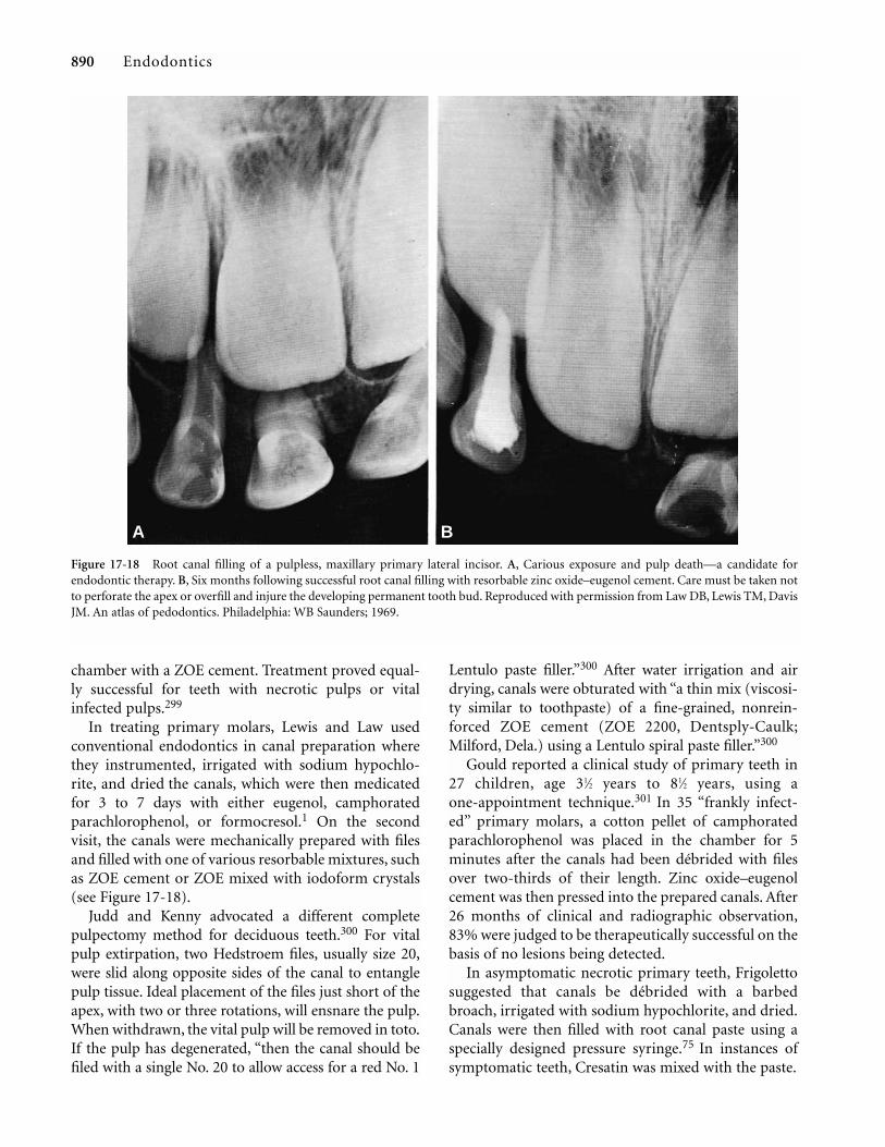
chamber with a ZOE cement. Treatment proved equal-ly successful for teeth with necrotic pulps or vitalinfected pulps.299
In treating primary molars, Lewis and Law usedconventional endodontics in canal preparation wherethey instrumented, irrigated with sodium hypochlo-rite, and dried the canals, which were then medicatedfor 3 to 7 days with either eugenol, camphoratedparachlorophenol, or formocresol.1 On the secondvisit, the canals were mechanically prepared with filesand filled with one of various resorbable mixtures, suchas ZOE cement or ZOE mixed with iodoform crystals(see Figure 17-18).
Judd and Kenny advocated a different completepulpectomy method for deciduous teeth.300 For vitalpulp extirpation, two Hedstroem files, usually size 20,were slid along opposite sides of the canal to entanglepulp tissue. Ideal placement of the files just short of theapex, with two or three rotations, will ensnare the pulp.When withdrawn, the vital pulp will be removed in toto.If the pulp has degenerated, “then the canal should befiled with a single No. 20 to allow access for a red No. 1
890 Endodontics
Lentulo paste filler.”300 After water irrigation and airdrying, canals were obturated with “a thin mix (viscosi-ty similar to toothpaste) of a fine-grained, nonrein-forced ZOE cement (ZOE 2200, Dentsply-Caulk;Milford, Dela.) using a Lentulo spiral paste filler.”300
Gould reported a clinical study of primary teeth in27 children, age 31⁄2 years to 81⁄2 years, using aone-appointment technique.301 In 35 “frankly infect-ed” primary molars, a cotton pellet of camphoratedparachlorophenol was placed in the chamber for 5minutes after the canals had been débrided with filesover two-thirds of their length. Zinc oxide–eugenolcement was then pressed into the prepared canals. After26 months of clinical and radiographic observation,83% were judged to be therapeutically successful on thebasis of no lesions being detected.
In asymptomatic necrotic primary teeth, Frigolettosuggested that canals be débrided with a barbedbroach, irrigated with sodium hypochlorite, and dried.Canals were then filled with root canal paste using aspecially designed pressure syringe.75 In instances ofsymptomatic teeth, Cresatin was mixed with the paste.
Figure 17-18 Root canal filling of a pulpless, maxillary primary lateral incisor. A, Carious exposure and pulp death—a candidate forendodontic therapy. B, Six months following successful root canal filling with resorbable zinc oxide–eugenol cement. Care must be taken notto perforate the apex or overfill and injure the developing permanent tooth bud. Reproduced with permission from Law DB, Lewis TM, DavisJM. An atlas of pedodontics. Philadelphia: WB Saunders; 1969.
A B

Pediatric Endodontics 891
Starkey has described a one-appointment andmultiappointment method of treating cariouslyinvolved primary pulp tissue.91 The one-appointmentmethod is used in cases with vital pulp tissue, in whichinflammation extends beyond the coronal pulp and noradiographic evidence of periradicular involvement ispresent. In these cases, Starkey recommended a partialpulpectomy to remove the coronal aspects of the radic-ular pulp, controlling hemorrhaging and filling thecanals and crown with a creamy mix of ZOE cement.
Starkey’s multiappointment method was advocatedfor cases with necrotic pulps and periradicularinvolvement.91 At the first appointment, coronal pulpdebris is removed, but the canals are not instrumented.A medicament such as formocresol or camphoratedmonochlorophenol is placed in the pulp chamber andsealed with IRM for 1 week. If the tooth and surround-ing gingival tissues are asymptomatic and clinicallynegative at the second visit, the canals are mechanical-ly cleansed and débrided and then filled with ZOEcement (Figure 17-19). Modifications of these proce-dures have been described by Cullen,302 Dugal andCurgon,303 Goerig and Camp,304 Kopel,305 Mathewsonand Primosch,89 and Spedding.306
It should be noted that some controversy exists withrespect to the relative effectiveness of the one-sitting and
two-sitting pulpectomy procedures. Coll et al. reportedan 80 to 86% success rate with the one-sitting tech-nique.307 Primosch et al. noted that 60% of US under-graduate dental programs teach the one-sitting techniqueversus 26% teaching the two-sitting technique.69
Extension of formocresol use to the pulpectomytechnique was a logical sequence. Vander Wall et al. haveshown formocresol to be more effective than eithercamphorated parachlorophenol or Cresatin as a rootcanal medicament for inhibiting bacterial growth.308
Several studies have evaluated the clinical and radi-ographic findings of the pulpectomy procedure fornonvital primary molars and primary anteriors usingformocresol. Coll et al. evaluated a one-appointmentformocresol pulpectomy technique for nonvital pri-mary molars. After a mean observation period of 70months, 86.1% were judged successful.307 They alsofound that successful pulpectomized primary molarswere not over-retained and the successor premolars hada very low incidence of hypoplastic defects.307
Barr et al., in a radiographic retrospective evaluationof primary molar pulpectomies performed in a privatepractice with a mean observation period of 40.2months, found an overall success rate of 85.5%.309
Noteworthy findings included 88% complete ZOEpaste resorption and a 25.8% reduction of preoperative
Figure 17-19 Three-year successful root canal filling ofmandibular second primary molar. (Courtesy of Dr. Paul E.Starkey.) The canals have been thoroughly filed and irrigat-ed at the first appointment and medicated with formocresolor camphorated parachlorophenol. At the second appoint-ment, the canals were filled with resorbable zincoxide–eugenol cement. Reproduced with permission fromLaw DB, Lewis TM, Davis JM. An atlas of pedodontics.Philadelphia: WB Saunders; 1969.
radiolucencies. These clinicians suggested that posteri-or primary molar pulpectomies have a relatively highsuccess rate in private practice.
Coll et al. and Flaitz et al. also evaluated the results ofpulpectomy treatment in primary anterior teeth.310,311
Using clinical and radiographic evaluations, Coll andcolleagues completed 27 pulpectomies in primary inci-sors and found that their 78% success rate did not differstatistically from comparable primary molar rates after amean of 45 months.310 Seventy-three percent were con-sidered to have exfoliated normally. These investigatorsconcluded, however, that documented success rates forindirect pulp capping and pulpotomies in primary ante-rior teeth were higher than for pulpectomies.
Flaitz et al.’s contrasting study compared 57 pulpo-tomies versus 87 pulpectomies in primary anteriorteeth followed for a mean of 37 months.311 Based onthe final radiographs in the study, treatment was suc-cessful in 68.5% of the pulpotomized group of anteri-or teeth versus 84% of the pulpectomized group. Theyconcluded overall that pulpectomy was a better treat-ment option for primary incisors even though theymay have shown more radiographic pathosis at thetime of the diagnosis.
Yacobi and Kenny have twice monitored their successrates in vital pulpectomy and immediate ZOE (ZOE2200, L. D. Caulk Co.; Milford, Dela.) filling. At 6 months,their success rate was comparable to the formocresolresults of 89% for anterior teeth and 92% for posteriorteeth.312 At 2 years, reporting on 81 patients and 253teeth, Payne et al., using ZOE, reported a mean successrate of 83% for anterior teeth and 90% for posteriorteeth. They conjectured that the discrepancy in ratesbetween anterior and posterior teeth was related to thefinal restorations—microleakage from composite resinin the anterior regions and stainless crowns in the poste-rior.313 They believed this to be a most acceptable alter-native method for saving primary teeth while avoidingthe compromising effects of the aldehydes.
Alternative Pulpectomy Canal Obturants
Zinc oxide–eugenol cement has been the most fre-quently used obturant in the pulpectomy technique.Primosch et al. noted that 90% of US pediatric den-tistry undergraduate programs teach ZOE as thepulpectomy obturant of choice.69 Although consideredto be resorbable, Coll et al., in a 6-year follow-up of 41pulpectomized primary molars, found that ZOE parti-cle retention in the gingival sulcus occurred in 8 of 17patients followed to the time of premolar eruption.307
Their technique included a 5-minute formocresol-blotted paper point treatment of the canals prior to
892 Endodontics
obturation. As previously mentioned, there has beenconcern about the use of formocresol in any form inpediatric endodontic therapy. Alternative pulpectomyagents have been proposed to improve on the biocom-patibility limitations of ZOE and formocresol.
Hendry et al. compared calcium hydroxide withZOE as a pulpectomy obturant in primary teeth ofdogs. At 4 weeks post-treatment, canals treated withcalcium hydroxide exhibited less inflammation, lesspathologic root resorption, and more hard tissue appo-sition than ZOE and control-treated teeth.314
Barker and Lockett identified the potential benefitsof Kri paste, an iodoform compound, also containingparachlorophenol, camphor, and menthol. The advan-tages of this material include bactericidal propertiesand excellent resorbability. Histologically, they foundthat this material easily resorbed even when extrudedbeyond the apex of the treated teeth. An ingress of con-nective tissue was seen in the apical portions of thetreated canals.315 Bactericidal iodoform pastes havebeen reintroduced as a root canal filling.245,316,317
Garcia-Godoy obtained a 95.6% success clinically andradiographically with Kri paste during a 24-monthperiod for 43 teeth.318 It was noted that this pastewould resorb within 2 weeks if found in the periradic-ular or furcation areas.319 Rifkin reported an 89% clin-ical and radiographic success rate after 1 year with Kripaste pulpectomies in primary teeth.296 Holan andFuks clinically and radiographically compared Kripaste with ZOE in human primary molars after 48months postoperatively. They found overall successrates of 84% with the Kri paste group versus 65% withthe ZOE group. Kri paste was almost twice as success-ful in primary first molars as ZOE. However, no signif-icant differences between these two agents occurred inprimary second molars. Overfills with Kri paste result-ed in 79% success versus 41% success with ZOE. Theyconcluded that iodoform-containing paste had apotential advantage over ZOE in the pulpectomy pro-cedure for primary teeth.319
Treatment Considerations
The preceding review demonstrates the varied tech-niques and successes for mastering pulp therapy fornonvital and irreversibly inflamed primary teeth.Before outlining treatment methods, special considera-tions, indications, and contraindications must beaddressed by the clinician.
General Considerations.
1. The patient should be healthy and cooperative. Ifany systemic disorders are present that would com-

Pediatric Endodontics 893
promise a child’s responses, the child’s physician ormedical team should be consulted.
2. Informed consent, with a clear explanation of theprocedure to the parents, must be obtained.
Dental Considerations.
1. The tooth must be restorable after the root canaltreatment.
2. Chronologic and dental age must be evaluated torule out teeth with eminent exfoliation.
3. Psychological or cosmetic factors (anterior primaryteeth) must be considered, which are often moreimportant to the parent than to the child.
4. The number of teeth to be treated and strategicimportance to the developing occlusion must beevaluated.
5. Primary molar root anatomy, along with the prox-imity of the underlying succedaneous tooth, must beevaluated.
Indications for a Pulpectomy Procedure.
1. Primary teeth with pulpal inflammation extendingbeyond the coronal pulp but with roots and alveo-lar bone free of pathologic resorption
2. Primary teeth with necrotic pulps, minimum rootresorption, and minimum bony destruction in thebifurcation area
3. Pulpless primary teeth with sinus tracts4. Pulpless primary teeth without permanent successors5. Pulpless primary second molars before the erup-
tion of the permanent first molar6. Pulpless primary teeth in hemophiliacs7. Pulpless primary anterior teeth when speech,
crowded arches, or esthetics are a factor8. Pulpless primary teeth next to the line of a palatal
cleft9. Pulpless primary molars supporting orthodontic
appliances10. Pulpless primary molars when arch length is defi-
cient11. Pulpless primary teeth when space maintainers or
continued supervision are not feasible (handi-capped or isolated children†)
Contraindications.
1. Teeth with nonrestorable crowns2. Periradicular involvement extending to the perma-
nent tooth bud3. Pathologic resorption of at least one-third of the
root with a fistulous sinus tract4. Excessive internal resorption5. Extensive pulp floor opening into the bifurcation6. Young patients with systemic illness such as congen-
ital or rheumatic heart disease, hepatitis, orleukemia and children on long-term corticosteroidtherapy or those who are immunocompromised
7. Primary teeth with underlying dentigerous or follic-ular cysts
Clinical Procedures: Partial Pulpectomy
Partial pulpectomy can be considered an extension ofthe pulpotomy procedure in that the coronal portion ofthe radicular pulp is amputated, leaving vital tissue inthe canal that is assumed to be healthy. Although dis-cussed in the context of nonvital pulp therapy, techni-cally, it is a vital pulp therapy technique. The decisionto implement the partial pulpectomy is made afterremoving the coronal pulp from the chamber andencountering difficulty with hemorrhage control fromthe radicular orifice.
Hemorrhage control is achieved with endodonticbroaches used to remove one-third to one half of thecoronal portion of the radicular pulp tissue from thecanals. The canals and chamber are irrigated withhydrogen peroxide followed by sodium hypochloriteand then dried with cotton pellets. If hemorrhage isstill impossible to control, all remaining radicular pulptissue is to be removed, and the complete pulpectomyprocedure must be implemented.
After successful hemorrhage control from theamputated radicular pulp, a formocresol-dampenedcotton pellet, squeezed dry, is placed in the pulp cham-ber for 1 to 5 minutes. The pellet is removed, and anonreinforced fast-setting ZOE cement is packed withpressure into the chamber and canals. A radiograph isthen taken, and if the canals appear to be adequatelyfilled, a stainless steel crown is placed as a permanentrestoration (Figure 17-20).
Clinic Procedures: Complete Pulpectomy
The child with a necrotic primary tooth presents a con-siderable challenge for the clinician. In some instances,the tooth may be totally asymptomatic from a clinicalstandpoint. In other instances, the tooth may be acute-ly or chronically abscessed, mobile and painful, with
†Owing to the isolation of the children involved in its Bureau ofIndian Health Affairs, the US Public Health Service has recom-mended root canal filling of primary teeth, whenever feasible,rather than space maintainers that require lengthy supervision.320

swollen periodontal tissues. In the latter case, the childmay be apprehensive and irritable, making relief ofpain and swelling the highest priority. In cases ofnondraining alveolar abscesses and cellulitis fromodontogenic origin, antibiotic therapy using first- orsecond-generation penicillins should be immediatelyprescribed for a period of 4 to 7 days. Canal instru-mentation can then be implemented.
The complete pulpectomy procedure involves thefollowing considerations. Under local anesthesia, thepulp chamber is carefully opened with a high-speedbur to relieve any pressure from the infected pulp. Alow-speed round bur or a spoon excavator may be usedto clean out the pulp chamber, which is then irrigatedwith sodium hypochlorite. In cases of acute inflamma-tion, a camphorated monochlorophenol-dampenedcotton pellet is placed in the coronal chamber as aninterim medicament to chemically sterilize the pulpcanals. In cases of chronic abscess formation, aformocresol-dampened cotton pellet is generally usedas the interim medicament. The chambers are thensealed with a fast-setting ZOE cement, and the toothmay need to be equilibrated to avoid hyperocclusion.
At the end of 1 week, if all acute symptoms includ-ing pain and soft tissue swelling have resolved, finalcanal preparation—careful enlargement and débride-ment—is completed with Hedstroem files. Canal irri-gation is accomplished with hydrogen peroxide andsodium hypochlorite. Tooth length should be carefully
894 Endodontics
determined, and instrumentation should not extendbeyond the apex. Fine files, in ultrasonic or sonicendodontic handpieces, with copious irrigation can beconsidered in this protocol.
The ribbon-shaped and tortuous root canals of pri-mary teeth present a time-consuming problem inobtaining adequate obturation. A pressure syringe wasdeveloped by Greenberg for filling primary canals.321
This technique has been described in detail bySpedding306 and by Krakow et al..322 The material ofchoice for filling the root canals of pulpectomized pri-mary teeth is pure ZOE, first mixed as a slurry and car-ried into the canals using either paper points, a syringe,a “Jiffy tube,” or a lentulo spiral root canal filler. Aylardand Johnson showed that the lentulo was the best over-all ZOE root canal–filling instrument for curved canalsand the pressure syringe technique was best for straightcanals.323 The slurry may be further compressed intothe canals by packing the chamber with a stiffer mix ofZOE. After the canals are estimated to be filled to thechamber floor, the chamber itself is filled with a suit-able cement such as a reinforced ZOE or a glassionomer. As previously mentioned, the tooth is pre-pared for the placement of a stainless steel crown afterevaluation of the canal filling by a radiograph.90
Mack and Halterman described the rationale andtechnique for an innovative approach to pulpectomy forprimary anterior teeth by using a labial entry to thecanal instead of the traditional lingual opening. Thisallows greater ease of instrumentation and providesincorporation of the access chamber in the esthetic labi-al veneer preparation. Bonded composite resin is used tocomplete the final esthetic restoration.324
Clinical Variation: Pulpotomy for Nonvital Primary Teeth
Less demanding techniques than the pulpectomy havebeen reported for treating irreversibly inflamed pri-mary teeth, usually involving the formocresol pulpoto-my technique. Ripa recommended that, owing to theanatomy of primary tooth canals, it would be mucheasier to perform complete débridement in nonvitalprimary molars using a pulpotomy technique, withappropriate medicaments.325 Although there is evi-dence to support such a concept, the consensus is thatthe pulpotomy technique should be confined to teethmeeting the selection criteria for vital pulp therapy.326
Velling327 and Droter328 reported high degrees of suc-cess in nonvital primary molars with a coronal pulpoto-my only, using formaldehyde-type medicaments in eitherone- or two-appointment visits. The final dressing in thecoronal pulp chamber was usually a modified ZOE
Figure 17-20 Root canal obturation and crown restoration for apulpless primary molar. At the first appointment, followingmechanical and chemical canal débridement, medicament is sealedin place for 1 week. The canal is obturated at the second appoint-ment with resorbable zinc oxide–eugenol cement. A, Zincoxide–eugenol cement root canal filling. B, Oxyphosphate of zinccement. C, Stainless steel crown.

Pediatric Endodontics 895
cement. Full described complete success in 20 childrenwith chronically abscessed primary molars and drainingfistulae in a two-appointment formocresol technique.329
All of the draining fistulae were reported to be resolved.As an extension of this technique, Meyer and Sayegh useda combination treatment of formocresol in the pulpchamber and curettage of the bifurcation to achieve an87% clinical success at 5 years postoperatively.330
In a survey of members of the American Academy ofPediatric Dentistry, success rates of 72% were reportedin nonvital primary molars that had been treated bycleaning only the coronal chamber and placingformocresol versus instrumenting the canals. Bothtreatments were concluded by filling the respectivelycleansed areas with a resorbable medicated cement andplacing a stainless steel crown.331
Myers et al., in a recent histologic study of failedpulpotomies in primary molars, stated that the devel-opment of a furcation lesion has the potential for cys-tic transformation and the tooth should be extract-ed.332 This conclusion also implies that pulpectomytreatment for nonvital primary molars with furcationlesions is contraindicated.
Pulpectomy Outcomes
Negative sequelae from endodontically treated primaryteeth in the form of accelerated resorption and exfolia-tion have been a major concern of many clinicians.Starkey felt that delayed eruption of the permanent suc-cessors sometimes followed pulpotomy and pulpecto-my treatment of primary molars, with some possibledeflection in the eruption path.333 This sequela was notseen in the studies by Barr et al.309 and Coll et al..310
Ankylosis of the primary tooth with a root canal filling
has also been noted. Erausquin and Devoto have shownthat formaldehyde-containing cements frequentlycaused partial ankylosis at different levels of the peri-odontal ligament.334 Coll and Sadrian, in a retrospectivestudy of pulpectomy outcomes, noted two parametersthat were the highest predictors of success.Pretreatment pathologic root resorption, when evi-dent, resulted in a 44.4% prevalence of enamel defectsin underlying permanent teeth after their eruption. Inthe absence of pretreatment pathologic root resorption,the pulpectomy success rate was 91.7%. The quality ofcanal fill relative to the apex was another outcomedeterminant, with 86.5% success rates occurring forcanals filled short of the apex, 88.9% success for canalsfilled to the apex, and 57.7% success for canals filledbeyond the apex.335
SUMMARY
The rationale for pediatric pulp therapy has developedout of extensive clinical studies and improved histolog-ic techniques. Ongoing research will result in modifica-tions that will enhance treatment outcomes.
A successful pediatric endodontic outcome shouldbe based on (1) re-establishment of healthy periodon-tal tissues; (2) freedom from pathologic root resorp-tion; (3) maintenance of the primary tooth in an infec-tion-free state to hold space for the eruption of its per-manent successor; (4) in the case of young permanentteeth, maintenance of the maximum amount of nonin-flamed portions of pulp tissue to enhance apexogenesisand root dentin formation. With adherence to soundprinciples in case selection and techniques, pediatricpulp therapy is a major health benefit to the child. Thetreatment modalities and medicaments that have been
Table 17-2 Pulp Treatment Summary: Current Recommendations
Indirect pulp cap Permanent teeth, primary teeth—calcium hydroxide glass ionomer cement, resin bonding agent
Direct pulp cap Permanent teeth—calcium hydroxide, mineral trioxide aggregate, resin bonding agent (?)
Direct pulp cap Primary teeth (mechanical exposures only)—calcium hydroxide
Pulpotomy Primary teeth—diluted and full-strength formocresol, glutaraldehyde, ferric sulfate (?),controlled energy techniques (?)
Pulpotomy Permanent teeth (apexogenesis)—calcium hydroxide, formocresol (?), glutaraldehyde(?)
Partial pulpectomy Primary teeth—zinc oxide–eugenol, zinc oxide–eugenol + formocresol
Complete pulpectomy Permanent teeth (apexification)—calcium hydroxide
Complete pulpectomy Primary teeth—zinc oxide–eugenol, zinc oxide–eugenol + formocresol, iodoform-containing pastes (?), calcium hydroxide (?)

discussed are summarized in Table 17-2, highlightingthe most substantiated and qualifying those that needfurther confirmation by additional research. The clini-cian must realize that these recommendations are notabsolute and will continue to be modified.
REFERENCES 1. Lewis TM, Law DC. Pulpal treatment of primary teeth. In:
Finn SB, editor. Clinical pedodontics. 4th ed. Philadelphia:WB Saunders; 1973.
2. Finn SB. Morphology of the primary teeth. In: Finn SB, editor.Clinical pedodontics. 4th ed. Philadelphia: WB Saunders;1973.
3. McDonald RE, Avery DR. Treatment of deep caries, vital pulpexposure and pulpless teeth. In: McDonald RE, Avery DR,editors. Dentistry for the child and adolescent. 6th ed. St.Louis: CV Mosby; 1995. p. 428–54.
4. Moss SJ, Addleston H. Histologic study of the pulpal floor ofdeciduous molars. J Am Dent Assoc 1965;70:372.
5. Rengelstein D, Slow WK. The prevalence of furcation forami-na in primary molars. Pediatr Dent 1989;11:198.
6. Wrbas KT, Kielbassa AM, Hellwig E. Microscopic studies ofaccessory canals in primary molar furcations. J Dent Child1997;64:118.
7. Greely CB. Pulp therapy for the primary and young perma-nent dentition. In: Forrester DJ, Wagner ML, Fleming J,editors. Pediatric dental medicine. Philadelphia: Lea &Febiger; 1981. p. 456–60.
8. Seltzer S, Bender IB. The dental pulp—biologic considerationsin dental procedures. 3rd ed. Philadelphia: JB Lippincott;1984.
9. Cox CF, Suzuki S. Re-evaluating pulp protection: calciumhydroxide liners vs. cohesive hybridization. J Am DentAssoc 1994;125:823.
10. Fox AG, Heeley JD. Histologic study of human primary teeth.Arch Oral Biol 1980;25:103.
11. Massler M. Preventive endodontics: vital pulp therapy. DentClin North Am 1967; p.670.
12. Bernick S. Innervation of the teeth and periodontium. DentClin North Am 1959; p.503.
13. Rapp R, et al.. The distribution of nerves in human primaryteeth. Anat Rec 1967;159:89.
14. Benzer G, Bevelander S. Morphology and incidence of second-ary dentin in human teeth. J Am Dent Assoc 1943;30:1075.
15. Ireland RI. Secondary dentin formation in deciduous teeth. JAm Dent Assoc 1941;28:1626.
16. Corbett MD. The incidence of secondary dentin in cariousteeth. Br Dent J 1963;114:142.
17. Sayegh FS. Qualitative and quantitative evaluation of newdentin in pulp capped teeth. J Dent Child 1968;35:7.
18. McDonald RE. Diagnostic aids and vital pulp therapy fordeciduous teeth. J Am Dent Assoc 1956;53:14.
19. Anderson AW, Sharav Y, Massler M. Reparative dentin forma-tion and pulp morphology. Oral Surg Oral Patts Oral Med1948;26:837.
20. Frankl SH. Pulp therapy in pedodontics. Oral Surg1972;34:192.
21. Eidelman E, et al. Pulp pathology in deciduous teeth: clinicaland histological correlations. Isr J Med Sci 1968;4:1244.
22. Prophet AS, Miller J. The effect of caries on the deciduouspulp. Br Dent J 1955;99:105.
896 Endodontics
23. Rayner JA, Southam II. Pulp changes in deciduous teeth asso-ciated with deep carious dentin. J Dent 1970;7:39.
24. Taylor B, et al. Response of the pulp and dentine to dentalcaries in primary molars. J Int Assoc Dent Child 1971;2:3.
25. Damele J. Clinical evaluation of indirect pulp capping: aprogress report. J Dent Res 1961;40:756.
26. Belanger G K. Pulp therapy for young permanent teeth. In:Pinkham JR, Cassamassimo PS, Fields HW, et al., editors.Pediatric dentistry—infancy through adolescence. 2nd ed.Philadelphia: WB Saunders; 1988. p. 483–91.
27. Troutman K, et al. Vital pulp therapy: part 1. In: Stewart RE, etal., editors. Pediatric dentistry. St. Louis: CV Mosby; 1982.p. 916.
28. DiMaggio JJ, et al. Histological evaluation of direct and indi-rect pulp capping [abstract]. J Dent Res 1963.
29. Trowbridge H, Berger J. The clinical management of deep car-ious lesions. Calif Dent J 1971;47:26.
30. Ishibaski M. A comparison between results of clinical andhisto-pathological diagnosis of disease of deciduous toothpulp. Dent Abstr 1958;3:547.
31. Reynolds RL. The determination of pulp vitality by means ofthermal and electrical stimuli. Oral Surg 1966;22:231.
32. Tomes J. A system of dental surgery. London: I. Churchill;1859.
34. Miller WD. What antiseptics should be used for sterilizing cav-ities before filling. Dent Cosmos 1891;33:337.
35. Black GV. A work on operative dentistry. Vol 2. Chicago:Medico-Dental Publishing; 1908.
36. Dorfman A, Stephan RM, Muntz JA. In vitro studies of cariousdentin. II. Extent of infection in carious lesions. J Am DentAssoc 1943;30:1901.
37. MacGregor AB, et al. Experimental studies of dental caries. I.The relationship of bacterial invasions to softening of thedentine. Br Dent J 1956;101:203.
38. Fusayama T, et al. Relationship between hardness, discol-oration and microbial invasion in carious dentin. J DentRes 1966;45:1033.
39. Fusayama T. Two layers of carious dentin: diagnosis and treat-ment. Oper Dent 1979;4:63.
40. Whitehead FI, et al. The relationship of bacterial invasion ofsoftening of the dentin in permanent and deciduous teeth.Br Dent J 1960;108:261.
41. Shovelton DS. A study of deep carious dentin. Int Dent J1968;18:392.
42. Seltzer S, Bender IB. The dental pulp. Philadelphia: JBLippincott; 1965. p. 184–98.
43. Reeves R, Stanley HR. The relationship of bacterial penetra-tion and pulpal pathosis in carious teeth. Oral Surg1966;22:59.
44. Shovelton DS. Studies of dentin and pulp in deep caries. IntDent J 1970;20:283.
45. Massler M. Op Cit. Ref no. 11.46. Massler M, Pawlak J. The affected and infected pulp. Oral Surg
1977;43:929.47. Canby CP, Bernier JL. Bacteriologic studies of carious dentin.
J Am Dent Assoc 1936;23:2083.48. Kuwabara RK, Massler M. Pulpal reactions to active and
arrested carious lesions. J Dent Child 1966;33:190.49. Bradford EW. The dentin, a barrier to caries. Br Dent J
1960;109:387.

Pediatric Endodontics 897
50. Young MA, Massler M. Some physical and chemical character-istics of carious dentin. Br Dent J 1963;115:406.
51. Leung RL, et al. Effect of Dycal on bacteria in deep cariouslesions. J Am Dent Assoc 1980;10:193.
52. Fairbourn DR, et al. Effect of improved Dycal and IRM on bac-teria in deep carious lesions. J Am Dent Assoc 1980;100:547.
53. Law DB, Lewis TM. The effects of calcium hydroxide on deepcarious lesions. Colorado Dent J 1964;42:27.
54. Held-Wydler E. Natural (indirect) pulp capping. J Dent Child1964;31:107.
55. Kerkhove BC, et al. A clinical and television densitometricevaluation of the indirect pulp capping technique. J DentChild 1967;34:192.
56. Ehrenreich DV. A comparison of the effects of zincoxide-eugenol and calcium hydroxide in carious dentin inhuman primary molars. J Dent Child 1968;35:451.
57. Stanley HR. Pulpal response to dental techniques and materi-als. Dent Clin North Am 1971;15:115.
58. Torstenson B, et al. Pulp reaction to IRM [abstract]. J Dent Res1982;61:573.
59. Nordstrom DO, et al. Use of stannous fluoride for indirectpulp capping. J Am Dent Assoc 1974;88:997.
60. King J, et al. Indirect pulp capping: a bacteriologic study ofdeep carious dentin in human teeth. Oral Surg 1965;20:663.
61. Aponte AJ, Hartsook JT, Crowley MC. Indirect pulp cappingverified. J Dent Child 1966;33:164.
62. Parikh SR, Massler M, Bahn A. Microorganisms in active andarrested carious lesions of dentin. N Y State Dent J1963;29:347.
63. Eidelman E, et al. Remineralization of carious dentin treatedwith calcium hydroxide. J Dent Child 1965;32:218.
64. Mjör IA, et al. The effect of calcium hydroxide and amalgamon noncarious vital dentin. Arch Oral Biol 1961;3:283.
65. Sawusch RH. Direct and indirect pulp capping with two newproducts. J Am Dent Assoc 1982;104:459.
66. Nirschl R, Avery D. Evaluation of a new pulp capping agent inindirect pulp therapy. J Dent Child 1983;50:25.
67. Coll JA, et al. An evaluation of pulp therapy in primary inci-sors. Pediatr Dent 1988;10:178.
68. Schroder A. Indirect capping and the treatment of deep cari-ous lesions. Int Dent J 1968;18:381.
69. Primosch RE, Glomb TA, Jerrell RG. Primary tooth pulp ther-apy as taught in predoctoral pediatric dental programs inthe United States. Pediatr Dent 1997;19:118–22.
70. Lado EA, Stanley HR. An in vitro study of bacterial inhibitionby VLC calcium hydroxide pulp cap compounds. PediatrDent 1987;9:292.
71. Jordan RE, Suzuki M. Conservative treatment of deep cariouslesions. Can Dent Assoc J 1971;37:337.
72. Fuks AB. Pulp therapy for the primary dentition. In: PinkhamJR, Cassamassimo PS, Fields HW, et al., editors. Pediatricdentistry—infancy through adolescence. 2nd ed. WBSaunders; 1988. p. 326–38.
73. Langeland K. Capping exposed pulpal tissue. Dent Clin NorthAm 1974;2:1.
74. Stanley HR. Criteria for standardizing and increasing credibil-ity of direct pulp capping studies. Am. J Dent 1998;11:S17.
75. Frigoletto RT. Pulp therapy in pedodontics. J Am Dent Assoc1973;86:1344.
76. Tronstad L, Mjör I.A. Capping of the inflamed pulp. Oral Surg1972;34:477.
77. Cox CF, Bergenholtz G. Healing sequence in capping inflameddental pulps of rhesus monkeys. Int Endod J 1986;19:113.
78. Heide S, Kerekes K. Delayed direct pulp capping in permanentincisors of monkeys. Int Endod J 1987;20:65.
79. Cotton WR. Bacterial contamination as a factor in healing ofpulp exposures. Oral Surg 1974;38:441.
80. Glass RL, Zander HA. Pulp healing. J Dent Res 1949;28:97.81. Lim KC, Kirk EE. Direct pulp capping: a review. Endod Dent
Traumatol 1987;3:213.82. Stanley HR, Lundy T. Dycal therapy for pulp exposures. Oral
Surg 1972;34:818.83. Weiss MD, Bjorvatn K. Pulp capping in deciduous and newly
erupted permanent teeth of monkeys. Oral Surg 1970;29:769.84. Kakehashi S, Stanley HR, Fitzgerald R J. The effects of surgical
exposures of the dental pulp in germ free and convention-al laboratory rats. Oral Surg 1965;20:340.
85. Langeland K, et al. Human pulp changes of iatrogenic origin.Oral Surg 1971;32:943.
86. Cox CF, Subay RK, Ostro E, et al. Tunnel defects in dentinbridges: their formation following direct pulp capping.Oper Dent 1996;21:4.
87. Baume L, Holz J. Long-term clinical assessment of direct pulpcapping. Int Dent J 1981;31:251.
88. Kennedy DB, Kapala JT. The dental pulp: biologic considera-tions of protection and treatment. In: Braham RL, MorrisME, editors. Textbook of pediatric dentistry. 2nd ed.Baltimore: Williams and Wilkins; 1985. p. 492–522.
89. Mathewson RJ, Primosch R. Pulp treatment. In: Fundamentalsof pediatric dentistry. 3rd ed. Chicago: Quintessence; 1995.p. 257–84.
90. Camp J. Pulp therapy for primary and young permanent teeth.Dent Clin North Am 1984;28:651.
91. Starkey PE. Treatment of pulpally involved primary molars. In:McDonald RE, et al., editors. Current therapy in dentistry.Vol VII. St. Louis: CV Mosby; 1980.
92. Weine DM. The effect of calcium hydroxide in direct pulp cap-ping in primary teeth [thesis]. Ann Arbor [MA]: Univ. ofMichigan School of Dentistry; 1963.
93. Jeppersen K. Direct pulp capping on primary teeth—along-term investigation. Int Assoc Dent Child 1971;12:10.
94. Jerrell RG, Courts FK, Stanley HR. A comparison of two calci-um hydroxide agents in direct pulp capping of primaryteeth. J Dent Child 1984;51:34.
95. Kalins V, Frisbee HE. The effect of dentine fragments on thehealing of the exposed pulp. Arch Oral Biol 1960;2:96.
96. Schröder U, et al. A one-year follow-up of partial pulpotomyand calcium hydroxide capping in primary molars. EndodDent Traumatol 1987;3:304.
97. Arnold DS. The use of formocresol as a pulp capping agent inhuman primary teeth [thesis]. Lincoln, NE: Univ. ofNebraska School of Dentistry; 1970.
98. Cvek M. A clinical report on partial pulpotomy and cappingwith calcium hydroxide in permanent incisors with com-plicated crown fracture. J Endod 1978;4:232.
99. Zilberman U, et al. Partial pulpotomy in carious permanentmolars. Am J Dent 1989;2:147.
100. Mejare I, Cvek M. Partial pulpotomy in young permanentteeth with deep carious lesions. Endod Dent Traumatol1993;9:238.
101. Fuks AB, Chosack A, Klein H, Eidelman E. Partial pulpotomy asa treatment alternative for exposed pulps in crown-fracturedpermanent incisors. Endod Dent Traumatol 1987;3:100.

898 Endodontics
125. Hembree JH, Andrews IT. Zinc oxide as a pulp capping agent.Miss Dent J 1974;30:10.
126. Watts A. Zinc oxide as a pulp capping agent [abstract]. J DentRes 1977;56:D104.
127. Holland R, et al. The influence of the sealing material in the heal-ing process of inflamed pulps capped with calcium hydroxideor zinc oxide-eugenol cement. Acta Odontol Pediatr 1981;2:5.
128. Sveen OB. Pulp capping of primary teeth with zincoxide-eugenol. Odontol Tidskr 1969;77:427.
129. Brosch JW. Capping pulps with a compound of calcium phos-phate, neomycin and hydrocortisone. J Dent Child1966;33:42.
130. Soldali GD. Pulp capping with antibiotics. Fla Dent J1975;46:18.
131. Bhaskar SH, et al. Tissue response to cortisone-containing andcortisone-free calcium hydroxide. J Dent Child 1969;36:1.
132. Ulmansky M, Sela J. Response of pulpotomy wounds in nor-mal human teeth to successively applied Ledermix andCalxyl. Arch Oral Biol 1971;16:1393.
133. Haskell EW, et al. Direct pulp capping treatment: a long-termfollow-up. J Am Dent Assoc 1978;97:607.
134. McWalter G, et al. Long-term study of pulp capping in mon-keys with three agents. J Am Dent Assoc 1976;933:105.
135. Watts A, Paterson RC. The response of the mechanicallyexposed pulp to prednisolone and triamcinolone ace-tonide. Int Endod J 1988;21:9.
136. Gardner DE, Mitchell DF, McDonald RE. Treatment of pulpsof monkeys with vancomycin and calcium hydroxide. JDR1971;50:1273.
137. Negm M, et al. Clinical and histologic study of human pulpalresponse to new cements containing calcium hydroxide.Oral Surg 1980;50:462.
138. Bhaskar SH, et al. Human pulp capping with isobutylcyano-acrylate. J Dent Res 1972;51:50.
139. Heys DR, Cox CF, Avery JK. Histological considerations ofdirect pulp capping agents. J Dent Res 1981;68:1371.
140. Subay RK, Asci S. Human pulpal response to hydroxyapatiteand a calcium hydroxide material as direct capping agents.Oral Surg 1993;76:485.
141. Dick HM, Carmichael DJ. Reconstituted antigen poor collagenpreparations as potential pulp capping agents. JOE1980;6:641.
142. Fuks AB, et al. Enriched collagen solution as a pulp dressing inpulpotomized teeth in monkeys. Pediatr Dent 1984;6:243.
143. Ibrahim SM, Mitchell DF, Healy HG. A histopathologic studyof the effects of formocresol in pulp capping of permanentteeth. Egypt Dent J 1970;16:219.
144. Garcia-Godoy F. Direct pulp capping and partial pulpotomywith diluted formocresol in primary molars. Acta OdontolPediatr 1984;5:57.
145. Bergenholtz G, et al. Bacterial leakage around dental restora-tions: its effect on the dental pulp. J Oral Pathol 1982;11:439.
146. Cox CF. Evaluation and treatment of bacterial microleakage.Am J Dent 1994;7:293–5.
147. Snuggs HM, Cox CF, Powell CS, White KC. Pulpal healing anddentinal bridge formation in an acidic environment.Quintessence Int 1993;24:501.
148. Barnes IE, Kidd EAM. Disappearing Dycal. Br Dent J1980;111:147.
149. Cox CF. Effects of adhesive resins and various dental cementson the pulp. Oper Dent 1992;55:165.
102. Watts A, Paterson RC. Bacterial contamination as a factorinfluencing the toxicity of materials to the exposed pulp.Oral Surg 1987;64:466.
103. Cox CF. Pulp capping of dental pulp mechanically exposed tooral microflora: a 1-2 year observation of wound healing inthe monkey. J Oral Pathol 1985;14:156.
104. Cox CF, et al. Biocompatibility of surface sealed dental materi-als against exposed pulps. J Prosthet Dent 1987;57:1.
105. Nyborg H. Capping of the pulp. The processes involved andtheir outcome. Odontol Tidskr 1958;66:296.
106. Phaneuf RA, Frankl S, Reuben M. A comparative histologicalevaluation of three calcium hydroxide preparations onhuman primary dental pulp. J Dent Child 1968;35:61.
107. Vermeersch AC. Pulp conservation. Oper Dent 1977;2:105.108. Joos RW. Calcium hydroxide as a pulp capping agent.
Northwest Dent 1974;53:362.109. Tamburic SD, Vuleta GM, Ognjanovic JM. In vitro release of
calcium and hydroxyl ions from two types of calciumhydroxide preparations. Int Endod J 1993;26:125.
110. Yoshiba K, Yoshiba N, Nakamura H, et al., editors.Immunolocalization of fibronectin during reparativedentinogenesis in human teeth after pulp capping with cal-cium hydroxide. J Dent Res 1996;75:1590.
111. Fisher FJ. The effect of a calcium hydroxide/water paste onmicro-organisms in carious dentine. Br Dent J1972;143:231.
112. Lado EA. In vitro antimicrobial activity of six pulp cappingagents. Oral Surg 1986;61:197.
113. Estrela C, Fabiana CP, Yoto I, Bammann LL. In vitro determi-nation of direct antimicrobial effect of calcium hydroxide.JOE 1998;24:15.
114. Sciaky I, Pisanti S. Localization of calcium placed over ampu-tated pulps in dogs teeth. J Dent Res 1960;39:1128.
115. Attalla MN, Noujaim AA. Role of calcium hydroxide in theformation of reparative dentin. Can Dent Assoc J1969;35:267.
116. Stark MM, et al. The localization of radioactive Ca(OH)2(Ca45) over exposed pulps in rhesus monkey teeth: a pre-liminary report. J Oral Ther Pharmacol 1964;3:290.
117. Holland R, et al. Histochemical analysis of the dog’s dentalpulp after pulp capping with calcium, barium, and stron-tium hydroxides. JOE 1982;8:444.
118. Turner C, Courts FJ, Stanley HR. A histological comparison ofdirect pulp capping agents in primary canines. J Dent Child1987;54:423.
119. Tronstad L. Reaction of the exposed pulp to Dycal treatment.Oral Surg 1974;38:945.
120. Drahein R, Murrey A. Compressive strength of two calciumhydroxide bases. J Prosthet Dent 1985;54:365.
121. Fuks AB, et al. Clinical and radiographic assessment of directpulp capping and pulpotomy in young permanent teeth.Pediatr Dent 1982;4:240.
122. Stanley HR, Pameijer CH. Pulp capping with a newvisible-light-curing calcium hydroxide composition. OperDent 1985;10:156.
123. Seale NS, Stanley HR. Pulpal response to a new visible lightcured composite calcium hydroxide pulp capping agent inhuman premolars. Pediatr Dent 1988;10:239.
124. Howerton B, Cox CF. Pulpal biocompatibility of twophoto-cured Ca(OH)2 containing materials [abstract].JOE 1987;13:129.

Pediatric Endodontics 899
150. Kanca J III. Bonding to tooth structure—a rationale for a clin-ical protocol. J Esthet Dent 1989;1:135–8.
151. Miyakoshi S, et al. Interfacial interaction of 4-META-MMA-TBBresin and the pulp [abstract]. J Dent Res 1993;72:220.
152. Cox CF, et al. Pulp response following in vivo etching and4-META bonding. J Dent Res 1993;72:213.
153. Cox CF, White KC. Biocompatibility of amalgam on exposedpulps employing a biological seal [abstract]. J Dent Res1992;71:187.
154. Tsuneda Y, Hayakawa T, Yamamoto H, et al. A histopathologi-cal study of direct pulp capping with adhesive resins. OperDent 1995;20:223.
155. Kashiwada T, Takagi M. New restoration and direct pulp cap-ping systems using adhesive composite resin. Bull TokyoMed Dent Univ 1991;38:45.
156. Heitman T, Unterbrink G. Direct pulp capping with a dentinaladhesive resin system—a pilot study. Quintessence Int1995;26:765.
157. Bazzuchi M, et al. Pulp response to direct capping of adhesiveresins and glass ionomer cements [abstract]. J Dent Res1995;74:555.
158. Kanca J III. Replacement of a fractured incisor fragment overpulpal exposure: a long-term case report. Quintessence Int1996;27:829.
159. Stanley HR. Pulpal consideration of adhesive materials. OperDent 1992;55:151.
160. Pameijer CH, Stanley HR. The disastrous effects of the “totaletch” technique in vital pulp capping in primates. Am JDent 1998;11:S45.
161. Araujo FB, Barata JS, Costa CAS, Garcia-Godoy F. Clinical,radiographical and histological evaluation of direct cap-ping with a resin adhesive in primary teeth [abstract]. JDent Res 1996;75:280.
162. Gwinnett AJ, Tay FR. Early and intermediate time response ofthe dental pulp to an acid etch technique in vivo. Am JDent 1998;11:S35.
163. Kopel HM. The pulp capping procedure in primary teeth“revisited.” J Dent Child 1997;64:327.
164. Cox CF, et al. Biocompatibility of various dental materials:pulp healing with a surface seal. Int J Periodont Rest Dent1996;16:241.
165. Stanley HR, Pameijer CH. Dentistry’s friend—calciumhydroxide. Oper Dent 1997; 22:1.
166. Katoh Y, et al. Direct capping effects on exposed pulp of maca-ca fascicularis [abstract]. J Dent Res 1996;75:34.
167. Katoh Y, Kimura T, Inaba T. Clinical prognosis of pulp tissuedirect-capping with adhesive resins [abstract]. J Dent Res1997;76:162.
168. Torabinejad M, et al. Physical and chemical properties of a newroot-end filling material. JOE 1995;21:349.
169. Pitt Ford TR, Torabinejad M, Abedi HR, et al. Using mineraltrioxide aggregate as a pulp capping material. J Am DentAssoc 1996;127:1491.
170. Chaung HM, et al. Comparison of calcium phosphate cementmixture and pure calcium hydroxide as direct pulp-cap-ping agents. J Formos Med Assoc 1996;95:545.
171. Yoshimine Y, Maeda K. Histologic evaluation of tetracalciumphosphate-based cement as a direct pulp-capping agent.Oral Surg Oral Med Oral Pathol Oral Radiol Endod1995;79:351.
172. Dannenberg JL. Pedodontic endodontics. Dent Clin North Am1974;18:367.
173. Mejare I. Pulpotomy of primary molars with coronal or totalpulpitis using formocresol technique. Scand J Dent Res1979;87:208.
174. Ranly DM. Pulpotomy therapy in primary teeth: new modali-ties for old rationales. Pediatr Dent 1994;16:403.
175. Berger JE. A review of the erroneously labeled “mummifica-tion” techniques of pulp therapy. Oral Surg 1972;34:131.
176. Urist M. Bone formation by autoinduction. Science1965;150:893.
177. Nakashima M. Induction of dentin formation on canineamputated pulp by recombinant human bone morpho-genetic proteins (BMP)-2 and -4. J Dent Res 1994;73:1515.
178. Fadhavi S, Anderson AW. A comparison of the pulpal responseto freeze-dried bone, calcium hydroxide, and zinc oxide-eugenol in primary teeth in two cynomolgus monkeys.Pediatr Dent 1996;18:52.
179. Buckley JP. The chemistry of pulp decomposition with arational treatment for this condition and its sequelae. J AmDent Assoc 1904;3:764.
180. Sweet CA. Procedure for treatment of exposed and pulplessdeciduous teeth. J Am Dent Assoc 1930;17:1150.
181. Sweet CA. Treatment of vital primary teeth with pulpalinvolvement—therapeutic pulpotomy. Colorado StateDent Assoc J 1955;33:10.
182. Doyle WA, McDonald RE, Mitchell DF. Formocresol versuscalcium hydroxide in pulpotomy. J Dent Child 1962;29:86.
183. Massler M, Mansukhani H. Effects of formocresol on the den-tal pulp. J Dent Child 1959;26:277.
184. Emmerson CC, Miyamoto O, Sweet CA Sr, Bhatia HD. Pulpalchanges following formocresol applications on rat molarsand human primary teeth. South Calif Dent Assoc J1959;27:309.
185. Spedding RH, Mitchell DF, McDonald RE. Formocresol andcalcium hydroxide therapy. J Dent Res 1965;44:1023.
186. Law DB, Lewis TM. Formocresol pulpotomy in deciduousteeth. J Am Dent Assoc 1964;69:601.
187. Berger J. Pulp tissue reaction from formocresol and zinc oxideand eugenol. J Dent Child 1965;32:13.
188. Spamer RG. The formocresol pulpotomy: a histological studyof a single application of formocresol on the dental pulp ofhuman primary teeth [thesis]. Seattle (WA): Univ. ofWashington School of Dentistry; 1965.
189. Rolling I, Thylstrup A. A three-year clinical follow up study ofpulpotomized primary molars treated with the formocre-sol technique. Scand J Dent Res 1975;83:47.
190. Rolling I, Hasselgren G, Tronstad L. Morphologic and enzymehistochemical observations on the pulp of human primarymolars 3 to 5 years after formocresol condition. Oral Surg1976;42:518.
191. Rolling I, Lambjerg-Hansen H. Pulp condition of successfullyformocresol-treated primary molars. Scand J Dent Res1978;86:267.
192. Magnusson BO. Pulpotomy in primary molars: long-termclinical and histological evaluation. Int Dent J 1980;13:143.
193. Ranly DM, Lazzari EP. The formocresol pulpotomy: the past,the present and the future. J Pedod 1978;3:115.
194. Beaver HA, Kopel HM, Sabes WR. The effect of zincoxide-eugenol cement on a formocresolized pulp. J DentChild 1966;33:381.
195. Garcia-Godoy F. Penetration and pulpal response by two con-centrations of formocresol using two methods of applica-tion. J Pedod 1981;5:102.

196. Hicks JH, et al. Formocresol pulpotomies in primary molars: aradiographic study in a pediatric dentistry practice. J.Pedod 1986;10:331.
197. Ranly DM, Pope HO. An in vitro comparison of the release of3H-formaldehyde and 14C-p-cresol from zinc oxideeugenol cement. Pharm Ther Dent 1979;4:53.
198. Venham LJ. Pulpal responses to variations on the formocresolpulpotomy technique [thesis]. Columbus, OH: Ohio StateUniv. College of Dentistry; 1967.
199. Straffon LH, Han SS. The effect of formocresol on hamsterconnective tissue cells: a histologic and quantitativeradioautographic study with proline H3. Arch Oral Biol1968;13:271.
200. Straffon LH, Han S S. Effects of varying concentrations offormocresol on RNA synthesis of connective tissue insponge implants. Oral Surg 1970;29:915.
201. Loos PJ, Straffon LH, Han SS. Biological effects of formocre-sol. J Dent Child 1973;40:193.
202. Morawa AP, et al. Clinical evaluation of pulpotomies usingdilute formocresol. J Dent Child 1975;45:28.
203. Fuks A, Bimstein E. Clinical evaluation of diluted formocresolpulpotomies in the primary teeth of school children.Pediatr Dent 1981;3:321.
204. Fuks A, et al. A radiographic and histologic evaluation of theeffect of two concentrations of formocresol on pulpo-tomized primary and young permanent teeth in monkeys.Pediatr Dent 1983;5:9.
205. Wright FA, Widmer RP. Pulpal therapy in primary molar teeth:a retrospective study. J Pedod 1979;3:195.
206. Willard RM. Radiographic changes following formocresolpulpotomies in primary molars. J Dent Child 1976;43:34.
207. Garcia-Godoy F. Radiographic evaluation of root canal “calci-fication” following formocresol pulpotomy. J Dent Child1983;50:430.
208. Koch G, Nyborg H. Correlation between clinical and histolog-ic indications for pulpotomy of deciduous teeth. J Int AssocDent Child 1970;1:3.
209. Mejare I. Pulpotomy of primary molars with coronal or totalpulpitis using formocresol technique. Scand J Dent Res1979;87:208.
210. Garcia-Godoy F, et al. Pulpal response to different applicationtimes of formocresol. J Pedod 1982;6:176.
211. Miyamoto O. Current status of pulpal therapy on primary andyoung permanent teeth. J Acad Dent Handicapped1974;1:27.
212. Verco PJ, Allen KR. Formocresol pulpotomies in primaryteeth. J Int A Dent Child 1984;15:51.
213. Avram DC, Pulver F. Pulpotomy medicaments for vital pri-mary teeth. J Dent Child 1989;56:426.
214. Ibrahim SM, et al. A histopathological study of the effects offormocresol in pulp capping of permanent teeth. EgyptDent J 1970;16:219.
215. Trask PA. Formocresol pulpotomy in young permanent teeth.J Am Dent Assoc 1972;85:1316.
216. Myers DA. Effects of formocresol on pulps of cariouslyexposed permanent molars [thesis]. Memphis, TN:University of Tennessee School of Dentistry; 1972.
217. Armstrong RL, et al. Comparison of Dycal and formocresolpulpotomies in young permanent teeth in monkeys. OralSurg 1979;48:160.
218. Fiskio HM. Pulpotomies in vital and non-vital permanentteeth. Gen Dent 1974;22:27.
900 Endodontics
219. Spedding RH. Formocresol pulpotomies for permanent teeth.In: Goldman HM, et al., editors. Current therapy in den-tistry. Vol 5. St. Louis: CV Mosby; 1975.
220. Rothman MS. Formocresol pulpotomy: a practical procedurefor permanent teeth. Gen Dent 1977;25:29.
221. Schwartz EA. Formocresol vital pulpotomy on permanentdentition. Can Dent Assoc J 1980;46:570.
222. Muniz MA, et al. The formocresol technique in young perma-nent teeth. Oral Surg 1983;55:611.
223. Akbar A. A five year clinical study of formocresol treatment in120 cases of pulpotomy in permanent molars. J Pedod1987;11:242.
224. Nishino PK. Apical histologic response to formocresol pulpo-tomy. Chron Omaha Dent Soc 1974;37:242.
225. Teuscher GW, Zander HA. A preliminary report on pulpoto-my. Northwest Univ Grad Bull 1938;39:4.
226. Ulmansky M, et al. Scanning electron microscopy of calciumhydroxide induced bridges. J Oral Pathol 1972;1:244.
227. Goldberg F, et al. Evaluation of the dentinal bridge after pulpo-tomy and calcium hydroxide dressing. JOE 1984;10:318.
228. Berk H. Effects of calcium hydroxide methyl-cellulose paste onthe dental pulp. J Dent Child 1950;17:65.
229. Brown WE Jr. The vital pulpotomy technique for the manage-ment of vital exposed pulps in primary and young perma-nent teeth. Univ Mich Alumni Bull 1947;48:14.
230. Via WR Jr. Evaluation of deciduous molars treated by pulpo-tomy and calcium hydroxide. J Am Dent Assoc 1955;50:34.
231. Law DB. An evaluation of the vital pulpotomy technique. JDent Child 1956;23:40.
232. Schröder U, Granath LE. On internal dentin resorption indeciduous molars treated by pulpotomy and capped withcalcium hydroxide. Odontol Revy 1971;22:179.
233. Schröder U. A 2-year follow-up of primary molars, pulpo-tomized with a gentle technique and capped with calciumhydroxide. Scand J Dent Res 1978;86:273.
234. Berk H, Krakow A. A comparison of the management of pul-pal pathosis in primary and permanent teeth. Oral Surg1952;34:944.
235. Berson RB, Good D. Pulpotomy and pulpectomy for primaryteeth. In: Stewart RL, et al. editors. Pediatric dentistry: sci-entific foundation and clinical practice. St. Louis: CVMosby; 1982. p. 917–26.
236. Patterson SS. Pulp calcification due to operative procedures—pulpotomy. Int Dent J 1967;17:490.
237. Bimstein CD, et al. Histologic evaluation of the effect of dif-ferent cutting techniques on pulpotomized teeth. Am JDent 1989;2:151.
238. Pashley EL, et al. Systemic distribution of 14C-formaldehydefrom formocresol-treated pulpotomy sites. J Dent Res1980;59:603.
239. Myers DR, et al. Tissue changes induced by the absorption offormocresol from pulpotomy sites in dogs. Pediatr Dent1983;5:6.
240. Ranly DM. Formocresol toxicity: current knowledge. ActaOdontol Pediatr 1984;5:93.
241. Ranly DM, Horn D. Assessment of the systemic distributionand toxicity of formaldehyde following pulpotomy treat-ment. Part Two. J Dent Child 1987;54:404.
242. Messer LB, et al. Long-term effects of primary molar pulpo-tomies on succedaneous bicuspids. J Dent Res 1980;59:116.
Pediatric Endodontics 901
243. Rollings I, Paulsen S. Formocresol pulpotomy of primary teethand occurrence of enamel defects of permanent successors.Acta Odontol Scand 1978;36:243.
244. Mulder GR, et al. Consequences of endodontic treatment ofprimary teeth. Part II. A clinical investigation into theinfluence of formocresol pulpotomy on the permanentsuccessor. J Dent Child 1987;54:35.
245. American Academy of Pediatric Dentistry. Use of formocresolin pedodontics: a committee report. Chicago: 1984.
246. Sadler ES, Frankl S, Ruben MP. The histological response ofthe dental pulp to Cresatin. J Dent Child 1971;38:49.
247. Nevins AJ, et al. Pulpotomy and partial pulpectomy proce-dures in monkey teeth using cross-linked collagen-calciumphosphate gel. Oral Surg 1980;49:360.
248. ’s-Gravenmade E J. Some biochemical considerations inendodontics. JOE 1975;1:233.
249. Hill SD, et al. Comparison of antimicrobial and cytotoxiceffects of glutaraldehyde and formocresol. Oral Surg1991;71:89.
250. Wernes JC, ‘s-Gravenmade EJ. Glutaraldehyde: a new fixativein endodontics [abstract]. JDR 1973;52:601.
251. Dankert J, et al. Diffusion of formocresol and glutaraldehydethrough dentin and cementum. JOE 1976;2:42.
252. Kopel HM, Bernick S, Zachrisson E. The effects of glutaralde-hyde on primary pulp tissue following coronal amputation:an in vivo histologic study. J Dent Child 1980;47:425.
253. Dilly G, Courts FJ. Immunological response to four pulpalmedicaments. Pediatr Dent 1981;3:179.
254. Lekka M, et al. Comparison between formaldehyde and glu-taraldehyde diffusion through root tissues of pulpotomytreated teeth. J Pedod 1984;8:185.
255. Garcia-Godoy F. A 42 month clinical evaluation of glutaralde-hyde pulpotomies in primary teeth. J Pedod 1986;10:148.
256. Fuks A, Bernstein E, Klein H. Assessment of a 2% buffered glu-taraldehyde solution in pulpotomized primary teeth ofchildren: a preliminary report. J Pedod 1986;10:323.
257. Alacam A. Pulpal tissue changes following pulpotomies withformocresol, glutaraldehyde-calcium hydroxide, glu-taraldehyde-zinc oxide eugenol pastes in primary teeth. JPedod 1989;13:123.
258. Davis M, et al. An alternative to formocresol for vital pulpoto-my. J Dent Child 1982;49:176.
259. Fuks AB, et al. Glutaraldehyde as a pulp dressing after pulpotomyin primary teeth of baboon monkeys. Pediatr Dent 1986;8:32.
260. Lloyd JM, et al. The effects of various concentrations andlengths of application of glutaraldehyde on monkey pulptissue. Pediatr Dent 1988;10:115.
261. Ranly DM, Garcia-Godoy F, et al. Time, concentration and pHparameters for the use of glutaraldehyde as a pulpotomyagent: an in vitro study. Pediatr Dent 1987;9:199.
262. Garcia-Godoy F, Ranly DM. Clinical evaluation of pulpo-tomies with ZOE as the vehicle for glutaraldehyde. PediatrDent 1987;9:144.
263. Hernandez JRP, et al. Evaluaion clinica y radiografica depulpotomies on formocresol o glutaraldehydo en dientesphmonentes [abstract]. Acta Odontol Pediatr 1987;8:59.
264. Myers DR, et al. Systemic absorption of 14C-glutaraldehydefrom glutaraldehyde-treated pulpotomy sites. Pediatr Dent1986;8:134.
265. Ranly DB, et al. Assessment of the systemic distribution andtoxicity of glutaraldehyde as a pulpotomy agent. PediatrDent 1989;11:8.
266. Kouri EM, Matthews JL, Taylor PP. Epinephrine in pulpotomy.J Dent Child 1969;36:123
267. Helig J, Yates J, Siskin M, et al. Calcium hydroxide pulpotomyfor primary teeth: a clinical study. J Am Dent Assoc1984;108:775–8.
268. Landau M, Johnson D. Pulpal responses to ferric sulfate inmonkeys [abstract]. J Dent Res 1988;67:215.
269. Fei AL, Udin RD, Johnson R. A clinical study of ferric sulfate asa pulpotomy agent in primary teeth. Pediatr Dent1991;13:327.
270. Fuks AB, Holan G, Davis JM, Eidelman E. Ferric sulfate versusdilute formocresol in pulpotomized primary molars: long-term follow-up. Pediatr Dent 1997;19:327.
271. Fuks AB, Eidelman E, Cleaton-Jones P, Michaeli Y. Pulpresponse to ferric sulfate, diluted formocresol, and IRM inpulpotomized primary baboon teeth. J Dent Child1997;64:254–9.
272. Abedi HR, Ingle JI. Mineral trioxide aggregate: a review of anew cement. Calif Dent Assoc J 1995;23:36.
273. Higashi T, Okamoto H. Influence of particle size of calciumphosphate ceramics as a capping agent on the formation ofa hard tissue barrier in amputated pulp. J Endod1996;22:281.
274. Yoshiba K, Yoshiba N, Iwaku M. Histological observations ofhard tissue barrier formation in amputated dental pulpcapped with alpha-tricalcium phosphate containing calci-um hydroxide. Endod Dent Traumatol 1994;10:113.
275. Ruemping DR, et al. Electrosurgical pulpotomy in primates—a comparison with formocresol. Pediatr Dent 1983;5:14.
276. Shaw DW, Sheller B, Barrus BD, Morton TH Jr. Electrosurgicalpulpotomy—a 6-month study in primates. JOE1987;13:500.
277. Shulman ER, et al. Comparison of electrosurgery andformocresol as pulpotomy techniques in monkey primaryteeth. Pediatr Dent 1987;9:189.
278. Sheller B, Morton TH. Electrosurgical pulpotomy: a pilotstudy in humans. JOE 1987;13:69.
279. Mack RB, Dean JA. Electrosurgical pulpotomy: a retrospectivehuman study. J Dent Child 1993;60:107.
280. Fishman SA, Udin RD, Good DL, Rodef F. Success of electro-fulguration pulpotomies covered by zinc oxide-eugenol orcalcium hydroxide: a clinical study. Pediatr Dent1996;18:385.
281. Adrian JC. Pulp effect of neodymium laser. Oral Surg OralMed Oral Pathol Oral Radiol Endod 1977;44:301.
282. Shoji S, Nakamura M, Horiuchi H. Histopathological changesin dental pulps irradiated by CO2 laser: a preliminaryreport on laser pulpotomy. JOE 1985;11:379.
283. Kato J, Hashimoto M, Ono H. Pulp reactions after pulpotomywith Nd:YAG laser irradiation [abstract]. J Dent Res1991;70:384.
284. McGuire S, et al. Comparison of Nd:YAG laser with formocre-sol in permanent tooth pulpotomies in dogs [abstract]. JDent Res 1995;74:160.
285. Hibbard ED, Ireland RL. Morphology of the root canals of theprimary molar teeth. J Dent Child 1957;24:250.
286. Barker BC, et al. Anatomy of root canals. IV. Deciduous teeth.Austr Dent J 1975;20:101.
287. Hartsook JT. Pulpal therapy in primary and young permanentteeth. Dent Clin North Am 1966; p.385.
288. Hallett GE. Endodontic treatment and conservation of tempo-rary teeth. Int Dent J 1968;18:520.

289. Matsumiya S. Experimental pathological study on the effect oftreatment of infected root canals in the deciduous tooth ongrowth of the permanent tooth germ. Int Dent J1968;18:546.
290. Valdenhaug J. Periradicular inflammation in primary teethand its effect on the permanent successors. Int J Oral Surg1974;3:171.
291. Brook AH, Winter GB. Developmental arrest of permanenttooth germs following pulpal infection of deciduous teeth.Br Dent J 1975;139:9.
292. Marsh SJ, Largent MD. A bacteriological study of the pulpcanals of infected primary molars. J Dent Child1967;34:460.
293. O’Riordan MW, Coll J. Pulpectomy procedure for deciduousteeth with severe pulpal necrosis. J Am Dent Assoc1979;99:480.
294. Tagger E, Sarnat H. Root canal therapy of infected primaryteeth. Acta Odontol Pediatr 1984;5:63.
295. Camp JH. Pedodontic-endodontic treatment. In: Cohen S,Burns RC, editors. Pathways of the pulp. St. Louis: CVMosby; 1987. p. 685–722.
296. Rifkin A. A simple, effective safe technique for the root canaltreatment of abscessed primary teeth. J Dent Child1980;47:435.
297. Rabinowitz BZ. Pulp management of primary teeth. Oral Surg1953; 6:542.
298. Rabinowitz BZ. Pulp management of primary teeth. Oral Surg1953;6:671.
299. Hobson P. Pulp treatment of deciduous teeth. Br Dent J1970;128:275.
300. Judd P, Kenny D. Non-aldehyde pulpectomy technique for pri-mary teeth. Ontario Dentist 1991;68:25.
301. Gould JM. Root canal therapy for infected primary molarteeth - preliminary report. J Dent Child 1970;34:23.
302. Cullen CL. Endodontic therapy of deciduous teeth. CompendContin Educ 1983;4:302.
303. Dugal MS, Curgon ME. Restoration of the broken down pri-mary molar: 1. Pulpectomy technique. Dent Update1989;16:26.
304. Goerig AC, Camp JH. Root canal treatment in primary teeth:a review. Pediatr Dent 1983;5:33.
305. Kopel HM. Root canal therapy for primary teeth. Mich DentAssoc J 1970;52:28.
306. Spedding R. Endodontic treatment of primary molars. In:Goldman HM, et al., editors. Current therapy in dentistry.St. Louis: CV Mosby; 1977. p. 558–69.
307. Coll JA, Josell S, Casper JS. Evaluation of a one appointmentformocresol pulpectomy technique for primary molars.Pediatr Dent 1985;7:123.
308. Vander Wall GL, et al. Endodontic medicaments. Oral Surg1972;25:238.
309. Barr ES, et al. Radiographic evaluation of primary molarpulpectomies in a pediatric dental practice [abstract].Pediatr Dent 1986;8:180.
310. Coll JA, et al. An evaluation of pulpal therapy in primary inci-sors. Pediatr Dent 1988;10:178.
311. Flaitz CM, et al. Radiographic evaluation of pulpal therapy forprimary anterior teeth. J Dent Child 1989;56:182.
312. Yacobi R, et al. Evolving primary pulp therapy techniques. JAm Dent Assoc 1991;122:83.
902 Endodontics
313. Payne RG, Kenny DJ, Johnston DH, Judd PL. Two year out-come study of zinc oxide-eugenol root canal treatment forvital primary teeth. J Can Dent Assoc 1993;59:1.
314. Hendry J, Jeansonne BG, Dummett CO Jr, Burrell W.Comparison of calcium hydroxide and zinc oxide andeugenol pulpectomies in primary teeth of dogs. Oral SurgOral Med Oral Pathol Oral Radiol Endod 1982;54:445.
315. Barker BC, Lockett BC. Endodontic experiments withresorbable paste. Aust Dent J 1971;16:364.
316. Rifkin A. The root canal treatment of abscessed primary teeth:a 3-4 year follow-up. J Dent Child 1982;49:428.
317. Zilberman UL, Mass E. Endodontic treatment of infected pri-mary teeth using Maisto’s paste. J Dent Child 1989;56:117.
318. Garcia-Godoy F. Evaluation of an iodoform paste in root canaltherapy for infected primary teeth. J Dent Child1987;54:30.
319. Holan G, Fuks AB. A comparison of pulpectomies using ZOEand Kri Paste in primary molars: a retrospective study.Pediatr Dent 1993;15:403.
320. Glauser RO. Preventive orthodontics and limited treatmentprocedures. US Public Health Service, Division of IndianHealth; 1967.
321. Greenberg M. Filling root canals of deciduous teeth by aninjection technique. Dent Dig 1961;67:574.
322. Krakow A, et al. Advanced endodontics in pedodontics. In:White G, editor. Clinical oral pediatrics. Chicago:Quintessence; 1981. p. 248–64.
323. Aylard S, Johnson R. Assessment of filling techniques for pri-mary teeth. Pediatr Dent 1987;9:195.
324. Mack RB, Halterman CW. Labial pulpectomy access followedby esthetic composite resin restoration for nonvital maxil-lary deciduous incisors. J Am Dent Assoc 1980;100:374.
325. Ripa LW. Pulp therapy for the primary dentition. II. The treat-ment of teeth with non-vital or degenerating pulps. ConnState Dent Assoc J 1970;44:210.
326. American Academy of Pediatric Dentistry guidelines for pulptherapy for primary and young permanent teeth. PediatrDent 1998;20:43.
327. Velling RJ. A study of the treatment of infected and necroticprimary teeth. J Dent Child 1961;28:213.
328. Droter JA. Pulp therapy in primary teeth. J Dent Child1967;34:507.
329. Full CA. Pulpotomy treatment of fistulated primary molars.Quintessence Int 1979;10:73.
330. Meyer FW, Sayegh FS. Wound healing following curettementof bifurcation abscesses of human primary molars. OralSurg 1979;47:267.
331. Parkins FM, et al. Coronal vs. canal therapy for non-vital pri-mary molars: a clinical trial [abstract]. JDR 1977;56:143.
332. Myers DR, et al. Histopathology of radiolucent furcationlesions associated with pulpotomy-treated primary molars.Pediatr Dent 1988;10:291.
333. Starkey P. Treatment of pulpally involved primary molars. In:McDonald RE, et al, editors. Current therapy in dentistry.St. Louis: CV Mosby; 1980. p. 414.
334. Erausquin J, Devoto FC. Alveolodental ankylosis induced byroot canal treatment in rat molars. Oral Surg 1970;30:105.
335. Coll JA, Sadrian R. Predicting pulpectomy success and its rela-tionship to exfoliation and succedaneous dentition. PediatrDent 1996;18:57.