chapter 1: cell structure national 5 biology unit 1: cell biology 7/2/20151mrs smith
TRANSCRIPT
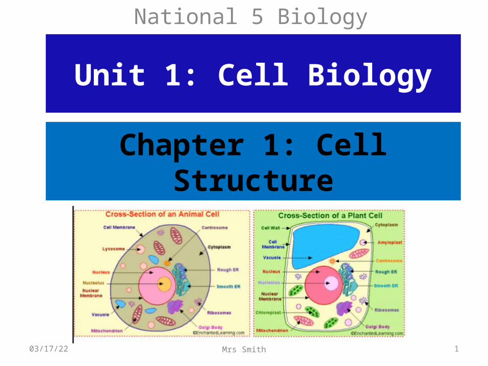
Chapter 1: Cell Structure
National 5 Biology
Unit 1: Cell Biology
04/19/23 1Mrs Smith
Learning Intentions
National 5 Biology
04/19/23 2Mrs Smith
By the end of this chapter, you should be able to describe cell structure - Specifically be able to discuss - Cell ultrastructure and functions to include: cell wall, mitochondrion, chloroplast, cell membrane, cytoplasm, vacuole, nucleus, ribosome and plasmid. - You should also be able to describe the ultrastructure by using examples from typical plant, animal, fungi and bacterial cells. - Explain fungal structure in terms of similarity to plant and animal cells but with a different cell wall structure. (Cell wall structure in fungal and bacterial cells is different from plant cells, ie chitin not cellulose)- Bacterial structure description should include the absence of organelles (no nucleus, mitochondria vacuole or chloroplasts) and a different cell wall structure to plant and fungal cells. -Bacterial structure should also include a description of there chromosomes and plasmid.
CELLS ARE THE STARTING POINT!
• All living organisms on Earth are divided into cells. The main concept of cell theory is that cells are the basic structural unit for all organisms. Nothing smaller than a cell can lead to independent life.
• Cells are small compartments that hold the biological equipment necessary to keep an organism alive and successful. Living things may be single-celled or they may be very complex such as a human being.
04/19/23 3Mrs Smith
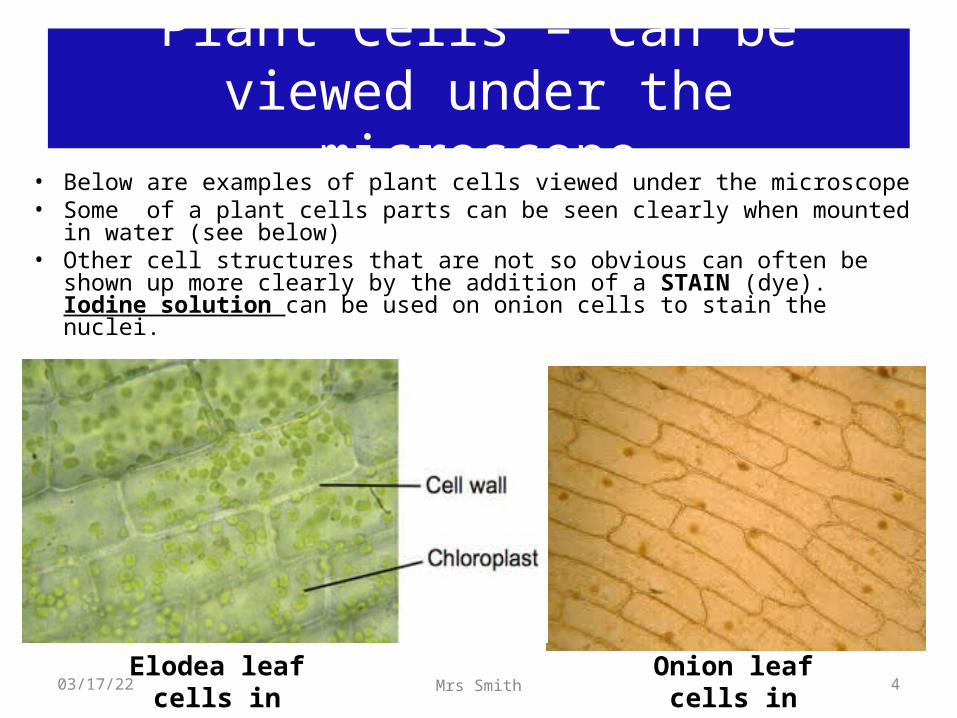
Plant Cells – Can be viewed under the microscope
• Below are examples of plant cells viewed under the microscope• Some of a plant cells parts can be seen clearly when mounted in water (see
below)• Other cell structures that are not so obvious can often be shown up more
clearly by the addition of a STAIN (dye). Iodine solution can be used on onion cells to stain the nuclei.
Onion leaf cells in iodine solution
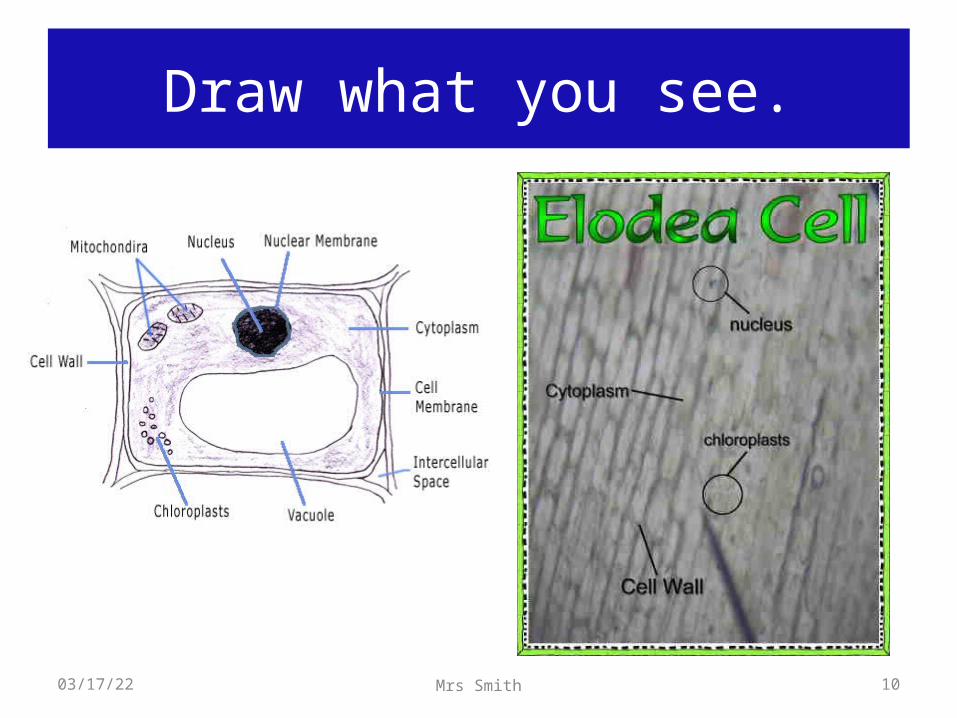
Elodea leaf cells in water
04/19/23 4Mrs Smith

Using stains with the microscope.
Stains can be used to make cells more visible under the microscope, e.g. Iodine solution.
Onion skin: 2 pieces were taken from the bottom layer of an onion and placed on 2 different slides.
With one, some iodine was placed n the slide to see the different layers, The other slide was left unstained.
04/19/23 5Mrs Smith
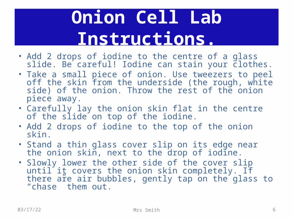
• Add 2 drops of iodine to the centre of a glass slide. Be careful! Iodine can stain your clothes.
• Take a small piece of onion. Use tweezers to peel off the skin from the underside (the rough, white side) of the onion. Throw the rest of the onion piece away.
• Carefully lay the onion skin flat in the centre of the slide on top of the iodine.
• Add 2 drops of iodine to the top of the onion skin. • Stand a thin glass cover slip on its edge near the onion skin,
next to the drop of iodine. • Slowly lower the other side of the cover slip until it covers
the onion skin completely. If there are air bubbles, gently tap on the glass to “chase” them out.
Onion Cell Lab Instructions.
04/19/23 6Mrs Smith
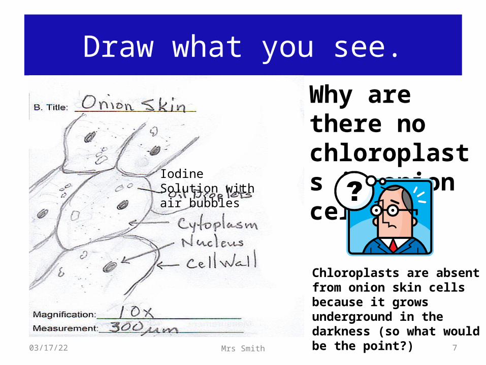
Draw what you see.
Why are there no chloroplasts in onion cells?
Iodine Solution with air bubbles
Chloroplasts are absent from onion skin cells because it grows underground in the darkness (so what would be the point?)
04/19/23 7Mrs Smith
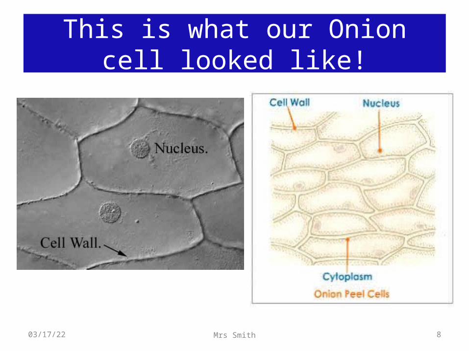
This is what our Onion cell looked like!
04/19/23 8Mrs Smith
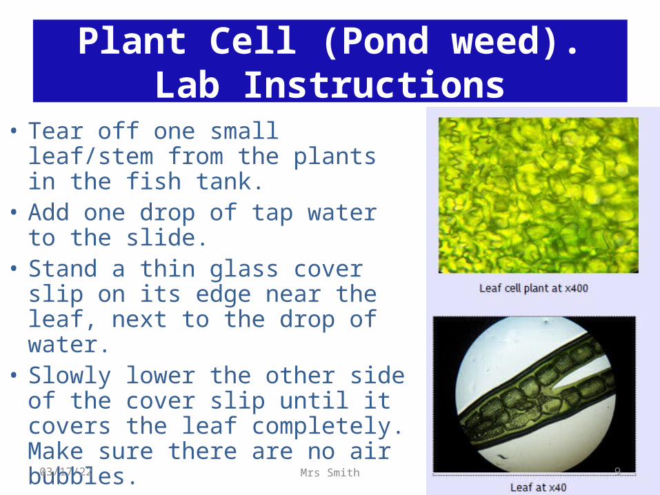
• Tear off one small leaf/stem from the plants in the fish tank.
• Add one drop of tap water to the slide.
• Stand a thin glass cover slip on its edge near the leaf, next to the drop of water.
• Slowly lower the other side of the cover slip until it covers the leaf completely. Make sure there are no air bubbles.
Plant Cell (Pond weed).Lab Instructions
04/19/23 9Mrs Smith
Draw what you see.
04/19/23 10Mrs Smith
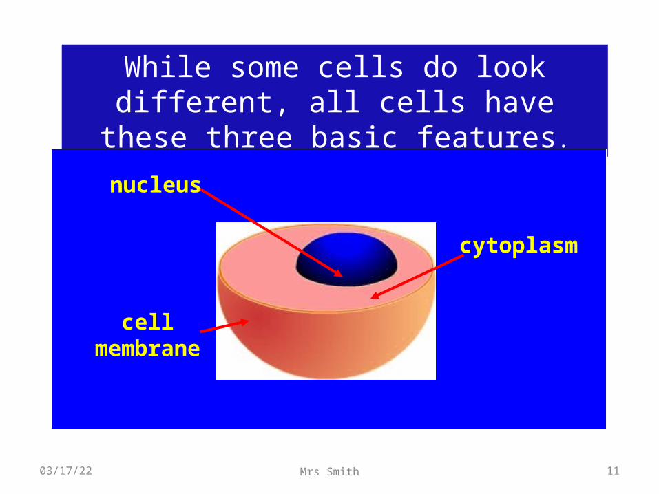
While some cells do look different, all cells have these three basic
features.
nucleus
cytoplasm
cell membra
ne
04/19/23 11Mrs Smith
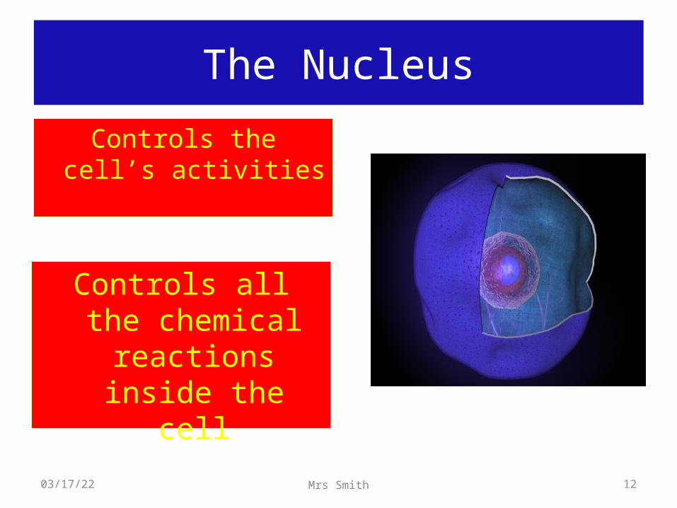
The Nucleus
Controls the cell’s activities
Controls all the chemical
reactions inside the cell
04/19/23 12Mrs Smith
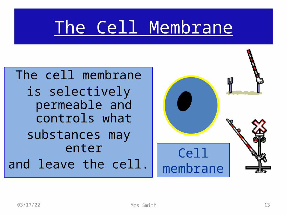
The Cell Membrane
The cell membraneis selectively permeable and controls what
substances may enterand leave the cell. Cell
membrane
04/19/23 13Mrs Smith
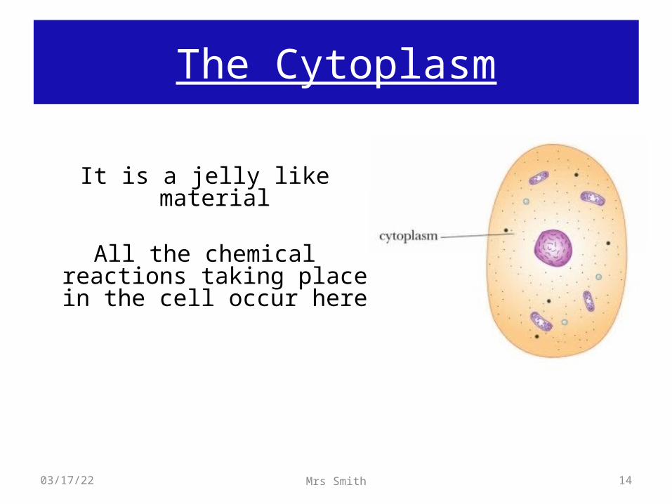
The Cytoplasm
It is a jelly like material
All the chemical reactions taking place in the cell occur here
04/19/23 14Mrs Smith
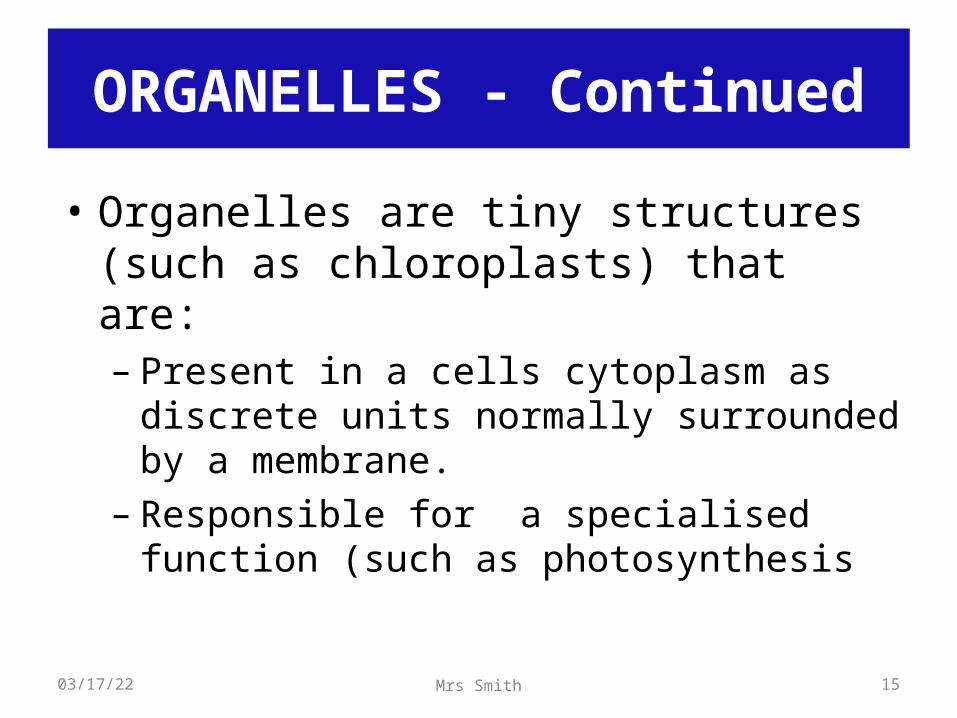
• Organelles are tiny structures (such as chloroplasts) that are:– Present in a cells cytoplasm as discrete units
normally surrounded by a membrane.– Responsible for a specialised function (such as
photosynthesis
ORGANELLES - Continued
04/19/23 15Mrs Smith
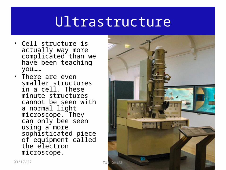
Ultrastructure• Cell structure is
actually way more complicated than we have been teaching you……
• There are even smaller structures in a cell. These minute structures cannot be seen with a normal light microscope. They can only bee seen using a more sophisticated piece of equipment called the electron microscope.
04/19/23 16Mrs Smith
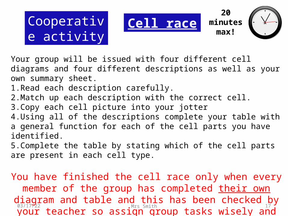
Your group will be issued with four different cell diagrams and four different descriptions as well as your own summary sheet.1.Read each description carefully.2.Match up each description with the correct cell.3.Copy each cell picture into your jotter4.Using all of the descriptions complete your table with a general function for each of the cell parts you have identified.5.Complete the table by stating which of the cell parts are present in each cell type.
You have finished the cell race only when every member of the group has completed their own diagram and table and this has been checked by your teacher so
assign group tasks wisely and remember to include everybody.
Cell raceCooperative activity
20 minutes max!
04/19/23 17Mrs Smith
Animal Cell
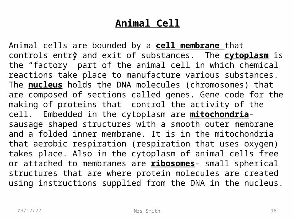
Animal cells are bounded by a cell membrane that controls entry and exit of substances. The cytoplasm is the “factory” part of the animal cell in which chemical reactions take place to manufacture various substances. The nucleus holds the DNA molecules (chromosomes) that are composed of sections called genes. Gene code for the making of proteins that control the activity of the cell. Embedded in the cytoplasm are mitochondria- sausage shaped structures with a smooth outer membrane and a folded inner membrane. It is in the mitochondria that aerobic respiration (respiration that uses oxygen) takes place. Also in the cytoplasm of animal cells free or attached to membranes are ribosomes- small spherical structures that are where protein molecules are created using instructions supplied from the DNA in the nucleus.
04/19/23 18Mrs Smith
Plant Cell
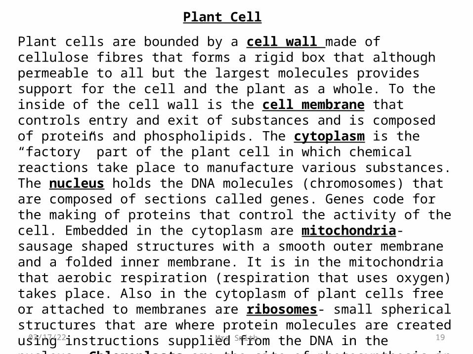
Plant cells are bounded by a cell wall made of cellulose fibres that forms a rigid box that although permeable to all but the largest molecules provides support for the cell and the plant as a whole. To the inside of the cell wall is the cell membrane that controls entry and exit of substances and is composed of proteins and phospholipids. The cytoplasm is the “factory” part of the plant cell in which chemical reactions take place to manufacture various substances. The nucleus holds the DNA molecules (chromosomes) that are composed of sections called genes. Genes code for the making of proteins that control the activity of the cell. Embedded in the cytoplasm are mitochondria- sausage shaped structures with a smooth outer membrane and a folded inner membrane. It is in the mitochondria that aerobic respiration (respiration that uses oxygen) takes place. Also in the cytoplasm of plant cells free or attached to membranes are ribosomes- small spherical structures that are where protein molecules are created using instructions supplied from the DNA in the nucleus. Chloroplasts are the site of photosynthesis in green plant cells. Vacuoles are fluid filled sac containing cell sap which are important in controlling water balance within the cell.04/19/23 19Mrs Smith
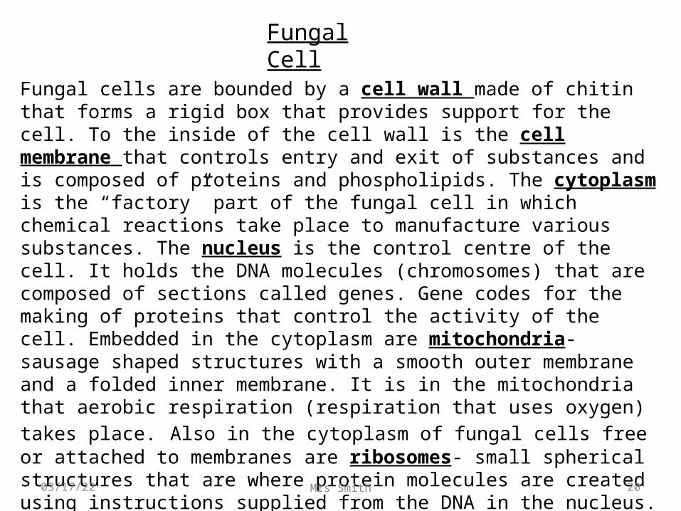
Fungal cells are bounded by a cell wall made of chitin that forms a rigid box that provides support for the cell. To the inside of the cell wall is the cell membrane that controls entry and exit of substances and is composed of proteins and phospholipids. The cytoplasm is the “factory” part of the fungal cell in which chemical reactions take place to manufacture various substances. The nucleus is the control centre of the cell. It holds the DNA molecules (chromosomes) that are composed of sections called genes. Gene codes for the making of proteins that control the activity of the cell. Embedded in the cytoplasm are mitochondria- sausage shaped structures with a smooth outer membrane and a folded inner membrane. It is in the mitochondria that aerobic respiration (respiration that uses oxygen) takes place. Also in the cytoplasm of fungal cells free or attached to membranes are ribosomes- small spherical structures that are where protein molecules are created using instructions supplied from the DNA in the nucleus. Vacuoles are fluid filled sac containing cell sap which are important in controlling water balance within the cell.
Fungal Cell
04/19/23 20Mrs Smith
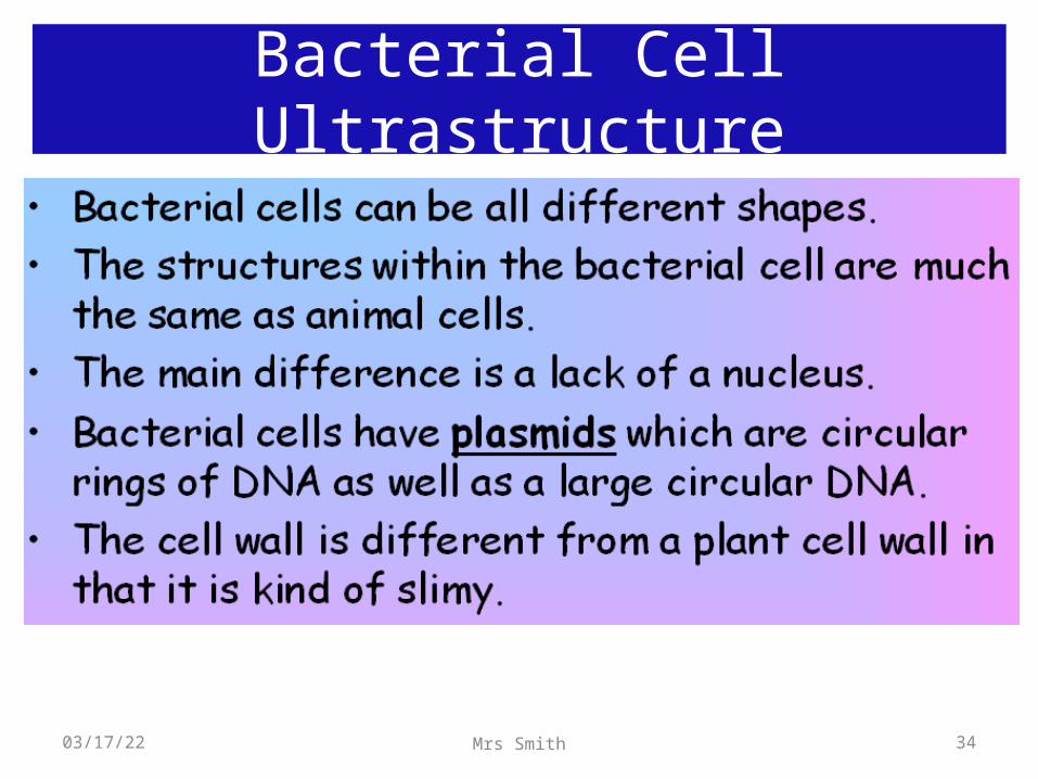
Bacterial Cell
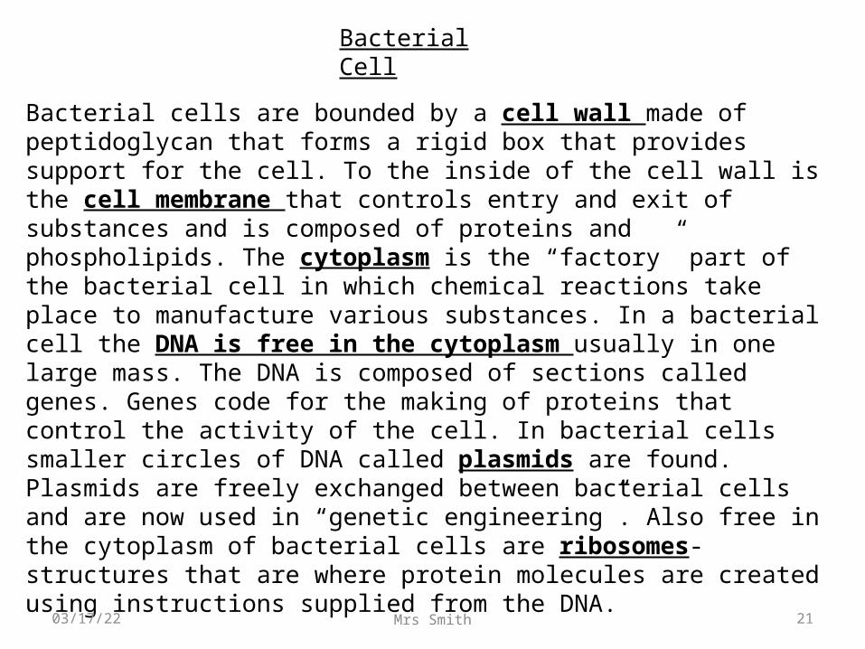
Bacterial cells are bounded by a cell wall made of peptidoglycan that forms a rigid box that provides support for the cell. To the inside of the cell wall is the cell membrane that controls entry and exit of substances and is composed of proteins and phospholipids. The cytoplasm is the “factory” part of the bacterial cell in which chemical reactions take place to manufacture various substances. In a bacterial cell the DNA is free in the cytoplasm usually in one large mass. The DNA is composed of sections called genes. Genes code for the making of proteins that control the activity of the cell. In bacterial cells smaller circles of DNA called plasmids are found. Plasmids are freely exchanged between bacterial cells and are now used in “genetic engineering”. Also free in the cytoplasm of bacterial cells are ribosomes- structures that are where protein molecules are created using instructions supplied from the DNA.
04/19/23 21Mrs Smith
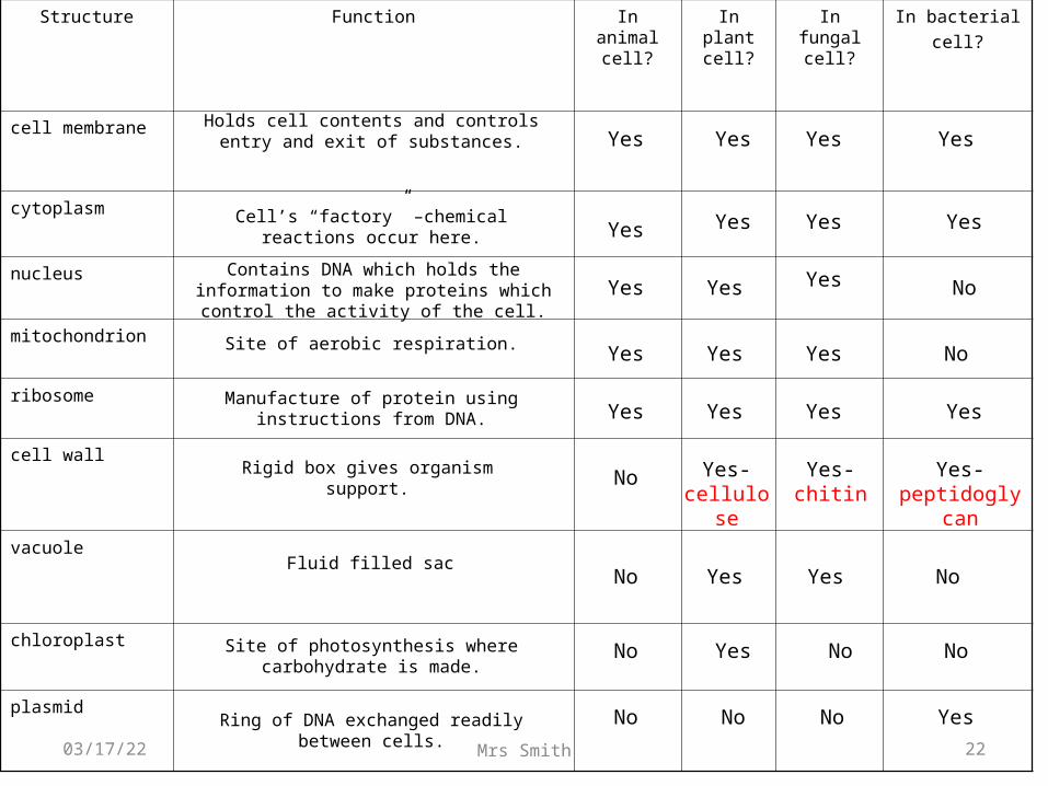
Structure Function In animal cell?
In plant cell?
In fungal cell?
In bacterialcell?
cell membrane
cytoplasm
nucleus
mitochondrion
ribosome
cell wall
vacuole
chloroplast
Holds cell contents and controls entry and exit of substances.
Cell’s “factory” –chemical reactions occur here.
Contains DNA which holds the information to make proteins which control the activity
of the cell.
Rigid box gives organism support.
Fluid filled sac
Site of photosynthesis where carbohydrate is made.
Yes Yes
Yes Yes
Yes- cellulos
e
Yes
Yes
No
No
No
Yes- chitin
Yes- peptidoglyc
an
Yes Yes
Yes Yes
Yes No
No No
Ring of DNA exchanged readily between cells.
No No No Yes
Yes Yes Yes No
Yes Yes Yes No
Yes Yes Yes YesManufacture of protein using instructions
from DNA.
plasmid
Site of aerobic respiration.
04/19/23 22Mrs Smith
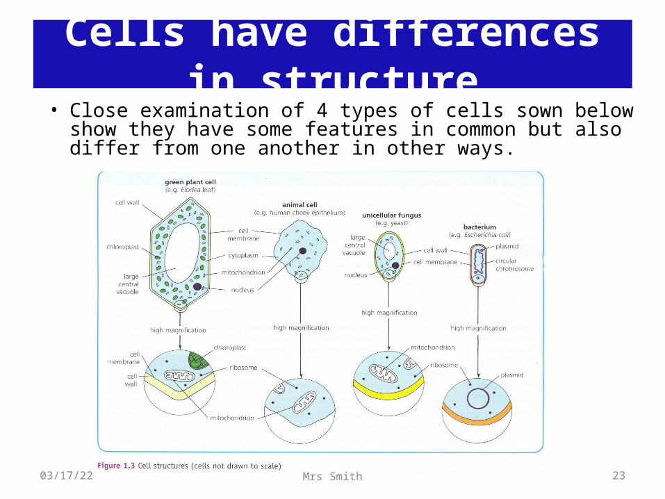
• Close examination of 4 types of cells sown below show they have some features in common but also differ from one another in other ways.
Cells have differences in structure
04/19/23 23Mrs Smith
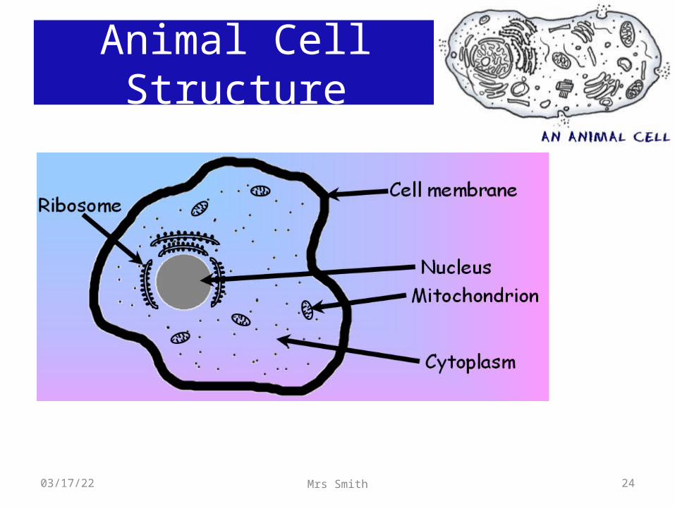
Animal Cell Structure
04/19/23 24Mrs Smith
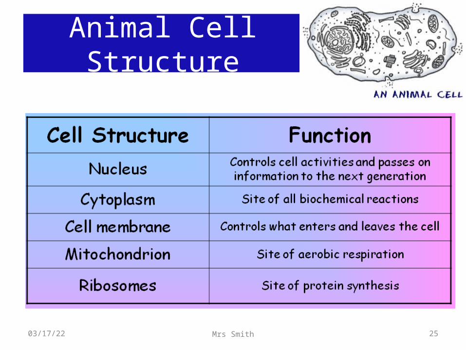
Animal Cell Structure
04/19/23 25Mrs Smith
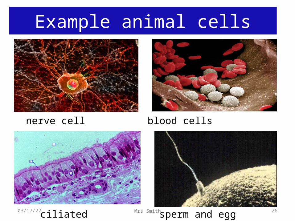
nerve cell blood cells
ciliated epithelial cells
sperm and egg cells
Example animal cells
04/19/23 26Mrs Smith
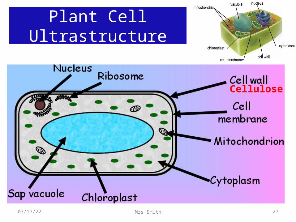
Plant Cell Ultrastructure
04/19/23 27Mrs Smith
Cellulose
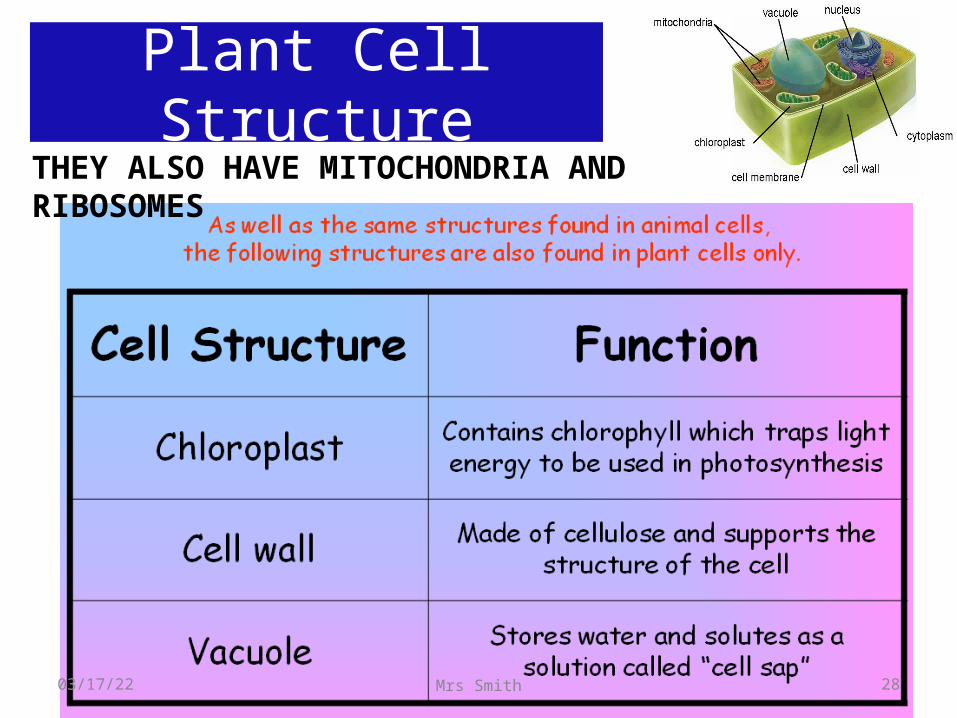
Plant Cell StructureTHEY ALSO HAVE MITOCHONDRIA AND RIBOSOMES
04/19/23 28Mrs Smith
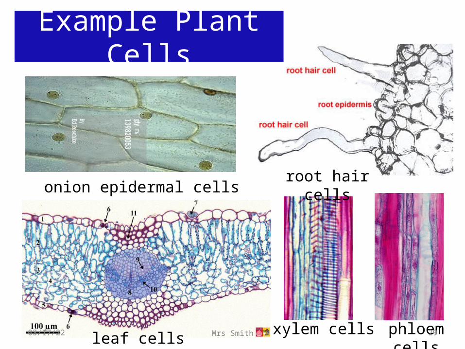
onion epidermal cellsroot hair cells
xylem cells phloem cells
leaf cells
Example Plant Cells
04/19/23 29Mrs Smith
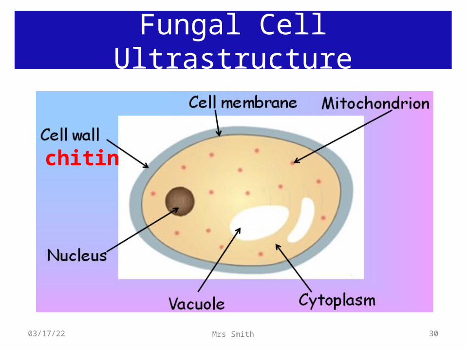
Fungal Cell Ultrastructure
04/19/23 30Mrs Smith
chitin
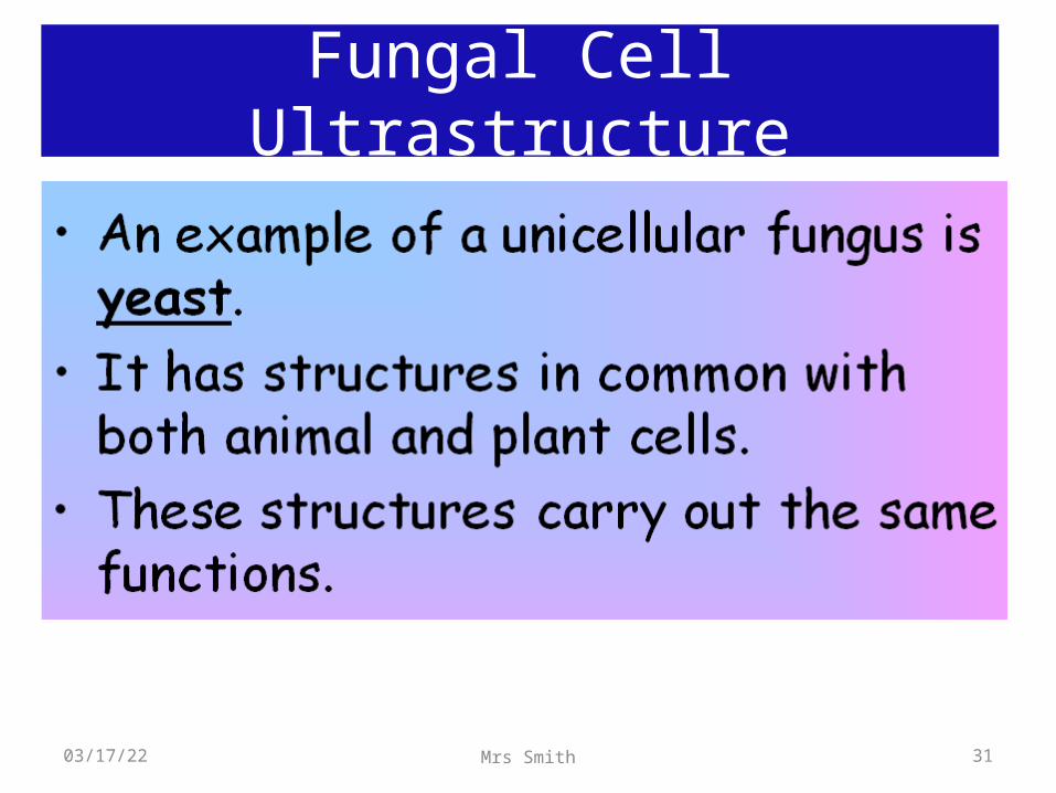
Fungal Cell Ultrastructure
04/19/23 31Mrs Smith
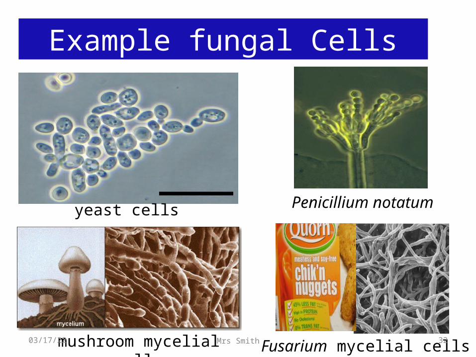
yeast cells
mushroom mycelial cells Fusarium mycelial cells
Penicillium notatum
Example fungal Cells
04/19/23 32Mrs Smith
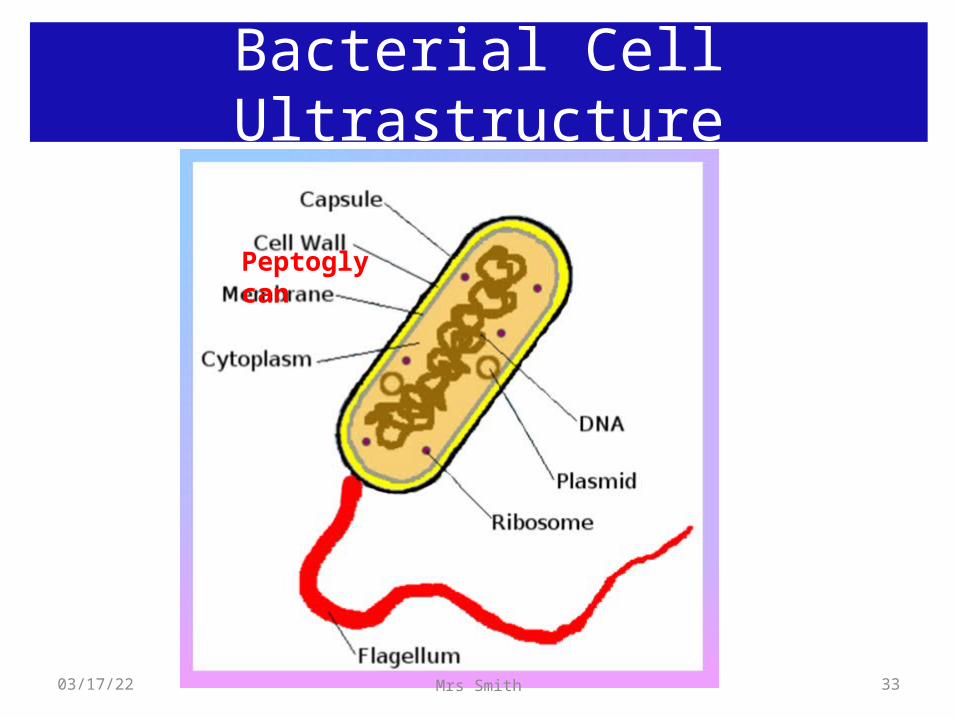
Bacterial Cell Ultrastructure
04/19/23 33Mrs Smith
Peptoglycan
Bacterial Cell Ultrastructure
04/19/23 34Mrs Smith
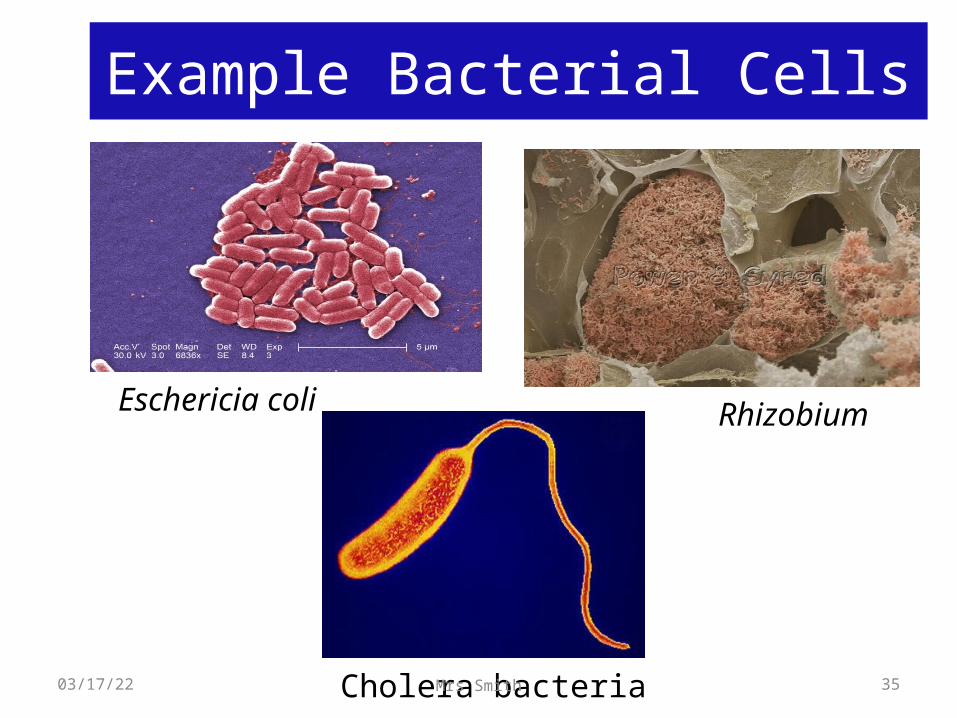
Eschericia coli Rhizobium
Cholera bacteria
Example Bacterial Cells
04/19/23 35Mrs Smith

Cell Rap
04/19/23 36Mrs Smith
The Cell Song
04/19/23 37Mrs Smith
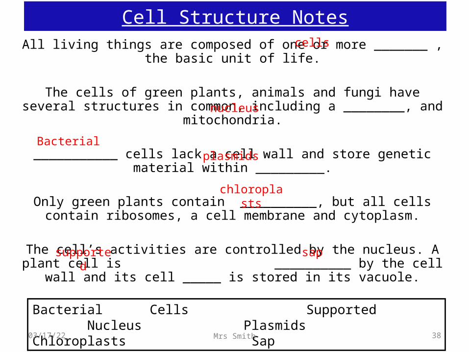
Cell Structure NotesAll living things are composed of one or more _______ , the basic
unit of life.
The cells of green plants, animals and fungi have several structures in common, including a ________, and mitochondria.
___________ cells lack a cell wall and store genetic material within
_________.
Only green plants contain __________, but all cells contain ribosomes, a cell membrane and cytoplasm.
The cell’s activities are controlled by the nucleus. A plant cell is __________ by the cell wall and its cell _____ is stored in
its vacuole.
Bacterial Cells Supported Nucleus Plasmids Chloroplasts Sap
cells
nucleus
Bacterialplasmids
chloroplasts
supported
sap
04/19/23 38Mrs Smith
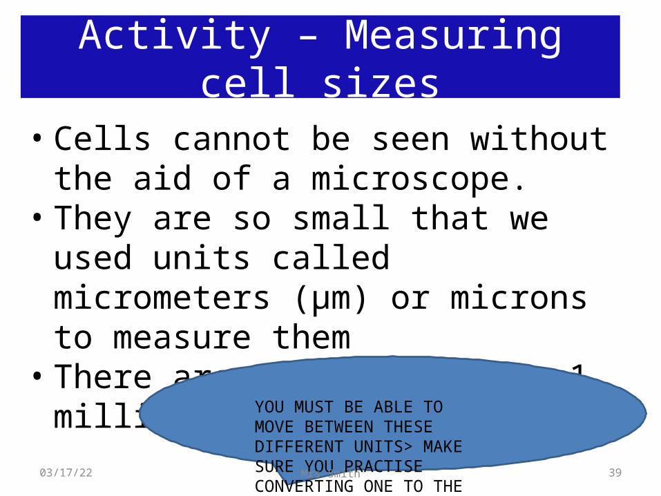
Activity – Measuring cell sizes
• Cells cannot be seen without the aid of a microscope.
• They are so small that we used units called micrometers (μm) or microns to measure them
• There are 1000 microns in 1 millimetre.
YOU MUST BE ABLE TO MOVE BETWEEN THESE DIFFERENT UNITS> MAKE SURE YOU PRACTISE CONVERTING ONE TO THE OTHER.
04/19/23 39Mrs Smith
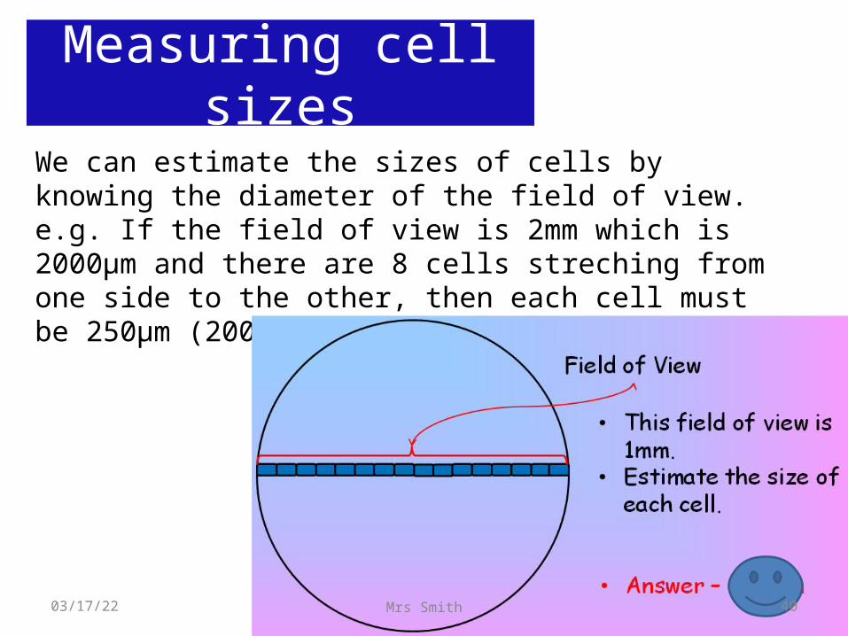
Measuring cell sizesWe can estimate the sizes of cells by knowing the diameter of the field of view.e.g. If the field of view is 2mm which is 2000μm and there are 8 cells streching from one side to the other, then each cell must be 250μm (2000/8)
04/19/23 40Mrs Smith
0.4
0.2
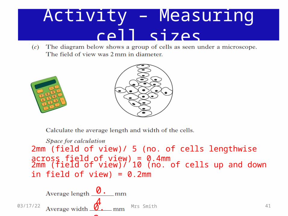
2mm (field of view)/ 5 (no. of cells lengthwise across field of view) = 0.4mm
2mm (field of view)/ 10 (no. of cells up and down in field of view) = 0.2mm
Activity – Measuring cell sizes
04/19/23 41Mrs Smith
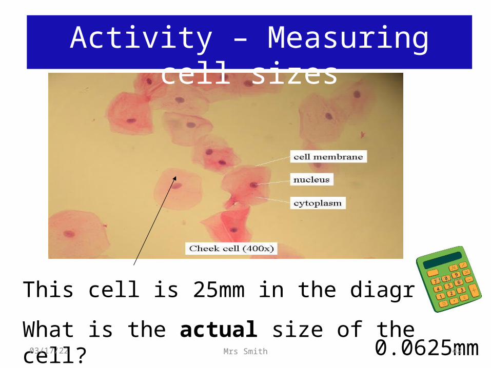
This cell is 25mm in the diagram.
What is the actual size of the cell?0.0625mm
Activity – Measuring cell sizes
04/19/23 42Mrs Smith
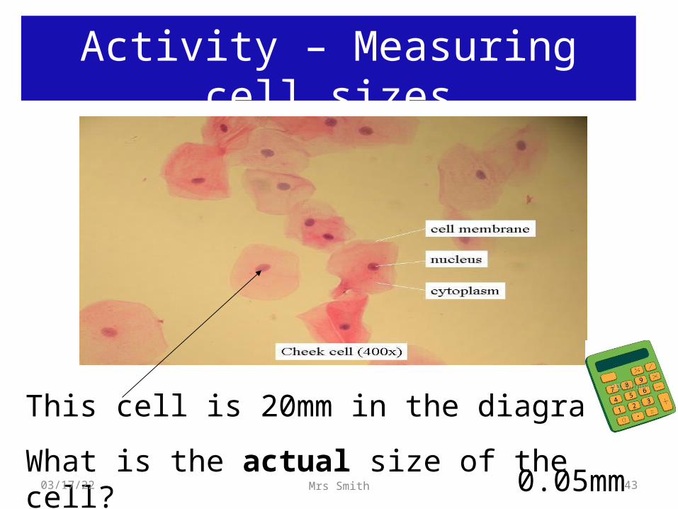
This cell is 20mm in the diagram.
What is the actual size of the cell?0.05mm
Activity – Measuring cell sizes
04/19/23 43Mrs Smith
Task TYK
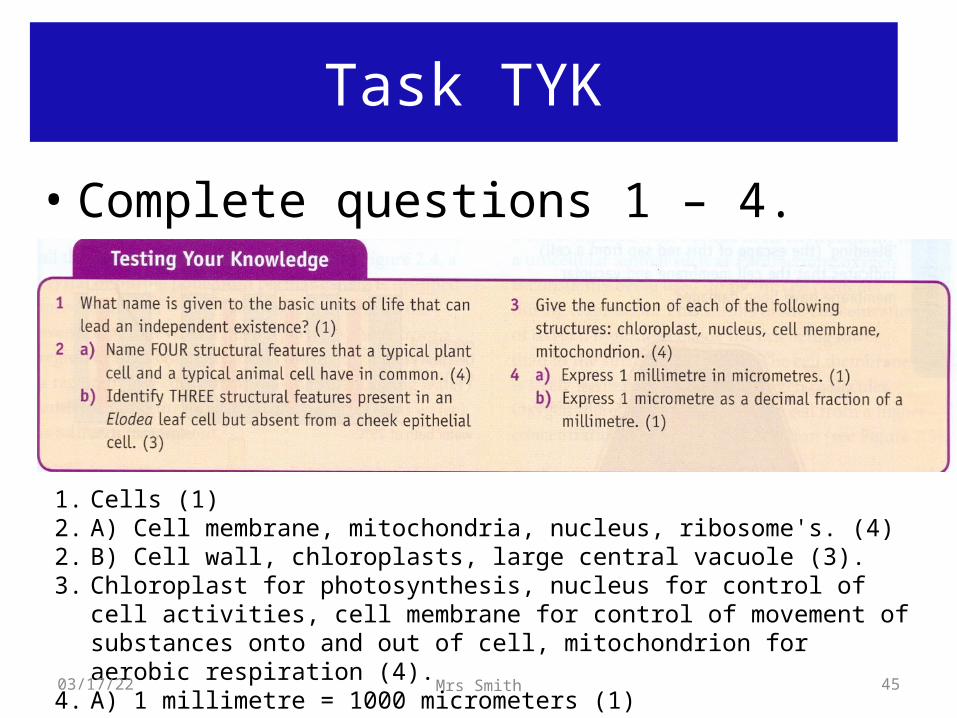
• Complete questions 1 – 4. Torrance p5
04/19/23 44Mrs Smith
Task TYK
• Complete questions 1 – 4. Torrance p5
1. Cells (1)2. A) Cell membrane, mitochondria, nucleus, ribosome's. (4)2. B) Cell wall, chloroplasts, large central vacuole (3).3. Chloroplast for photosynthesis, nucleus for control of cell activities, cell membrane
for control of movement of substances onto and out of cell, mitochondrion for aerobic respiration (4).
4. A) 1 millimetre = 1000 micrometers (1)4. B) 1 micrometer = 0.001 millimetres (1).04/19/23 45Mrs Smith
Quick test
1. How is the cell wall of a plant cell different from that of a yeast cell?
2. Name two cell structures that cannot be seen with a light microscope.
3. What features do yeast and p[lant cells have in common?
04/19/23 46Mrs Smith
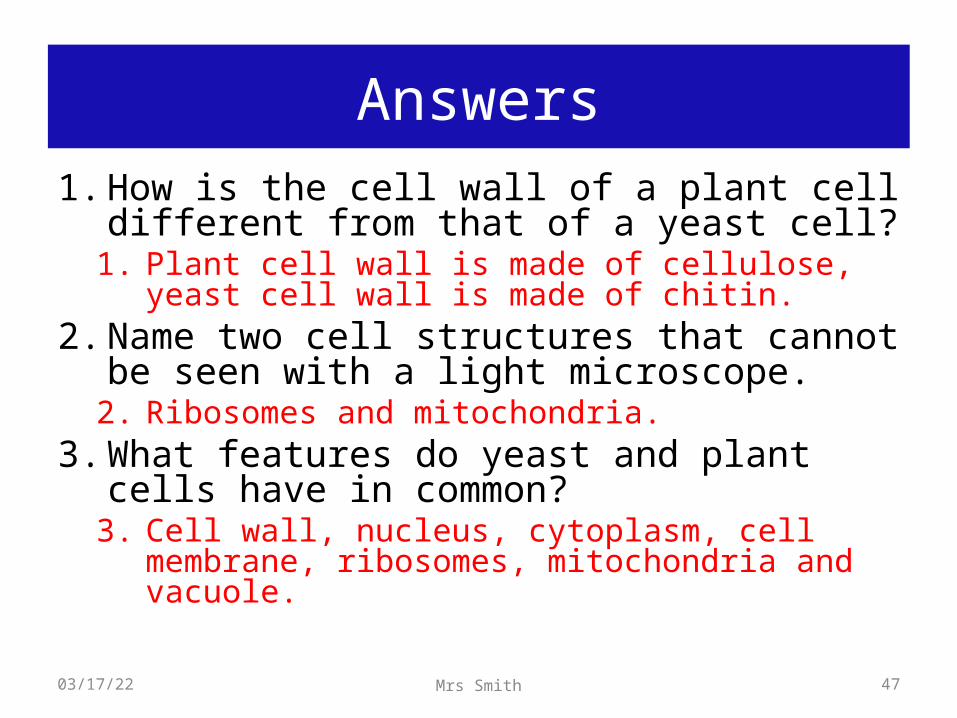
Answers1. How is the cell wall of a plant cell different from
that of a yeast cell?1. Plant cell wall is made of cellulose, yeast cell wall is
made of chitin.2. Name two cell structures that cannot be seen
with a light microscope.2. Ribosomes and mitochondria.
3. What features do yeast and plant cells have in common?
3. Cell wall, nucleus, cytoplasm, cell membrane, ribosomes, mitochondria and vacuole.
04/19/23 47Mrs Smith
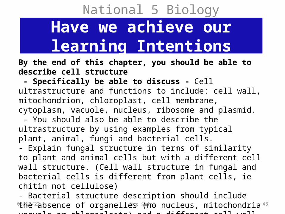
Have we achieve our learning Intentions
National 5 Biology
04/19/23 48Mrs Smith
By the end of this chapter, you should be able to describe cell structure - Specifically be able to discuss - Cell ultrastructure and functions to include: cell wall, mitochondrion, chloroplast, cell membrane, cytoplasm, vacuole, nucleus, ribosome and plasmid. - You should also be able to describe the ultrastructure by using examples from typical plant, animal, fungi and bacterial cells. - Explain fungal structure in terms of similarity to plant and animal cells but with a different cell wall structure. (Cell wall structure in fungal and bacterial cells is different from plant cells, ie chitin not cellulose)- Bacterial structure description should include the absence of organelles (no nucleus, mitochondria vacuole or chloroplasts) and a different cell wall structure to plant and fungal cells. -Bacterial structure should also include a description of there chromosomes and plasmid.