chapter - 1 introduction - information and...
TRANSCRIPT

1
CHAPTER - 1
INTRODUCTION
“One surprising reason to save your Banana peels”
1.1 GENERAL
Diabetes mellitus is the most prevalent metabolic syndrome world wide with
an incidence varying between 1 to 8%[1,2]. The disease arises when insufficient
insulin is produced, or when the available insulin does not function properly. This
diabetes is characterized by hyperglycemia (elevation of Blood sugar level)
resulting in various short term metabolic changes in lipid and protein metabolism and
long term irreversible vascular changes. The long term manifestation of diabetes
can result in the development of some complications, broadly classified as micro
vascular or macro vascular disease.
Micro vascular complications include Neuropathy (Nerve damage),
Nephropathy (Renal disease) and Retinopathy (vision disorders), while macro
vascular complications include heart disease, stroke and peripheral vascular disease,
which can lead to ulcers, gangrene and amputation [3]. These complications are also
found in non diabetic population, but have a two to five fold increase in diabetic
subjects [4].The incidence of diabetes mellitus is increasing worldwide and rapidly
assuming epidemic proportions. India is no exception, and about 25 million Indians
are estimated to be suffering from diabetes [1]. Further projections indicate that India
will have maximum number of diabetic patients by the year 2025 [2].
There are two major categories of diabetes,insulin dependent diabetes
mellitus (IDDM, Type 1 diabetes mellitus) and Non-insulin dependent diabetes
mellitus (NIDDM, Type-2 diabetes mellitus). Type 1 diabetes occurs due to almost
95% destructions of -cells of islets of Langerhans in the endocrine pancreas caused by an
autoimmune process, usually leading to absolute insulin deficiency, this type has an
2
early onset, most often between the ages of 10 to16 yrs. Insulin resistance in
peripheral tissue and an insulin secretive defect of the -cells characterizes Type 2
diabetes mellitus (NIDDM). It is the most common form of diabetes mellitus constituting
above 90% of the diabetic population and highly associated with a family history of
diabetes, older age, obesity and lack of exercise[3].
The global prevalence of diabetes is estimated to increase, from 4% in 1995
to 5.4% by the year 2025[4]. The World Health Organization (WHO) has predicted
that the major burden will occur in the developing countries, there will be a 42%
increase from 51 to 72 million in the developed countries while 170% increase
from 84 to 228 million, in the developing countries[5]. Prevalence of the
complications is greater among the lower socio economic people due to lack of
good control of blood glucose level and hypertension and also due to behavioral
factors. The direct and indirect costs involved in the treatment of the chronic disease
especially when associated with the vascular complications are enormous. The
overall global scenario urges to implement cost effective and at the same time
efficacious preventive measures against diabetes to reduce the high morbidity and
mortality [4] .
1.1.1 Currently available therapies
Currently available therapies for diabetes include Insulin and various oral Anti
diabetic agents such as Sulfonylureas, Biguanides, -Glucosidase inhibitors, and
Glinides, which are used as mono therapy or in combination to achieve better
glycemic regulation. Many of these oral antidiabetic agents suffer from various
adverse effects, thus, managing diabetes without any side effects is still a challenge
[6], and hence the search for more effective and safer therapeutic agents in
eradicating diabetic syndromes has continued to be an important area of
investigation. Both fasting and postprandial impaired glucose tolerance are
associated with an increased risk of developing Type 2 diabetes mellitus and
therefore form an important target group for interventions aimed at preventing
diabetes [7].
3
The pharmacological agents with the greatest effect on postprandial
hyperglycemia include Insulin lispro, Amylin analogues, and -glucosidase inhibitors.
In hyperglycemia associated with diabetes, the use of Aldose reductase inhibitors
has been reported for the treatment of diabetic complications [8].
Aldose reductase as a key enzyme in the polyol pathway has been reported to
catalyze the reduction of glucose to S-orbitol. S-orbitol does not readily diffuse
across cell membranes, and the intracellular accumulation of S-orbitol has been
implicated in the chronic complications of diabetes such as peripheral neuropathy,
retinopathy, and cataracts [9]. A recent study reported that Aldose reductase may also
be involved with another signal transduction pathway in the pathogenesis of diabetic
nephropathy [10].
1.1.2 Back to Herbal medicine.
In the India, indigenous remedies have been used in the treatment of diabetes
since the time of Charaka and Sushruta (6th century B.C.) [11]. Plants have always
been exemplary source of drugs and many of the currently available drugs have been
derived directly or indirectly from them. The Ethnobotanical information reports about
800 plants that may possess anti diabetic potential [12].
Many of such plants have exhibited antidiabetic activity when assessed using
presently available experimental techniques [13-16]. It may be mentioned in this
connection that the discovery of widely used hypoglycemic drug, Metformin came
from the traditional approach of using Galega officinalis. In spite of all these, the
indigenous system has not yet gained enough momentum in the scientific
community. The reasons may be many including lack of belief among the
practitioners of conventional medicine over traditional medicine, traditional form
of medicine are not very well defined and natural drug may vary tremendously in
content, quality and safety. To scope with severe problems associated with using
synthetic antidiabetic drugs, there is a need to look for more efficacious drugs with
lesser side effects and also of low cost.
4
It is the high time to turn our attention to the plant kingdom in search of
natural drugs for diabetes following an integrated approach and using correct
procedures. The hypoglycemic effect of several plants used as antidiabetic remedies
has already been confirmed, and the mechanisms of hypoglycemic activity of these
plants are being studied; if even a single plant material stands the acid test of efficacy
comparable to commonly used synthetic oral drugs already marketed, it will herald
the discovery of cheap and relatively non toxic drug [17].
1.1.3Global endorsement of Herbal medicine
Plants have been the basis of many traditional medicine systems throughout
the world for thousands of years and continue to provide mankind with new
remedies. Use of plants as a source of medicine has been inherited and is an
important component of the health care system in India. In the Indian systems of
medicine, most practitioners formulate and dispense their own receipes; hence this
requires proper documentation and research. In western world also, the use of herbal
medicines is steadily growing with approximately 40 % of population reporting use
of herbs to treat medical illnesses [18] Public, academic and government interest in
traditional medicines is growing exponentially due to the increased incidence of the
adverse drug reactions and economic burden of the modern system of medicine
[19].
1.1.4 India - Treasures of medicinal plants
There are about 45,000 plant species in India, with concentrated hotspots in
the region of Eastern Himalayas, Western Ghats and Andaman & Nicobar Island.
The officially documented plants with medicinal potential are 3000 but traditional
practitioners use more than 6000. India is the largest producer of medicinal herbs
and is appropriately called the botanical garden of the world [20]. In rural India, 70
% of the population is dependent on the traditional system of medicine, the
Ayurveda [18]. Three of the ten most widely selling herbal medicines in the
developed countries, namely preparations ofAllium sativum, Aloe barbedensis and
Panax spp. are available in India. There are about 7000 firms manufacturing
traditional medicines with or without standardization [19] .

5
1.1.5 Drug discovery from medicinal plants
Plants form a dominant part of Ayurvedic pharmacopoeia where drugs have
been Classified on the basis of their physiological action [21]. Herbal drug is
estimated that approximately one quarter of prescribed drugs contain plant extracts
or active ingredients obtained from plant substances. Aspirin, Atropine,
Artimesinin, Colchicine, Digoxin, Ephedrine, Morphine, Physostigmine,
Pilocarpine, Quinine, Quinidine, Reserpine, Taxol, Tubocurarine, Vincristine and
Vinblastine are a few important examples of what medicinal plants have given us in
the pastof many strategies for selection of plants as drug source, the most rewarding
has been the criteria of their use in folklore medicine[22].For example, Rauwellia
serpentina (L.) Benth ex. Kurz provided the hypotensive alkaloids reserpine,
reseinnamine and deserpedine; Digitalis purpurea L. and Digitalis lantana Ehrh.
provided digitoxin and digoxin both powerful diatonic agents; Papaver somniferum
L. provides opium alkaloids, the analgesics codeine and morphine as well as the
antitussive noscapine and smooth muscle relaxant, papaverine; Atropha belladonna
provided the parasympatholytic atropine, scopolamine and 1-hyos-cyamine, Taxol
(bark of Taxus brevifolia Nutt.) is the most recent anticancer drug discovered from
plant source [21] .
1.1.6 Global challenges
The current lifestyle of humans almost everywhere in the world is in sharp
contrast than earlier time, and as a consequence, humans suffer from a large number
of chronic diseases. In the past, infectious diseases killed our ancestors early, often
younger than age 40, so they did not display the current epidemic of chronic
diseases as obesity, diabetes, hypertension, coronary heart disease, and cancer[23].
1.1.6.1 Inflammation
Chronic inflammation, induced by biological, chemical, and physical
factors, is associated with increased risk of human cancer at various sites. Chronic
inflammatory processes induce oxidative/nitrosative stress and lipid per oxidation
(LPO), thereby generating excess reactive oxygen species (ROS), reactive nitrogen
species (RNS), and DNA reactive aldehydes [24] .

6
1.1.6.2 Atherosclerosis
Atherosclerosis is a chronic vascular disease in which inflammation and
oxidative stress has important role at every stage. The disease process develops and
progresses in response to abnormal cholesterol deposits in the intima of large
arteries [25].
1.1.6.3 Cancer
Most tumors form discrete masses but in the leukemias, the tumor cells are
spread through the bone marrow or lymphoid tissues and circulate in the blood.
DNA damage plays a very important role in carcinogenesis and any agent, which is
capable of chemically modifying DNA could be carcinogenic. Hydroxyl radical
attack upon DNA generates a whole series of modified purine and pyrimidine bases
many of which are known to be mutagenic[26] .
1.1.6.4 Diabetes
It has been postulated that the etiology of the complications of diabetes
involves oxidative stress perhaps as a result of hypoglycemia [27]. Glucose itself and
hyperglycemia related increased protein glycosylation are important sources of free
radicals [28]. Elevated glucose causes slow but significant non enzymatic
glycosylation of proteins in diabetes [29] .
1.2 D IABETES
Diabetes mellitus often referred to simply as diabetes is a condition in which
the body does not produce enough or properly respond to insulin, a hormone
produced in the pancreas. Insulin enables cells to absorb glucose in order to turn it
into energy. In diabetes, the body either doesn't respond properly to its own insulin
ordoesn't make enough insulin, or both. This causes glucose to accumulate in the
blood, often leading to various complications [30].

7
1.2.1 Classification of Diabetes mellitus
Based on etiology diabetes mellitus is classified as (Endotex.com).
I Type 1 diabetes ( -cell destruction, usually leading to absolute insulin deficiency).
Immune mediated idiopathic
II Type 2 diabetes (may range from predominantly insulin resistance with
relative insulindeficiency to a predominantly insulin secretary defect
with insulin resistance).
III Gestational Diabetes Melllitus (GDM) .
IV Other specific types
1.2.1. 1 Genetic defects of -cell function
1. Chromosome 20q, Hepatocyte Nuclear Transcripter Factor HNF-4 (MODY1).
2. Chromosome 7p, glucokinase (MODY2).
3. Chromosome 12q, HNF-1 (MODY3).
4. Chromosome 13q, insulin promoter factor (MODY4).
5. Chromosome 17q, HNF-1 (MODY5).
6. Chromosome 2q, Neurogenic differentiation 1/b-cell e-box
transactivator 2 (MODY6).
7. Mitochondrial DNA
1.2.1.2 Genetic defects in insulin action
1. Type A insulin resistance
2. Leprechaunism
3. Rabson-Mendenhall syndrome
4. Lipoatrophic diabetes
8
1.2.1.3 Diseases of the exocrine pancreas
1. Pancreatitis
2. Trauma / Pancreatectomy
3. Neoplasia
4. Cystic fibrosis
5. Hemochromatosis
6. Fibrocalculous pancreatopathy
1.2.1 .4 Endocrinopathies
1. Acromegaly
2. Cushing's syndrome
3. Glucogoma
4. Pheochromocytoma
5. Hyperthyrodism
6. Somatostatinoma
7. Aldosteronoma
1.2.1 .5 Drug or Chemical induced Diabetes
1. Vacor
2. Pentamidine
3. Nicotinic acid
4. Glucocorticoids
5. Thyroid hormone
6. Diazoxide
7. -adrenergic agonists
8. Thiazides
9. Phenytoin
9
10. -interferon
11. Streptozotocin
12. Alloxan
1.2.1.6 Infections
1. Congential rubella
2. Cytomegalovirus
1.2.1 .7 Other genetic syndromes associated with diabetes
1. Down's syndrome
2. Klinefelter's syndrome
3. Turner's syndrome
4. Wolfram's syndrome
5. Friedreich's ataxia
6. Huntington's chorea
7. Laurence-Moon-Bieldel syndrome
8. Myotonic dystrophy
9. Porphyria
10. Prader-Willi syndrome
1.2.2 Pathophysiology of Diabetes Mellitus
Insulin Dependent Diabetes Mellitus
Type 1 diabetes is the form of diabetes in which there is destruction of the
insulin producing cells of the pancreas which are located in the islets of pancreas.
The process of destruction can occur over a period of several years. This phase being
called the pre diabetic phase. It is a result from an interaction between
environmental and genetic factors that trigger selective cell destruction [31].

10
In the first phase, leukocytes invade the islets[32]. Diabetes progresses when
most islets have been killed and there is no longer sufficient insulin production to
regulate blood glucose levels. Patients inject insulin to compensate for insulin
deficiency, but the effort and practice needed to mimic the normal -cell function,
which precisely adjusts the rate of insulin secretion to the actual circulating blood glucose
level is enormous.Massive, specific cell destruction, mainly by apoptosis, is the hallmark of
type 1 diabetes [33,34].
Diabetes is primarily mediated by T -lymphocytes. T-cells with
diabetogenic properties fall into both the CD4+ helper and the CD8+ killer classes.
The disease results from T-cell activation by recognition of islet -cell antigens of the
major histocompatibility complex (MHC) molecules presented on -cells ( Figure 1.1 A
& B ). Detection of apoptotic -cells in vivo and the study of their characteristics in animal
models are very difficult due to the asynchronism of the apoptotic process. The
clearance of apoptotic -cells by macrophages has been estimated to occur from 1.7 to 11 min
[35].
In pancreas of type 1 diabetic patients where -cell destruction proceeds for
months or years it is nearly impossible to study ongoing apoptotic pathways. However,
apoptosis is the main form of -cell death in diabetes. Two major pathways have been
considered to occur at the onset of type 1 diabetes; the perforin / granzyme and the
Fas / FasL system [36] .The perforin/granzyme pathway, which could be the effector
pathway utilized by CD8+ T-cells, leading to insertion of perforin complexes into
the cell membrane and osmotic lysis, is the main cause of cell death induced by
cytotoxic T-cells. However, observations of different type 1 diabetic animal models
contradict the hypothesis of the perforin/granzyme mechanism being a main death
pathway in type 1 diabetes. A second hypothesis, also under debate, suggests that the
interaction of activated Fas receptor with its ligand (FasL) is the initiating pathway of
-cell apoptosis. FasL is expressed primarily on activated T-lymphocyte [37].

11
Figure A 1.1: Proposed model of -cell death in autoimmune diabetes [32].
(A) The T-cell is activated by direct recognition of islet -cell antigens (dots)
presented by MHC molecules (in this case class I molecules) on -cells. Interaction
activates the apoptotic machinery via the Perforin or the Fas / FasL pathway
Figure.B. 1.1: Proposed model of -cell death in autoimmune diabetes [32].
(B) T-cells recognize the MHC moleculesIndirectly via antigen presenting cells
(macrophages). The resulting activation initiates - cell death mediated by surface
receptors (Fas / FasL, TNF- / TNF-R (i), cytokines (ii), activation of macrophages (iii)
and activation of the -cell and their production of cell death mediators (iv).
12
Another initiating pathway of cell apoptosis is the TNF- / TNF-receptor
interaction, reported in a CD8+ T-cell dependent diabetes model [38].Macrophages
and activated T- cells are important effector cells leading to cell destruction, inducing
apoptosis via the synthesis of pro inflammatory cytokines, such as IL-1 and TNF- as
well as nitric oxide and other free radicals. T-cells also express the Fas receptor ligand
(FasL), the tumor necrosis factor related apoptosis inducing ligand (TRAIL) and the
TNF- -receptor, all leading to apoptosis.
1.2.3 The major metabolic derangements which result from insulin deficiency
in IDDM are impaired glucose, lipid and protein metabolism.
1.2.3 .1 Glucose metabolism
Uncontrolled IDDM leads to increased hepatic glucose
output.First,liverglycogen stores are mobilized then hepatic gluconeogenesis is used
to produce n deficiency also impairs non-hepatic tissue utilization of glucose. In
particular in adipose tissue and skeletal muscle, insulin stimulates glucose uptake.
This is accomplished by insulin mediated movement of glucose transporter proteins
to the plasma membrane of these tissues. Reduced glucose uptake by peripheral
tissues in turn leads to a reduced rate of glucose metabolism.
In addition, the level of hepatic glucokinase is regulated by insulin.
Therefore, a reduced rate of glucose phosphorylation in hepatocytes leads to increased
delivery to the blood. Other enzymes involved in anabolic metabolism of glucose are
affected by insulin (primarily through covalent modifications). The combination of
increased hepatic glucose production and reduced peripheral tissues metabolism
leads to elevated plasma glucose levels. When the capacity of the kidneys to absorb
glucose is surpassed, glycosuria ensues. Glucose is an osmotic diuretic and an
increase in renal loss of glucose is accompanied by loss of water and electrolytes,
termed polyuria. The result of the loss of water and overall volume leads to the
activation of the thirst mechanism (polydipsia). The negative caloric balance which
results from the glycosuria and tissue catabolism leads to an increase in appetite and
food intake (polyphagia).
13
1.2.3 .2 Lipid metabolism
One major role of insulin is to stimulate the storage of food energy following
the consumption of a meal. This energy storage is in the form of glycogen in
hepatocytes and skeletal muscle. Additionally, insulin stimulates hepatocytes to
synthesize triglycerides and storage of triglycerides in adipose tissue. In opposition to
increased adipocyte storage of triglycerides is insulin-mediated inhibition of lipolysis.
In uncontrolled IDDM there is a rapid mobilization of triglycerides leading to
increased levels of plasma free fatty acids. The free fatty acids are taken up by
numerous tissues (however, not the brain) and metabolized to provide energy.
Free fatty acids are also taken up by the liver. Normally, the levels of
malonyl-CoA are high in the presence of insulin. These high levels of malonyl-CoA
inhibit carnitine palmitoyltransferase I, the enzyme required for the transport of fatty
acyl-CoA's into the mitochondria where they are subjected to oxidation for energy
production. Thus, in the absence of insulin, malonyl-CoA levels fall and transport of
fatty acyl-CoA's into the mitochondria increases [39].
Mitochondrial oxidation of fatty acids generates acetyl CoA, which can be
further oxidized in the TCA cycle. However, in hepatocytes the majority of the acetyl
CoA is not oxidized by the TCA cycle but is metabolized into the ketone bodies,
acetoacetate and hydroxybutyrate. These ketone bodies leave the liver and are used
for energy production by the brain, heart and skeletal muscle. In IDDM, the
increased availability of free fatty acids and ketone bodies exacerbates the reduced
utilization of glucose furthering the ensuing hyperglycemia. Production of ketone
bodies in excess of the body's ability to utilize them leads to ketoacidosis.
In diabetics, this can be easily diagnosed by smelling the breath. A
spontaneous breakdown product of acetoacetate in acetone, which is volatilized by
the lungs producing a distinctive odor. Normally, plasma triglycerides are acted upon
by lipoprotein lipase (LPL), an enzyme on the surface of the endothelial cells lining
the vessels. In particular, LPL activity allows fatty acids to be taken from
circulating triglycerides for storage in adipocytes. The activity of LPL requires
insulin and in its absence a hyper triglyceridemia results [39].
14
1.2.2 .3 Protein metabolism
Insulin regulates the synthesis of many genes, either positively or negatively
that then affect overall metabolism. Insulin has a global effect on protein
metabolism increasing the rate of protein synthesis and decreasing the rate of protein
degradation.Thus,insulindeficiency will lead to increased catabolism of protein. The
increased rate of proteolysis leads to elevated concentrations in plasma amino acids.
These amino acids serve as precursors for hepatic and renal gluconeogensis. In
liver, the increased gluconeogenesis further contributes to the hyperglycemia seen in
IDDM [39] .
1.2.4 Type 2 Diabetes Mellitus (NIDDM)
In Type 2 diabetes both insulin resistant and deficient secretion are present.
This shows the heterogenecity of the disease, on one hand the impaired cell function
and on the other hand the impaired insulin stimulation of glucose uptake in the
muscle, adipocytes and liver by insulin are present. The development of type 2
diabetes also depends on the degree to which environmental and genetic factors may
contribute.
At the initial stages of the disease, individuals with Type 2 diabetes lose their
ability to produce sufficient quantities of insulin to maintain normoglycemia in the
face of insulin resistance and chronic hyperglycemia develops. Furthermore, insulin
resistance may cause secondary insulin deficiency and insulin deficiency tends to
lead to insulin resistance. They are reinforcing defects, partly through an effect
referred as glucotoxicity .Some period of hyperglycemia has a secondary noxious
effect that aggravates both insulin resistance and insulin deficiency[39].
The interrelations of insulin resistance, insulin deficiency and glucose
toxicity that create overall hyperglycemia in type2 diabetes are depicted. Insulin
resistance and insulin deficiency are mutually reinforcing factors. Glucose toxicity
refers to the secondary aggravating effects of hyperglycemia that both increase
insulin resistance & beta cell function. The glucose toxicity diminished or eliminated
by any therapy that lowers blood glucose[39].

15
1.2.4.1 Insulinthe main regulator of energy metabolism
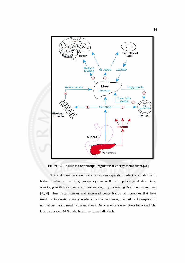
Glucose stimulates pancreatic cells and is the main physiological regulator of acute
insulin secretion & biosynthesis. Normal glucose regulation range(3 to 5.5 mM) is
dependent on closed feedback loop relationship between liver, peripheral tissues
(primarily muscle) and pancreatic islets as shown in Figure (1.20 [40].
When glucose and other nutrients are absorbed from the gastrointestinal tract,
this elicits insulin secretion. Insulin regulates the metabolism of multiple fuels
(indicated in blue). Selected actions of insulin are indicated in red (+, activation; -,
inhibition). Insulin activates transport of glucose into muscle & adipose tissue and also
promotes synthesis of glycogen & triglycerides. Insulin inhibits lipolysis in adipose
tissue, ketogenesis in liver, & proteolysis in muscle. Insulin also inhibits hepatic
glucose production by inhibiting both glycogenolysis and gluconeogenesis.
Insulin does not directly regulate the metabolism of red blood cells which use
glycolysis to provide energy. Although the brain uses glucose in the fed state, it can
also use ketone bodies (Acetoacetate and 3- hydroxybutyrate) when levels rise high
enough (e.g., during fasting). As in type 1 diabetes mellitus, the loss of effective
insulin action directly leads to unrestrained hepatic glucose production and
inefficient peripheral glucose utilization. Excessive hepatic glucose output largely
accounts for elevated fasting plasma glucose (FPG) levels.
Resistance to the antilipolytic action of insulin in adipose tissue leads to
elevated plasma free fatty acid (FFA) levels and increased FFA delivery to the liver.
There, the oxidation of FFA generates energy (ATP) needed to sustain
gluconeogenesis. In addition, the latter process is stimulated by FFA metabolites such
as acyl coenzyme A [39]. In this indirect manner, insulin resistance also contributes
to elevated glucose production in the liver [42]. Moreover, the elevation of FFA
levels also contributes to insulin resistance in muscle.
16
Figure 1.2: Insulin is the principal regulator of energy metabolism.[41]
The endocrine pancreas has an enormous capacity to adapt to conditions of
higher insulin demand (e.g. pregnancy), as well as to pathological states (e.g.
obesity, growth hormone or cortisol excess), by increasing cell function and mass
[43,44]. These circumstances and increased concentration of hormones that have
insulin antagonistic activity mediate insulin resistance, the failure to respond to
normal circulating insulin concentrations. Diabetes occurs when cells fail to adapt. This
is the case in about 10 % of the insulin resistant individuals.
17
1.2.4 .2 Insulin Resistance
Insulin resistance describes an insufficient action of insulin on target tissues
(muscle, liver and adipose tissue) and requires an adaptation by the cells to increase
insulin production. Insulin resistance triggers the development of type 2 diabetes in
most patients. It is present in type 2 diabetic and in obese individuals.
In about 90% of patients with insulin resistance, the cell can adapt to this higher
insulin demand, but the other 10% become diabetic with time. A correlation between
increasing body mass index and decreasing insulin sensitivity has been
demonstrated. These results show that insulin resistance in obese diabetic patients
is mainly a consequence of obesity. Weight loss can restore normal glucose
tolerance in obese individuals with impaired glucose tolerance and prevent
progression of diabetes in obese individuals with insulin resistance[45].
There are many individuals, obese and insulin resistant, but non diabetic,
being able to secrete sufficient insulin to compensate for the insulin resistance. The
inability to do this may reflect essential genetic defects in those who develop type 2
diabetes, as a defect in the functional capacity of pancreatic cells to secrete insulin is
undoubtedly necessary for the development of overt hyperglycemia [46].
Type 2 diabetes is characterized by a progressive decrease in insulin action,
followed by an inability of the cell to compensate for insulin resistance. Insulin
resistance is the first lesion, due to interactions among genes, aging, and metabolic
changes produced by obesity. Insulin resistance in visceral fat leads to increased
fatty acid production, which exacerbates insulin resistance in liver and muscle.
The cell compensates for insulin resistance by secreting more insulin. Ultimately,
the cell can no longer compensate, leading to impaired glucose tolerance and diabetes.
1.2.5 Gestational Diabetes mellitus (GDM)
Gestational diabetes mellitus (GDM) is defined as glucose intolerance, which
is first recognized during pregnancy. In most women who develop GDM, the
disorder has its onset in the third trimester of pregnancy. At least 6 weeks after the
pregnancy ends, the woman should receive an oral glucose tolerance test and be
18
reclassified as having diabetes, normal glucose tolerance, impaired glucose tolerance
or impaired fasting glucose. Gestational diabetes complicates about 4% of all
pregnancies.
1.2.6 Specific types of Diabetes
1.2.6.1 Genetic Defects
Maturity onset diabetes of the young (MODY) is characterized by impaired
insulin secretion with minimal or no insulin resistance [47].Patients typically exhibit
mild hyperglycemia at an early age. The disease is inherited in an autosomal
dominant pattern and, at present, six different genetic abnormalities have been
identified [48].
Genetic inability to convert proinsulin to insulin results in mild
hyperglycemia and is inherited an autosomal dominant pattern.Similarly, the
production of mutant insulin molecules has been identified in a few families and
results in mild glucose intolerance. Several genetic mutations have been described in
the insulin receptor and are associated with insulin resistance. Type A insulin
resistance refers to the clinical syndrome of Acanthosis Nigricans, Virilization in
women, polycystic ovaries, and hyperinsulinemia. Lipo atrophic diabetes results from
post receptor defects in insulin signaling [49].
1.2.6.2 Diseases of the Exocrine Pancreas
Damage of the pancreas must be extensive for diabetes to occur. The most
common causes are pancreatitis, trauma, and carcinoma. Cystic fibrosis and
hemochromatosis also have been associated with impaired insulin secretion.
1.2.6 .3 Endocrinopathies
Since growth hormone, cortisol, glucagons and epinephrine increase hepatic
glucose production and induce insulin resistance in peripheral (muscle) tissues;
excess production of these hormones can cause or exacerbate underlying diabetes
[50,51].Although the primary mechanism of action of these counter regulatory
hormones is the induction of insulin resistance in muscle and liver, overt type 2
diabetes mellitus does not develop in the absence of beta cell failure.

19
1.2.6.4 Infections
A variety of infections have been etiologically related to the development of
diabetes mellitus. Of these, the most clearly established is congenital rubella
[52].Approximately 20% of infants who are infected with the rubella virus at birth
develop autoimmune type 2 diabetes later in life. These individuals have the typical
type 1 susceptibility genotype, DR3 / DR4 [53].
1.2.6.5 Drugs
A large number of commonly used drugs have been shown to induce insulin
resistance and /or impair cell function and can lead to the development of diabetes
mellitus in susceptible individuals. An extensive review of these drugs and their
mechanism of action have been published [54].
1.2.7 Causes of Diabetes
• Hereditary and genetics factors
• Infections caused by viruses
• Stress
• Obesity
• Increased cholesterol level
• High carbohydrate diet
• Nutritional deficiency
• Excess intake of oil and sugar
• No physical exercise
• Over eating
• Tension and worries
• High blood pressure
• Insulin deficiency
• Insulin resistance

20
1.2.8 Symptoms of Diabetes
• Increased thirst
• Frequent urination
• Extreme hunger
• Unexplained weight loss
• Fatigue
• Blurred vision
• Slow healing of injuries
• Frequent infections, like gums, skin, vaginal or bladder infection
• Weakness or loss of strength
1.3 OXIDATIVE STRESS
Oxidative Stress depicts the existence of products called free radicals
(moleculespossessing an unpaired electron) and reactive oxygen species, which are
formed in normal physiology but become deleterious when not being quenched by a
cascade of antioxidants systems. This can result either from an overproduction of
ROS or from the inactivation of the AOS, thus shifting the OS / AOS balance in
favour of stress. ROS oxidize various types of biomolecules, finally leading to
cellular lesions by damaging DNA or stimulating apoptosis for cell death.
Some ROS are considered more important than others, such as superoxide,
hydroxyl radicals or peroxides. However not all oxygencontaining radicals have high
oxidative potential. ROS are neutralized by a battery of AOS, which can be divided
into mainly two categories: enzymes (e.g.Superoxide Dismutase SOD, Glutathione
Peroxidase GPX and Catalase CAT) and non enzymatic systems (e.g.: glutathione
GSH, vitamins A, C and E). Some are located in cell membranes, others in the
cytosol, and others in blood plasma. Due to its location in mitochondria and its
position in the antioxidant chain, SOD is usually considered as particularly
important since even modest decreases in SOD are sufficient to provoke cell damage.
Quantitatively, however, albumin and uric acid are the main AOS [55].
21
1.3.1 Oxidative stress and Diabetes
Oxidative stress plays a major role in the pathogenesis of both types of
diabetes mellitus. Free radicals are formed in diabetes by glucose oxidation, protein
glycation, and the subsequent degradation of glycated proteins. High levels of free
radicals and the simultaneously declined antioxidant enzyme levels lead to cell
damage, inactivation of enzymes, and lipid peroxidation. Accumulated evidence also
indicates that oxidative stress activated signaling pathways thatmediate insulin
resistance and cell dysfunction. These consequences of oxidative stress can promote
the development of diabetes complications. Therefore, oxidative stress, antioxidant
defenses, cellular redox status should be regarded as the central players in diabetes
and its complications.
Experimental diabetes can be induced in rodents by feeding alloxan or
streptozotocin. It is well established that alloxan works by generating reactive
oxygen species and the -cells of pancreatic islets are specifically destroyed .It is assumed
that free radicals that Alloxan generated kill the islet cells. Increased oxidative stress,
defined as a persistent imbalance between the production of highly reactive
molecular species (chiefly oxygen and nitrogen) and antioxidant defenses, is a
widely accepted participant in the development and progression of diabetes and its
complications[56,57].Diabetes is usually accompanied by increased production of
free radicals or impaired antioxidant defenses. Glucose oxidation is the main source
of free radicals. Mechanisms by which increased oxidative stress is involved in the
diabetic complications are partly known, including activation of transcriptional
factors, advanced glycated end products (AGEs) and protein kinase C.
1.3.2 Oxidative stress and Type 1 Diabetes mellitus
It is known that STZ or Alloxan diabetes in rodent is the result of destruction
of Pancreatic cells by free radicals, the combined result of active redox enzymes and
inadequate antioxidant defenses in those cells[55] and it is very difficult to induce
alloxan and Streptozotocin diabetes in human pancreatic cells because these cells
have very higher levels of SOD and Catalase compared with rodent pancreatic
cells[58].Nevertheless, certain xenobiotics can induce diabetes in human, which can

22
be due to the generation of free radicals. Vacor, which could block complex I of the
mitochondrial respiratory chain, can lead to generation of O2. By causing leakage of
reducing electrons on to O2, possibly via ubisemiquinone. It is worthwhile taking a
look at maternally inherited diabetes. This type of disease is triggered by defects in
mitochondrialy coded gene (commonly in tRNALeu (UUR) ) [59].
This defect seems to produce defective or insufficient complex I. High levels
of heteroplasmy in cells would produce defective mitochondria, which presumably
would generate radicals. It is pretty interesting to look closely the underlying
mechanisms of streptozotocin induced diabetes. There are two ways in which
streptozotocin can be used to induce diabetes in rodent. The traditional procedure is
to use a single toxic dose which causes cell death in 2-4 days. The alternative is to
apply multiple low doses, which leads to a more immunologically based disease with
insulitis and the activation of C-type retroviruses, perhaps resembling more closely
type 1 diabetes in human. It was showed that streptozotocin leads directly to
generation of H2O2 in cells, more actively indeed than that of alloxan [60]. It is
reported that SOD prevents the impairment of islet microcirculation that is an early
consequence of streptozotocin in rats [61]. It is apparent that streptozotocin works
by a free radical mechanism like alloxan.
1.3.3 Oxidative stress and Type II Diabetes mellitus
A large genetic component also exists in type 2 diabetes mellitus and the
concordance rate in identical twins is around 90%. This suggests that genes
essentially determine the disease in the appropriate environment. Insulin resistance is
one of the major characteristics of type 2 diabetes mellitus. If the insulin resistance
can result from oxidative damage, then a prediction would be that chronic oxidative
stress would lead to hyperinsulinaemia if plasma glucose is clamped at normal level
by infusing the required insulin. Following experiment supports this hypothesis. Fat
fed mice, infused with insulin and glucose, showed impaired glucose clearance.
However, glucose clearance was slowed 2-3 fold further by prior feeding with low-
dose of streptozotocin, which had little hyperglycemic effect by itself and led to
chronic oxidative stress[62].

23
The streptozotocin effect was presumably not on the cell with continuous
supplement with external insulin, but seemed to have caused insulin resistance directly.
Evidence showed that membrane proteins are early targets of oxidative stress. An
early event in the induction of the multiple low dose of streptozotocin diabetes is the
gradual loss of GLUT2 glucose transporter from islet cell membranes, which could
cause insulin resistance [63].
1.3.4 Hyperglycemia and oxidative stress signaling
As discussed above, diabetic complications come from chronic elevated
glucose levels in both type 1 and 2 diabetes. The pathogenic effects of high glucose
are mediated via ROS generated by glucose oxidation and protein glycation. In
addition to their ability to directly inflict macromolecular damage, ROS can function
as signaling molecules to activate a number of cellular stress-sensitive pathways that
cause cellular damage, which ultimately lead to late complications of diabetes.
Furthermore, these same pathways are also showed to link to insulin resistance and
cell dysfunction[64]. These pathways are involved in the pathogenesis of diabetes.
Free radicals can be generated in glucose oxidation, which is believed to be
the main source of free radicals. In its enediol form, glucose is oxidized in a
transition metal dependent reaction to an enediol radical anion that is converted into
reactiveketoaldehydes and to superoxide anion radicals. The superoxide anion
radicals undergo dismutation to hydrogen peroxide, which if not degraded by
catalase or glutathione per oxidase, and in the presence of transitional metals, can lead
to production of extremely reactive hydroxyl radicals [65]. Superoxide anion radicals
can also react with nitric oxide to form reactive per oxy nitrite radicals.
Hyperglycemia is also found to promote lipid per oxidation of low density
lipoprotein (LDL) by a superoxide-dependent pathway to generate free radicals [66].
Another important source of free radicals in diabetes is interaction of glucose
with proteins which leads to protein glycation. Glycation involves the condensation of
glucose with the -amino group of lysine, the -amino group of an N terminal amino acid or the
amines of nucleic acids, which will result in the formation of AGEs. The increase
availability of glucose in diabetes mellitus induces enhanced production of AGEs.
24
This process has been described as glycosylation, and is probably the major source
of increased generation of ROS in diabetic patients [67]. AGEs are believed to be
involved in the genesis of many of the irreversible complications of diabetes,
including expanded extracellular matrix, cellular hypertrophy, hyperplasia, and
vascular complication [68].The formation of glycoxidation products is not only the
result of glucose induced oxidative stress. Fructose, which is increased as a
consequence of activation of the polyol pathway, leads to the formation of AGE
precursors methylglyoxal and 3-deoxyglucosone [69].
These AGEs, via their receptors (RAGEs), inactivate enzymes and alter their
structures and functions, promote free radicals formation, and quench and block
antiproliferative effects of nitric oxide. By increasing intracellular oxidative stress,
AGEs activate the transcription factor NF-kB, thus promoting up-regulation of
various NF-kB controlled target genes[70].NF-kB enhances production of nitric
oxide, which is believed to be a mediator of islet cell damage. In addition,
hyperglycaemia leads to glycation of antioxidant enzymes, which could alter the structure
and function of antioxidant enzymes such that they are unable to detoxify free
radicals, exacerbating oxidative stress in diabetes. Therefore, the process of glucose
oxidation might be responsible not only for increased ROS products but also for
decrease availability of antioxidant enzymes.
1.3.5 Oxidative stress and Insulin Resistance
ROS and oxidative stress can lead to the activation of multiple serine kinase
cascadesin vitro. The insulin signaling pathway provides a number of potential targets
of these activated kinases, including the insulin receptor (IR) and the family of IR
substrate (IRS) proteins. For IRS-1 and 2, an increase in serine phosphorylation
decreases the extent of tyrosine phosphorylation and is consistent with the attenuation
of insulin action. The role of serine kianse activation in oxidative stress induced
insulin resistance. A variety of stimuli increase ROS production and oxidative stress.
This results in the activation of multiple stress sensitive serine/threonine kinase
signaling cascades. Once activated, these kinases are able to phosphorylate multiple
targets, such as the IR and IRS proteins. Increased phosphorylation of IR or IRS

25
proteins on discrete serine or threonine sites decreases the extent of insulin-
stimulated tyrosine phosphorylation. Consequently, the association and/or activities
of downstream signaling molecules (e.g. PI-3 kinase) are decreased, resulting in
reduced insulin action[65].
1.3.6 Oxidative stress and -cell dysfunction
cells are responsible for sensing and secreting insulin in response to glucose
stimulation. -cells are sensitive to ROS because they are low in antioxidant enzymes such as
Catalase, GPX, SOD. over expression of the antioxidant enzymes in islets or transgenic
mice prevents many of the deleterious effects discussed above. Oxygen stress
generated by short exposure of cells proparations to H2O2 increases production of
p21, an inhibitor of cyclin dependent kinase, decreases insulin mRNA, cytosolic
ATP and calcium flux in cytosol and mitochondria and cause apoptosis. Insulin
secretion stimulated by glucose or methyl succinate can be inhibited shortly,
whereas the response to K+ remains normal. These results suggest that the
mitochondrial processes involved in glucose mediated insulin secretion are
particularly affected by oxidative stress [71].
1.4 NATURAL DEFENSE AGAINST OXIDATIVE STRESS
Reactive species can be eliminated by a number of enzymatic and non
enzymatic antioxidant mechanisms. Enzyme SOD immediately convertsO2 to H2O2,
which is then detoxified to water either by catalase in the lysosomes or by glutathione
peroxidase in the mitochondria. Another enzyme that is important is glutathione
reductase, which glutathione that is used as a hydrogen donor by glutathione
peroxidase during the elimination of H2O2. Recent reviewes shows that diabetes has
multiple effects on the protein levels and activity of these enzymes, which further
augment oxidative stress by causing a suppressed defense response[72].
Increased isoprostane levels in diabetic patients with chronic heart failure are
correlated with antioxidant status and disease severity[73,74]. Thus, modulation of
these enzymes in target organs prone to diabetic complications such as heart and
kidney may prove beneficial in the prevention and management of heart failure and

26
kidney failure. Non enzymatic antioxidants include vitamins A, C and E; glutathione,
-lipoic acid, carotenoids, trace elements like copper, zinc and selenium; coenzyme
Q10 (Co Q10); and cofactors like folic acid, uric acid, albumin, and vitamins B1,
B2, B6 and B12. Alterations in the antioxidant defense system in diabetes have
recently been reviewed [75].
Glutathione (GSH) acts as a direct scavenger as well as a co substrate for
GSH peroxidase. It is a major intracellular redox tampon system. Vitamin E is a fat-
soluble vitamin that prevents lipid peroxidation. It exists in 8 different forms, of
which - tocopherol is the most active form in humans. Hydroxyl radical reacts with
tocopherol forming a stabilized phenolic radical which is reduced back to phenol by
ascorbate and NAD(P)H dependent reductase enzymes [76].
CoQ10 is an endogenously synthesized compound that acts as an electron
carrier in the Complex II of the mitochondrial electron transport chain. CoQ10 is a
lipid soluble antioxidant, and in higher concentrations, it scavengesO2 and improves
endothelial dysfunction in diabetes. Vitamin C (ascorbic acid) increases NO
production in endothelial cells by stabilizing NOS cofactor BH4 [77].
-Lipoic acid is a hydrophilic antioxidant and can therefore exert beneficial effects in
both aqueous and lipid environments. -lipoic acid is reduced to another active compound
dihydrolipoate. Dihydrolipoate is able to regenerate other antioxidants such as
vitamin C, vitamin E and reduced glutathione through redox cycling. Thus, both
experimental and clinical studies summarized in the next sections utilized these
naturally occurring antioxidants, especially vitamins C, E and -lipoic acid, in order to
delineate the role of oxidative stress in the development of vascular complications of
diabetes.
Role of antioxidant in treatment of diabetes.
The clinical trials conducted to date failed to provide adequate support for the
use of antioxidants in diabetes, it is still too early to reach a definitive conclusion on
this issue. As discussed above, with the exception of -lipoic acid studies in diabetic
neuropathy, data from clinical trials are limited. The majority of studies were not
27
designed to assess the effect of antioxidant use specifically in diabetic patients. This
is an important point because diabetic patients represent a population in whom
oxidative stress is much higher than in the general population. As in the SPACE
trial of patients on hemodialysis, patients exposed to very high oxidative stress
responded favorably to vitamin E supplementation [78].
It is possible that antioxidants would be more demonstrably effective in a
patient population chosen on the basis of elevated levels of oxidative stress.
Unfortunately, none of the studies to date effectively assessed the baseline oxidative
stress of the enrolled patients using any of the commonly accepted markers of
inflammation.
In all likelihood, the choice and dose of antioxidant might be very important.
The clinical trials focused mainly on the use of vitamin E. Negative results with
vitamins cannot be generalized to all antioxidants. As has been eloquently argued
elsewhere, treating the antioxidant vitamins as a single class of compounds with
expected similar effects inappropriately disregards their wide range of chemical
properties and pharmodyanimics[79].
Recently, it has been postulated that antioxidant potency of vitamins such as
C and E is limited because these antioxidants work as scavengers of existing excess
reactive species in a stoichiometric manner and this approach represents a
symptomatic approach to oxidative stress-associated clinical problem[80]. Based on
the new developments in our understanding of the pathophysiology of oxidative
stress, it is clear that strategies to block the formation of reactive radicals will
provide a targeted and causal approach to provide conclusive evidence whether
antioxidants should be part of the cardiovascular treatment plan in diabetes.
Cytosolic SOD and catalase mimetics, L-propionyl carnitine, PKC- inhibitor
LY- 333531, peroxynitrite catalyst FP15 and mitochondrial uncoupler DNP [81,82].
Given the number of shortcomings in the clinical trials, it seems clear that more
research on the use of antioxidants in the prevention of cardiovascular complications
in diabetes is necessary and strongly encouraged. From a clinical view point,
however, efforts for the prevention of diabetic complications should seek to
28
maximize the benefits of proven therapeutic strategies including appropriate life
style changes and controlling blood pressure, blood glucose and lipids.
1.5 THERAPEUTIC INTERVENTIONS IN DIABETES MELLITUS
The care of diabetes on self-management is based on the patient's clinical status
and his/her ability to participate in self care. Insulin replacement therapy is the main
stay for patients with type 1 DM while diet and lifestyle modifications are
considered the corner stone for the treatment and management of type 2 DM. Insulin
is also important in type 2 DM when blood glucose levels cannot be controlled by
diet, weight loss, exercise and oral medications. Oral hypoglycemic agents are also
useful in the treatment of type 2 DM. Oral hypoglycemic agents include
Sulphonylureas, Biguanides, Glucosidase inhibitors and glitazones.
The main objective of these drugs is to correct the underlying metabolic
disorder, such as insulin resistance and inadequate insulin secretion. Theyshould be
prescribed in combination with an appropriate diet and lifestyle changes. Diet and
lifestyle strategies are to reduce weight, improve glycaemic control and reduce the
risk of cardiovascular complications, which account for 70% to 80% of deaths
among those with diabetes. Diabetes is best controlled by either diet alone and
exercise (non- pharmacological), or diet with herbal or oral hypoglycaemic agents
or insulin (pharmacological).
1.5.1. Pharmacological intervention-Type II Diabetes Mellitus
Insulin
The introduction of insulin to treat diabetes has saved an estimated 5 million
years of life for patients with type 1 diabetes during the year 2000. Considerable
progress has been made, in recent years, in the production, formulation and delivery
of insulin preparations, as well as the development of insulin treatment regimens
which maintain long term normoglycemia, with a low risk of hypoglycemia. The
importance of the aim of preventing or slowing the progression of chronic
microvascular complications has been conclusively proven during the last decade, in
both type 1 and type 2 diabetes.
29
Insulin analogues have an alteration in the amino acid sequence of human
insulin, which change the rate of insulin absorption, or some other structural change
like being linked to a fatty acid chain, that alters the insulin time action curve[83].
Regular insulin is modified to result in the various short-acting insulin analogus,
Insulin lispro (Humalog),Insulin aspart (Novolog) and Insulin glulisine (Apidra);
intermediate (Isophane, Lente) long-acting analogues: Insulin glargine and insulin
detemir.
Many insulin preparations are available and are grouped according to their
ion: a rapid acting formulation to cover meals, intermediate and longer acting
preparations to provide steady (background) basal levels between meals and
overnight. Insulin is prepared either from human, or porcine, or bovine or a mixture
of bovine and porcine. Human insulin (Humulin, Novolin) is now widely available
prepared by recombinant DNA techniques. The physicochemical properties of human,
porcine and bovine insulins differ owing to their different amino acid sequences.
Human insulin is more soluble than porcine insulin in aqueous solutions. It is
supplied at neutral pH to make it more stable. Insulin is the mainstay for treatment of
virtually all type 1 DM and many type 2 DM patients. Insulin may be administered
intravenously (IV), or intramuscularly (IM); however for long-term treatment,
subcutaneous (SC) route is preferred. Short and rapid acting insulin preparations:
have the most rapid onset of action but the shortest duration. Short-acting insulin
(i.e. regular or soluble) usually should be injected 30 to 45 min before meals [84].
Regular insulin may also be given IV or IM. After IV injection, there is a
rapid fall in blood glucose concentration within 30-45 (5-15 min for lispro, aspart and
glulisine insulins), reaches its peak in 1.5 to 4 hours (30-90 min for Lispro, Aspart and
Glulisine) and the duration of its action is 5-8 hours (2-5 hours for Lispro, Aspart and
Glulisine). Intravenous infusions of insulin are useful in patients with ketoacidosis or
during the perioperative period, during labour and delivery, and in intensive care
situations. Regular insulin is present in solution for injection as a hexamer and to be
efficiently absorbed into the circulation the insulin hexamer must dissociate into
dimmers or monomers.

30
It is this dissociation process that takes 30-60 min that determines the onset
and ultimately the time action curve of regular insulin. Unlike regular insulin, the
insulin analogues (Lispro, Aspart and Glulisine) dissociate into monomers almost
instantaneously following injection. This property results in rapid absorption and
shorter duration of action compared to regular insulin. Lispro has two advantages
over regular insulin: first, the prevalence of hypoglycemia is reduced by 20% to 30
%; second, glucose control, as assessed by HbA1c is modestly but significantly
improved (0.3% to 0.5%). Aspart insulin and Glulisine insulins are similar to Lispro.
Several shortacting human regular insulin (dry powder or liquid suspension)
preparations are available as inhalations and when delivered have an onset and peak
action time similar to that of a rapid acting insulin but a duration of action is slightly
longer then that of the currently available rapid acting insulin analogues.
1.5.2 Oral Hypoglycemic agents
1.5.2.1 Sulfonylureas
Sulfonylureas are structurally related to sulphonamides and were discovered
accidentally, in 1942 when it was noted that some sulphonamides caused
hypoglycemia in experimental animals. These observations were extended, and 1-
butyl-3-sulfonylurea (carbutamide) became the first clinically useful sulfonylurea for
the treatment of diabetes. This compound was later withdrawn because of adverse
effects on the bone marrow but led to the discovery of the entire class of
sulfonylureas. In the 1950s tolbutamide was widely used in type 2 DM and
subsequently 20 different agents of this class have been in use worldwide. This was
followed by the introduction of biguanides, phenformin, which was later withdrawn
because of an increase in the frequency of lactic acidosis associated with it use. Later
on metformin was introduced and this drug has been used extensively in Europe
without the side effects of phenformin.
1.5.2.2 Biguanides
Metformin (Glucophage) and phenformin were introduced in 1957 and
buphormin was introduced in 1958. They were widely used in Europe for treating

31
type 2 diabetes for nearly 20 years. The latter two were withdrawn in many countries
in the 1970s because of an association with fatal lactic acidosis [85].
Additionally an increased risk of cardiovascular mortality was seen with oral
hypoglycemic agents compared with insulin. Metformin has a very low rate of lactic
acidosis compared with phenformin and has been widely used in Europe, Canada,
Middle East and other countries; it became available in the united states in 1995.
Metformin given alone or in combination with a sulfonylurea improves glycaemic
control and lipid concentrations in patients who respond poorly to diet or to a
sulfonylurea alone[86].Sulfonylureas in reducing fasting plasma glucose (FPG) and
postprandial glucose concentrations, but caused no weight gain or hypoglycaemia in
contrast to sulfonylurea therapy[87].
1.5.2.3 Thiazolidinediones
Thiazolidinediones (TZDs) are chemically and functionally unrelated to the
other classes of oral antidiabetic agents. A thiazolidine-2, 4-dione structure is
common to all agents. Two compounds in this class are currently in use.
Rosiglitazone (Avandia) and pioglitazone (Actos) are the two thiazolidinediones in
use. The third, troglitazone, was withdrawn from use because of its association with
severe hepatic toxicity[89].
1.5.2.4 Meglitinide analogues
The meglitinide analogues are a new class of drugs developed to improve
early phase insulin secretion, which is one of the earliest pathophysiologal
manifestations of type 2 DM. These are derived from the meglitinide portion of
sulfonylureas. Examples of this group are repaglinide and nateglinide. Another
meglitinide known as mitiglinide is undergoing clinical trials. Repaglinide is derived
from the non-sulfonylurea moiety of glibenclamide whereas nateglinide is derived
from the amino acid D-phenylalanine.
The S-enantiomer of repaglinide is the pharmacologically active part of the
racemic molecule. In the rat model, this enantiomer has more than 100 times greater
hypoglycaemic potency than the R-enantiomer. Clinically available repaglinide is

32
about 98% pure for the Senantiomer. The meglitinides are rapid-acting insulin
secretagogues (also known as prandial glucose regulators) that have a fast onset and
short duration of action resulting in more physiological secretion of insulin from the
-cell without causing continued elevation of insulin in the post-absorptive phase, thus
reducing glycemia without increasing the risk of hypoglycemia.
1.5.2.5 -Glucosidase inhibitors
-Glucosidase inhibitors have been developed specifically to delay the digestion of
complex carbohydrates and decrease the postprandial rise in plasma glucose, thus
reproducing the effect of a low glycogenic index/high fibre diet. These actions
significantly reduce postprandial glycaemic and insulinaemic increase whether they
are used as monotherapy or combined in the treatment of type 1 and type 2 diabetes.
These drugs have an excellent safety profile.
1.6 HERBS USED IN TREATMENT OF DIABETES
In the last few years there has been an exponential growth in the field of
herbal medicine and these drugs are gaining popularity both in developing and
developed countries because of their natural origin and less side effects. Many
traditional medicines in use are derived from medicinal plants, minerals and organic
matter[90]. A number of medicinal plants, traditionally used for over 1000 years
named rasayana are present in herbal preparations of Indian traditional health care
systems[91].
1.7 STREPTOZOTOCIN
Streptozotocin (Streptozocin, STZ) is a naturally occurring chemical that is
particularly toxic to the insulin-producing beta cells of the pancreas in mammals. It is
used in medicine for treating certain cancers of the Islets of langerhans and used in
medical research to produce an animal model for diabetes. Streptozotocin is broad
spectrum antibiotic from streptomyces achromogenes. Since the finding that STZ
possess diabetogenic properties mediated by pancreatic cell destruction, this
compound has been widely used to induce diabetes in experimental mode l[92]. The
cell specific toxin STZ, an analogue of glynac, has been used to create animal

33
models of diabetes, despite an incomplete understanding of how STZ actually
Cause cell death.
Streptozotocin action in cells is accompanied by characteristic alterations in blood
insulin and glucose concentrations. Two hours after injection, the hyperglycemia is
observed with a concomitant drop in blood insulin. About six hours later,
hypoglycemia occurs with high levels of blood insulin. Finally, hyperglycemia
develops and blood insulin levels decreases [93].
These changes in blood glucose and insulin concentrations reflect
abnormalities in cell function. STZ impairs glucose oxidation and decreases insulin
biosynthesis and secretion. It was observed that STZ at first abolished the cell
response to glucose. Temporary return of responsiveness then appears which is
followed by its permanent loss and cells are damaged [94].
STZ is taken up by pancreatic cells via glucose transporter glut2. A reduced
expression of glut2 has been found to prevent the diabetogenic action of STZ
observed that STZ itself restricts glut2 expression in vivo and in vitro when
administered in multiple doses. Intracellular action of STZ results in changes of DNA
in pancreatic cells comprising its fragmentation. Recent experiments have proved that the
main reason for the STZ induced cell death isalkylation of DNA [95].
The alkylating activity of STZ is related to its nitrosourea moiety, especially
at the o6 position of guanine. After STZ injection to rats, different methylated purines
were found in tissues of these animal [96]. Since STZ is a nitric oxide (NO) donor
and no was found to bring about the destruction of pancreatic islet cells, it was
proposed that this molecule contributes to STZ induced dna damage [97]. The
participation of (NO) in the cytotoxic effect of STZ was confirmed in several
experiments [97].
Pancreatic cells exposed to STZ manifested changes characteristic for no
action, i.e. increased activity of guanylyl cyclase and enhanced formation of cgmp
[98]. STZ is, however, not a spontaneous nitric oxide donor. This molecule is
liberated when stz is metabolized inside cells, but NO synthase is not required for this
34
effect [97]. On the other hand, the lowering of NO concentration in pancreatic islet
cells by inhibition of the inducible form of nitric oxide synthase partially
counteracted DNA cleavage induced by STZ [97].
A similar effect can be attained by no scavengers [97]. However, the results
of several experiments provide the evidence that no is not the only molecule
responsible for the cytotoxic effect of STZ. STZ was found to generate reactive
oxygen species, which also contribute to DNA fragmentation and evoke other
deleterious changes in the cells[99].
The formation of superoxide anions results from both STZ action on
mitochondria and increased activity of xanthine oxidase. It was demonstrated that
STZ inhibits the krebs cycle and substantially decreases oxygen consumption by
mitochondria[100]. These effects strongly limit mitochondrial ATP production and
cause depletion of this nucleotide in cells. Restriction of mitochondrialATP generation
is partially mediated by NO.
This molecule was found to bind to the iron containing aconitase inhibiting
enzyme activity. Augmented ATP dephosphorylation increases the supply of substrate
for xanthine oxidase ( cells possess high activity of this enzyme) and enhances the
production of uric acid the final product of ATP degradation. Then, xanthine oxidase
catalyses reaction in which the superoxide anion is formed [101].
As a result of superoxide anion generation hydrogen peroxide and hydroxyl
radicals are formed [102]. The inhibition of xanthine oxidase by allopurinol restricts
the cytotoxic effect of STZ in vitro. Pretreatment of cells with this inhibitor prevented the
STZ induced decrease of insulin secretion[102]. It can be stated that potent alkylating
properties of STZ are the main reason of its toxicity. However, the synergistic action
of both NO and reactive oxygenspecies may also contribute to DNA fragmentation
and other deleterious changes caused by STZ. NO and reactive oxygen species can
act separately or form the highly toxic peroxynitrate (Fig.1.3). Therefore,
intracellular antioxidants or NOscavengers substantially attenuate STZ toxicity.

35
Fig.1.3 The mechanism of Streptozotocin (STZ)-induced toxic events in
cells of rat pancreas. mit - mitochondria; xod - xanthine oxidase
1.8 Musa sapientum
Musa sapientum which is commonly called banana is a herbaceous plant of
the family Musaceae. It is known to have originated from the tropical region of
Southern Asia. The Musa sapientum grows up to a height of about 2-8m with leaves
of about 3.5m in length. The stem which is also called pseudostem produces a single
bunch of banana before dying and replaced by new pseudostem. The fruit grows in
hanging cluster, with twenty fruits to a tier and 3 – 20 tiers to a bunch. The fruit is
protected by its peel which is discarded as waste after the inner fleshy portion is
eaten.Banana is a familiar tropical fruit. From its native Southwestern Pacific home,
the banana plant spread to India by about 600 BC and later on it spread all over the

36
tropical world. It is possibly the world's oldest cultivated crop. It even spread into the
Islands of the Pacific and to the West Coast of Africa as early as 200-300 BC [103].
1.8.1 Taxonomical classification
Kingdom : Plantae
Division : Magnoliophyta
Class : Liliopsida
Order : Zingiberales
Family : Musaceae
Genus : Musa
Species : Musa sapientum.
1.8.2Cultivation and Distribution
In different countries about 300 varieties of bananas are grown, of which a
vast majority have been growing in Asian, IndoMalaysian and Australian tropics and
are now widely found throughout the tropical and subtropicalCountries. India,
Philippines, China, Ecuador, Brazil, Indonesia, Mexico, Costa Rica, Colombia.
Fig 1.4 Plants with fruits of Musa sapientum.
37
1.8.3 Varieties of Banana in India.
1. Monthan – Musa spp- Blueggoe-AAB
2. Karpooravalli - Musa spp- karpooravalli-ABB
3. Nendaran - Musa spp- fresh plantain-AAB
4. Kadali- Musa spp- Ney pooven-AB
5. Pachainadan - Musa spp- pachainadan-AABS
6. Poovan - Musa spp- Mysore-AAB
7. Rasthali- Musa spp- Rasthali-AAB
8. Robusta- Musa spp- robusta-AAB
9. Sevvazhai- Musa spp- Red banana-AAA.
1.8.4 Traditional Uses
Thailand is one of the top banana producing countries[103]. The fruit of
Musa paradisiaca and Musa sapientum is traditionally used in diarrhoea(unripe),
dysentery, intestinal lesions in ulcerative colitis, diabetes (unripe), in sprue, uremia,
nephritis, gout, hypertension, cardiac disease [104]. Musa sapientum is also used in
the treatment of excess menstruation with Canna indica L. var. speciosa [105].
Banana leaves (ashes) are used in eczema [105], as cool dressings for blister and
burns [104]. Flowers are used in dysentery and menorrhagia. Stem juice of fruited
plant is used for treating diarrhoea, dysentery, cholera, otalgia, haemoptysis and
flower is used in dysentery, diabetes and menorrhagia [104]. The root is used in
blood disorders, venereal diseases [104].
1.9 PHARMACOGNOSY
Pharmacognosy basically deals with the standardization, authentication and
study of natural drugs. It is closely involved with allied fields, viz. phytochemistry
and toxicological screening of natural products. Much of the research in
pharmacognosy has been done in identifying controversial species of plants,
authentication of commonly used traditional medicinal plants through
38
morphological, histological, physic chemical and toxicological parameters,
especially heavy metal estimation and radiobiological contamination in plants,
prescribed by an authoritative source. The importance of pharmacognosy has been
widely felt in recent times [107].
The herbal drug industry is considered to be a high growth industry of the late
90s and seeing the growing demand, it is all set to grow in the next century. The
trend for the increasing popularity of medicinal herbs in countries like America,
Australia and Germany is well supported by statistical data. Ayurveda strongly
believes in polyherbal formulations and scientists of modern era often ask for
scientific validation of herbal remedies. The efficacy of some herbal products is
beyond doubt, the most recent examples being Taxus brevifolia Nutt. (Taxols) and
Silybium marianum (L.)Garetn.(Silymarin). Hypericum perforatum (hypericin &
hyperforin), Allium sativum L. (allicin or allin), Ginkgo biloba L. (Ginkgolic acid)
are popularly used herbal remedies among people.
All these herbals are standardized for active constituent. Standardization
means adjusting the herbal drug preparation to a defined content of the active
constituent. Extract refers to a concentrated preparation of active constituent of a
medicinal herb. The concept of standardized extracts definitely provides a solid
platform for scientific validation of herbals [108].
Some drugs of plant origin in conventional medical practice are not pure
compounds but direct extracts or plant materials that have been suitably prepared and
standardized [109]. The World Health Organization has recommended the use of
arthemisinin derivatives from Artemisia annua (Composite), a Chinese herb with
established pharmacognostic data, as a first line drug in the treatment of malaria
[110]. Most of the cases of accidental herbal medicine misuse start with wrong
identification of a medicinal plant prescribed.
Many of the traditional systems have records where one common vernacular
name is supplied in place of two or more entirely different species. Ginseng, which
is a common Indian drug, is sold under 13 different names in the market. For
example Chinese or Asiatic ginseng (Panax ginseng), American ginseng (Panax
39
quinquefolius), Siberian ginseng (Eleutherococcus senticosus), Ayurvedic ginseng
(Withania somnifera Dunal.) and Russian ginseng (Acanthopanax senticosus) [111].
Such names could create confusion over prescription, which may eventually lead to
serious consequences. With this backdrop, it becomes extremely important to make
an effort towards standardization of the plant material to be used as medicine. The
process of standardization can be achieved by stepwise pharmacognostic studies
[107].
1.10 THE NEED OF THE HOUR
A majority of the present day diseases are reported to be due to the shift in the
balance of the proxidant and the antioxidant homeostatic phenomenon in the body.
Pro- oxidant conditions dominate either due to the increased generation of the free
radicals caused by excessive oxidative stress of the present day life, or due to the
poor scavenging/quenching in the body caused by depletion of the dietary
antioxidants [112]. In other words, the root cause of all diseases (acute or chronic) is
generation of free radicals. Therefore, the dire need of the hour is to discover or
identify medicinal plants, rich in antioxidants. Medicinal plants can be economic,
natural and easily affordable by all the people.
1.11 ANTIOXIDANTS
The term "antioxidant" refers to any molecule capable of stabilizing or
deactivating free radicals before they attack cells. There are also molecules
deserving the "antioxidant" term, because they act as chelating agents binding metal
ions (redox activity). Antioxidants are absolutely critical for maintaining optimal
cellular and systemic health and well being [113]. To protect the cells and organ
systems of the body against reactive oxygen species, humans have evolved a highly
sophisticated and complex antioxidant protection system. It involves a variety of
components, both endogenous and exogenous in origin, that function interactively
and synergistically to neutralize free radicals.
40
These components include,
1) Nutrient- derived antioxidants like ascorbic acid (vitamin C), tocopherols
and tocotrienols (vitamin E), carotenoids, and other low molecular weight
compounds such as glutathione and lipoic acid.
2) Antioxidant enzymes, e.g., superoxide dismutase, glutathione peroxidase,
and glutathione reductase, which catalyze free radical quenching reactions.
3) Metal binding proteins, such as ferritin, lactoferrin, albumin, and
ceruloplasmin that sequester free iron and copper ions that are capable of
catalyzing oxidative reactions.
4) Numerous other antioxidant phytonutrients present in a wide variety of plant
foods. In nature there are a wide variety of naturally occurring antioxidants
which are different in their composition, physical and chemical properties,
mechanisms and site of action [114].
1.11.1 Essential water soluble Antioxidants
Vitamin C
Ascorbic acid (vitamin C) is the major essential water soluble antioxidant in
human serum. Vitamin C in humans must be ingested for survival. Vitamin C is an
electron donor, and this property accounts for all its known functions. It is present in
relatively high concentrations extracellularly in the blood plasma Vitamin C can
function as an antioxidant and scavenge the O2, 1O2,OH, neutralize hypochlorous
acid (HOCl), and prevent lipid peroxidation [115].
1.11.2 Non-essential water soluble Antioxidants
1.11.2.1 Glutathione
Glutathione (GSH) in its reduced form is a good scavenger of many free
radicals like O2,OH and various lipid hydroperoxides and may help to detoxify
many inhaled oxidizing air pollutants like ozone, NO2 and free radicals in cigarette

41
smoke in respiratory tract [116]. GSH is an important water-soluble antioxidant
present ubiquitously in cells. Its main function is to detoxify xenobiotic toxins by
conjugation giving rise to oxidized glutathione disulfide (GSSG) [117].
1.11.2.2 Lipoic acid
-Lipoic acid (8-thioctic acid) is another thiol with antioxidant properties . It has been
reported to participate along with vitamin C, glutathione, vitamin E, and -carotene in
what is defined by Packer as a biological "redox antioxidant network"[118]. Lipoic acid is
involved in recycling oxidized vitamin C and/or vitamin E and yields either
dehydrolipoic acid (the oxidized form) or another radical. -Lipoic acid and GSH can
scavenge cigarette smoke-contained reactive oxygen species and preferentially react
with aldehydes, thus protecting proteins from oxidation [119].
1.11.3 Essential Lipid soluble Antioxidants
Vitamin E
Vitamin E ( -tocopherol) is the major intracellular lipophilic, chain breaker, and
efficient antioxidant capable of trapping peroxyl radicals intermediates in lipid
peroxidation and is responsible for protecting PUFA (Poly unsaturated fatty acid)
present in cell membrane and low density lipoprotein and quenching free radicals
and reactive oxygen species. It is also essential for structural membrane
stability[116].
1.11.4 Non-essential Lipid soluble Antioxidants
-carotene
Carotenoids such as -carotene are suggested to have antioxidant properties capable
of quenching free radicals such as singlet oxygen (1O2) [120]. It was also suggested
that a cooperative interaction exists between fat soluble antioxidants; the relation
between carotene and vitamin E was reported to be synergistic.

42
1.11.5 Secondary metabolites as Antioxidants
1.11.5.1 Phenolic compounds
Medicinal plant parts (roots, leaves, branches/stems, barks, flowers, and
fruits) are commonly rich in phenolic compounds, such as flavonoids, phenolic
acids, stilbenes, tannins, coumarins, lignans and lignins .Phenolic compounds are
ubiquitous bioactive compounds and a diverse group of secondary
metabolites.Accordingly,bioactive polyphenols have attracted special attention
because they can protect the human body from the oxidative stress which may cause
many diseases, including cancer, cardiovascular problems, and aging [121]..
1.11.5..2 Flavonoids
Flavonoids, which are partly responsible for the pigmentation of flowers,
fruits and leaves, are subdivided into flavanols, flavonols, flavones, flavanones
and anthocyanins based on the saturation of the flavan ring and also their
hydroxylation.They occur mostly as glycosylated derivatives, sometimes conjugated
with sulphate or organic acids [122].
1.12I IMPORTANCEOF SERUM BIOCHEMICAL ANALYSIS
Liver cell damage is characterized by a rise in serum enzymes like SGOT,
SGPT, ALP, etc. In general, SGOT concentrations are consistently higher than
SGPT levels which are expected since body cells contain more SGOT than SGPT.
Usually, about 80 % of SGOT is found in the mitochondria whereas SGPT
is purely cytosolic enzyme. Therefore, SGOT appears in higher concentrations in a
number of tissues (Liver, Kidney, heart and pancreas) and is released slowly in
comparison to SGPT. But since SGPT is localized primarily in the cytosol of
hepatocytes, this enzyme is considered a more sensitive marker of hepatocellular
damage than SGOT and within limits can provide a quantitative assessment of the
degree of damage sustained by the liver [123].
The urea and creatinine are good indicatiors for renal function. If kidney
function falls, the urea and creatinine levels will rise [124]. In preclinical safety
43
studies of new compounds, organ weight changes are often difficult to interpret in
relation to primary compound effects when reductions in food consumption is also
present. By gaining a better understanding of tissue changes caused solely by feed
restriction, it may be possible to differentiate direct compound effects from those of
inadequate nutrition. Various studies have yielded information about the effects of
inadequate nutrition on body weights, organ weights, histologic tissue changes, and
clinical pathology data in rats [125]. On a body weight basis, it is assumed for
toxicity data extrapolation that humans are usually about 10 times more sensitive
than rodents.
On a body surface area basis, humans usually show about the same
sensitivity as test mammals, i.e. the same dose per unit of body surface area will
give the same given defined effect, in about the same percentage of the population.
Knowing the above relationships, it is possible to estimate the exposure to a
chemical that humans should be able to tolerate [126].
1.13HISTOPATHOLOGICAL STUDY
1.13.1 Liver
The liver has a broad range of functions, such as detoxification, protein
synthesis, and production of chemicals which are necessary for digestion Liver is a
target organ and primary site of detoxification. Liver is the major site of
metabolism and is therefore prone to various disorders as a consequence of
exposure to the toxins of extrinsic as well as intrinsic forms. Liver plays an
important role in metabolism to maintain energy level and structural stability of
body [127]. It is also a site for biotransformation by which a toxic compound gets
transformed to less harmful form and reduces toxicity. However, toxic compound
damages the liver cells and produce hepatotoxicity.
The liver is surrounded by a thin connective tissue layer, Glisson's capsule
which becomes thicker around the inferior cava vein and in hepatic hilum. The duct,
vein, and artery divide liverv into left and right lobes. The right lobe is further
divided into an anterior and posterior segment by the right hepatic vein. The left

44
lobe is divided into the medial and lateral segments by the left hepatic vein. The
connective tissue divides the hepatic parenchyma in lobules and receives the name
of periportal connective tissue, since it surrounds the portal triads. Within the
lobule, a rigid network of reticular fibers is observed that in the periphery are
continued by the interlobular periportal connective tissue [128].
1.13.2 Kidney
The kidney is divided into two regions, an outer cortex and an inner
medulla. The nephron (the functional unit of the kidney) is organized so that
Bowman's capsule and the proximal and distal tubules are located in the cortex and
the loop of Henle and the collecting tubules are located primarily in the medulla. In
the cortical region Bowman's capsules are relatively abundant and appear as
spherical structures with a coiled mass of capillaries, the glomerulus in the center.
Surrounding the Bowman's capsule are the elements of proximal and distal
convoluted tubules cut in various plains of section. The cells making up the walls of
the tubules are cuboid in shape and contain a prominent nucleus. In the medulla there
are elements from the loop of Henle and the collecting tubules which cut mostly in
longitudinal section
1.13.3 Retina
Retina consist of ten layers such as, nerve fibre layer,inner limiting
membrane, ganglionic cell layer, inner plexiform layer, inner nuclear layer, outer
plexiform layer, outer nuclear layer, outerlimiting memdrane layers of rods and
cones, pigment cell layer.
1.13.4 Pancreas
Pancreas shows pancreatic lobules separated by connective tissue septa. The
pancreatic lobules consist largely of the exocrine acini and their intralobular ducts.
Most of the lobules show small, round, light-staining islets of langerhans.
The center of islet cells consist of aggregates of small cells (70%) while the
periphery comprises of large -cells(25%).Intervening these cells are seen thin walled
45
capillaries. Immunohistochemical staining of normal adult human islets with anti
insulin antibodies show a diffuse mass of cells with other islet cell types distributed
among them. In contrast, islets of nondiabetic strains of rodent show a large cell core,
with other islet cells forming an enveloping mantle that is sometimes continuous but
often is discontinuous.
The islet is an important controlling unit of metabolism and the endocrine
cellular mass is dynamic and reactive to changes in secretory demand. Systemic
metabolic changes due to insulin insensitivity and loss of glucose control are mirrored
by changes in islet structure. The islet increases (hypertrophy) and decreases (atrophy)
in size and function with worsening diabetic state. Islet hypertrophy and insulin
hypersecretion are stimulated by an increased requirement for insulin, which may be
due to hyperphagia, insulin resistance, or, most commonly, a combination of the two.
1.14SELECTION OF THE PLANT FOR THE PRESENT STUDY
When selecting a plant for pharmacological activities, four basic methods are
usually followed[127]
a) Random choice of plant species
b) Choice based on ethnomedical use
c) Follow up of existing literature on the use of the species
d) Chemotaxonomic approaches
Comparison of the four methods showed that the choice based on folklore has
given about 25% more positive leads than other methods. Musa sapientum showed
anti hyperglycemic effect in hyperglycemic rabbit . The chloroform extract of
flowers of Musa sapientum showed blood glucose and glycosylated haemoglobin
reduction and total hemoglobin increase after oral administration in rats
[129].However, Musa paradisiaca stem juice showed hyperglycemic
activity.Isolated pectin from the juice of the inflorescence stalk of Musa sapientum
increases the glycogen synthesis, decreases glycogenolysis and gluconeogenesis
[130].
46
The Anti diabetic effect of the leaves, stem, fruit, root and flower has been
demonstrated. Despite this wide use as Anti diabetic, no study has been performed so
far on the Anti diabetic properties of this fruit peel. This is why the objective of this
study is to investigate the effect of Musa sapientum Fruit peel (Banana) on
biochemical and histological parameters in STZ induced diabetic rats in order to
understand its mechanism of action.