chapter 1 the structure and function of prokaryotes eukaryotes eukaryotes prokaryotes...
TRANSCRIPT
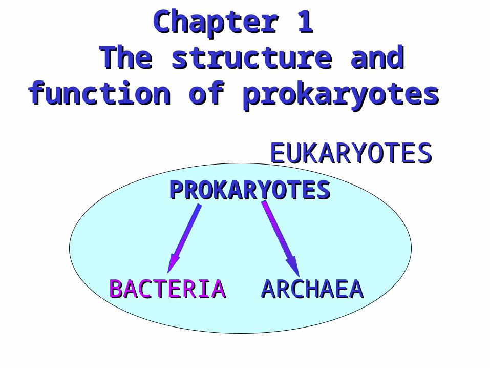
Chapter 1Chapter 1 The structure and function of The structure and function of
prokaryotesprokaryotes
EUKARYOTESEUKARYOTESPROKARYOTESPROKARYOTES
BACTERIABACTERIA ARCHAEAARCHAEA
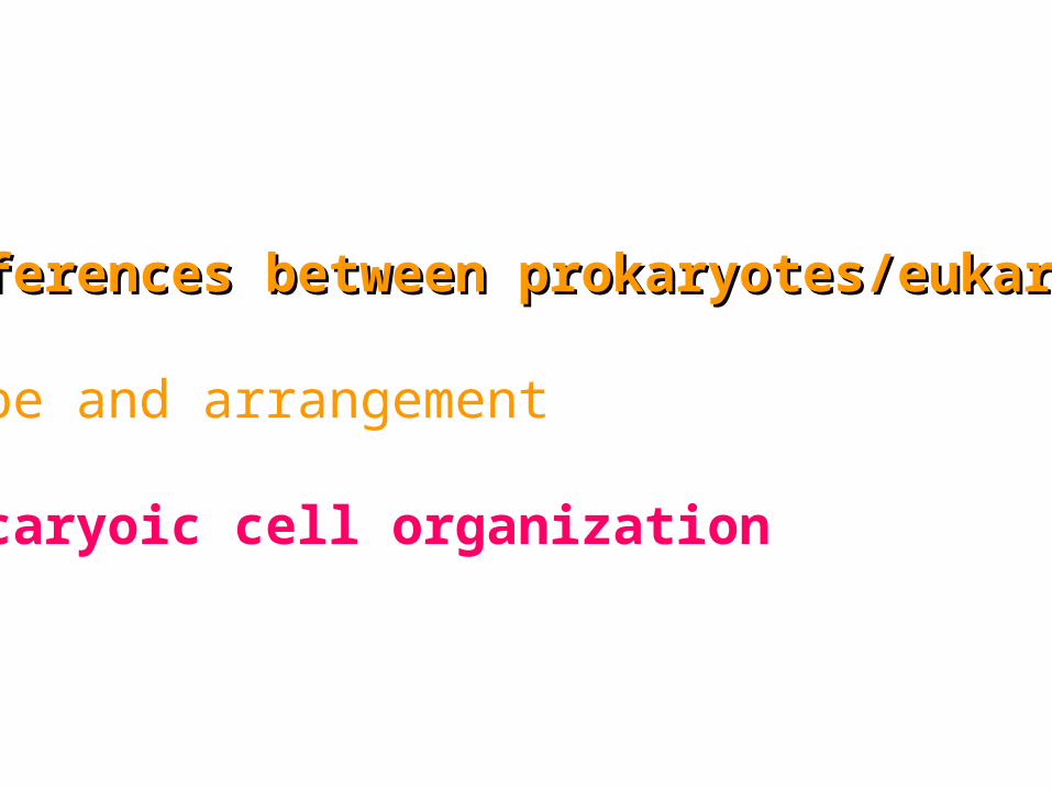
1.Differences between prokaryotes/eukaryotes1.Differences between prokaryotes/eukaryotes
2.Shape and arrangement
3.Procaryoic cell organization
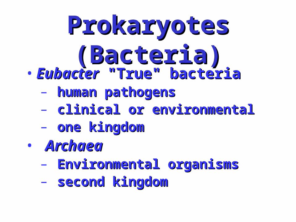
Prokaryotes (Bacteria)Prokaryotes (Bacteria)• EubacterEubacter "True" bacteria "True" bacteria
– human pathogenshuman pathogens– clinical or environmentalclinical or environmental– one kingdomone kingdom
• ArchaeaArchaea – Environmental organisms Environmental organisms – second kingdomsecond kingdom
EukaryotesEukaryotes
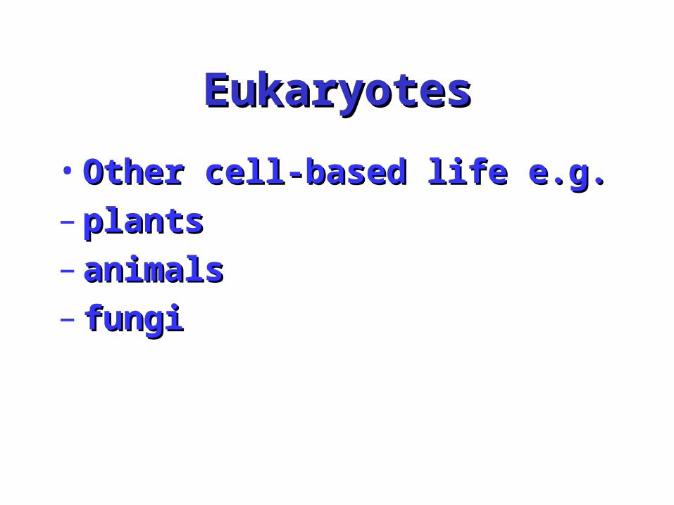
• Other cell-based life e.g.Other cell-based life e.g.
– plantsplants
– animalsanimals
– fungi fungi
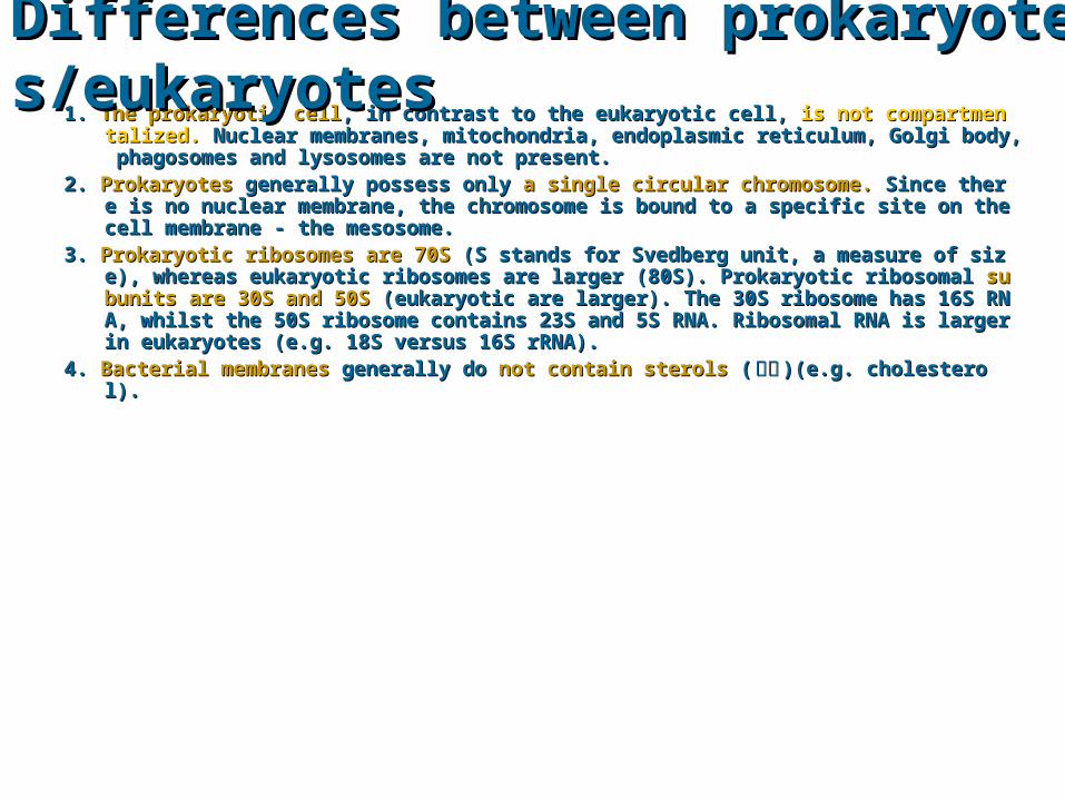
1. 1. The prokaryotic cellThe prokaryotic cell, in contrast to the eukaryotic cell, , in contrast to the eukaryotic cell, is not compartmentalized.is not compartmentalized. Nuclear membr Nuclear membranes, mitochondria, endoplasmic reticulum, Golgi body, phagosomes and lysosomes are not anes, mitochondria, endoplasmic reticulum, Golgi body, phagosomes and lysosomes are not present.present.
2. 2. ProkaryotesProkaryotes generally possess only generally possess only a single circular chromosome.a single circular chromosome. Since there is no nuclear me Since there is no nuclear membrane, the chromosome is bound to a specific site on the cell membrane - the mesosome.mbrane, the chromosome is bound to a specific site on the cell membrane - the mesosome.
3. 3. Prokaryotic ribosomes are 70SProkaryotic ribosomes are 70S (S stands for Svedberg unit, a measure of size), whereas eukaryo (S stands for Svedberg unit, a measure of size), whereas eukaryotic ribosomes are larger (80S). Prokaryotic ribosomal tic ribosomes are larger (80S). Prokaryotic ribosomal subunits are 30S and 50Ssubunits are 30S and 50S (eukaryotic ar (eukaryotic are larger). The 30S ribosome has 16S RNA, whilst the 50S ribosome contains 23S and 5S RNA. e larger). The 30S ribosome has 16S RNA, whilst the 50S ribosome contains 23S and 5S RNA. Ribosomal RNA is larger in eukaryotes (e.g. 18S versus 16S rRNA).Ribosomal RNA is larger in eukaryotes (e.g. 18S versus 16S rRNA).
4. 4. Bacterial membranesBacterial membranes generally do generally do not contain sterolsnot contain sterols ( ( 甾醇甾醇 )(e.g. cholesterol).)(e.g. cholesterol).
Differences between prokaryotes/eukaryotesDifferences between prokaryotes/eukaryotes
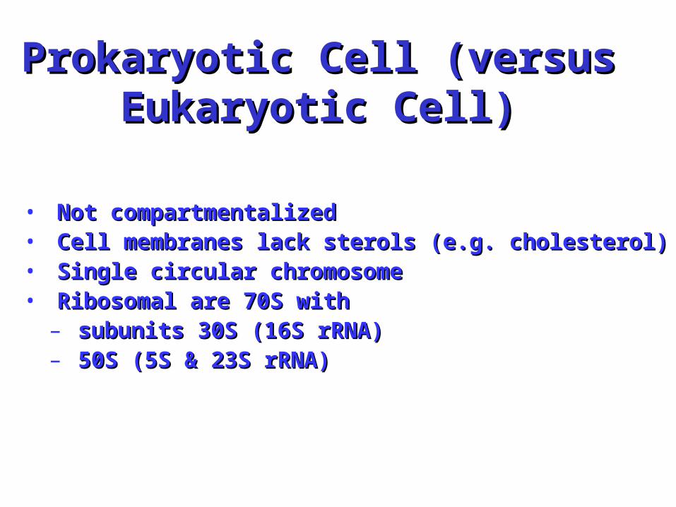
Prokaryotic Cell (versus EukaryoProkaryotic Cell (versus Eukaryotic Cell)tic Cell)
• Not compartmentalized Not compartmentalized • Cell membranes lack sterols (e.g. cholesterol)Cell membranes lack sterols (e.g. cholesterol)• Single circular chromosomeSingle circular chromosome• Ribosomal are 70S with Ribosomal are 70S with
– subunits 30S (16S rRNA) subunits 30S (16S rRNA) – 50S (5S & 23S rRNA)50S (5S & 23S rRNA)
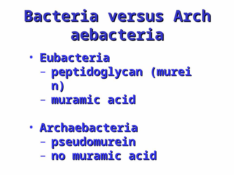
Bacteria versus ArchaebacteBacteria versus Archaebacteriaria
• EubacteriaEubacteria– peptidoglycan (murein)peptidoglycan (murein)– muramic acid muramic acid
• Archaebacteria Archaebacteria – pseudomureinpseudomurein– no muramic acid no muramic acid
Bacteria versus ArchaebacteBacteria versus Archaebacteriaria
• 16S rRNA 16S rRNA
– sequence very differentsequence very different
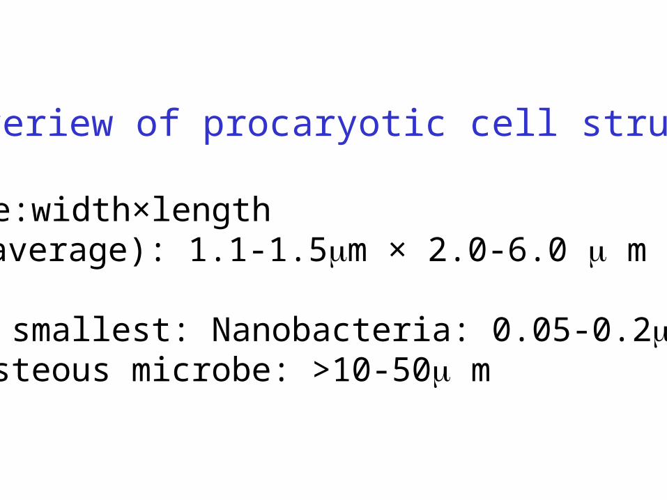
An overiew of procaryotic cell structre
1.size:width×lengthE.coli(average): 1.1-1.5m × 2.0-6.0 m
? The smallest: Nanobacteria: 0.05-0.2 m? monsteous microbe: >10-50 m
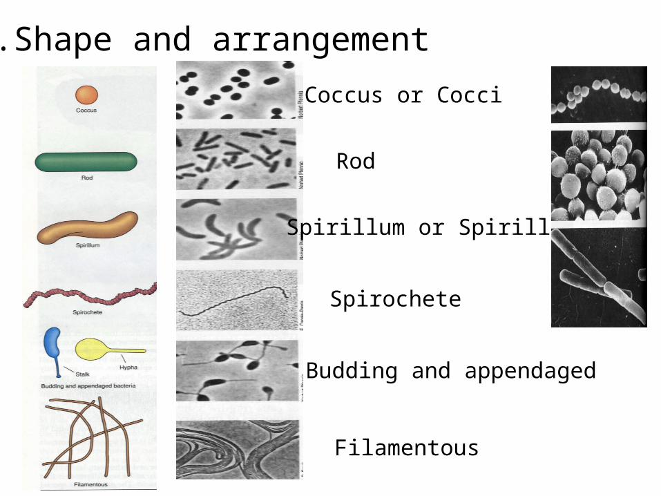
Coccus or Cocci
Rod
Spirillum or Spirilla
Spirochete
Budding and appendaged
Filamentous
2.Shape and arrangement
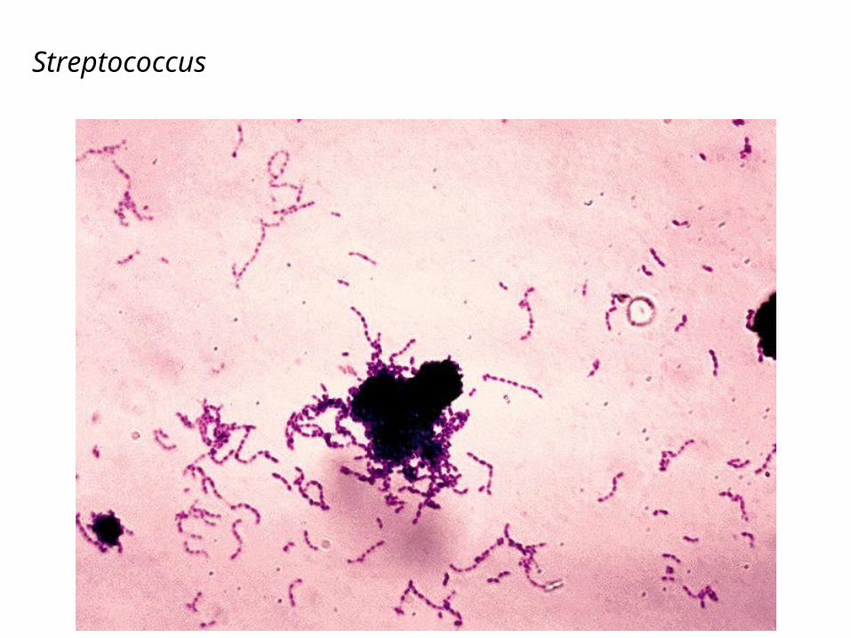
Streptococcus
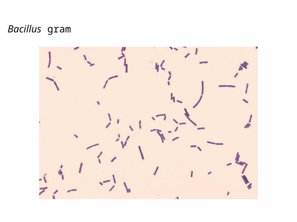
Bacillus gram
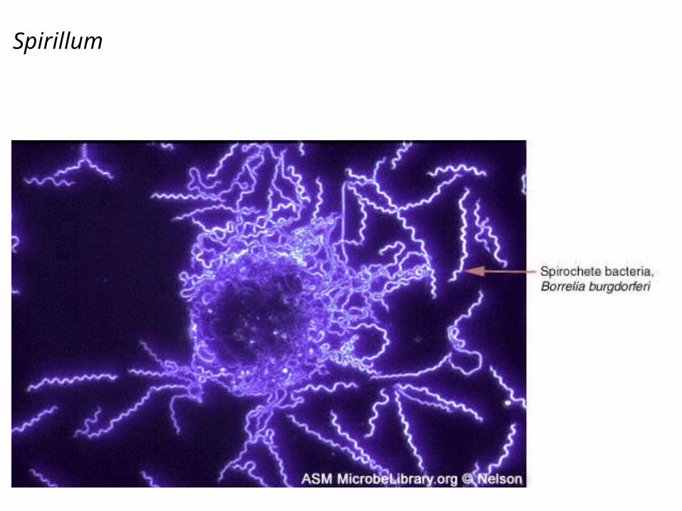
Spirillum
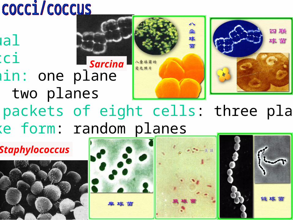
IndividualDiplococciLong chain: one planeTetrads: two planesCubical packets of eight cells: three planes Grapelike form: random planes
Sarcina
Staphylococcus
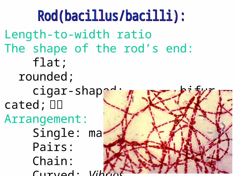
Length-to-width ratioThe shape of the rod’s end: flat; rounded; cigar-shaped; bifurcated; 分叉Arrangement: Single: many Pairs: Chain: Curved: Vibrios
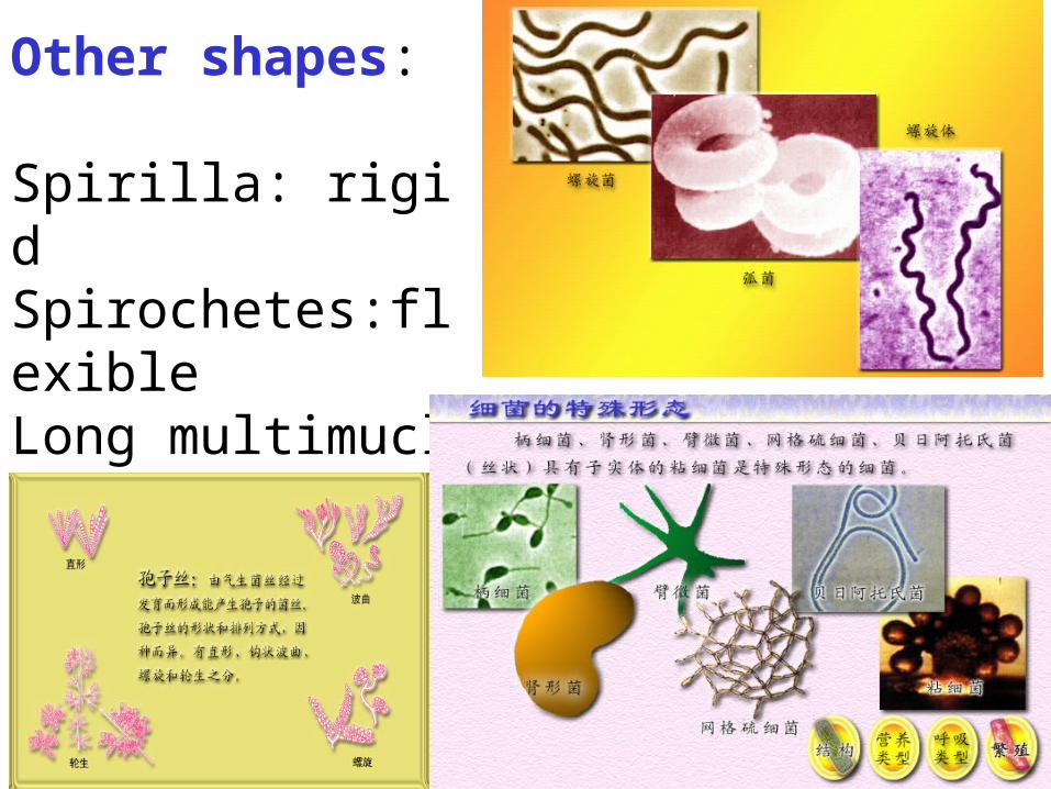
Other shapes:
Spirilla: rigidSpirochetes:flexible Long multimucleate filaments: Actinomycetes

Procaryoic cell organization
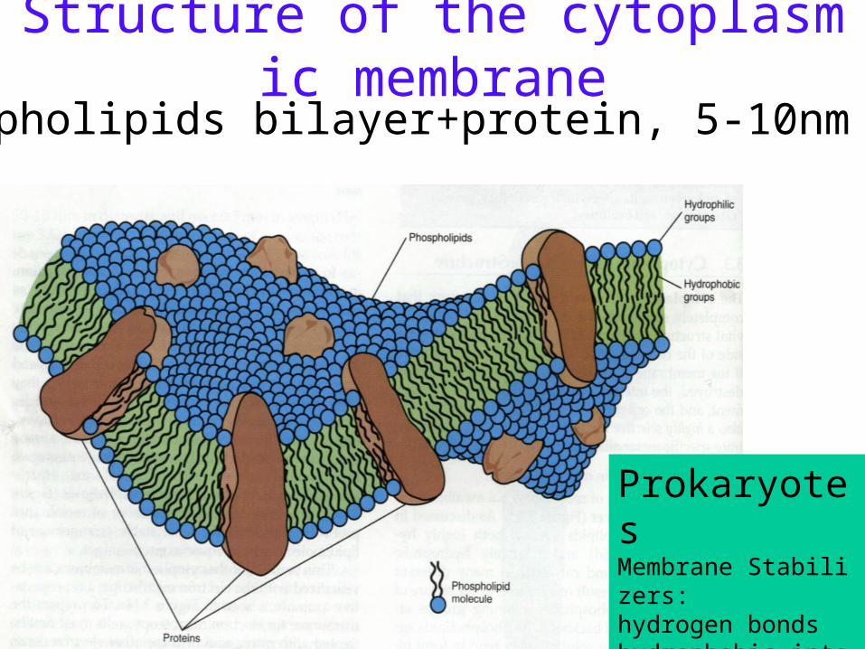
Structure of the cytoplasmic membrane
ProkaryotesMembrane Stabilizers:hydrogen bondshydrophobic interactionsMg2+, Ca2+, hopanoids
Phospholipids bilayer+protein, 5-10nm thick
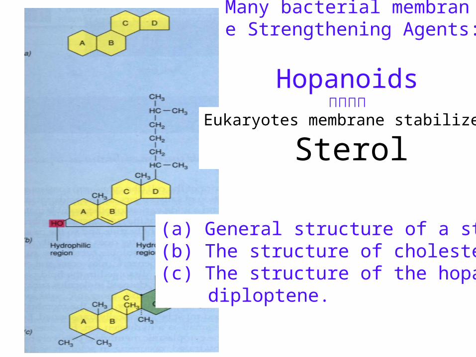
Many bacterial membrane Strengthening Agents:
Hopanoids类何帕烷
(a) General structure of a sterol;(b) The structure of cholesterol;(c) The structure of the hopanoid
diploptene.
Eukaryotes membrane stabilizer:
Sterol
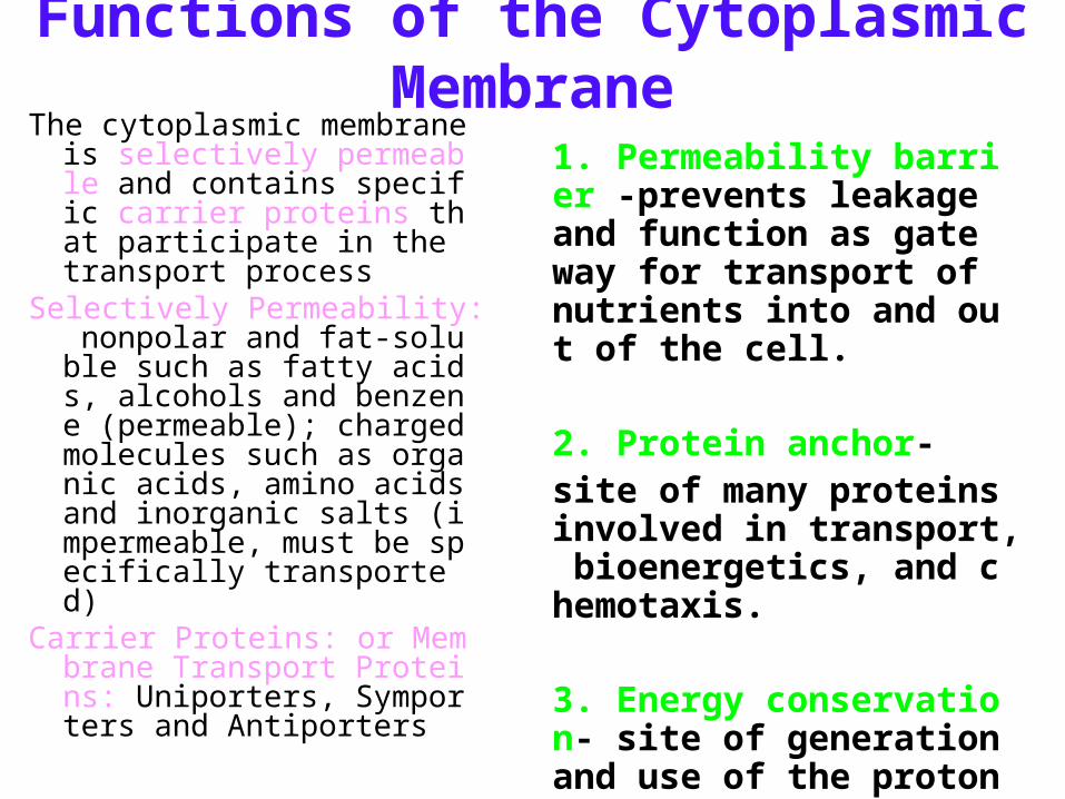
Functions of the Cytoplasmic MembraneThe cytoplasmic membrane is
selectively permeable and contains specific carrier proteins that participate in the transport process
Selectively Permeability: nonpolar and fat-soluble such as fatty acids, alcohols and benzene (permeable); charged molecules such as organic acids, amino acids and inorganic salts (impermeable, must be specifically transported)
Carrier Proteins: or Membrane Transport Proteins: Uniporters, Symporters and Antiporters
1. Permeability barrier -prevents leakage and function as gate way for transport of nutrients into and out of the cell.
2. Protein anchor- site of many proteins involved in transport, bioenergetics, and chemotaxis.
3. Energy conservation- site of generation and use of the proton motive force.
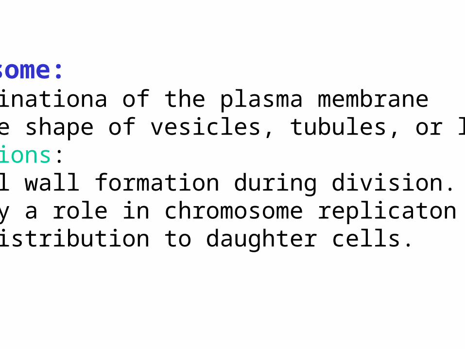
Mesosome: invaginationa of the plasma membrane in the shape of vesicles, tubules, or lamellae.Functions:1.cell wall formation during division.2.play a role in chromosome replicaton and distribution to daughter cells.
The cytoplasmic matrix: substance lying between the plasma membrane and the nucleoid.
Protoplast: the plasma membrane and everything within.

Making Wall-less formsMaking Wall-less forms• Result from action of:Result from action of:
–enzymes lytic for cell wall enzymes lytic for cell wall –antibiotics inhibiting peptidoglycan biosantibiotics inhibiting peptidoglycan bios
ynthesisynthesis• Wall-less bacteria that don’t replicate:Wall-less bacteria that don’t replicate:
–spheroplasts (with outer membrane)?spheroplasts (with outer membrane)?–protoplasts (no outer membrane). ?protoplasts (no outer membrane). ?
• Wall-less bacteria that replicateWall-less bacteria that replicate–L formsL forms
Naturally Wall-less GenusNaturally Wall-less Genus MycloplasmaMycloplasma

Within the cytoplasm of procaryotic (and eucaryotic) cells are several kinds of reserve deposits, known as inclusions. Some inclusions are common to a wide variety of bacteria, whereas others are limited to a small number of species and therefore serve as a basis for identification. Among the more prominent bacterial inclusions are the following:
Carbon storage polymers – PHB and glycogenPhosphate polymersSulfur Granules Gas Vacuoles
INCLUSIONS
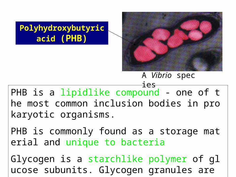
Polyhydroxybutyric acid (PHB)
PHB is a lipidlike compound - one of the most common inclusion bodies in prokaryotic organisms.
PHB is commonly found as a storage material and unique to bacteria
Glycogen is a starchlike polymer of glucose subunits. Glycogen granules are usually smaller than PHB granules.
A Vibrio species
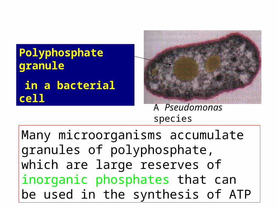
Many microorganisms accumulate granules of polyphosphate, which are large reserves of inorganic phosphates that can be used in the synthesis of ATP
Polyphosphate granule
in a bacterial cell
A Pseudomonas species
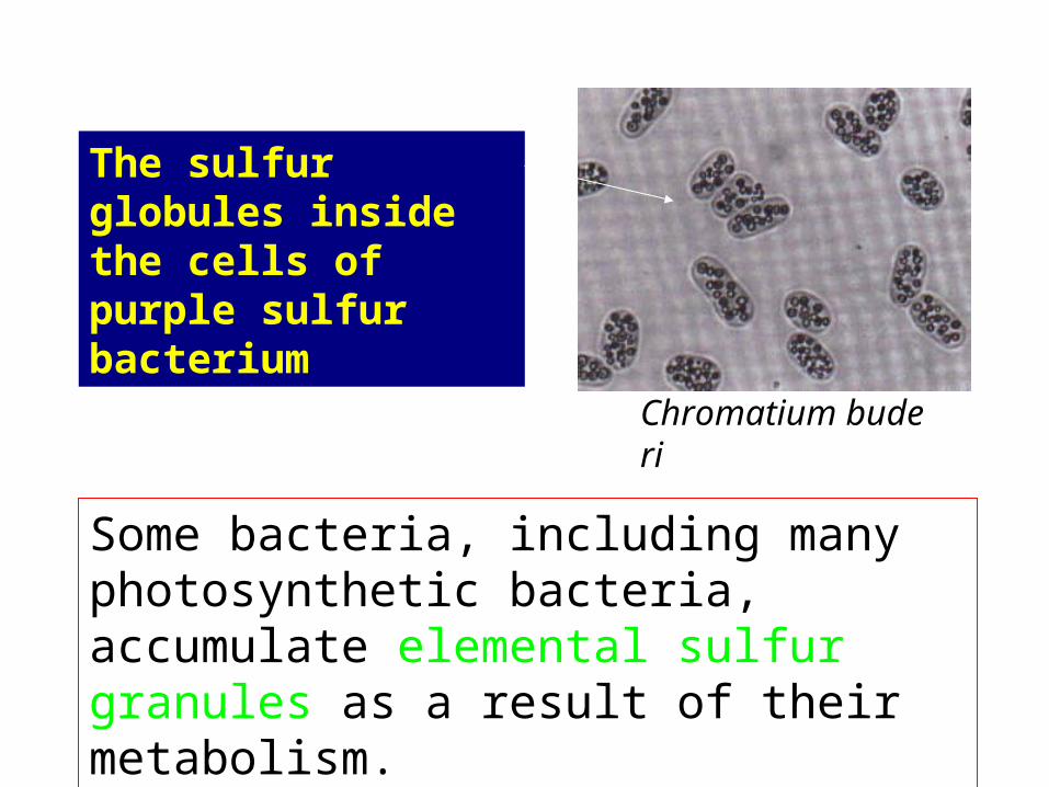
The sulfur globules inside the cells of purple sulfur bacterium
Chromatium buderi
Some bacteria, including many photosynthetic bacteria, accumulate elemental sulfur granules as a result of their metabolism.
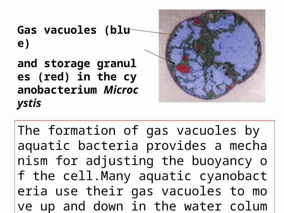
Gas vacuoles (blue)
and storage granules (red) in the cyanobacterium Microcystis
The formation of gas vacuoles by aquatic bacteria provides a mechanism for adjusting the buoyancy of the cell.Many aquatic cyanobacteria use their gas vacuoles to move up and down in the water column.

Magnetosomes
Magnetic particles of Fe3O4 Isolated from Aquaspirillum magnetotacticum
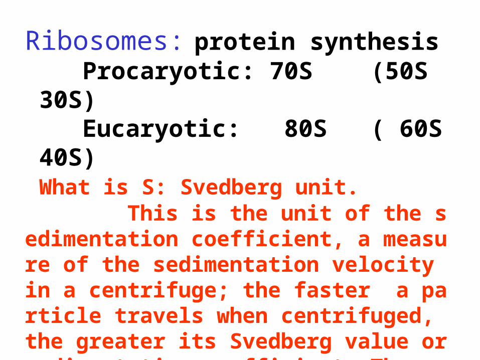
Ribosomes: protein synthesis Procaryotic: 70S (50S 30S) Eucaryotic: 80S ( 60S 40S) What is S: Svedberg unit. This is the unit of the sedimentation coefficient, a measure of the sedimentation velocity in a centrifuge; the faster a particle travels when centrifuged, the greater its Svedberg value or sedimentation coefficient. The sedimentation coefficient is a function of a particle’s molecular weigh, volume, and shape.
The Nucleoid Usually procaryotics contain a single circle of double-stranded DNA.But some: a linear DNA chromosomeRecently: Vibrio cholerae: >one chromosome.Nucleoid: 60% DNA, 30% RNA, 10% protein (by weight)Exceptions: 1. Pirellula: a single membrane 2. Gemmata obscuriglobus: two membrane
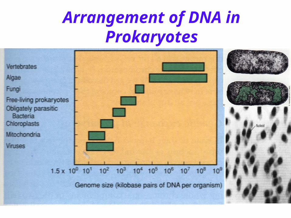
Arrangement of DNA in Prokaryotes

DNA Supercoiling
To package the DNA into the cell requires that the DNA be supercoiled.
There are over 50 supercoiled domains in the E. coli chromosome, they are stabilized by association with the structural proteins.
• Plasmids: Plasmids: ds, circular or linear Dds, circular or linear DNA, exist and replicate independently NA, exist and replicate independently of the chromosome or may be integratof the chromosome or may be integrated with it, not required for host growted with it, not required for host growth and reproduction.h and reproduction.• extrachromosomal DNA• multiple copy number• coding pathogenesis and antibiotic resistance factors• bacterial replication