chapter 1 vertebrates originate - wiley
TRANSCRIPT

Vertebrate Palaeontology, Fourth Edition. Michael J. Benton.© 2015 Michael J. Benton. Published 2015 by John Wiley & Sons, Ltd.Companion Website: www.wiley.com/go/benton/vertebratepalaeontology
C HA P T E R 1
Vertebrates Originate
0002125262.INDD 1 6/26/2014 3:49:18 PM
COPYRIG
HTED M
ATERIAL

2 Chapter 1
INTRODUCTION
Vertebrates are the animals with backbones, the fishes, amphibians, reptiles, birds, and mammals. We have always been especially interested in vertebrates because this is the animal group that includes humans. The efforts of generations of vertebrate palae-ontologists have been repaid by the discovery of countless spec-tacular fossils: heavily armoured fishes of the Ordovician and Devonian, seven- and eight-toed land animals, sail-backed mammal-like reptiles, early birds and dinosaurs with feathers, giant rhinoceroses, rodents with horns, horse-eating flightless birds, and sabre-toothed cats. These fossils tell us where the liv-ing vertebrates have come from, and they show us glimpses of different worlds that seem so bizarre that they would defy the imagination of a science fiction writer. Despite all this informa-tion that has accumulated over the past 200 years, the origin of vertebrates is hotly debated.
One thing is clear from the biology of living animals. Vertebrates are members of a larger group, termed the Phylum Chordata, which also includes their closest living relatives, marine animals such as the sea squirts and amphioxus (see below). These creatures do not have a skeleton, but they share other features, such as a notochord, a flexible, tough rod that runs along the length of the body down the back. The noto-chord in living chordates is generally made from an outer sheath of collagen, a tough fibrous connective tissue that encloses tur-gid, fluid-filled spaces. Invertebrate chordates also have V-shaped muscle blocks (myomeres) along the length of their body. The question about the origin of vertebrates then broad-ens out to include the origin of chordates.
Looked at more widely, vertebrates are a minor twig in the ‘Tree of Life’ (Figure 1.1). It is common to think of the major divisions of life as being animals, plants, protists, and simple organisms classed broadly as bacteria and viruses. However, molecular studies since the 1990s (e.g. Woese, 2000; Wolf et al., 2002) have shown that the fundamental splits were between Bacteria, Archaea, and Eukaryota. The familiar plants, animals and fungi are members of Eukaryota, all diagnosed by complex cells with a membrane-bound nucleus and the primitive pres-ence of mitochondria. Within Eukaryota are various protist groups, as well as plants, fungi, and animals, and of course ver-tebrates are animals. Among animals, it has always been assumed that chordates are closely related to hemichordates
(acorn worms and pterobranch worms) and echinoderms (starfish, sea lilies, and sea urchins), and this is now widely confirmed, based on morphological, developmental and molecular evidence.
The purpose of this chapter is to explore the various lines of evidence that can be used to reconstruct the origin of the verte-brates: the study of modern animals that are vertebrate-like in some features, the study of molecular relationships, and fossils.
1.1 SEA SQUIRTS AND THE LANCELET
There are two key groups of living non-vertebrate chordates, the sea squirts and the cephalochordates (amphioxus). The amphi-oxus certainly looks superficially fish-like, but adult sea squirts look like rubbery bottles, and so would hardly seem to be sensible candidates for close relatives of the vertebrates!
1.1.1 Urochordata: sea squirts
A typical sea squirt, or tunicate, is Ciona (Figure 1.2(a)), which lives attached to rocks in seas around the world. It is a 100–150 mm tall bag-shaped organism with a translucent outer skin (the tunic) and two openings, or siphons, at the top. The body is firmly fixed to a hard substrate.
The internal structure is fairly complex (Figure 1.2(b)). A large pharynx fills most of the internal space, and its walls are perforated by hundreds of gill slits, each of which bears a fringe of cilia, fine hair-like vibratile structures. Seawater is pumped through the inhalant siphon into the pharynx by beating movements of the cilia, and the water is then passed through a surrounding cavity, the atrium, and ejected through the exhal-ant siphon. The pharynx serves mainly to capture food particles
KEY QUESTIONS IN THIS CHAPTER
1 What are the closest living relatives of vertebrates?2 When did deuterostomes and chordates originate?3 What are the key characters of chordates?4 How do embryology and morphology, combined with new phylogenomic studies, inform us about the evolution of animals and the origin of vertebrates?5 How do extraordinary new fossil discoveries from China help us understand the ancestry of vertebrates?
Plants
AnimalsFungi
ProtistsPlantchloroplasts
BacteriaMitochondria
Archaea
Eukaryota
Figure 1.1 The ‘Tree of Life’, the commonly accepted view of the relationships of all organisms. Note the location of ‘Animals’, a minor twig in the tree, close to plants and Fungi. Source: Adapted from various sources.
0002125262.INDD 2 6/26/2014 3:49:18 PM

___________________________________________________________________________________ Vertebrates Originate 3
from the stream of seawater that flows through it. The seawater is drawn into a filter bag of mucus, which is produced inside the pharynx by an organ called the endostyle. During feeding, the endostyle continuously secretes mucus into the oesophagus, together with the food particles that it has filtered from the sea-water, and the food is passed to the stomach for digestion. Tunicates also have a heart that pumps the blood around the body; an intriguing aspect is that the heart stops beating every few minutes and the direction of blood flow reverses.
Why is Ciona identified as a chordate? The pharynx and other structures are in fact very like those of the cephalochor-dates and lamprey larvae, but further evidence is to be found in the larval stage, when the sea squirt is a tiny free-swimming tadpole-shaped animal with a head and a tail. The larval sea squirt (Figure 1.2(c)) has a notochord that runs along the tail, and this identifies it as a chordate. There are muscles on either side of the notochord that contract alternately, causing the tail to beat from side to side, and this drives the animal forward in the water. The larva has a dorsal nerve cord, running along the tail
just above the notochord, and this expands at the front into a very simple brain that includes a light sensor (an ‘eye’) and a tilt detector.
The larva then settles on a suitable surface. It up-ends onto the tip of its ‘snout’ and attaches itself by means of adhesive suckers (Figure 1.2(d)). The notochord and tail portion wither away, and the pharynx and gut expand to fill up the body cavity. This extraordinary metamorphosis occurs rapidly to allow the adult to start feeding in its new way as soon as possible.
1.1.2 Cephalochordata: amphioxus
Another chordate generally reckoned to be related closely to the vertebrates is the amphioxus or lancelet, Branchiostoma, a repre-sentative of the Cephalochordata (or Acraniata). The adult amphioxus is convincingly chordate-like, being a 50 mm long paperknife-shaped animal that looks like a young lamprey or eel, yet lacking a head (Holland, 2010; Bertrand and
Tunic
(a)
(c)
(b) (d)Inhalant siphon
Exhalantsiphon
Degenerating tail
Tunic
Pharynx
Atrium
Anus
Intestine
Level ofcross-sectionOesophagus
Stomach
Nervoussystem
5 cm
5 cm
Statocyst
Mouth ‘Eye’ Exhalant siphon
Nerve cord
Stomach Notochord
Notochordremnants
Pharynx
Adhesivesurfaces
Tail
HeartGill slits
0.1 mm
Endostyle
Adhesivedisc
Pharynx
Endostyle
Gill slits
Atrium
Heart
Figure 1.2 The sea squirts: (a) Ciona, external view; (b) internal anatomy and cross-section of an adult; (c) swimming larva; (d) metamorphosing form. Source: Adapted from Jefferies (1986) and other sources.
0002125262.INDD 3 6/26/2014 3:49:19 PM

4 Chapter 1
Escriva, 2011). Amphioxus swims freely by undulating its whole body from side to side, and it burrows in the sediment on the sea floor (Figure 1.3(a)).
Amphioxus feeds by filtering food particles out of the seawa-ter. Water is pumped into the mouth and through the pharynx by cilia or the gill slits, and food particles are caught up in a bag of mucus produced by the endostyle, the feeding system seen also in tunicates and in the larvae of the lamprey. The mucus with its contained food particles is pulled into the gut for diges-tion, whereas the seawater passes through the gill slits into the atrium. Oxygen is also extracted, and the waste water then exits through the atriopore.
The anatomy of amphioxus, with its pharynx, notochord, dorsal nerve cord, myotomes, and endostyle (Figure 1.3(b)) is typically chordate. Swimming and burrowing are by means of lateral contractions of the myomeres acting against the stiff rod-like notochord.
1.2 AMBULACRARIA: ECHINODERMS AND HEMICHORDATES
Unexpected relatives of chordates are the Ambulacraria, a clade consisting of echinoderms and hemichordates. The living members of these groups do not look much like modern verte-brates, but there is considerable evidence for the relationship (see Box 1.1).
Echinoderms today include such familiar animals as starfish and sea urchins, as well as ophiuroids (brittle stars), crinoids (‘sea lilies’) and holothurians (‘sea cucumbers’). There are some 7000 species of living echinoderms and 13,000 extinct species. Echinoderms all share four key features: (1) a calcite skeleton made from many ossicles, each composed of many aligned small crystals in a somewhat spongy arrangement called stereom; (2) a water vascular system that functions in locomotion, respiration,
and feeding; (3) ossicles are linked by mutable collagen, liga-ments that are normally rigid, but can be loosened; and (4) pen-taradial (five-fold) symmetry. Most of these special features of echinoderms do not show close similarities to other deuteros-tomes, but the water vascular system may have evolved from simple tentacular systems, such as those of pterobranch hemichordates.
The first putative echinoderms include Arkarua from the Vendian of Australia, a disc-shaped organism with radial ridges and a five-pointed central depression, but it has no stereom or evidence of a water vascular system and the identification is inconclusive. The first definitive echinoderms appeared in the Early Cambrian as part of the Cambrian Explosion, and these included some close relatives of living forms, as well as other entirely extinct groups, some of them lacking pentaradial symmetry.
The hemichordates (Röttinger and Lowe, 2012) include two superficially very different kinds of marine animals. The first, the pterobranchs such as Cephalodiscus (Figure 1.4(a,b)), are small animals that live in loose colonies on the seabed in the southern hemisphere and in equatorial waters. Cephalodiscus has a plate-like head shield, a collar with five to nine pairs of feeding arms, and a sac-like trunk perforated by a pair of gill slits and containing the gut and gonads, and the body ends in a contractile stalk. Cilia on the arms produce a feeding current, and food particles are captured by mucus on the arms, while water passes out of the pharynx through the gill slits. The ani-mal lives in or around a group of horny tubes that the colony has constructed, and it attaches itself inside these tubes by means of a sucker on the end of the stalk.
The second hemichordate group, the acorn worms, or enteropneusts, such as Saccoglossus, are worm-like animals varying in length from 20 mm to 2.5 m. They live in burrows low on the shore in Europe and elsewhere. Saccoglossus (Figure 1.4(c)) has a long muscular proboscis that fits into a
Figure 1.3 Amphioxus, a cephalochordate: (a) modes of life, including swimming and burrowing into sand for protection; (b) internal anatomy. Source: Adapted from Pough et al. (2012) and other sources.
Muddy sediment
(a)
Sand
(b)
Dorsal nerve cord
Fin-ray boxesAnus
IntestineAtriopore
Gonads 10 mmEndostyle
Buccal cirri
Notochord Diverticulum Myotomes
Pharynx
0002125262.INDD 4 6/26/2014 3:49:20 PM

___________________________________________________________________________________ Vertebrates Originate 5
fleshy ring or collar behind. The mouth is placed beneath this collar, and seawater and sand are pumped through the gut and expelled through an anus at the posterior end of the body. The long body is pierced by small holes at the front end, homolo-gous with the gill slits of Cephalodiscus, sea squirts, amphi-oxus, and vertebrates, based on morphology and expression of developmental genes (Cannon et al., 2013). Developmental genes also indicate homology of the postanal tail regions in Saccoglossus and vertebrates.
The fossil record of enteropneusts has been debated. It is widely assumed that the extinct, colonial graptolites were a clade of hemichordates, and particularly allied with ptero-branchs, based on similarities in the ultrastructure of their wall structures (Sato et al., 2008). However, fossils of the two extant clades have been restricted to rare forms in the Carboniferous and Jurassic until reports (Caron et al., 2013; Maletz, 2014) of Cambrian specimens from Chengjiang and the Burgess Shale respectively. The latter example, the worm-like Spartobranchus,
shows a fibrous tube that might be a precursor of the pterobranch periderm, suggesting that pterobranchs arose from enteropneust-like ancestors.
The phylogeny of hemichordates is actively debated. However, morphological (Smith et al., 2004) and molecular (Röttinger and Lowe, 2012; Cannon et al., 2013) data now concur that Hemichordata is a valid phylum. Hemichordates do not have a notochord at any stage, but they possess gill slits, as in chordates, and giant nerve cells in the nerve cord of the collar region that are probably equivalent to similar nerve cells in amphioxus and primitive vertebrates. Both pterobranchs and enteropneusts share morphological characters indicating mono-phyly of the Hemichordata, such as the stomochord (an anterior buccal tube on the dorsal part of the pharynx) and mesocoe-lomic ducts. Earlier molecular phylogenetic studies suggested that enteropneust worms were either monophyletic (based on 28S rDNA) or not (based on 18S rDNA), but micro-RNAs provide strong evidence for monophyly (Peterson et al., 2013).
Two substantially different schemes for deuterostome relationships have been proposed. The ‘traditional’ view (e.g. Maisey, 1986; Donoghue et al., 1998; illustration (a)) was to place the hemichordates as basal to chordates since they both share ciliated gill slits and giant nerve cells, as well as other features, which are not seen in echinoderms. Enteropneusts were sometimes said to be closer relatives of chordates since their gill slits are similar, they have a very short dorsal hollow nerve cord, and a number of other features of the gut not seen in pterobranchs. Most authors regarded amphioxus as the closest relative of the Vertebrata on the basis of 10–15 features that are not seen in tunicates.
The second view (illustration (b)) is supported by morphological and molecular data and is now widely accepted (Swalla and Smith, 2008; Edgecombe et al., 2011). The first molecular studies, in which the 18S rRNA genes of echinoderms, hemichordates, and chordates were compared were inconclusive, but newer work (e.g. Eernisse and Peterson, 2004; Delsuc et al., 2006; Swalla and Smith, 2008; Edgecombe et al., 2011; Röttinger and Lowe, 2012; Cannon et al., 2013) pairs hemichordates with echinoderms as the clade Ambulacraria, and within the clade Chordata places cephalochordates as the basal clade, and pairs Urochordata and Vertebrata, as clade Olfactores because of shared characters in the olfactory region. See Box 3.1 for phylogeny of Vertebrata.
BOx 1.1 DEUTEROSTOME RELATIONSHIPS
Cladograms showing the relationships of the main deuterostome groups: (a) the ‘traditional’ model, and (b) molecular model. Synapomorphies: A DEUTEROSTOMIA, blastopore becomes anus during development, bipartite mesocoel, mesocoelomic ducts; B, stomochord, paired gill slits; C, multiple pairs of gill slits, pharyngeal slits U-shaped, dorsal hollow nerve cord, preoral ciliary organ, mouth anterior and ventral and anus posterior and ventral or dorsal, multiciliated cells; D CHORDATA, notochord present and not attached to gut, dorsal hollow nerve cord with neural-plate stage in development, endostyle organ, a true tail used in swimming; E, digestive caecum, open capillary junctions, somites present, lateral-plate mesoderm, neural tube differentiated into grey and white matter, cerebral vesicle in brain; F OLFACTORES, specialized olfactory areas in buccal cavity, hind-tail tripartite, dorsal longitudinal canal connected with notochord; G AMBULACRARIA, trimeric arrangement of the adult coelom, axial complex with hydropore, dipleureula larva with neotroch.
Protos
tomes
E
chino
derm
ata
H
emich
orda
ta
U
roch
orda
ta
Cep
haloc
hord
ata
Ver
tebra
ta
(b)
G
AMBULACRARIA
D CHORDATAF OLFACTORES
A DEUTEROSTOMIA
Protos
tomes
E
chino
derm
ata
P
terob
ranc
hia
E
ntero
pneu
sta
Uro
chor
data
Cep
haloc
hord
ata
Ver
tebra
ta
A DEUTEROSTOMIAB
CD CHORDATA
E
HEMICHORDATA(a)
0002125262.INDD 5 6/26/2014 3:49:21 PM

6 Chapter 1
1.3 DEUTEROSTOME RELATIONSHIPS
The relationships of chordates used to be rather problematic, but intensive analyses of molecular data have provided a clearer picture (Eernisse and Peterson, 2004; Swalla and Smith, 2008; Edgecombe et al., 2011). The Phylum Chordata is part of a larger clade, the Deuterostomia, comprising chordates, hemichordates, and echinoderms, which in turn is part of a yet larger clade of all the bilaterally symmetrical animals, the Bilateria, and these in turn fall within Metazoa, the animals. But what exactly diagnoses the Deuterostomia, and how can some of our closest relatives be sea urchins, starfish, and worm-like animals? The clues come from embryology, the study of the early phases of development in, and just out of, the egg, and from molecular phylogenetic analysis.
1.3.1 Embryology and the position of the anus
In early development each animal starts as a single cell. Soon this cell begins to divide, first into two cells, then four, then eight, sixteen, and so on (Figure 1.5(a–c)). Eventually a hollow ball of cells is produced, called the blastula stage (Figure 1.5(d)). A pocket of cells then moves inwards, forming the precursor of the gut and other internal structures. The opening of this deep pocket is called the blastopore. You can imagine pushing in the walls of a hollow rubber squash ball with your thumb to produce a model of this embryonic pattern, known as the gastrula stage (Figure 1.5(e–g)).
Embryologists noticed some time ago that animals fall into two large groups depending on the relative orientation of the mouth and anus. The classic story is that in most invertebrates (the protostomes), the blastopore becomes the mouth (Figure 1.5(h)),
Figure 1.4 Typical hemichordates: (a) the pterobranch Cephalodiscus, internal anatomy and (b) mode of life; (c) the enteropneust Saccoglossus, mode of life and external anatomy. Source: Adapted from Jefferies (1986) and other sources.
Heart
Headshield
Mouth
Gill slit
Attachment sucker
Stalk
Stomach
Arm
Anus
Gonad
IntestinePharynx
1 mm
(a)
5 mm
(b)
5 mmCollar
ProboscisMouth
Gonad
Gill slits
Anus
(c)
0002125262.INDD 6 6/26/2014 3:49:22 PM

___________________________________________________________________________________ Vertebrates Originate 7
whereas in others (the deuterostomes), including the chordates, this opening becomes the anus (Figure 1.5(i)), and the mouth is a secondary perforation. Such a dramatic turnaround, a switch from mouth to anus, seems incredible. Note, however, that many proto-stomes show deuterostomy, and this condition may be primitive and shared by all Bilateria (Eernisse and Peterson, 2004). This peculiarity of embryological development was noted over a cen-tury ago, and the group Deuterostomia named in 1908; but does it stand up to the scrutiny of modern molecular phylogenetics?
1.3.2 Animal phylogenomics
Numerous zoologists have contributed over the years to disentan-gling the relationships of animals. All creatures from sponges and corals to crabs, clams, and birds, are animals, members of the clade Metazoa, diagnosed by a combination of feeding, being motile, lacking rigid cell walls, and passing through the blastula embryonic stage. These characteristics are not all exclusive, how-ever. First, metazoans are distinguished from most plants and algae by being heterotrophs, meaning they feed on other organ-isms to acquire carbon, which is digested in an internal chamber
(gut), whereas plants and algae are able to fix carbon from the atmosphere or water. Fungi and many bacteria, however, are also heterotrophs. Secondly, metazoans are motile, meaning they use energy to move spontaneously and actively, at least at some stage in their lives (larval stages in ‘fixed’ forms such as sponges and corals can swim), although some bacteria and protists are also motile, moving by means of a flagellum. Thirdly, animals lack the rigid cell walls seen in plants, fungi, and algae, and fourthly most pass through the blastula embryonic stage (see Section 1.3.1).
Metazoa, Bilateria, and Deuterostomia are monophyletic groups, or clades. A clade is a group that has a single common ancestor, and that includes all of the descendants of that ancestor (see Section 2.5.1). Before the advent of molecular phylogenetics (see Section 2.5.2), and even after, there has been active debate about the relationships of the various animal clades. It is usually easy to determine membership of these major clades, the phyla (see Box 2.4) – such as arthropods, molluscs, or sponges – but determining how the phyla relate to each other within Metazoa has been difficult. However, by 2010, a consensus about the major outlines of animal relationships had been reached (Figure 1.6).
The fundamental division of Metazoa distinguishes six early-branching clades (including sponges and corals) from the Bilateria, supported by both morphological and molecular evi-dence (Eernisse and Peterson, 2004; Halanych, 2004; Philippe et al., 2009; Edgecombe et al., 2011; Nielsen, 2012). The Bilateria have bilateral symmetry primitively, and most are triploblastic, meaning they have three fundamental body wall tissues that arise from the ectoderm, mesoderm, and endoderm in the embryo. Non-bilaterian metazoans may be diploblastic, lacking the mesoderm, or monoblastic like sponges and placozoans. Within Bilateria, most animals are Nephrozoa, taxa that are characterized by the possession of an excretory system. Finally, Nephrozoa is divided into the two major clades Protostomia and Deuterostomia, long recognized on embryological grounds. Protostomes include the Ecdysozoa (animals that moult, such as nematodes, arthropods, priapulids, and some minor groups) and Spiralia (animals with spiral development, such as bryozo-ans, annelids, molluscs, brachiopods, rotifers, and other phyla). Most spiralians belong to the clade Lophotrochozoa.
The monophyly of Deuterostomia is confirmed both by mor-phology and by phylogenomics. All deuterostomes share the posterior blastopore that generally becomes the anus, as well as gill slits (present only in precursors of the echinoderms). Further, most molecular phylogenetic analyses indicate monophyly (e.g. Eernisse and Peterson, 2004; Swalla and Smith, 2008; Edgecombe et al., 2011; Röttinger and Lowe, 2012; Cannon et al., 2013), although this is queried in some studies (e.g. Delsuc et al., 2006; Mallatt et al., 2010). Some recent phylogenomic studies have suggested the addition of two further clades to Deuterostomia, the Xenoturbellida and the Acoelomorpha, simple worms with no through gut and a simple nervous system. However, these assignments are controversial (Edgecombe et al., 2011; Röttinger and Lowe, 2012). Further, there has been some dispute over the interrelationships among these deuterostome taxa (see Box 1.1).
Cavityof gut
(a) (b) (c)
(d)
(g)
(h)
(e)
(i)
(f)
Blastopore –mouth
Secondary mouth Blastopore – anus
Secondary anus
Figure 1.5 Embryonic development: (a–g) sequence of cell division in amphioxus, from the single-cell stage (a), through the blastula stage (d), to the gastrula stage (g). (h) Fate of the blastophore in protostomes, and (i) in deuterostomes. Source: Adapted from Jefferies (1986) and other sources.
0002125262.INDD 7 6/26/2014 3:49:23 PM

8 Chapter 1
1.4 CHORDATE ORIGINS
Among morphological characters, the chordates all share sev-eral unique features such as a notochord, a dorsal hollow nerve cord with a shared developmental pattern, an endostyle organ (equivalent to the thyroid gland of vertebrates), and a tail used for swimming. It is generally accepted that only chordates have true tails. A tail technically may be defined as a distinct region extending behind the visceral cavity, and in particular located
entirely behind the anus; hence the term ‘postanal tail’, to be quite precise. Non-chordates, such as insects, worms, molluscs, jellyfish, and sea urchins, do not have tails. What of the fossil evidence?
There are many putative early fossil chordates, and their numbers have grown hugely since 1995, with the announcement of remarkable new finds from the Chengjiang biota of China, an Early Cambrian deposit (see Box 1.2). These new specimens, combined with studies of modern forms, give clues about the
Protostomia
Deuterostomia
BilateriaMetazoa
Ecdysozoa
TrochozoaPlatyzoa
Spiralia
Nephrozoa
Gnathifera
Scalidophora
Nematoida
Ambulacraria
Homoscleromorpha
Choanozoa
Silicea
Calcarea
Cnidaria
Ctenophora
Placozoa
Acoela
Echinodermata
Hemichordata
Chaetognatha
Entoprocta
Cycliophora
Priapulida
Kinorhyncha
Loricifera
Onychophora
Tardigrada
Arthropoda
Bryozoa
Brachiopoda
Nemertea
Annelida
Mollusca
Xenoturbellida
M
B D
N
P
Am
Ac
Ne
Sc
S T
Gastrotricha
Gnathostomulida
Micrognathozoa
Rotifera
Gn
Pl
E
Phoronida
Urochordata
CephalochordataC
Chordata
Polyzoa
Nemertodermatida
Po
Acoelomorpha
Platyhelminthes
Nematomorpha
Ol
Olfactores
Craniata (incl. Vertebrata)
Pr
Porifera
Pa
Panarthropoda
P
D
BM
E
TPl
S
N
Gn
Sc
Ne
AmC
Po
Ac
Ol
Pr
Pa
Nematoda
Figure 1.6 Relationships of the major phyla of animals, based on accumulated knowledge from anatomy and embryology, combined with current phylogenomic work. Source: G. Edgecombe, The Natural History Museum, London, UK. Reproduced with permission.
0002125262.INDD 8 6/26/2014 3:49:26 PM
___________________________________________________________________________________ Vertebrates Originate 9
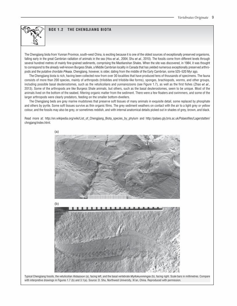
BOx 1.2 THE CHENGJIANG BIOTA
The Chengjiang biota from Yunnan Province, south-west China, is exciting because it is one of the oldest sources of exceptionally preserved organisms, falling early in the great Cambrian radiation of animals in the sea (Hou et al., 2004; Shu et al., 2010). The fossils come from different levels through several hundred metres of mainly fine-grained sediments, comprising the Maotianshan Shales. When the site was discovered, in 1984, it was thought to correspond to the already well-known Burgess Shale, a Middle Cambrian locality in Canada that has yielded numerous exceptionally preserved arthro-pods and the putative chordate Pikaia. Chengjiang, however, is older, dating from the middle of the Early Cambrian, some 525–520 Myr ago.
The Chengjiang biota is rich, having been collected now from over 30 localities that have produced tens of thousands of specimens. The fauna consists of more than 200 species, mainly of arthropods (trilobites and trilobite-like forms), sponges, brachiopods, worms, and other groups, including possible basal deuterostomes, such as the vetulicolians and yunnanozoons (see Figure 1.7), as well as the first fishes (Zhao et al., 2013). Some of the arthropods are like Burgess Shale animals, but others, such as the basal deuterostomes, seem to be unique. Most of the animals lived on the bottom of the seabed, filtering organic matter from the sediment. There were a few floaters and swimmers, and some of the larger arthropods were clearly predators, feeding on the smaller bottom-dwellers.
The Chengjiang beds are grey marine mudstones that preserve soft tissues of many animals in exquisite detail, some replaced by phosphate and others by pyrite. Some soft tissues survive as thin organic films. The grey sediment weathers on contact with the air to a light grey or yellow colour, and the fossils may also be grey, or sometimes reddish, and with internal anatomical details picked out in shades of grey, brown, and black.
Read more at: http://en.wikipedia.org/wiki/List_of_Chengjiang_Biota_species_by_phylum and http://palaeo.gly.bris.ac.uk/Palaeofiles/Lagerstatten/chngjang/index.html.
(a)
(b)
Typical Chengjiang fossils, the vetulicolian Xidazoon (a), facing left, and the basal vertebrate Myllokunmingia (b), facing right. Scale bars in millimetres. Compare with interpretive drawings in Figures 1.7 (b) and 3.1(a). Source: D. Shu, Northwest University, Xi’an, China. Reproduced with permission.
0002125262.INDD 9 6/26/2014 3:49:29 PM
10 Chapter 1
early evolution of chordates, but there are many debates (Donoghue and Purnell, 2009).
1.4.1 Diverse early chordates
There are three main categories of possible early chordates: pos-sible urochordates, possible cephalochordates, and vetulico-lians. At one time, conodonts, represented in the fossil record generally only by their tooth elements, were treated as dubious chordates. Conodonts are now placed firmly within the Vertebrata, as jawless fishes, as are some of the basal chordate taxa from Chengjiang, such as Haikouichthys (see Chapter 3).
Urochordates have a patchy fossil record. Isolated impres-sions of sac-like bodies, and trace fossils, markings made in or on the sediment by the activities of animals, have been ascribed to tunicates. The best fossils are small sac-like specimens from Chengjiang, Shankouclava, which shows a large perforated branchial basket, branchial slits, and an elongate endostyle (Chen et al., 2003). There is also a possible degenerating tail, suggesting this might be a larva that had just settled (cf. Figure 1.2(d)).
The fossil record of cephalochordates is not much better. The Chengjiang biota includes a superficially amphioxus-like cepha-lochordate, Cathaymyrus, as well as the yunnanozoons, which have also been identified as cephalochordates, although most assign them to other positions among deuterostomes (see below). In the absence of hard tissues such as bone, these non-vertebrate chordates are not often preserved.
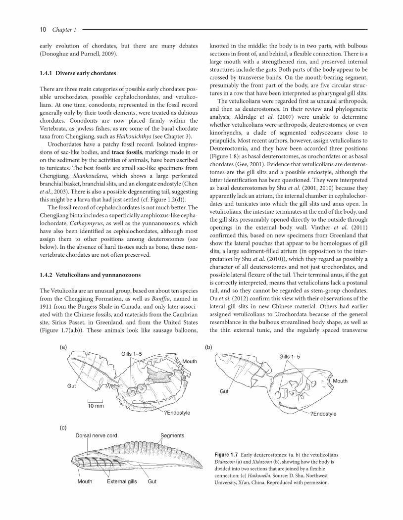
1.4.2 Vetulicolians and yunnanozoons
The Vetulicolia are an unusual group, based on about ten species from the Chengjiang Formation, as well as Banffia, named in 1911 from the Burgess Shale in Canada, and only later associ-ated with the Chinese fossils, and materials from the Cambrian site, Sirius Passet, in Greenland, and from the United States (Figure 1.7(a,b)). These animals look like sausage balloons,
knotted in the middle: the body is in two parts, with bulbous sections in front of, and behind, a flexible connection. There is a large mouth with a strengthened rim, and preserved internal structures include the guts. Both parts of the body appear to be crossed by transverse bands. On the mouth-bearing segment, presumably the front part of the body, are five circular struc-tures in a row that have been interpreted as pharyngeal gill slits.
The vetulicolians were regarded first as unusual arthropods, and then as deuterostomes. In their review and phylogenetic analysis, Aldridge et al. (2007) were unable to determine whether vetulicolians were arthropods, deuterostomes, or even kinorhynchs, a clade of segmented ecdysozoans close to priapulids. Most recent authors, however, assign vetulicolians to Deuterostomia, and they have been accorded three positions (Figure 1.8): as basal deuterostomes, as urochordates or as basal chordates (Gee, 2001). Evidence that vetulicolians are deuteros-tomes are the gill slits and a possible endostyle, although the latter identification has been questioned. They were interpreted as basal deuterostomes by Shu et al. (2001, 2010) because they apparently lack an atrium, the internal chamber in cephalochor-dates and tunicates into which the gill slits and anus open. In vetulicolians, the intestine terminates at the end of the body, and the gill slits presumably opened directly to the outside through openings in the external body wall. Vinther et al. (2011) confirmed this, based on new specimens from Greenland that show the lateral pouches that appear to be homologues of gill slits, a large sediment-filled atrium (in opposition to the inter-pretation by Shu et al. (2010)), which they regard as possibly a character of all deuterostomes and not just urochordates, and possible lateral flexure of the tail. Their terminal anus, if the gut is correctly interpreted, means that vetulicolians lack a postanal tail, and so they cannot be regarded as stem-group chordates. Ou et al. (2012) confirm this view with their observations of the lateral gill slits in new Chinese material. Others had earlier assigned vetulicolians to Urochordata because of the general resemblance in the bulbous streamlined body shape, as well as the thin external tunic, and the regularly spaced transverse
Figure 1.7 Early deuterostomes: (a, b) the vetulicolians Didazoon (a) and Xidazoon (b), showing how the body is divided into two sections that are joined by a flexible connection; (c) Haikouella. Source: D. Shu, Northwest University, Xi’an, China. Reproduced with permission.
Gut
Gills 1–5Mouth
?Endostyle
(a)
10 mm
Gills 1–5
Gut
Mouth
?Endostyle
(b)
(c)Dorsal nerve cord Segments
Mouth External gills Gut
0002125262.INDD 10 6/26/2014 3:49:30 PM

__________________________________________________________________________________ Vertebrates Originate 11
bands, which might be muscles that ran round the body in rings (Lacalli, 2002). The absence of a notochord in vetulicolians was said not to be critical, since most adult tunicates also have lost this structure, and Gee (2001) suggested that these unusual fos-sils are just what would be expected as the ancestral vertebrate, long predicted to have emerged from a sac-like animal that is all guts (like a tunicate), which then became surrounded by mus-culature, nerves, and sensory systems to enable locomotion.
The yunnanozoons, also from Chengjiang, such as Yunnanozoon and Haikouella (Figure 1.7(c)) look like much more convincing basal chordates, perhaps even close to vertebrates, with their fish-like form, dorsal fin, postanal tail, notochord, gill slits, and even some head structures. Nonetheless, they have been interpreted as occupying many different positions in deuterostome phylogeny (Figure 1.8) by rival researchers. One team identified these ani-mals first as possible cephalochordates (Chen et al., 1995), and then upwards as vertebrates (Chen et al., 1999; Holland and Chen, 2001; Mallatt and Chen, 2003). The other team preferred to regard the yunnanozoons first as hemichordates (Shu et al., 1996), and then downwards as basal deuterostomes allied to the vetulicolians (Shu et al., 2003b). The problems revolve around different inter-pretations of coloured blobs, lines, and squiggles in the fossils. There are plenty of fossils – literally thousands – but anatomical interpretation is critical (Donoghue and Purnell, 2009).
Haikouella and Yunnanozoon are 25–40 mm long, and pre-served as flattened bluish-grey to black films on the rock. Chen et al. (1995) were able to see a notochord, a filter-feeding pharynx with an endostyle, segmented musculature, and branchial arches, all chordate characters. Chen et al. (1999) and Mallatt and Chen (2003) went further, identifying an enlarged, possibly three-part, brain and paired lateral eyes in Haikouella, hence indicating it might have had a distinctive, enlarged head, a key feature of verte-brates. Shu et al. (1996) argued, however, that there is no notochord, and that this tubular structure is actually the gut. In addition, they
suggested that the segmented musculature was wrongly identified. In contrast, they claimed to see key hemichordate features in Yunnanozoon, and especially that the body is divided into three parts from front to back, a proboscis, a collar, and a trunk that is divided into a branchial and a gut region, just as in the living acorn worm (see Figure 1.4(c)). Shu et al. (2003, 2010) subsequently noted similarities between the yunnanozoons and the vetulico-lians, and moved them down from the hemichordates to a basal position among deuterostomes (Figure 1.8): they could see no evidence of a notochord, segmented muscles, a large brain, lateral eyes, or any of the other chordate features previously reported.
The final early chordate to consider is Pikaia from the Burgess Shale in Canada, named in 1911 as an annelid, but subsequently widely regarded as a basal chordate or even basal vertebrate (Figure 1.9). In a thorough redescription of 114 specimens, Conway Morris and Caron (2012) highlight its chordate characteristics: a laterally compressed, hydrodynamic body with about 100 myomeres, a thin dorsal fin, a small bilobed head with tentacles but no eyes, possible pharyngeal pores, a pharyngeal cavity, an almost terminal mouth, a proba-ble terminal anus (and hence no postanal tail), a dorsal nerve cord, a possible notochord, and a blood vascular system. As with the yunnanozoons, however, determining the phylogenetic
CEPHALOCHORDAT
A
Yu
nnan
ozoo
ns (C
hen e
t al.,
1995
)
Yun
nano
zoon
s (Che
n et a
l., 19
99;
Swalla a
nd S
mith, 2
008)
CRANIA
TA
Vetulic
olian
s (La
calli,
2002
)
UROCHORDATA
Vetulic
olian
s (Gee
, 200
1)
Yunn
anoz
oons
(Shu
et al
., 199
6)
HEMIC
HORDATA
ECHINODERMATA
Yunn
anoz
oons
(Shu
et al
., 200
3)
Vetulic
olian
s (Shu
et al
., 200
1;
Vint
her e
t al.,
2011
; Ou e
t al.,
2012
)
Vetulic
olian
s (Shu
et al
., 200
4)
Yunn
anoz
oons
(Mor
ris an
d Car
on, 2
012)
Figure 1.8 Phylogenetic tree of the extant deuterostomes, with suggested locations of the major fossil groups. Source: Adapted from various sources.
Figure 1.9 The early chordate Pikaia from the Burgess Shale, Canada. Source: J-B. Caron, Smithsonian Institution, Washington , DC, USA. Reproduced with permission.
0002125262.INDD 11 6/26/2014 3:49:32 PM

12 Chapter 1
When an organism dies its carcass decays, and information is lost. Until recently, such loss of information was assumed to be random, but taphonomic experiments on modern amphioxus and lampreys (Sansom et al., 2010) show that the first tissues to rot away take with them key diagnostic characters. In fact, through the process of decay over a few weeks, tissues are lost in such a way that the specimens become more and more primitive in appearance.
The rather smelly experiments on lamprey and amphioxus juveniles were run for up to 200 days, with dead specimens decaying in normal seawater and at reasonable temperatures. Tissues began to be lost quickly. In the case of amphioxus, the eye spot was lost after 11 days, the atriopore after 15, the anterior bulb after 21, and the midgut caecum and storage organ after 28. Most resilient to decay were the myomeres and the notochord, and before those the endostyle, pharyngeal arches, and gonads. Sansom et al. (2010) noted that these last tissues are those most commonly seen in exceptionally preserved basal chordate and deuterostome fossils from the Chengjiang and Burgess Shale biotas.
The initial suite of characters that disappeared in the decaying amphioxus specimens were those diagnostic of Cephalochordata, and the myomeres and notochord are the most general chordate characters. Normal decay processes then favour preservation of primitive characters, and phylogenetic analysis of chordate fossils will position the fossils in a more basal position than is correct. These decay experiments strongly suggest that the fossil record of non-vertebrate chordates is affected by a systematic bias of stem-ward slippage down the cladogram, and that some Cambrian chordate fossils are placed too deep in the phylogeny. These experiments partly explain why palaeontologists have had such a hard time in finding the diagnostic characters that would help them to identify the true phylogenetic positions of vetulicolians, yunnanozoons, Pikaia, and early vertebrates such as Haikouichthys (see Chapter 3).
Crown–cephalochordate
Stem–cephalochordate
Stem–cephalochordate
Crown–chordate
Stem–chordate
VertebrataCephalochordata
Stem–chordate
None None
Stage 1 Stage 1
Stage 2 Stage 2
Stage 3 Stage 3
Stage 4
Stage 5
DECAY
Crown–petromyozontid (juvenile)
Stem–petromyozontid (juvenile)
Crown–vertebrate
Stem–vertebrate
Stem–chordate
Stem–chordate
CHORDATA
Petromyzontida
Morphological decay stages of amphioxus (left) and larval lamprey (right) and the phylogenetic position of each stage if interpreted as a fossil. Rectangles on branches of the phylogeny are morphological characters, their shade indicating the order of loss (white, early; dark, late). As each organism decays, its phylogenetic position moves down the tree; this is evidence for taphonomic bias in the identification of fossil chordates. Characters are colour coded according to the hierarchical level for which they are informative (green, chordate; yellow, cephalochordate; blue, vertebrate; purple, cyclostome and vertebrate; red, petromyzontid – see Colour plate 1.1). Source: Sansom et al. (2010). Reproduced with permission from Nature Publishing Group.
BOx 1.3 ROTTING BIAS
position of Pikaia is problematic. It is a chordate because of the sigmoidal (S-curved) myomeres and the putative notochord. Some would classify it as a chordate, or even a vertebrate, on the basis of the head and putative sensory organs, but Conway Morris and Caron (2012) see it as allied with yunnanozoons, at the base of Chordata (see Figure 1.8). In a revision of the new morphological data, Mallatt and Holland (2013) cannot resolve the phylogenetic position of Pikaia, but
find it located higher in the tree, either as sister group to Chordata or to Vertebrata.
An important note of caution about the interpretation of Pikaia and the other early deuterostome fossils is that their phylogenetic placement depends on the identification of key diagnostic characters of the various subclades, such as ambulacrarians, ceph-alochordates, urochordates, and chordates, and yet taphonomic experiments (see Box 1.3) suggest the need for extreme caution.
0002125262.INDD 12 6/26/2014 3:49:36 PM

__________________________________________________________________________________ Vertebrates Originate 13
1.4.3 Development and vertebrate origins
The development of living vertebrates and other chordates indicates a great deal about their ancestry. Traditionally, embryos are sliced thinly on a microtome, rather like a mini salami-slicer, and three-dimensional reconstructions are made from scans of the thin-sections. In addition, and most impor-tantly, studies of the genome allow developmental biologists to relate specific anatomical structures to genes. In many cases, they have found that genes that code for particular organs or functions are shared among widely different species that may have had enormously long independent histories. So, hypothe-ses of homology between organs can be tested by identifying
shared genes, and recent work on amphioxus has been remark-ably informative (see Box 1.4).
These recent studies shed light on an older theory for the ori-gin of vertebrates, which proposes that we arose ultimately from the sea squirt tadpole. In the 1920s, the distinguished zoologist Walter Garstang noted the similarities between the larval sea squirt (see Figure 1.2(c)), adult amphioxus (see Figure 1.3(b)) and vertebrates. The sea squirt tail seemed to him to be a tran-sient appendage that evolved as an outgrowth from the body to ensure wide dispersal of the larvae before they settled. Garstang (1928) proposed that the evolutionary link between the sea squirts and all higher chordates is through a process termed paedomorphosis, the full development of the gonads and
New work on amphioxus has given clues about the origin of vertebrate characters, particularly the head. Amphioxus, the classic cepha-lochordate (see Figure 1.3), looks superficially like a rather simple fish, but it lacks the vertebrate hallmarks of a true head with well-defined sensory organs and the three-part brain (see Section 1.5). So how could the head and the sense organs and the three-part brain have arisen from the first chordates?
Anatomists have for a long time sought evidence for homologies between the cerebral vesicle of amphioxus and the three-part brain of ver-tebrates, the frontal eye of amphioxus and the paired eyes of vertebrates and other such structures. New studies by three developmental biolo-gists, who rather confusingly share the homologous surname of Holland – Linda Holland and Nicholas Holland (both at the Scripps Institute of Oceanography, San Diego) and Peter Holland (at the University of Oxford) – have revealed amphioxus homologues of developmental genes on the basis of amino acid sequences of conserved regions (Holland and Chen, 2001; Holland and Holland, 2001; Holland et al., 2001; Koop and Holland, 2008; Holland et al., 2008a, 2008b; Holland, 2009, 2013; Holland, 2010; Holland and Onai, 2011). It turns out that developmental genes show remarkable conservation across a wide range of animal phyla – in sequence, expression and in function. In other words, when the Hollands sequence particular segments of the chromosomes of amphioxus and of vertebrates, they find the same developmental genes (genes that regu-late fundamental aspects of an animal’s orientation and key organs), and these genes express themselves in comparable parts of the body, hence pointing to potential homologies.
Of particular interest is that, despite over 500 Myr of independent evolution, the amphioxus genome contains a basic set of chordate genes involved in development and cell signalling, including a fifteenth Hox gene (Holland et al., 2008b). It turns out that, in places where amphioxus has a single gene, vertebrates often have two, three, or four equivalent genes as a result of two intervening whole-genome duplication events. As examples of homologous genes and functions, the expression patterns of amphioxus homologues of the genes called Dlx, Otx, Hox-1 and Hox-3 indicate that the amphioxus nerve cord, which has no obvious divisions except for a slight anterior swelling, has counterparts in the vertebrate forebrain and hindbrain. Further, expression of the genes Pax-1, Pax-2/5/8 and Brachyury homologues support homologies of amphioxus and vertebrate gill slits and notochord.
So even though amphioxus adults have a very simple brain, and simple sense organs (the ‘eye spot’), the genes are shared, and phylogenetic precursors of vertebrate brain regions, eyes, and other organs, are there in amphioxus. Even that most typical of vertebrate organ systems, the skeleton, has its gene and morphological precursors in amphioxus.
It had been argued that amphioxus shares the fundamentals of the vertebrate neural crest, and this was supported by discovery of shared gene expression. However, this is now regarded as over-interpretation (Donoghue et al., 2008). First, the neural crest has been regarded as a unique feature of vertebrates, and indeed it is a developmental precursor of virtually all the distinctive vertebrate characters. The neural crest starts as a group of cells that forms on either side of the developing spinal cord and migrates to all areas of the body, providing the starting point for much of the head and face, and contributes to many other parts of the body such as the skin, nervous system and limbs, producing the cranial nerves, the fin rays, the pharyngeal gill skeleton, and other key vertebrate characters. The neural crest is preceded in development by the neural plate, a feature that occurs in the embryos of all bilaterians: this forms as a thickening of the embryonic ectodermal cells, and the borders push up as the neural folds on either side to form an elongate neural tube, precursor of the brain and spinal cord. All aspects of this process are guided by particular developmental genes shared among all bilaterians (Donoghue et al., 2008). Genomic studies do not show that amphioxus and vertebrates share unique neural crest specifiers, although some, such as the SoxE family of transcription factors were co-opted to the neural plate and act to specify development of some neural crest derivatives in the lamprey.
Read more about neural crest development, with movies, at: http://php.med.unsw.edu.au/embryology/index.php?title=Neural_Crest_Development, developmental (homeobox) genes at: http://ghr.nlm.nih.gov/geneFamily/homeobox and http://www.nature.com/scitable/ topicpage/hox-genes-in-development-the-hox-code-41402, and the song ‘It’s a long way to amphioxus’, sung to the tune of ‘It’s a long way to Tipperary’, with audio performance, at: http://evolution.gs.washington.edu/amphioxus/amphioxus.html.
BOx 1.4 GENES AND BRAINS
Continued
0002125262.INDD 13 6/26/2014 3:49:36 PM

14 Chapter 1
reproductive abilities in an essentially juvenile body. According to his view, an ancient sea squirt larva failed to metamorphose and became adult (i.e. reproductively mature) as a swimming larval form. This elegant theory, however, is rejected by recent molecular phylogenies of tunicates that suggest their developmen-tal characters are unique and did not give rise to the vertebrates.
1.5 VERTEBRATES AND THE HEAD
The vertebrates, the major group of chordates, form the subject of this book. They have sometimes been termed craniates since all forms, including the hagfishes and lampreys, have special-ized head features (the cranium, the skull). The term vertebrate is better known, so will be used here, following recommenda-tions by Donoghue et al. (1998).
The basic vertebrate body plan (Figure 1.10) shows all of the chordate characters so far described – notochord, dorsal nerve cord, pharyngeal gill slits, postanal tail, myomeres, and
so on. The additional synapomorphies of vertebrates include a range of features that make up a true head: well-defined sensory organs (nose, eye, ear) with the necessary nervous connections, the cranial nerves, and the olfactory, optic, and auditory (otic) regions that make up a true brain. Larval sea squirts and amphioxus have an expansion of the nerve cord at the front end and all the vertebrate cell and sensory organ systems, as we have seen, but these are not developed to the same level as in vertebrates. Also, as we have seen, palaeon-tologists continue to debate whether Cambrian fossils such as the yunnanozoons and Pikaia did or did not have a true head with sensory organs.
1.6 FURTHER READING
You can read more about the palaeontological, embryological, and molecular debates concerning the origins of chordates and vertebrates in Gee (1996). Jefferies (1986) provides the fullest
r1r2
r3r4
r5r6
r7r8
1
2
3
4
5
6
7
89
10
11
Otx2
BF1
BF1
Hox b1
Otx
Hox1
Hox b3 Hox b4
Hox4
Hox3
Islet1
Islet
Spinal cord
Spinal cord
Hindbrain
Hindbrain
MidbrainEye
Anterior limit of primarymotor centre
Optic tectum?
OT
OT
Lamellar body
Pineal photoreceptorsin lamellar body or epiphysis
Epiphysis
Source of Reissner's fibre
TelencephalonI II
III
IV VVI
Dorsalnerves
Rostralnerves
SRF
SRF
PMC
PMC
PPR
PPR
LB
LB
E
E
Lamprey
Amphioxus
The front part of the developing nerve cords of amphioxus (left) and a vertebrate (right), viewed from above. In amphioxus, the cerebral vesicle, the brain region, is stippled and the numbered rectangles represent the muscular segmentation. Key features of the head and brain of the vertebrate are labelled, and possible homologies with amphioxus are indicated. The zones of expression of developmental genes are indicated to the side, confirming that amphioxus has morphological homologues of the three-part vertebrate brain (forebrain, midbrain, hindbrain), and a segmental structure to the hindbrain, which is composed of eight segments in each case. Source: Adapted from Holland and Chen (200) and Holland and Holland (1999).
0002125262.INDD 14 6/26/2014 3:49:36 PM

__________________________________________________________________________________ Vertebrates Originate 15
Tail Trunk
StomachNotochord
Liver
Dorsalnervecord
Pharynxwith gillslits Muscle
blocksNostril
Hornyteeth
EyeEar
Head
HeartGonadKidneyAnus
Non-mobilecartilaginousfin rays
Figure 1.10 The hypothetical ’basic’ vertebrate body plan, shown in longitudinal section. Source: Adapted from Jefferies (1986).
account of basal chordate anatomy, and makes an impassioned case for the generally rejected role of carpoids in linking echinoderms and chordates. Edgecombe et al. (2011) provide a thorough overview of current evidence on metazoan relation-ships, and the current position and debates over Cambrian deuterostome fossils are presented in excellent review papers by Holland and Chen (2001), Halanych (2004), Chen (2008), Swalla and Smith (2008), and Shu et al. (2010). You can find out more about modern invertebrates, and in particular those clas-sified as deuterostomes in Barnes et al. (2001), Brusca and Brusca (2003), and Nielsen (2012). The embryology and anatomy of modern vertebrates is covered by many zoology texts, such as the classic by Romer and Parsons (1986), and more recent textbooks such as Hildebrand and Goslow (2001), Liem et al. (2001), Kardong (2011), and Pough et al. (2012). Waegele et al. (2014) provides review papers on all aspects of current metazoan phylogenomics.
Useful web sites include the interactive Tree of Life pages at: http://tolweb.org/Animals/2374, the Berkeley phylogeny pages at: http://www.ucmp.berkeley.edu/exhibit/phylogeny.html, an interactive tree at: http://www.onezoom.org/, and the Encyclopedia of Life, a summary of all named species, at: http://eol.org/.
1.7 REFERENCES
Aldridge, R.J., Hou, X.G., Siveter, D.J., Siveter, D.J. and Gabbott, S.E. (2007) The systematics and phylogenetic relationships of vetulico-lians. Palaeontology, 50, 131–168.
Barnes, R., Calow, P., Olive, P.J.W., Golding, G.W. and Spicer, J.I. (2001) The Invertebrates: a Synthesis, 3rd edn. John Wiley & Sons, Oxford.
Bertrand, S. and Escriva, H. (2011) Evolutionary crossroads in develop-mental biology: amphioxus. Development, 138, 4819–830.
Brusca, R.C. and Brusca, G. J. (2003) The Invertebrates, 2nd edn. Sinauer, Sunderland, MA.
Cannon, J.T., Swalla, B.J. and Halanych, K.M. (2013) Hemichordate molecular phylogeny reveals a novel cold-water clade of harrimaniid acorn worms. Biological Bulletin, 225, 194–204.
Caron, J.-B., Morris, S.C. and Cameron, C.B. (2013) Tubicolous entero-pneusts from the Cambrian period. Nature, 495, 503–6.
Chen, J.Y. (2008) Early crest animals and the insight they provide into the evolutionary origin of craniates. Genesis, 46, 623–39.
Chen J.Y., Dzik, J., Edgecombe, G.D., Ramsköld, L. and Zhou G.Q. (1995) A possible Early Cambrian chordate. Nature, 377, 720–22.
Chen J.Y., Huang S.Y. and Li C.W. (1999) An early Cambrian craniate-like chordate. Nature, 402, 518–22.
Chen J.Y., Huang S.Y., Peng Q.P., Chi H.M., Wang X.Q. and Feng M. (2003) The first tunicate from the Early Cambrian of South China. Proceedings of the National Academy of Sciences, USA, 100, 8314–318.
Conway Morris, S. and Caron, J.-B. (2012) Pikaia gracilens Walcott, a stem-group chordate from the Middle Cambrian of British Columbia. Biological Reviews, 87, 480–512.
Delsuc, F., Brinkmann, H., Chourrout, D. and Philippe, H. (2006) Tunicates and not cephalochordates are the closest living relatives of vertebrates. Nature, 439, 965–68.
Donoghue, P.C.J. and Purnell, M.A. (2009) Distinguishing heat from light in debate over controversial fossils. BioEssays, 31, 178–89.
Donoghue, P.C.J., Purnell, M.A. and Aldridge, R.J. (1998) Conodont anatomy, chordate phylogeny and vertebrate classification. Lethaia, 31, 211–19.
Donoghue, P.C.J., Graham, A. and Kelsh, R.N. (2008) The origin and evolution of the neural crest. BioEssays, 30, 530–41.
Edgecombe, G.D., Giribet, G., Dunn, C.W., Hejnol, A., Kristensen, R.M., Neves, R.C., Rouse, G.W., Worsaae, K. and Sørensen, M.V. (2011) Higher-level metazoan relationships: recent progress and remaining questions. Organisms, Diversity and Evolution, 11, 151–72.
QUESTIONS FOR FUTURE RESEARCH
1 What are the closest relatives of chordates among other animal groups?2 When did the first chordates and the first vertebrates arise?3 Are there ways to improve interpretation of soft-tissue characters in Cambrian deuterostome fossils from Chengjiang, the Burgess Shales, and other fossil lagerstätten?4 How does the anatomy and physiology of living deuteros-tomes inform us about early deuterostome and chordate adaptations?5 Can different phylogenomic analyses be rationalized, for exam-ple to understand why different phylogenetic conclusions may emerge from studies of whole mitochondrial genomes, collections of nuclear genes, and micro-RNAs?
0002125262.INDD 15 6/26/2014 3:49:37 PM

16 Chapter 1
Eernisse, D.J. and Peterson, K.J. (2004) The interrelationships of animal phyla, in Assembling the Tree of Life (eds J. Cracraft and M.J. Donoghue). Oxford University Press, New York, pp. 197–208.
Garstang, W. (1928) The morphology of Tunicata and its bearing on the phylogeny of the Chordata. Quarterly Journal of the Microscopical Society, 72, 51–187.
Gee, H. (1996) Origin of Vertebrates. Chapman & Hall, London.Gee, H. (2001) On being vetulicolian. Nature, 414, 407–9.Halanych, K.M. (2004) The new view of animal phylogeny. Annual
Review of Ecology, Evolution and Systematics, 35, 229–56.Hildebrand, M. and Goslow, G.E. (2001) Analysis of Vertebrate Structure,
5th edn. John Wiley & Sons, Chichester.Holland, L.Z. (2009) Chordate roots of the vertebrate nervous system:
expanding the molecular toolkit. Nature Reviews Neuroscience, 10, 736–46.
Holland, L.Z. (2013) Evolution of new characters after whole genome duplications: insights from amphioxus. Seminars in Cell and Developmental Biology, 24, 101–9.
Holland, L.Z. and Holland, N.D. (2001) Amphioxus and the evolution-ary origin of the vertebrate neural crest and midbrain/ interbrain boundary, in Major Events in Early Vertebrate Evolution (ed. P.E. Ahlberg). Taylor & Francis, London, pp. 15–32.
Holland, L.Z. and Onai, T. (2011) Early development of cephalochor-dates (amphioxus). Wiley Interdisciplinary Reviews: Developmental Biology, 1, 167–83.
Holland, L.Z., Holland, N.D. and Gilland, E. (2008a) Amphioxus and the evolution of head segmentation. Integrative and Comparative Biology, 48, 630–46.
Holland, L.Z. and 63 other authors. (2008b) The amphioxus genome illuminates vertebrate origins and cephalochordate biology. Genome Research, 18, 1100–111.
Holland, N.D. and Chen J.Y. (2001) Origin and early evolution of the vertebrates: new insights from advances in molecular biology, anatomy, and palaeontology. BioEssays, 23, 142–51.
Holland, P.W.H. (2010) From genomes to morphology: a view from amphioxus. Acta Zoologica, 91, 81–6.
Holland, P.W.H., Wada, H., Manzanares, S.M., Krumlauf, R. and Shimeld, S.M. (2001) The origin of the neural crest, in Major Events in Early Vertebrate Evolution (ed. P.E. Ahlberg). Taylor & Francis, London, pp. 33–9.
Hou, X.G., Aldridge, R.J., Bergstrom, J. and Siveter, D.J. (2004) The Cambrian Fossils of Chengjiang, China: the Flowering of Early Animal Life. Blackwell Science, Oxford.
Jefferies, R.P.S. (1986) The Ancestry of the Vertebrates. British Museum (Natural History), London.
Kardong, K.V. (2011) Vertebrates: Comparative Anatomy, Function, Evolution, 6th edn. McGraw-Hill, New York.
Koop, D. and Holland, L.Z. (2008) The basal chordate amphioxus as a simple model for elucidating developmental mechanisms in verte-brates. Birth Defects Research (Part C), 84, 175–87.
Lacalli, T. C. (2002) Vetulicolians – are they deuterostomes? chordates? BioEssays, 24, 208–11.
Liem, K., Bemis, W., Walker, W.F., Jr. and Grande, L. (2001) Functional Anatomy of the Vertebrates: an Evolutionary Perspective, 3rd edn. Thomson Brooks/Cole, Philadelphia.
Maisey, J.G. (1986) Heads and tails: a chordate phylogeny. Cladistics, 2, 201–56.
Maletz, J. (2013) Hemichordata (Pterobranchia, Enteropneusta) and the fossil record. Palaeogeography, Palaeoclimatology, Palaeoecology, 398, 16–27.
Mallatt, J. and Chen, J.-Y. (2003) Fossil sister group of craniates: predicted and found. Journal of Morphology, 258, 1–31.
Mallatt, J. and Holland, N. (2013) Pikaia gracilens Walcott: stem chordate, or already specialized in the Cambrian? Journal of Experimental Zoology. Part B. Molecular and Developmental Evolution, 320, 247–71.
Mallatt, J., Craig, C.W. and Yoder, M.J. (2010). Nearly complete rRNA genes assembled from across the metazoan animals: effects of more taxa, a structure-based alignment, and paired-sites evolutionary models on phylogenetic reconstruction. Molecular Phylogenetics and Evolution, 55, 1–17.
Nielsen, C. (2012) Animal Evolution: Interrelationships of the Living Phyla, 3rd edn. Oxford University Press, Oxford.
Ou, Q.A., Morris, S.C., Han, J., Zhang, Z.F., Liu, J.N., Chen, A.L., Zhang, X.L. and Shu, D.G. (2012) Evidence for gill slits and a pharynx in Cambrian vetulicolians: Implications for the early evolution of deu-terostomes. BMC Biology, 10, 81.
Peterson, K.J., Su, Y.H., Arnone, M.I., Swalla, B. and Kiong, B.L. (2013) MicroRNAs support the monophyly of enteropneust hemichordates. Journal of Experimental Zoology, B Molecular and Developmental Evolution, 320, 368–74.
Philippe, H. and 19 other authors. (2009) Phylogenomics revives traditional views on deep animal relationships. Current Biology, 19, 706–12.
Pough, F.H., Janis, C.M. and Heiser, J.B. (2012) Vertebrate Life, 9th edn. Pearson, New York.
Romer, A.S. and Parsons, T.S. (1986) The Vertebrate Body, 6th edn. W.B. Saunders, Philadelphia.
Röttinger, E. and Lowe, C.J. (2012) Evolutionary crossroads in develop-mental biology: hemichordates. Development, 139, 2463–475.
Sansom, R.S., Gabbott, S.E. and Purnell, M.A. (2010) Non-random decay of chordate characters causes bias in fossil interpretation. Nature, 463, 797–800.
Sato, A., Rickards, B. and Holland, P.W.H. (2008) The origins of grapto-lites and other pterobranchs: a journey from ‘Polyzoa’. Lethaia, 41, 303–16.
Shu, D.G., Zhang, X.L. and Chen, L. (1996) Reinterpretation of Yunnanozoon as the earliest known hemichordate. Nature, 380, 428–30.
Shu, D.G., Conway Morris, S., Han, J., Chen, L., Zhang, X.-L., Zhang, Z.-F., Liu, H.-Q. and Liu, J.-N. (2001) Primitive deuterostomes from the Chengjiang Lagerstätte (Lower Cambrian, China). Nature, 414, 419–24.
Shu, D.G., Conway Morris, S., Zhang, Z.F., Liu, J.N., Han, J., Chen, L., Zhang, X.L., Yasui, K. and Li, Y. (2003) A new species of Yunnanozoon with implications for deuterostome evolution. Science, 299, 1380–384.
Shu, D.G., Conway Morris, S., Zhang, Z.F. and Han, J. (2010) The earliest history of the deuterostomes: the importance of the Chengjiang Fossil-Lagerstätte. Proceedings of the Royal Society B, 277, 165–74.
Smith, A.B., Peterson, K.J., Wray, G. and Littewood, D.T.J. (2004) From bilateral symmetry to pentaradiality, in Assembling the Tree of Life (eds J. Cracraft and M.J. Donoghue). Oxford University Press, Oxford, pp. 365-83.
Swalla, B.J. and Smith, A.B. (2008) Deciphering deuterostome phylog-eny: molecular, morphological, and palaeontological perspectives. Philosophical Transactions of the Royal Society B, 363, 1557–568.
0002125262.INDD 16 6/26/2014 3:49:37 PM

__________________________________________________________________________________ Vertebrates Originate 17
Vinther, J., Smith, M.P. and Harper, D.A.T. (2011) Vetulicolians from the Lower Cambrian Sirius Passet Lagerstätte, North Greenland, and the polarity of morphological characters in basal deuterostomes. Palaeontology, 54, 711–19.
Waegele, J.W., Bartholomaeus, T.W. and Misof, B. (eds.) (2014) Deep Metazoan Phylogeny: The Backbone of the Tree of Life: New Insights from Analyses of Molecules, Morphology, and Theory of Data Analysis. Walter de Gruyter, Berlin.
Woese, C.R. (2000) Interpreting the universal phylogenetic tree. Proceedings of the National Academy of Sciences, USA, 97, 8392–396.
Wolf, Y.I., Rogozin, I.B., Grishin, N.V. and Koonin, E.V. (2002) Genome trees and the tree of life. Trends in Genetics, 18, 472–79.
Zhao, F.C., Caron, J.-B., Bottjer, D.J., Hu, S.X., Yin, Z.J. and Zhu, M.Y. (2013) Diversity and species abundance patterns of the Early Cambrian (Series 2, Stage 3) Chengjiang Biota from China. Paleobiology, 40, 50–69.
0002125262.INDD 17 6/26/2014 3:49:37 PM