chapter 10 lecture outline - palm beach state college · –describe the connective tissue...
TRANSCRIPT
1
Chapter 10
Lecture Outline
Copyright © McGraw-Hill Education. Permission required for reproduction or display.
See separate PowerPoint slides for all figures and tables pre-
inserted into PowerPoint without notes.
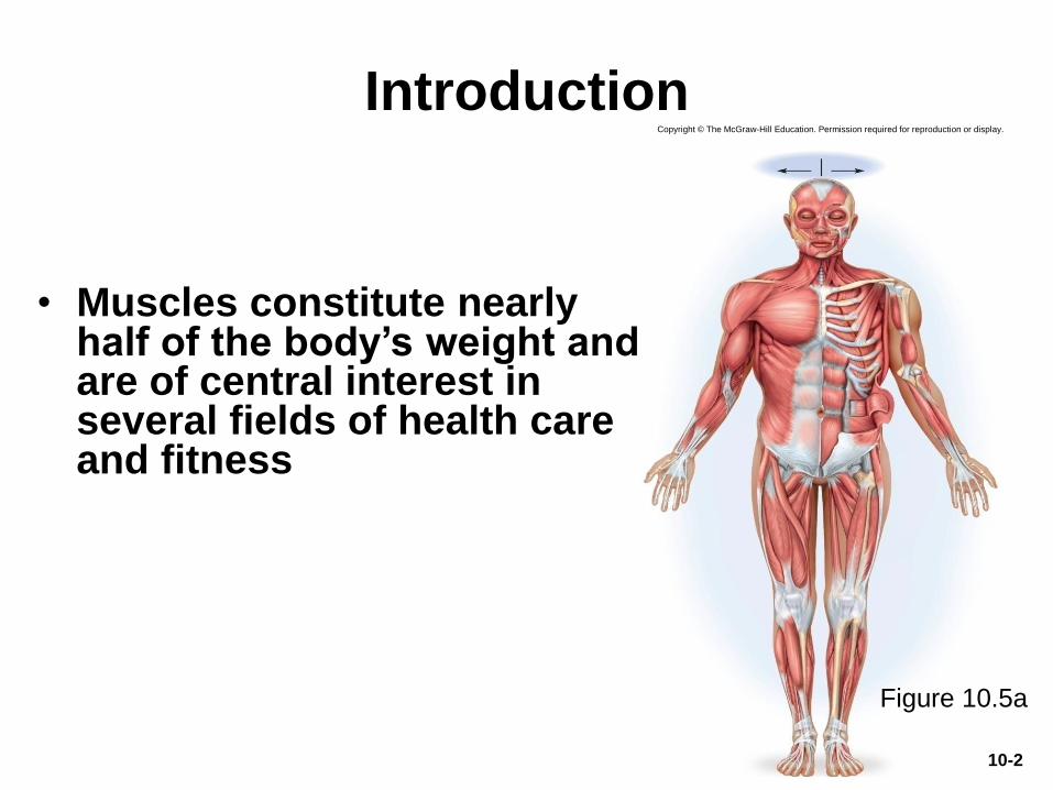
Introduction
• Muscles constitute nearly half of the body’s weight and are of central interest in several fields of health care and fitness
10-2
Figure 10.5a
Copyright © The McGraw-Hill Education. Permission required for reproduction or display.
The Structural and Functional
Organization of Muscles
• Expected Learning Outcomes
– Describe the varied functions of muscles.
– Describe the connective tissue components of a muscle
and their relationship to the bundling of muscle fibers.
– Describe the various shapes of skeletal muscles and
relate this to their functions.
– Explain what is meant by the origin, insertion, belly,
action, and innervation of a muscle.
10-3
The Structural and Functional
Organization of Muscles
(Continued)
– Describe the ways that muscles work in groups to aid,
oppose, or moderate each other’s actions.
– Distinguish between intrinsic and extrinsic muscles.
– Describe, in general terms, the nerve supply to the
muscles and where these nerves originate.
– Explain how the Latin names of muscles can aid in
visualizing and remembering them.
10-4
The Structural and Functional
Organization of Muscles
• About 600 human skeletal muscles
• Constitute about half of our body weight
• Three kinds of muscle tissue
– Skeletal, cardiac, smooth
• Specialized for one major purpose
– Converting the chemical energy in ATP into the
mechanical energy of motion
• Myology—the study of the muscular system
10-5
The Functions of Muscles
• Muscle functions include: movement, stability, control of openings, heat production, and glycemic control
• Movement– Move from place to place; move body parts; move body
contents in breathing, circulation, and digestion
– In communication: speech, writing, facial expressions and other nonverbal communications
• Stability– Maintain posture by preventing unwanted movements
– Antigravity muscles: prevent us from falling over
– Stabilize joints by maintaining tension
10-6
The Functions of Muscles
• Control of openings and passageways– Sphincters: internal muscular rings that control the
movement of food, blood, and other materials within body
• Heat production by skeletal muscles– As much as 85% of our body heat
• Glycemic control– Muscles absorb and store glucose which helps regulate
blood sugar concentration within normal range
10-7
Connective Tissues of a MuscleCopyright © The McGraw-Hill Companies, Inc. Permission required for reproduction or display.
Skeletal
muscle
Perimysium
Endomysium
(a)
Muscle
fascicle
Perimysium
Epimysium
Nerve
Blood vessels
Muscle fiber
Tendon
Fascia
Muscle fiber
Muscle fascicle
10-8
Figure 10.1a
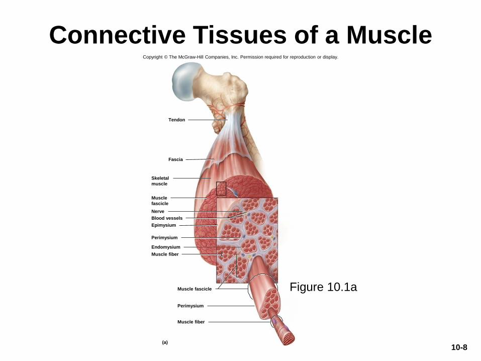
Connective Tissues and Fascicles
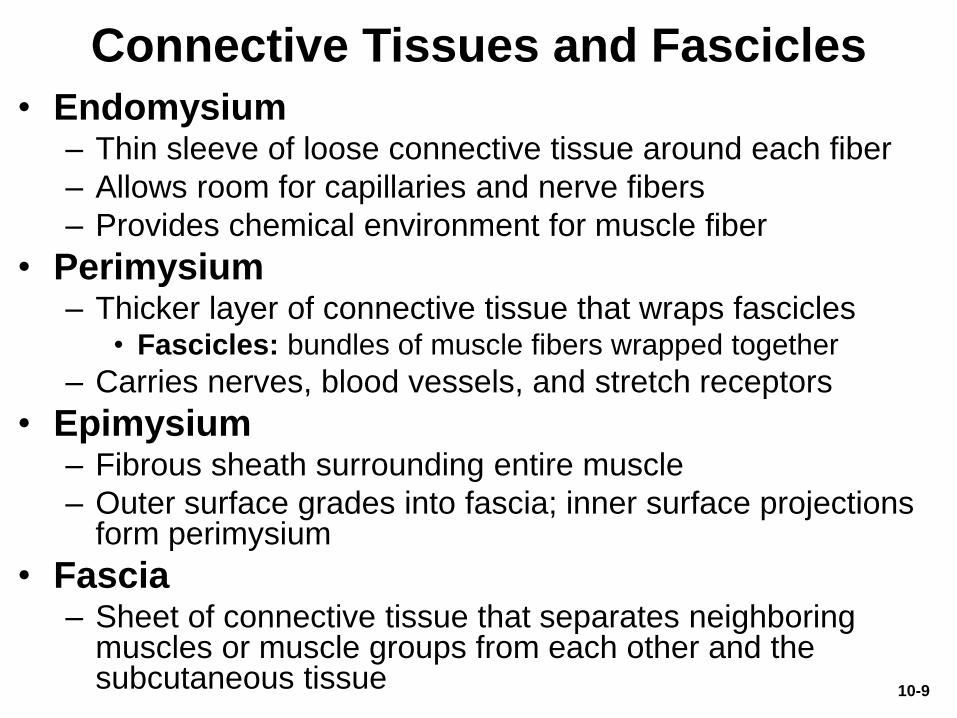
• Endomysium– Thin sleeve of loose connective tissue around each fiber
– Allows room for capillaries and nerve fibers
– Provides chemical environment for muscle fiber
• Perimysium– Thicker layer of connective tissue that wraps fascicles
• Fascicles: bundles of muscle fibers wrapped together
– Carries nerves, blood vessels, and stretch receptors
• Epimysium– Fibrous sheath surrounding entire muscle
– Outer surface grades into fascia; inner surface projections form perimysium
• Fascia– Sheet of connective tissue that separates neighboring
muscles or muscle groups from each other and the subcutaneous tissue
10-9
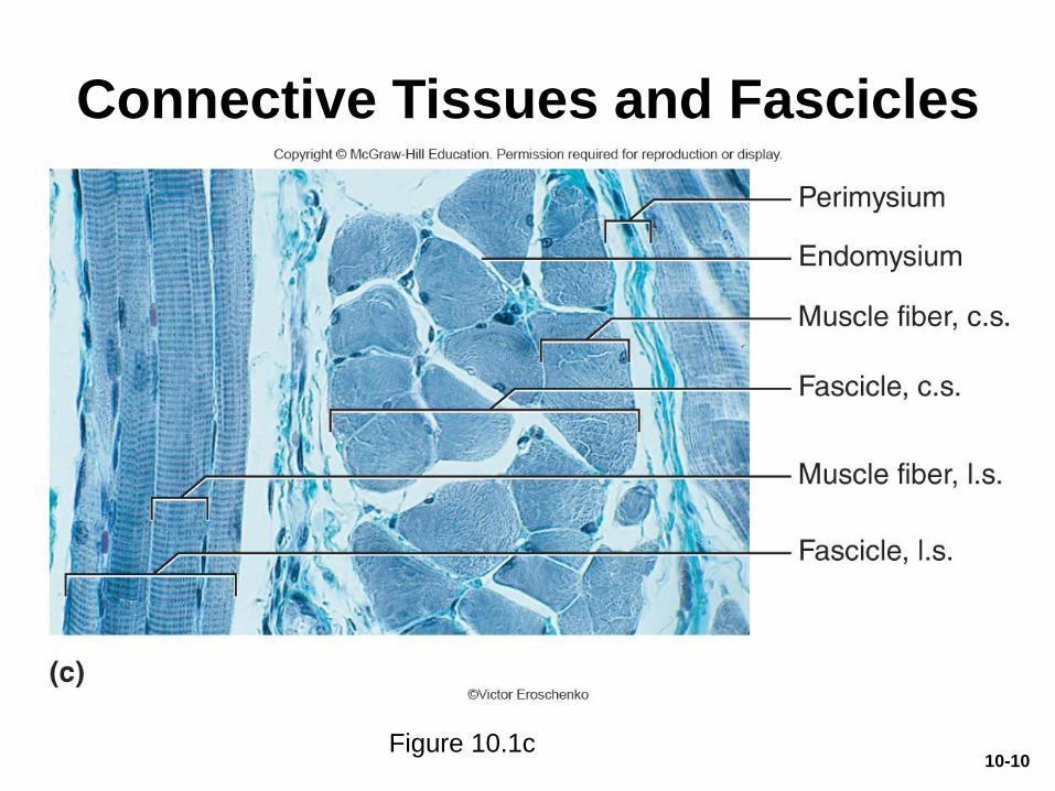
Connective Tissues and Fascicles
Figure 10.1c10-10
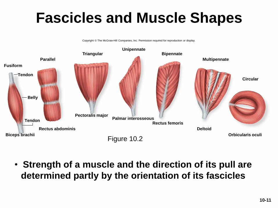
Fascicles and Muscle Shapes
Figure 10.2
• Strength of a muscle and the direction of its pull are
determined partly by the orientation of its fascicles
Fusiform
Parallel
TriangularUnipennate
Bipennate
Multipennate
Circular
Biceps brachii
Rectus abdominis
Pectoralis majorPalmar interosseous
Rectus femoris
Deltoid
Orbicularis oculi
Tendon
Tendon
Belly
Copyright © The McGraw-Hill Companies, Inc. Permission required for reproduction or display.
10-11
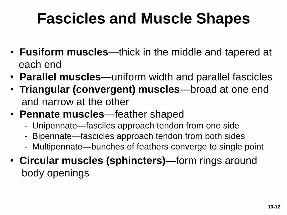
Fascicles and Muscle Shapes
• Fusiform muscles—thick in the middle and tapered at
each end
• Parallel muscles—uniform width and parallel fascicles
• Triangular (convergent) muscles—broad at one end
and narrow at the other
• Pennate muscles—feather shaped- Unipennate—fasciles approach tendon from one side
- Bipennate—fascicles approach tendon from both sides
- Multipennate—bunches of feathers converge to single point
• Circular muscles (sphincters)—form rings around
body openings
10-12
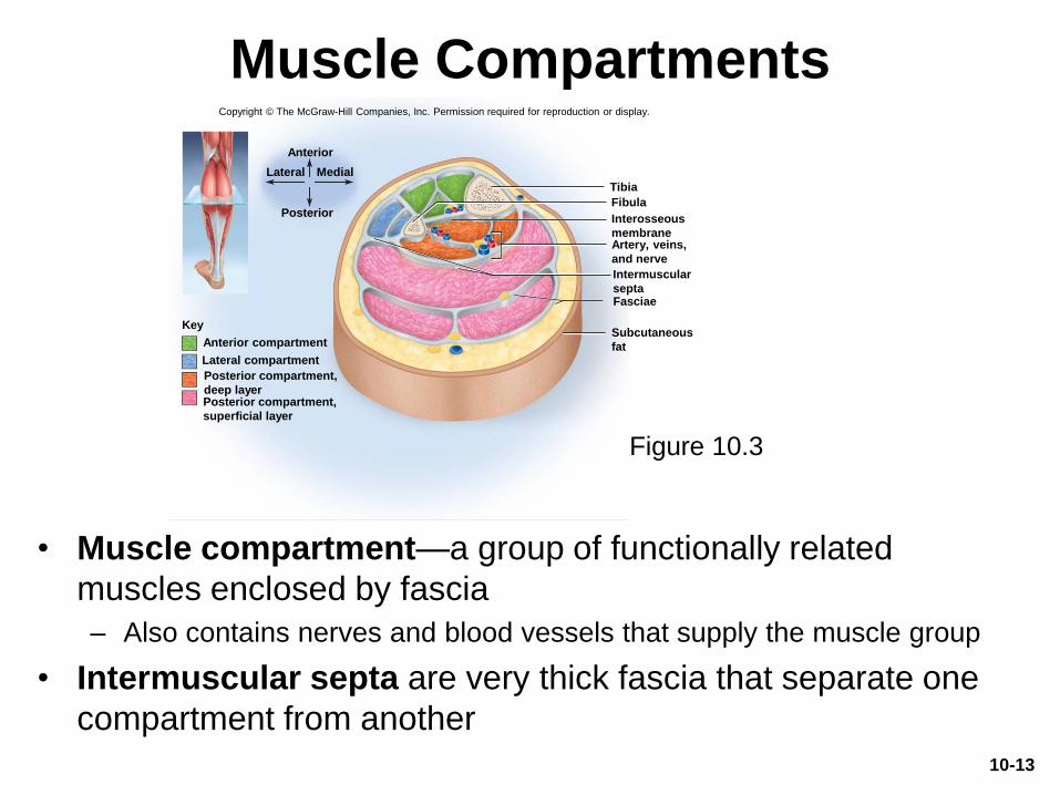
Muscle Compartments
• Muscle compartment—a group of functionally related
muscles enclosed by fascia
– Also contains nerves and blood vessels that supply the muscle group
• Intermuscular septa are very thick fascia that separate one
compartment from another
10-13
Figure 10.3
Anterior
Lateral Medial
Posterior
Key
Posterior compartment,
superficial layer
Posterior compartment,
deep layer
Lateral compartment
Anterior compartmentSubcutaneous
fat
Fasciae
Intermuscular
septa
Artery, veins,
and nerve
Interosseous
membrane
Fibula
Tibia
Copyright © The McGraw-Hill Companies, Inc. Permission required for reproduction or display.
Muscle Attachments
• Indirect attachment to bone
– Tendons connect muscle to bone
• Collagen fibers of the endo-, peri-, and epimysium
continue into the tendon and from there into periosteum
and matrix of bone
• Aponeurosis—tendon is a broad, flat sheet (palmar
aponeurosis)
• Retinaculum—connective tissue band that tendons from
separate muscles pass under
• Direct (fleshy) attachment to bone
– Little separation between muscle and bone
– Muscle seems to emerge directly from bone
10-14
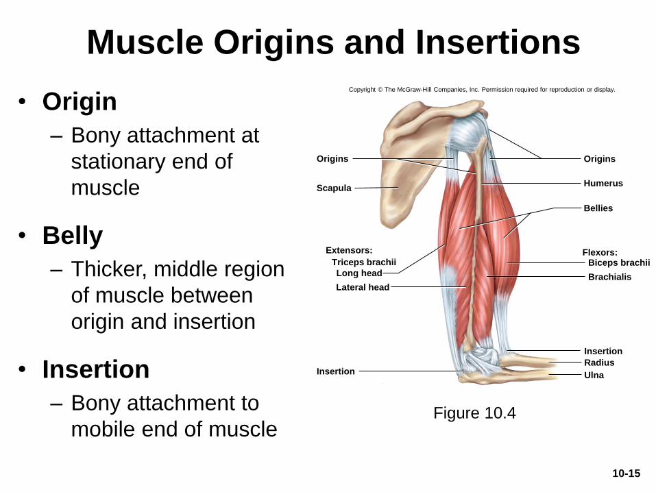
Muscle Origins and Insertions
• Origin
– Bony attachment at
stationary end of
muscle
• Belly
– Thicker, middle region
of muscle between
origin and insertion
• Insertion
– Bony attachment to
mobile end of muscleFigure 10.4
Scapula
Bellies
Radius
Insertion
Humerus
UlnaInsertion
Origins Origins
Long head
Extensors:
Lateral head
Flexors:Biceps brachii
Brachialis
Copyright © The McGraw-Hill Companies, Inc. Permission required for reproduction or display.
Triceps brachii
10-15
Muscle Origins and Insertions
• Some anatomists prefer nontraditional descriptions of attachments by proximal vs. distal or superior vs. inferior
• Some muscles insert not on bone but on the fascia or tendon of another muscle or on collagen fibers of the dermis– Example: many facial muscles insert in the skin
10-16
Functional Groups of Muscles
• Action—effect produced by a muscle to produce or
prevent movement
• Four categories of muscle action: prime mover,
synergist, antagonist, and fixator
– Prime mover (agonist)
• Muscle that produces most of force during a particular
joint action
– Synergist: muscle that aids the prime mover
• May contribute additional force, modify the direction of
movement, or stabilize a nearby joint
10-17
Functional Groups of Muscles
(Continued)
– Antagonist: opposes the prime mover
• Prevents excessive movement
• Sometimes relaxes to give prime mover control over
an action
• Antagonistic pairs—muscles that act on opposite
sides of a joint
– Fixator: muscle that prevents movement of bone
10-18
Functional Groups of Muscles
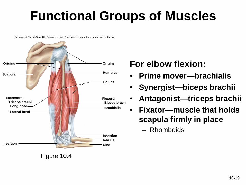
For elbow flexion:
• Prime mover—brachialis
• Synergist—biceps brachii
• Antagonist—triceps brachii
• Fixator—muscle that holds
scapula firmly in place
– Rhomboids
Figure 10.4
Scapula
Bellies
Radius
Insertion
Humerus
UlnaInsertion
Origins Origins
Extensors:
Lateral head
Flexors:Biceps brachii
Brachialis
Copyright © The McGraw-Hill Companies, Inc. Permission required for reproduction or display.
Long head
Triceps brachii
10-19
Intrinsic and Extrinsic Muscles
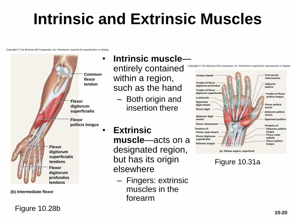
• Intrinsic muscle—entirely contained within a region, such as the hand
– Both origin and insertion there
• Extrinsic muscle—acts on a designated region, but has its origin elsewhere
– Fingers: extrinsic muscles in the forearm
Figure 10.28b
Figure 10.31a
(b) Intermediate flexor
Flexor
digitorum
superficialis
Flexor
pollicis longus
Flexor
digitorum
superficialis
tendons
Flexor
digitorum
profundus
tendons
Common
flexor
tendon
Opponens pollicis
Abductor pollicis
brevis
Flexor pollicis
brevis
Adductor
pollicis
Tendon of flexor
digitorum superficialis
Tendon of flexor
digitorum profundus
Abductor pollicis
longus
Palmaris longus
Flexor digitorum
superficialis
Flexor carpi ulnaris
Flexor retinaculum
Abductor digiti
minimi
Flexor digiti
Lumbricals
Opponens
digiti minimi
Tendon sheath
Flexor pollicis
longus
Flexor carpi
radialis
Tendon of flexor
pollicis longus
First dorsal
interosseous
Tendons of:
Tendons of:
(a) Palmar aspect, superficial
Copyright © The McGraw-Hill Companies, Inc. Permission required for reproduction or display.
Copyright © The McGraw-Hill Companies, Inc. Permission required for reproduction or display.
10-20
Muscle Innervation
• Innervation of a muscle—refers to the identity of
the nerve that stimulates it
– Knowing innervation enables diagnosis of nerve, spinal
cord, and brainstem injuries from muscle tests
• Spinal nerves arise from the spinal cord
– Emerge through intervertebral foramina
– Immediately branch into posterior and anterior rami
– Innervate muscles below the neck
– Plexus: web-like network of spinal nerves adjacent to
the vertebral column
10-21
Muscle Innervation
• Cranial nerves arise from the base of the brain
– Emerge through skull foramina
– Innervate the muscles of the head and neck
– Numbered CN I to CN XII
10-22
Blood Supply
• Muscular system receives about 1.24 L of
blood per minute at rest (one-quarter of the
blood pumped by the heart)
• During heavy exercise, total cardiac output
rises and the muscular system’s share is
more than three-quarters (11.6 L/min)
• Capillaries branch extensively through the
endomysium to reach every muscle fiber
10-23
How Muscles Are Named
• Latin names– Depressor labii inferioris, flexor digiti minimi brevis
• Describes distinctive aspects of the structure, location, or action of a muscle
• Footnotes throughout chapters show interpreted names of muscles
• Pronunciation of muscles available online at www.aprevealed.com
10-24
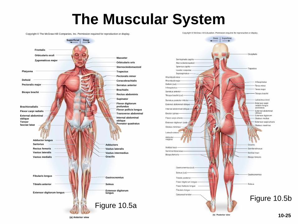
The Muscular System
Figure 10.5bFigure 10.5a
Frontalis
Orbicularis oculiMasseter
Orbicularis oris
Trapezius
External abdominal
oblique
Pronator quadratus
Gastrocnemius
Soleus
Adductor longus
Rectus abdominis
Serratus anterior
Sternocleidomastoid
Deltoid
Pectoralis major
Biceps brachii
Brachioradialis
Sartorius
Tensor
fasciae latae
Rectus femoris
Fibularis longus
Extensor digitorum longus
Tibialis anterior
Zygomaticus major
Vastus lateralis
Gracilis
Vastus intermedius
Adductors
Extensor digitorumlongus
Supinator
Flexor digitorum
profundus
Flexor pollicis longus
Transverse abdominal
Brachialis
Coracobrachialis
Platysma
Flexor carpi radialis
(a) Anterior view
Vastus medialis
Pectoralis minor
Internal abdominal
oblique
DeepSuperficial
Vastus lateralis
Copyright © The McGraw-Hill Companies, Inc. Permission required for reproduction or display.
10-25
A Learning Strategy
• Examine models, cadavers, dissected animals,
or a photographic atlas
• Palpate muscles on yourself if possible
• Locate origins and insertions of muscles on an
articulated skeleton
• Study derivation of each muscle name
– Usually describes the muscle’s location, appearance,
origin, insertion, or action
• Say the names aloud to yourself or study
partner, and spell them correctly
10-26
Muscles of the Head and Neck
• Expected Learning Outcomes
– Name and locate the muscles that produce facial
expression.
– Name and locate the muscles used for chewing and
swallowing.
– Name and locate the neck muscles that move the
head.
– Identify the origin, insertion, action, and innervation of
any of these muscles.
10-27
Muscles of Facial Expression
• Muscles that insert in the dermis and
subcutaneous tissues
• Tense the skin and produce facial
expressions
• Innervated by facial nerve (CN VII)
• Paralysis causes face to sag
• Found in scalp, forehead, around the eyes,
nose, and mouth, and in the neck
10-28

Muscles of Facial Expression
Figure 10.8a
Frontalis
Galea aponeurotica
Orbicularis oculi
Platysma
(a) Anterior view
Mentalis (cut)
Orbicularis oris
Masseter
Zygomaticus minor
Levator labii superioris
Zygomaticus major
Risorius
Depressor anguli oris
Depressor labii inferioris
Nasalis
Corrugator supercilii
Buccinator
Modiolus
Levator anguli oris
Superficial Deep
Copyright © The McGraw-Hill Companies, Inc. Permission required for reproduction or display.
10-29
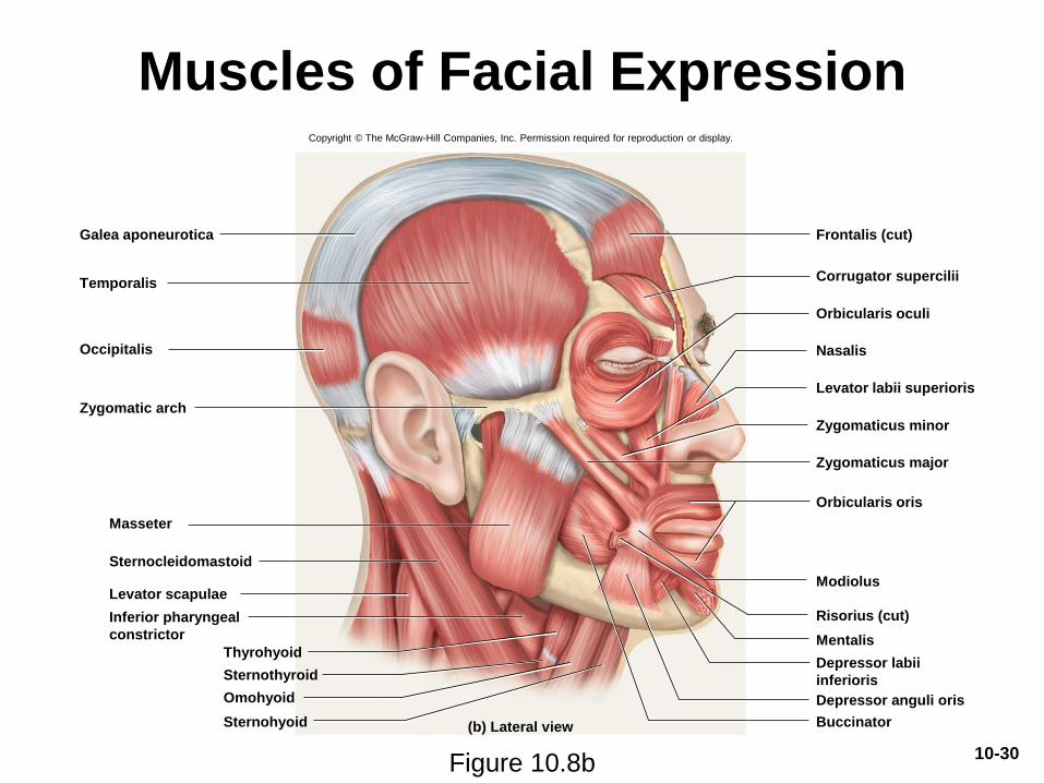
Muscles of Facial Expression
Frontalis (cut)Galea aponeurotica
Orbicularis oculi
Sternohyoid
Orbicularis oris
Occipitalis
Omohyoid
Sternothyroid
Inferior pharyngeal
constrictor
Sternocleidomastoid
Masseter
Thyrohyoid
Zygomatic arch
Levator labii superioris
Zygomaticus minor
Zygomaticus major
Depressor labii
inferioris
Depressor anguli oris
Buccinator
Risorius (cut)
Nasalis
Corrugator supercilii
Mentalis
Levator scapulae
(b) Lateral view
Modiolus
Temporalis
Copyright © The McGraw-Hill Companies, Inc. Permission required for reproduction or display.
Figure 10.8b10-30
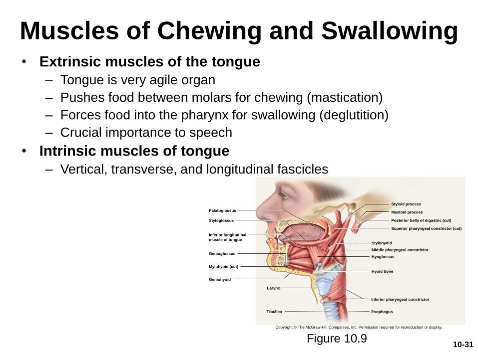
Muscles of Chewing and Swallowing• Extrinsic muscles of the tongue
– Tongue is very agile organ
– Pushes food between molars for chewing (mastication)
– Forces food into the pharynx for swallowing (deglutition)
– Crucial importance to speech
• Intrinsic muscles of tongue
– Vertical, transverse, and longitudinal fascicles
Figure 10.9
Genioglossus
Styloglossus
Palatoglossus
Posterior belly of digastric (cut)
Stylohyoid
Superior pharyngeal constrictor (cut)
Middle pharyngeal constrictor
Inferior pharyngeal constrictor
Hyoglossus
Hyoid bone
Styloid process
Mastoid process
Mylohyoid (cut)
Geniohyoid
Esophagus
Inferior longitudinal
muscle of tongue
Larynx
Copyright © The McGraw-Hill Companies, Inc. Permission required for reproduction or display.
Trachea
10-31
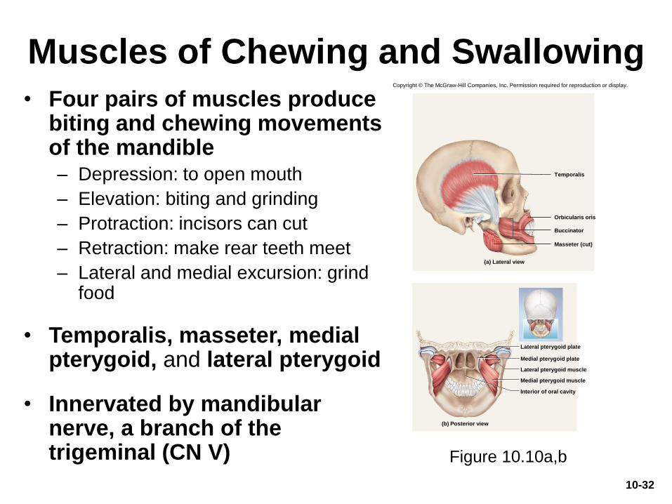
Muscles of Chewing and Swallowing
• Four pairs of muscles produce biting and chewing movements of the mandible– Depression: to open mouth
– Elevation: biting and grinding
– Protraction: incisors can cut
– Retraction: make rear teeth meet
– Lateral and medial excursion: grind food
• Temporalis, masseter, medial pterygoid, and lateral pterygoid
• Innervated by mandibular nerve, a branch of the trigeminal (CN V) Figure 10.10a,b
Temporalis
Orbicularis oris
Masseter (cut)
(a) Lateral view
Buccinator
Copyright © The McGraw-Hill Companies, Inc. Permission required for reproduction or display.
(b) Posterior view
Medial pterygoid muscle
Medial pterygoid plate
Lateral pterygoid plate
Interior of oral cavity
Lateral pterygoid muscle
10-32
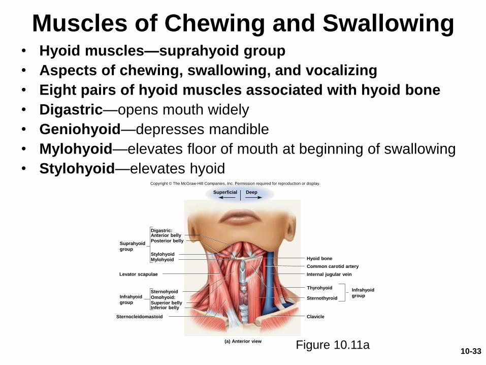
Muscles of Chewing and Swallowing• Hyoid muscles—suprahyoid group
• Aspects of chewing, swallowing, and vocalizing
• Eight pairs of hyoid muscles associated with hyoid bone
• Digastric—opens mouth widely
• Geniohyoid—depresses mandible
• Mylohyoid—elevates floor of mouth at beginning of swallowing
• Stylohyoid—elevates hyoid
Figure 10.11a
Levator scapulae
Sternocleidomastoid
Digastric:Anterior bellyPosterior belly
StylohyoidMylohyoid
Sternohyoid
Omohyoid:
Suprahyoid
group
Infrahyoid
group
(a) Anterior view
Hyoid bone
Common carotid artery
Internal jugular vein
Clavicle
Thyrohyoid
Superior bellyInferior belly
Superficial Deep
Infrahyoid
groupSternothyroid
Copyright © The McGraw-Hill Companies, Inc. Permission required for reproduction or display.
10-33

Muscles of Chewing and Swallowing• Hyoid muscles—infrahyoid group
• Fix hyoid bone from below, allowing suprahyoid muscles to open mouth
• Omohyoid—depresses hyoid after elevation
• Sternohyoid—depresses hyoid after elevation
• Thyrohyoid—depresses hyoid and elevates larynx
• Sternothyroid—depresses larynx after elevation
Figure 10.11b 10-34
Stylohyoid
Hyoglossus
Mylohyoid
Digastric
(anterior belly)
Hyoid bone
Thyrohyoid
Omohyoid
(superior belly)
Sternothyroid
Sternohyoid
(b) Lateral view
Digastric (posterior belly)
Splenius capitis
Inferior pharyngeal constrictor
Sternocleidomastoid
Trapezius
Levator scapulae
Scalenes
Omohyoid (inferior belly)
Copyright © The McGraw-Hill Companies, Inc. Permission required for reproduction or display.
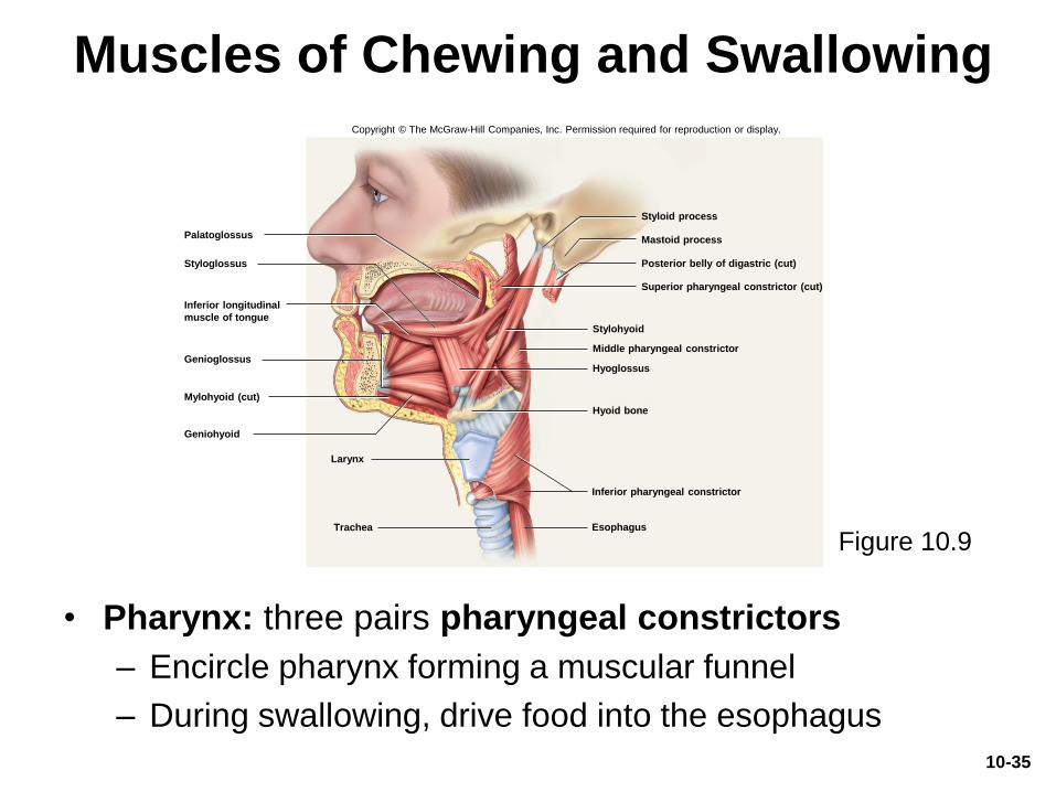
Muscles of Chewing and Swallowing
• Pharynx: three pairs pharyngeal constrictors
– Encircle pharynx forming a muscular funnel
– During swallowing, drive food into the esophagus
Figure 10.9
Genioglossus
Styloglossus
Palatoglossus
Posterior belly of digastric (cut)
Stylohyoid
Superior pharyngeal constrictor (cut)
Middle pharyngeal constrictor
Inferior pharyngeal constrictor
Hyoglossus
Hyoid bone
Styloid process
Mastoid process
Mylohyoid (cut)
Geniohyoid
Esophagus
Inferior longitudinal
muscle of tongue
Larynx
Copyright © The McGraw-Hill Companies, Inc. Permission required for reproduction or display.
Trachea
10-35

Muscles Acting on the Head
• Originate on vertebral column, thoracic cage,
and pectoral girdle
• Insert on the cranial bones
• Actions
– Flexion (tipping head forward)
– Extension (holding the head erect)
– Lateral flexion (tipping head to one side)
– Rotation (turning the head to the left and right)
10-36
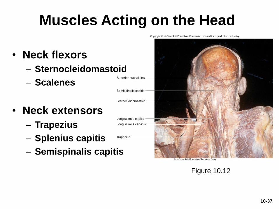
Muscles Acting on the Head
• Neck flexors
– Sternocleidomastoid
– Scalenes
• Neck extensors
– Trapezius
– Splenius capitis
– Semispinalis capitis
10-37
Figure 10.12
Muscles Acting on the Head
• May cause contralateral movement:
movement of the head toward the opposite
side
• May cause ipsilateral movement: movement
of the head toward the same side
10-38
Muscles of the Trunk
• Expected Learning Outcomes
– Name and locate the muscles of respiration and
explain how they affect airflow and abdominal
pressure.
– Name and locate the muscles of the abdominal wall,
back, and pelvic floor.
– Identify the origin, insertion, action, and innervation of
any of these muscles.
10-39
Muscles of the Trunk
• Three functional groups
– Muscles of respiration
– Muscles that support abdominal wall and pelvic floor
– Movement of vertebral column
10-40
Muscles of Respiration
• Breathing requires the use of muscles enclosing thoracic cavity– Diaphragm, external intercostal, internal intercostal,
and innermost intercostal muscles
• Inspiration—air intake
• Expiration—expelling air
10-41
Muscles of Respiration
• Other muscles of chest and abdomen that contribute to breathing– Sternocleidomastoid, scalenes of neck
– Pectoralis major and serratus anterior of chest
– Latissimus dorsi of back
– Abdominal muscles: internal and external obliques, and transverse abdominis
– Even some anal muscles
10-42
Muscles of Respiration
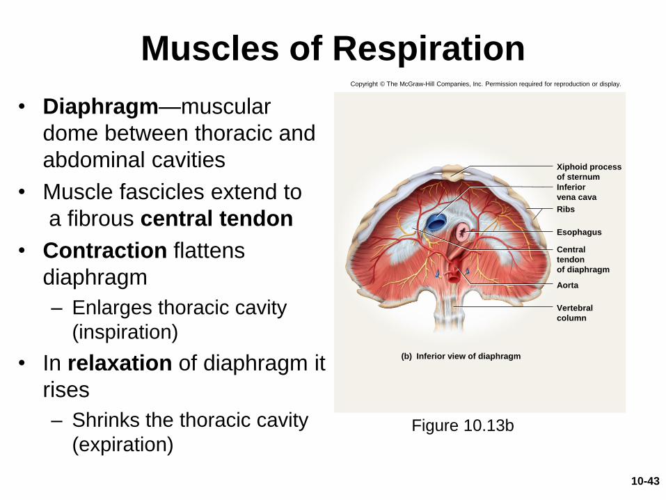
Figure 10.13b
Xiphoid process
of sternum
Esophagus
Aorta
Inferior
vena cava
Central
tendon
of diaphragm
(b) Inferior view of diaphragm
Vertebral
column
Ribs
• Diaphragm—muscular
dome between thoracic and
abdominal cavities
• Muscle fascicles extend to
a fibrous central tendon
• Contraction flattens
diaphragm
– Enlarges thoracic cavity
(inspiration)
• In relaxation of diaphragm it
rises
– Shrinks the thoracic cavity
(expiration)
Copyright © The McGraw-Hill Companies, Inc. Permission required for reproduction or display.
10-43
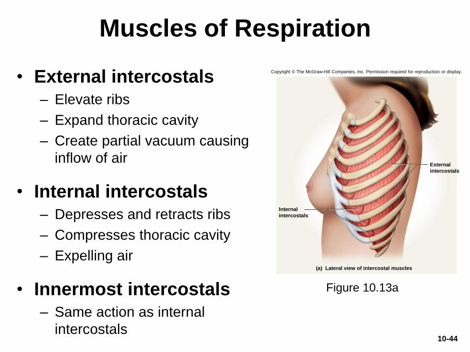
Muscles of Respiration
• External intercostals
– Elevate ribs
– Expand thoracic cavity
– Create partial vacuum causing
inflow of air
• Internal intercostals
– Depresses and retracts ribs
– Compresses thoracic cavity
– Expelling air
• Innermost intercostals
– Same action as internal
intercostals
Figure 10.13a
External
intercostals
Internal
intercostals
(a) Lateral view of intercostal muscles
Copyright © The McGraw-Hill Companies, Inc. Permission required for reproduction or display.
10-44
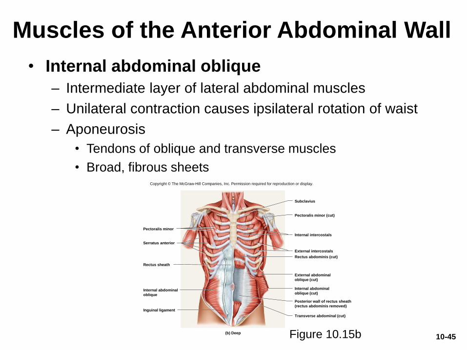
Muscles of the Anterior Abdominal Wall
• Internal abdominal oblique
– Intermediate layer of lateral abdominal muscles
– Unilateral contraction causes ipsilateral rotation of waist
– Aponeurosis
• Tendons of oblique and transverse muscles
• Broad, fibrous sheets
Subclavius
Pectoralis minor (cut)
Internal intercostals
External intercostals
Rectus abdominis (cut)
External abdominal
oblique (cut)
Internal abdominal
oblique (cut)
Transverse abdominal (cut)
Posterior wall of rectus sheath
(rectus abdominis removed)
Internal abdominal
oblique
Inguinal ligament
Rectus sheath
Serratus anterior
Pectoralis minor
(b) Deep
Copyright © The McGraw-Hill Companies, Inc. Permission required for reproduction or display.
Figure 10.15b 10-45
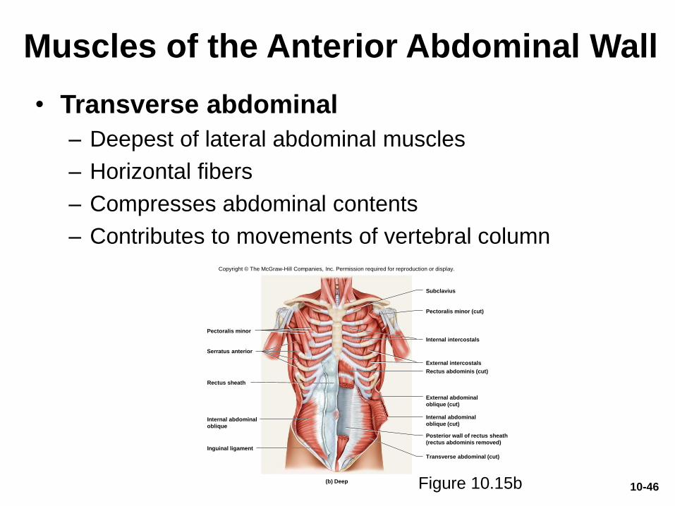
Muscles of the Anterior Abdominal Wall
• Transverse abdominal
– Deepest of lateral abdominal muscles
– Horizontal fibers
– Compresses abdominal contents
– Contributes to movements of vertebral column
Subclavius
Pectoralis minor (cut)
Internal intercostals
External intercostals
Rectus abdominis (cut)
External abdominal
oblique (cut)
Internal abdominal
oblique (cut)
Transverse abdominal (cut)
Posterior wall of rectus sheath
(rectus abdominis removed)
Internal abdominal
oblique
Inguinal ligament
Rectus sheath
Serratus anterior
Pectoralis minor
(b) Deep
Copyright © The McGraw-Hill Companies, Inc. Permission required for reproduction or display.
Figure 10.15b 10-46
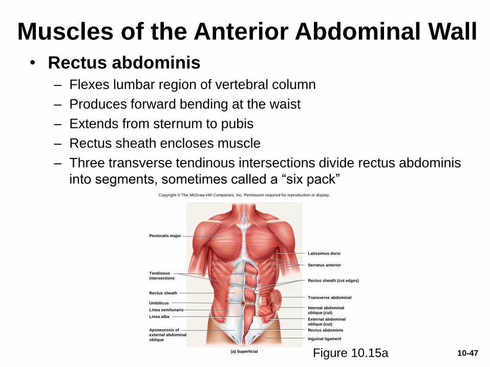
Muscles of the Anterior Abdominal Wall
• Rectus abdominis– Flexes lumbar region of vertebral column
– Produces forward bending at the waist
– Extends from sternum to pubis
– Rectus sheath encloses muscle
– Three transverse tendinous intersections divide rectus abdominis
into segments, sometimes called a “six pack”
Figure 10.15a
Pectoralis major
Tendinous
intersections
Linea alba
Latissimus dorsi
Rectus abdominis
Inguinal ligament
Rectus sheath (cut edges)
Serratus anterior
Aponeurosis of
external abdominal
oblique
Umbilicus
Linea semilunaris
Transverse abdominal
Internal abdominal
oblique (cut)
External abdominal
oblique (cut)
Rectus sheath
(a) Superficial
Copyright © The McGraw-Hill Companies, Inc. Permission required for reproduction or display.
10-47
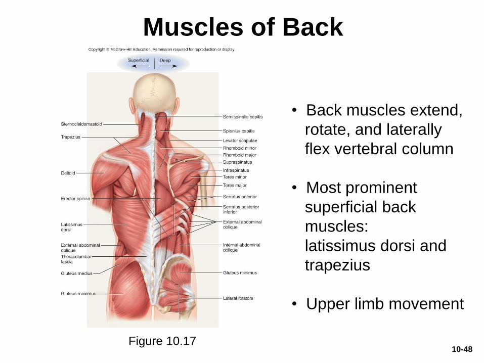
Muscles of Back
• Back muscles extend,
rotate, and laterally
flex vertebral column
• Most prominent
superficial back
muscles:
latissimus dorsi and
trapezius
• Upper limb movement
Figure 10.1710-48
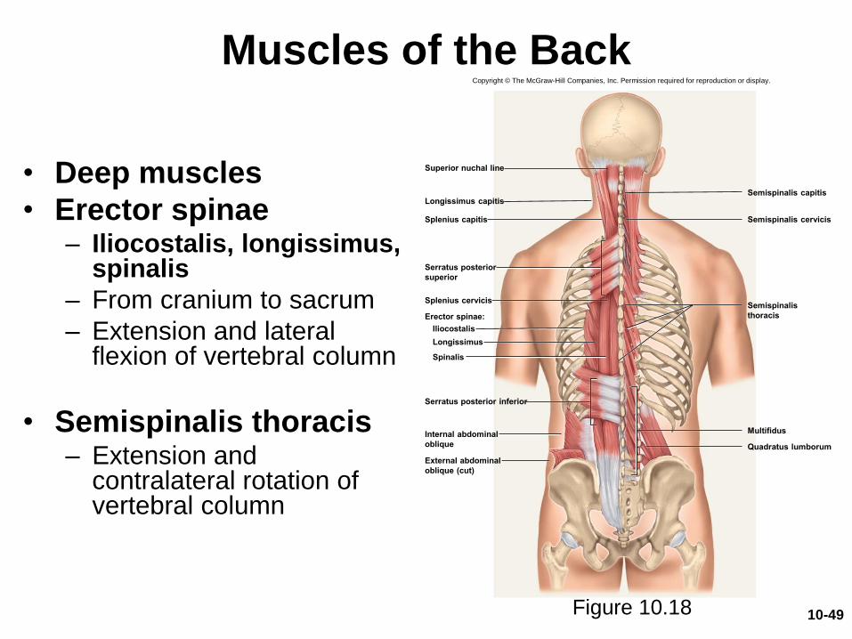
Muscles of the Back
• Deep muscles
• Erector spinae– Iliocostalis, longissimus,
spinalis
– From cranium to sacrum
– Extension and lateral flexion of vertebral column
• Semispinalis thoracis– Extension and
contralateral rotation of vertebral column
Figure 10.18
Longissimus capitisSemispinalis capitis
Internal abdominal
oblique
Erector spinae:
Semispinalis
thoracis
Multifidus
Quadratus lumborum
Superior nuchal line
Splenius capitis
Serratus posterior
superior
Splenius cervicis
External abdominal
oblique (cut)
Semispinalis cervicis
Serratus posterior inferior
Iliocostalis
Longissimus
Spinalis
Copyright © The McGraw-Hill Companies, Inc. Permission required for reproduction or display.
10-49
Muscles of the Back
(Continued)
• Quadratus lumborum– Aids respiration
– Ipsilateral flexion of lumbar vertebral column
• Multifidus– Stabilizes adjacent vertebrae
– Maintains posture
10-50
Muscles of the Pelvic Floor
• Layers of muscles and fasciae that span pelvic outlet– Penetrated by anal canal, urethra, and vagina
• Perineum—diamond-shaped region between the thighs – Bordered by four bony landmarks
• Pubic symphysis anteriorly
• Coccyx posteriorly
• Ischial tuberosities laterally
– Urogenital triangle: anterior half of perineum
– Anal triangle: posterior half of perineum
10-51
Muscles of the Pelvic Floor
• Layers or compartments of the perineum
– Superficial perineal space• Ischiocavernosus, bulbospongiosus
– Deep perineal space• Deep transverse perineal, compressor urethrae
– Anal triangle• External anal sphincter
– Pelvic diaphragm: deepest (most superior) layer • Levator ani
10-52
Muscles of the Pelvic Floor
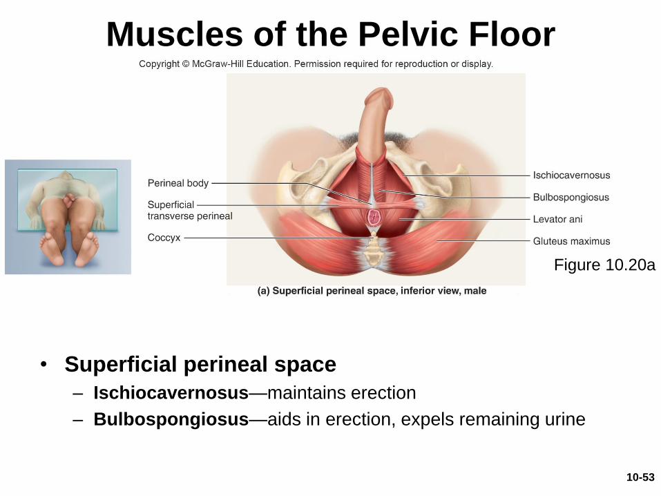
• Superficial perineal space
– Ischiocavernosus—maintains erection
– Bulbospongiosus—aids in erection, expels remaining urine
Figure 10.20a
10-53
Muscles of the Pelvic Floor
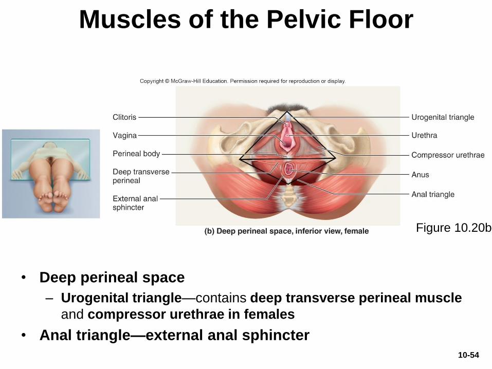
• Deep perineal space
– Urogenital triangle—contains deep transverse perineal muscle
and compressor urethrae in females
• Anal triangle—external anal sphincter
Figure 10.20b
10-54
Muscles of the Pelvic Floor
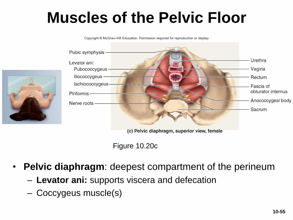
• Pelvic diaphragm: deepest compartment of the perineum
– Levator ani: supports viscera and defecation
– Coccygeus muscle(s)
Figure 10.20c
10-55
Copyright © The McGraw-Hill Companies, Inc. Permission required for reproduction or display.
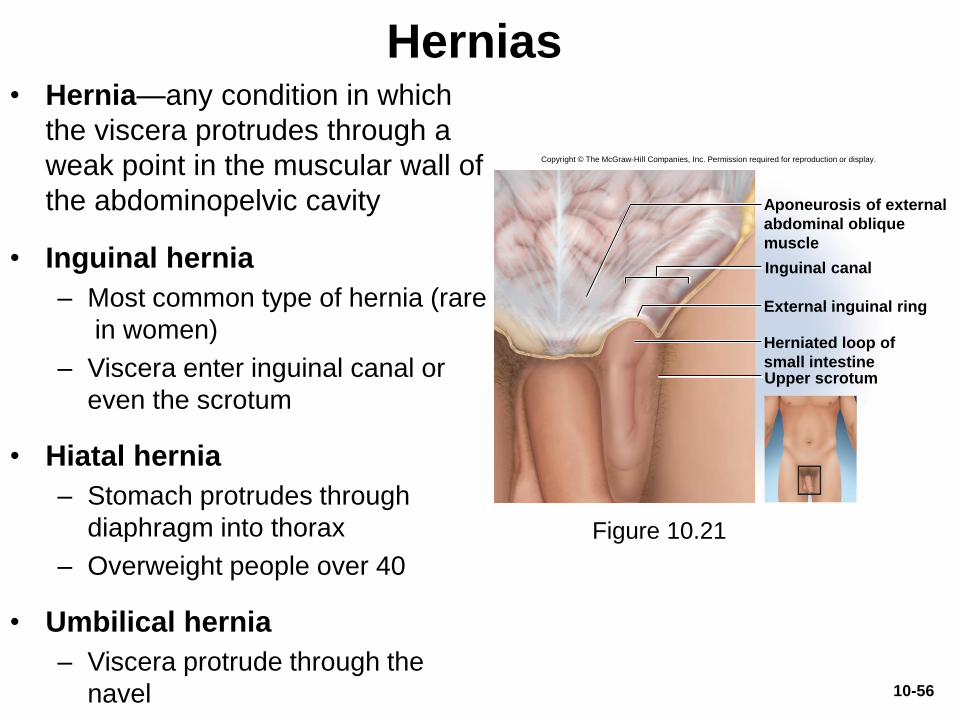
Aponeurosis of external
abdominal oblique
muscle
Inguinal canal
External inguinal ring
Herniated loop of
small intestineUpper scrotum
Hernias• Hernia—any condition in which
the viscera protrudes through a
weak point in the muscular wall of
the abdominopelvic cavity
• Inguinal hernia
– Most common type of hernia (rare
in women)
– Viscera enter inguinal canal or
even the scrotum
• Hiatal hernia
– Stomach protrudes through
diaphragm into thorax
– Overweight people over 40
• Umbilical hernia
– Viscera protrude through the
navel 10-56
Figure 10.21
Muscles Acting on the Shoulder
and Upper Limb
• Expected Learning Outcomes
– Name and locate the muscles that act on the pectoral
girdle, shoulder, elbow, wrist, and hand.
– Relate the actions of these muscles to the joint
movements described in chapter 9.
– Describe the origin, insertion, and innervation of each
muscle.
10-57
Muscles Acting on the Shoulder
and Upper Limb
• Compartments—spaces where muscles are separated by fibrous connective tissue sheets (fasciae)
– Each compartment contains one or more functionally related muscles along with their nerve and blood supplies
• Muscles of upper limbs divided into anterior and posterior compartments
• Intermuscular septa (thick fascia) separates compartments
• Compartment syndrome—one of the muscles or blood vessels in a compartment is injured
10-58
Compartment Syndrome• If a blood vessel in a compartment is damaged, blood
and tissue fluid accumulate
• Fasciae enclose muscle compartments snugly and prevent expansion
• Compartment syndrome—mounting pressure triggers a sequence of degenerative events– Blood flow to compartment is obstructed by pressure
– If ischemia (poor blood flow) persists for more than 2 to 4 hours, nerves begin to die
– After 6 hours, muscles begin to die
• Nerves can regenerate after pressure relieved, but muscle damage is permanent
• Myoglobin in urine indicates compartment syndrome
• Treatment: immobilization of limb and fasciotomy (incision to relieve compartment pressure)
10-59
Muscles Acting on the Shoulder
and Upper Limb
• Upper limb is used for a broad range of powerful
and subtle actions
– Climbing, grasping, throwing, writing, playing musical
instruments, and manipulating small objects
• Muscles that act on the scapula
• Muscles that act on the humerus and shoulder
joint
• Muscles that act on the forearm and elbow joint
• Muscles that act on the wrist, hand, and fingers
10-60
Muscles Acting on the Shoulder
• A group of muscles originate on the axial
skeleton and insert on clavicle or scapula
• Scapula loosely attached to thoracic cage
– Capable of great movement
– Rotation, elevation, depression, protraction, retraction
• Clavicle braces the shoulder and moderates
movements
10-61
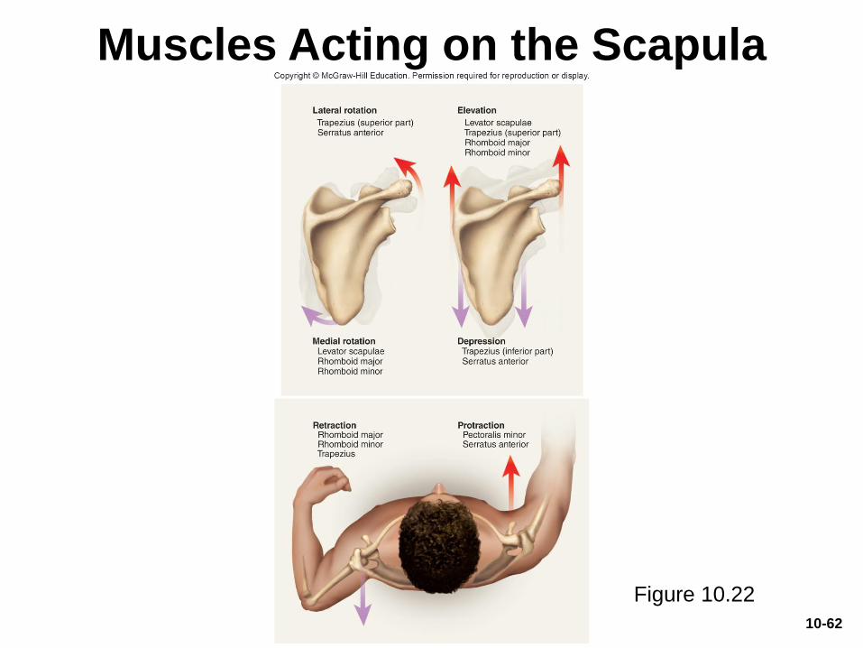
Muscles Acting on the Scapula
Figure 10.22
10-62
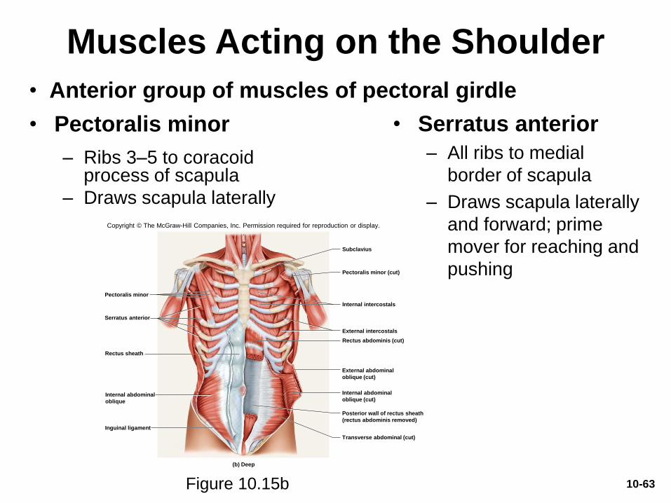
Muscles Acting on the Shoulder
• Pectoralis minor
– Ribs 3–5 to coracoid process of scapula
– Draws scapula laterally
• Serratus anterior
– All ribs to medial
border of scapula
– Draws scapula laterally
and forward; prime
mover for reaching and
pushing
Subclavius
Pectoralis minor (cut)
Internal intercostals
External intercostals
Rectus abdominis (cut)
Inguinal ligament
Rectus sheath
Serratus anterior
Pectoralis minor
(b) Deep
Internal abdominal
oblique
Transverse abdominal (cut)
Posterior wall of rectus sheath
(rectus abdominis removed)
Internal abdominal
oblique (cut)
External abdominal
oblique (cut)
Copyright © The McGraw-Hill Companies, Inc. Permission required for reproduction or display.
Figure 10.15b 10-63
• Anterior group of muscles of pectoral girdle

Muscles Acting on the Shoulder
• Four muscles of posterior group
– Trapezius: superficial
– Levator scapulae, Rhomboid minor, and Rhomboid major: deep
• Trapezius
– Stabilizes scapula and shoulder
– Elevates and depresses shoulder apex
Figure 10.17
Semispinalis capitis
Sternocleidomastoid
Deltoid
Levator scapulae
Rhomboid minor
Rhomboid major
Infraspinatus
Gluteus maximus
Gluteus medius Gluteus minimus
Lateral rotators
Serratus anterior
Supraspinatus
Erector spinae
Splenius capitis
Superficial Deep
Trapezius
Latissimus
dorsi
External abdominal
obliqueThoracolumbar
fascia
Teres minor
Teres major
Serratus posterior
inferior
External abdominal
oblique
Internal abdominal
oblique
Copyright © The McGraw-Hill Companies, Inc. Permission required for reproduction or display.
10-64
• Posterior group of muscles
of pectoral girdle
Muscles Acting on the Shoulder
(Continued from slide 170)
• Levator scapulae
– Elevates scapula
– Flexes neck laterally
• Rhomboid minor
– Retracts scapula and braces shoulder
• Rhomboid major
– Same as Rhomboid minor
10-65
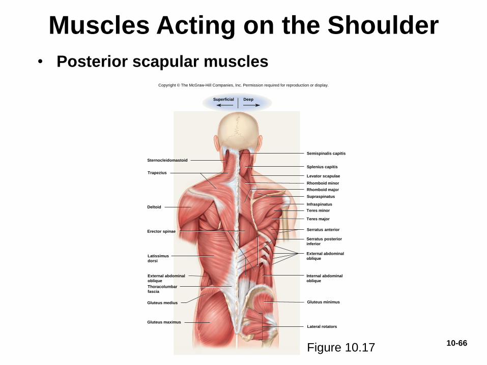
Muscles Acting on the Shoulder
Figure 10.17
Semispinalis capitis
Sternocleidomastoid
Deltoid
Levator scapulae
Rhomboid minor
Rhomboid major
Infraspinatus
Gluteus maximus
Gluteus medius Gluteus minimus
Lateral rotators
Serratus anterior
Supraspinatus
Erector spinae
Splenius capitis
Superficial Deep
Trapezius
Latissimus
dorsi
External abdominal
oblique
Thoracolumbar
fascia
Teres minor
Teres major
Serratus posterior
inferior
External abdominal
oblique
Internal abdominal
oblique
Copyright © The McGraw-Hill Companies, Inc. Permission required for reproduction or display.
10-66
• Posterior scapular muscles
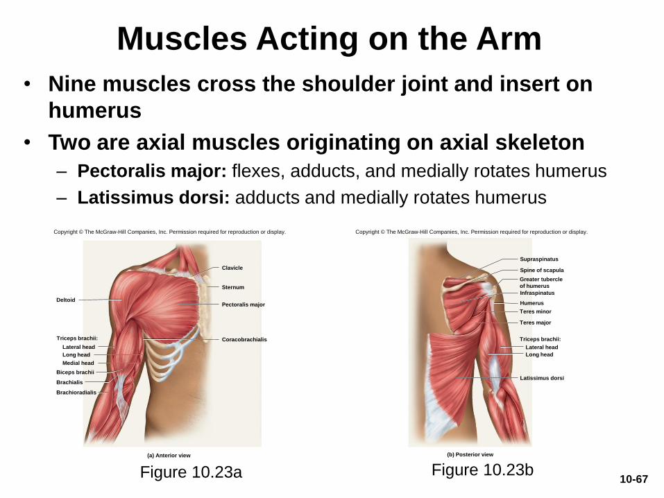
Muscles Acting on the Arm
• Nine muscles cross the shoulder joint and insert on
humerus
• Two are axial muscles originating on axial skeleton
– Pectoralis major: flexes, adducts, and medially rotates humerus
– Latissimus dorsi: adducts and medially rotates humerus
Figure 10.23a Figure 10.23b
Clavicle
Deltoid
Sternum
Pectoralis major
Coracobrachialis
Lateral head
Long head
Medial head
Biceps brachii
Brachialis
Brachioradialis
Triceps brachii:
(a) Anterior view
Copyright © The McGraw-Hill Companies, Inc. Permission required for reproduction or display.
(b) Posterior view
Supraspinatus
Spine of scapula
Infraspinatus
Lateral head
Long head
Latissimus dorsi
Humerus
Greater tubercle
of humerus
Teres minor
Teres major
Triceps brachii:
Copyright © The McGraw-Hill Companies, Inc. Permission required for reproduction or display.
10-67
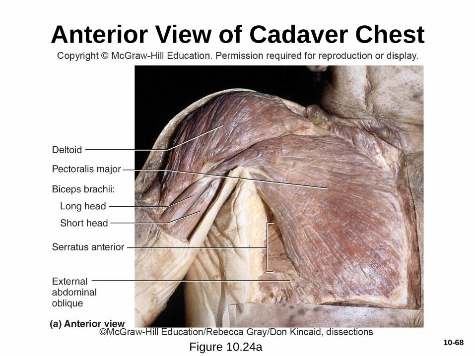
Anterior View of Cadaver Chest
Figure 10.24a10-68
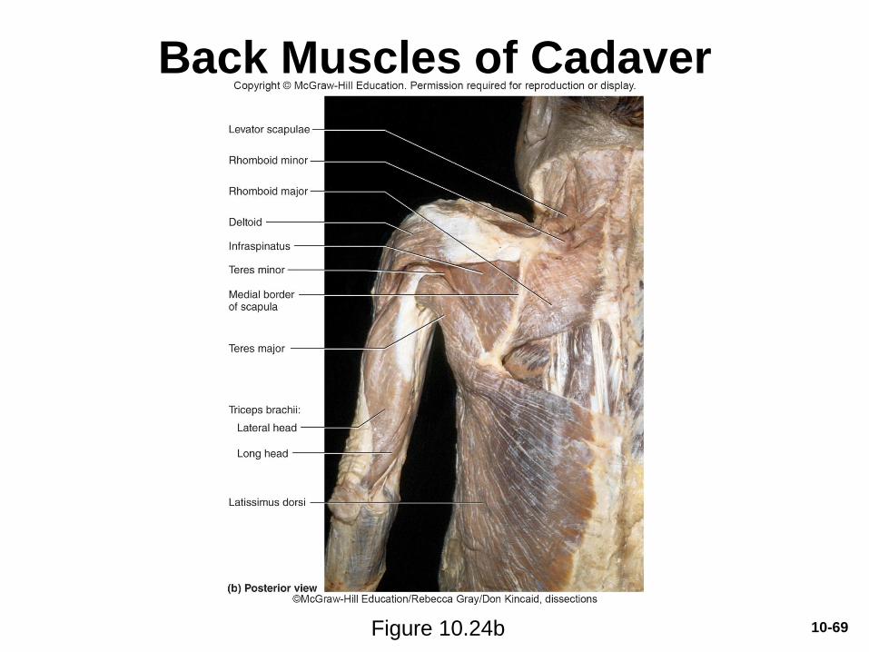
Back Muscles of Cadaver
Figure 10.24b 10-69
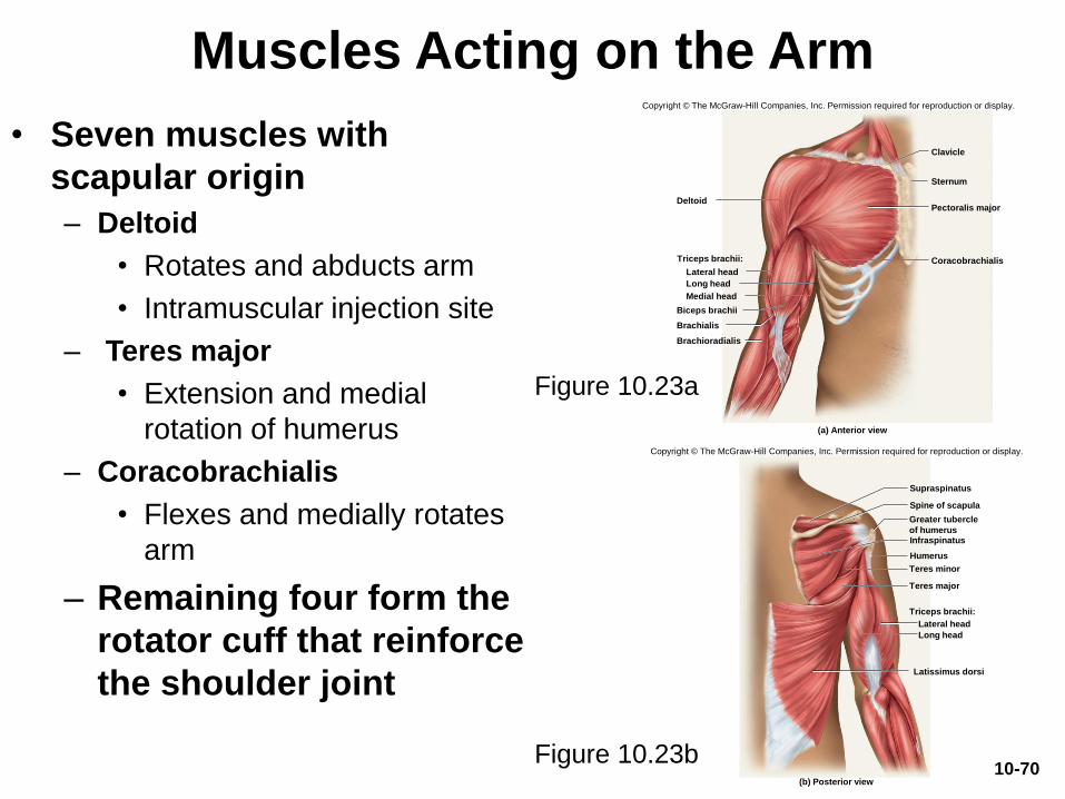
• Seven muscles with
scapular origin
– Deltoid
• Rotates and abducts arm
• Intramuscular injection site
– Teres major
• Extension and medial
rotation of humerus
– Coracobrachialis
• Flexes and medially rotates
arm
– Remaining four form the
rotator cuff that reinforce
the shoulder joint
Muscles Acting on the Arm
Figure 10.23b
Figure 10.23a
Clavicle
Deltoid
Sternum
Pectoralis major
Coracobrachialis
Lateral head
Long head
Medial head
Biceps brachii
Brachialis
Brachioradialis
Triceps brachii:
(a) Anterior view
Copyright © The McGraw-Hill Companies, Inc. Permission required for reproduction or display.
(b) Posterior view
Supraspinatus
Spine of scapula
Infraspinatus
Lateral head
Long head
Latissimus dorsi
Humerus
Greater tubercle
of humerus
Teres minor
Teres major
Triceps brachii:
Copyright © The McGraw-Hill Companies, Inc. Permission required for reproduction or display.
10-70
Muscles Acting on the Arm
• Rotator cuff muscles
• Tendons of the remaining four scapular muscles form the
rotator cuff
• Acronym “SITS muscles”
– Supraspinatus
– Infraspinatus
– Teres minor
– Subscapularis
• Tendons of these muscles merge with the joint capsule of
the shoulder as they cross it in route to the humerus
• Holds head of humerus into glenoid cavity
• Supraspinatus tendon easily damaged10-71
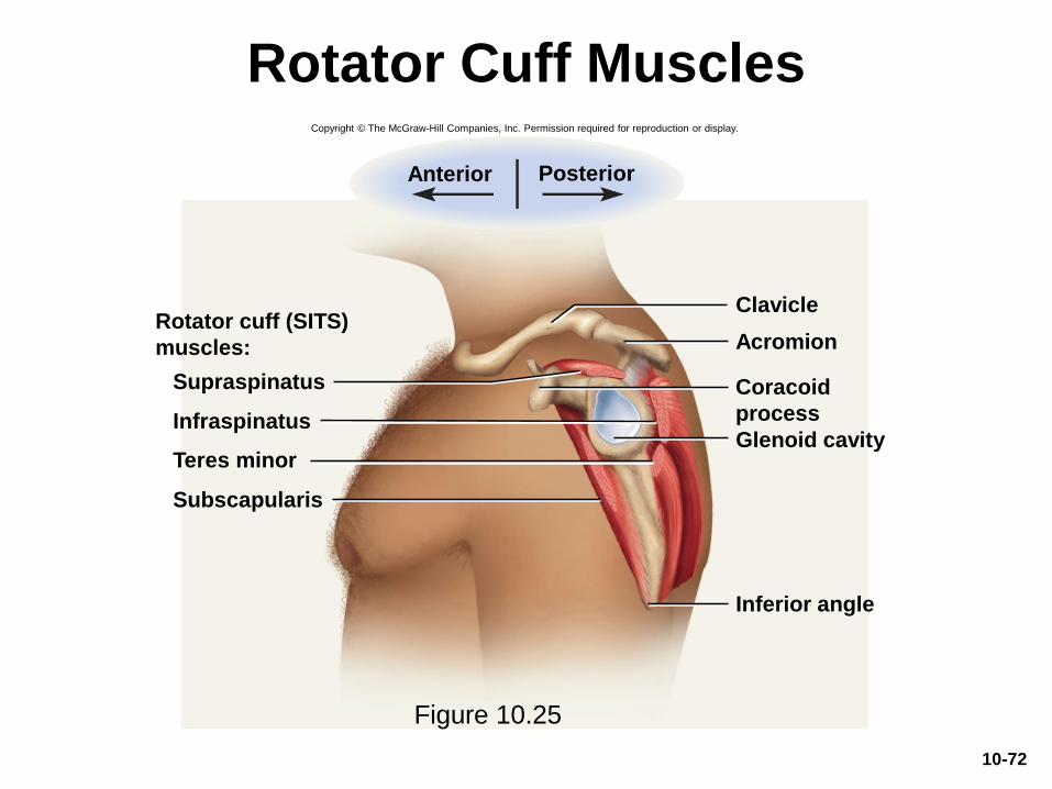
Rotator Cuff Muscles
Figure 10.25
Acromion
Inferior angle
Clavicle
Infraspinatus
Subscapularis
Glenoid cavity
Supraspinatus
Anterior Posterior
Rotator cuff (SITS)
muscles:
Teres minor
Coracoid
process
Copyright © The McGraw-Hill Companies, Inc. Permission required for reproduction or display.
10-72
Muscles Acting on the Forearm
• Elbow and forearm capable of flexion, extension,
pronation, and supination
– Carried out by muscles in both brachium (arm) and
antebrachium (forearm)
• Muscles with bellies in the arm (brachium)
– Principal elbow flexors: anterior compartment
• Brachialis and biceps brachii
– Brachialis produces 50% more power than biceps brachii
– Brachialis is prime mover of elbow flexion
– Principal elbow extensor: posterior compartment
• Triceps brachii
– Prime mover of elbow extension
10-73
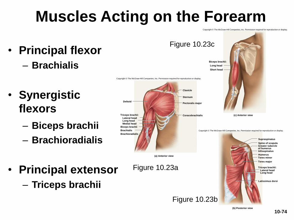
Muscles Acting on the Forearm
• Principal flexor
– Brachialis
• Synergistic
flexors
– Biceps brachii
– Brachioradialis
• Principal extensor
– Triceps brachii
Figure 10.23b
Figure 10.23a
Clavicle
Deltoid
Sternum
Pectoralis major
CoracobrachialisLateral headLong head
Medial head
Biceps brachii
Brachialis
Brachioradialis
Triceps brachii:
(a) Anterior view
Copyright © The McGraw-Hill Companies, Inc. Permission required for reproduction or display.
(b) Posterior view
Supraspinatus
Spine of scapula
Infraspinatus
Lateral headLong head
Latissimus dorsi
Humerus
Greater tubercleof humerus
Teres minor
Teres major
Triceps brachii:
Copyright © The McGraw-Hill Companies, Inc. Permission required for reproduction or display.
Biceps brachii:
Long head
Short head
(c) Anterior view
Copyright © The McGraw-Hill Companies, Inc. Permission required for reproduction or display.
Figure 10.23c
10-74
Muscles Acting on the Forearm
• Muscles with bellies in the forearm
(antebrachium)
– Brachioradialis: flexes elbow
– Anconeus: extends elbow
– Pronator quadratus: prime mover in forearm
pronation
– Pronator teres: assists pronator quadratus in
pronation
– Supinator: supinates the forearm
10-75
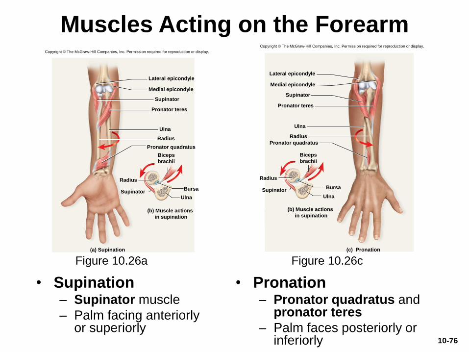
Muscles Acting on the Forearm
• Supination– Supinator muscle
– Palm facing anteriorly or superiorly
• Pronation– Pronator quadratus and
pronator teres
– Palm faces posteriorly or inferiorly
Figure 10.26a Figure 10.26c
Copyright © The McGraw-Hill Companies, Inc. Permission required for reproduction or display.
Medial epicondyle
Lateral epicondyle
Ulna
Pronator quadratus
Pronator teres
Supinator
Radius
Radius
(a) Supination
Ulna
Bursa
Biceps
brachii
Supinator
(b) Muscle actions
in supination
Copyright © The McGraw-Hill Companies, Inc. Permission required for reproduction or display.
Medial epicondyle
Lateral epicondyle
Ulna
Pronator quadratus
Pronator teres
Supinator
Radius
Radius
(c) Pronation
Ulna
Bursa
Biceps
brachii
Supinator
(b) Muscle actions
in supination
10-76
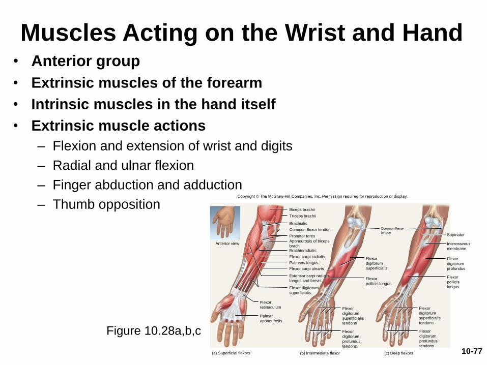
Muscles Acting on the Wrist and Hand• Anterior group
• Extrinsic muscles of the forearm
• Intrinsic muscles in the hand itself
• Extrinsic muscle actions
– Flexion and extension of wrist and digits
– Radial and ulnar flexion
– Finger abduction and adduction
– Thumb opposition
Figure 10.28a,b,c
Copyright © The McGraw-Hill Companies, Inc. Permission required for reproduction or display.
(c) Deep flexors(b) Intermediate flexor(a) Superficial flexors
Flexor
digitorum
superficialis
Flexor
digitorum
profundus
Supinator
Flexor
pollicis
longus
Biceps brachii
Brachialis
Pronator teres
Brachioradialis
Flexor carpi radialis
Palmaris longus
Flexor digitorum
superficialis
Extensor carpi radialis
longus and brevis
Triceps brachii
Common flexor tendon
Aponeurosis of biceps
brachii
Flexor carpi ulnaris
Interosseous
membrane
Flexor
pollicis longus
Flexor
digitorum
superficialis
tendons
Flexor
digitorum
profundus
tendons
Common flexor
tendon
Palmar
aponeurosis
Flexor
digitorum
superficialis
tendons
Flexor
digitorum
profundus
tendons
Flexor
retinaculum
Anterior view
10-77
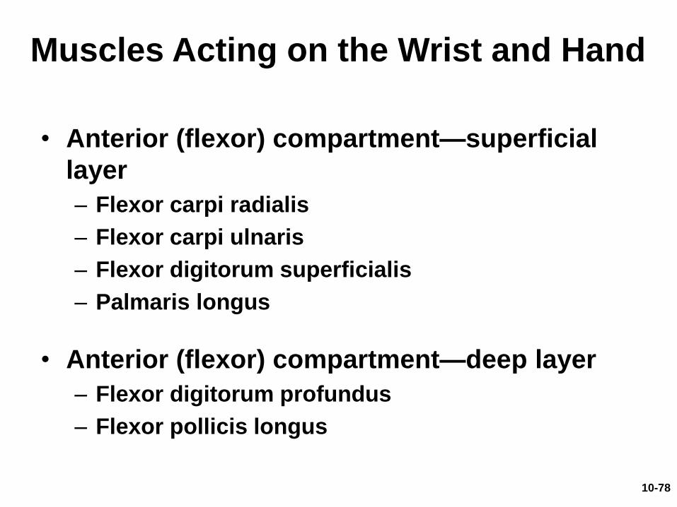
• Anterior (flexor) compartment—superficial
layer
– Flexor carpi radialis
– Flexor carpi ulnaris
– Flexor digitorum superficialis
– Palmaris longus
• Anterior (flexor) compartment—deep layer
– Flexor digitorum profundus
– Flexor pollicis longus
Muscles Acting on the Wrist and Hand
10-78
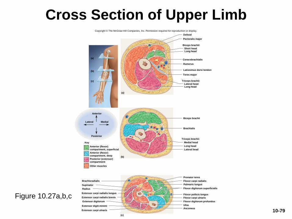
Cross Section of Upper Limb
Figure 10.27a,b,c
Copyright © The McGraw-Hill Companies, Inc. Permission required for reproduction or display.
Flexor carpi radialis
Flexor carpi ulnaris
Palmaris longus
Flexor pollicis longus
Flexor digitorum profundus
Extensor carpi radialis longus
Extensor carpi radialis brevis
Extensor digitorum
Extensor digiti minimi
Extensor carpi ulnaris
(c)
Pronator teres
Radius
Ulna
Anconeus
Brachioradialis
Flexor digitorum superficialis
Supinator
Short headLong head
Pectoralis major
Triceps brachii:
Coracobrachialis
Humerus
Deltoid
Teres major
Latissimus dorsi tendon
Biceps brachii:
Lateral headLong head
(a)
Brachialis
Biceps brachii
Triceps brachii:
Medial head
Long head
Lateral head
(b)
Anterior (flexor)
compartment, superficial
Anterior (flexor)
compartment, deep
Posterior (extensor)
compartment
Other muscles
Key
(a)
(b)
(c)
Posterior
Anterior
Lateral Medial
10-79
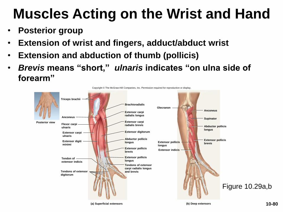
Muscles Acting on the Wrist and Hand• Posterior group
• Extension of wrist and fingers, adduct/abduct wrist
• Extension and abduction of thumb (pollicis)
• Brevis means “short,” ulnaris indicates “on ulna side of
forearm”Copyright © The McGraw-Hill Companies, Inc. Permission required for reproduction or display.
Anconeus
Supinator
Abductor pollicis
longus
Extensor pollicis
longus
Extensor pollicis
brevis
Extensor indicis
Olecranon
Tendons of extensor
digitorum
(a) Superficial extensors (b) Deep extensors
Posterior view
Extensor carpi
ulnaris
Anconeus
Triceps brachii
Flexor carpi
ulnaris
Extensor digiti
minimi
Tendon of
extensor indicis
Extensor carpi
radialis longus
Extensor carpi
radialis brevis
Extensor digitorum
Abductor pollicis
longus
Extensor pollicis
brevis
Extensor pollicis
longus
Brachioradialis
Tendons of extensor
carpi radialis longus
and brevis
Figure 10.29a,b
10-80
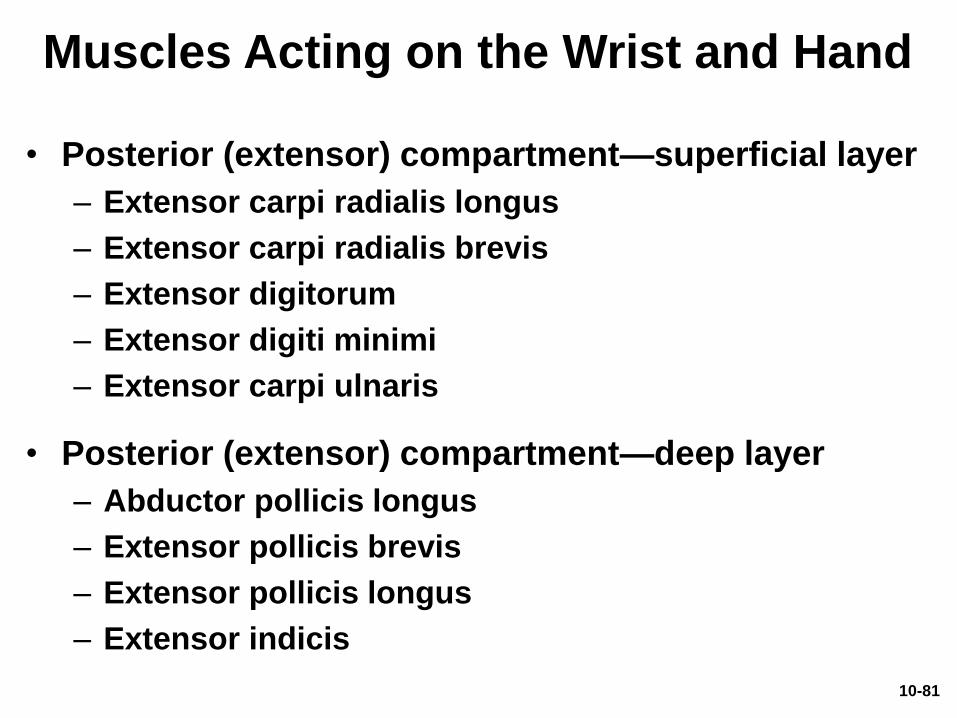
Muscles Acting on the Wrist and Hand
• Posterior (extensor) compartment—superficial layer
– Extensor carpi radialis longus
– Extensor carpi radialis brevis
– Extensor digitorum
– Extensor digiti minimi
– Extensor carpi ulnaris
• Posterior (extensor) compartment—deep layer
– Abductor pollicis longus
– Extensor pollicis brevis
– Extensor pollicis longus
– Extensor indicis
10-81
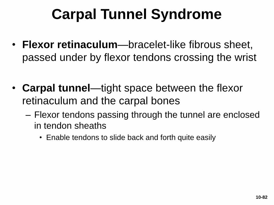
Carpal Tunnel Syndrome
• Flexor retinaculum—bracelet-like fibrous sheet,
passed under by flexor tendons crossing the wrist
• Carpal tunnel—tight space between the flexor
retinaculum and the carpal bones
– Flexor tendons passing through the tunnel are enclosed
in tendon sheaths
• Enable tendons to slide back and forth quite easily
10-82
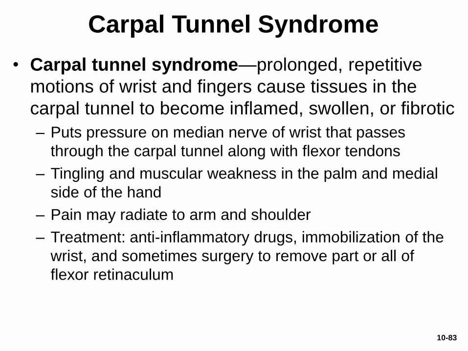
Carpal Tunnel Syndrome
• Carpal tunnel syndrome—prolonged, repetitive
motions of wrist and fingers cause tissues in the
carpal tunnel to become inflamed, swollen, or fibrotic
– Puts pressure on median nerve of wrist that passes
through the carpal tunnel along with flexor tendons
– Tingling and muscular weakness in the palm and medial
side of the hand
– Pain may radiate to arm and shoulder
– Treatment: anti-inflammatory drugs, immobilization of the
wrist, and sometimes surgery to remove part or all of
flexor retinaculum
10-83
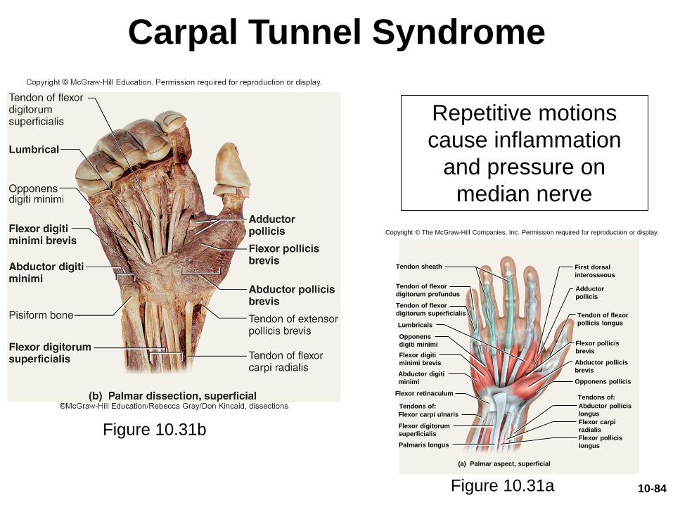
Carpal Tunnel Syndrome
Repetitive motions
cause inflammation
and pressure on
median nerve
Figure 10.31a
Figure 10.31b
Copyright © The McGraw-Hill Companies, Inc. Permission required for reproduction or display.
Opponens pollicis
Abductor pollicis
brevis
Flexor pollicis
brevis
Adductor
pollicis
Tendon of flexor
digitorum superficialis
Tendon of flexor
digitorum profundus
Palmaris longus
Flexor digitorum
superficialis
Flexor retinaculum
Abductor digiti
minimi
Flexor digiti
minimi brevis
Lumbricals
Opponens
digiti minimi
Tendon sheath
Flexor pollicis
longus
Flexor carpi
radialis
Tendon of flexor
pollicis longus
First dorsal
interosseous
Flexor carpi ulnaris
Abductor pollicis
longus
(a) Palmar aspect, superficial
Tendons of:
Tendons of:
10-84
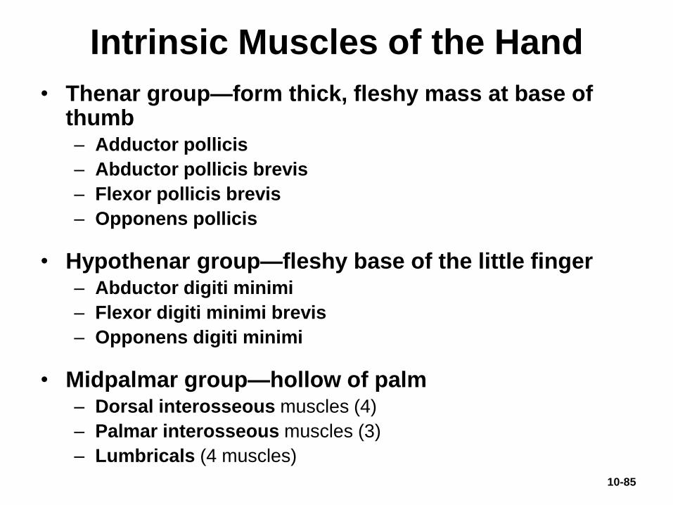
Intrinsic Muscles of the Hand
• Thenar group—form thick, fleshy mass at base of thumb– Adductor pollicis
– Abductor pollicis brevis
– Flexor pollicis brevis
– Opponens pollicis
• Hypothenar group—fleshy base of the little finger– Abductor digiti minimi
– Flexor digiti minimi brevis
– Opponens digiti minimi
• Midpalmar group—hollow of palm– Dorsal interosseous muscles (4)
– Palmar interosseous muscles (3)
– Lumbricals (4 muscles)
10-85
Muscles Acting on the Hip
and Lower Limb
• Expected Learning Outcomes
– Name and locate the muscles that act on the hip,
knee, ankle, and toe joints.
– Relate the actions of these muscles to the joint
movements described in chapter 9.
– Describe the origin, insertion, and innervation of each
muscle.
10-86
Muscles Acting on the Hip
and Lower Limb
• Body’s largest muscles found in lower limb
• Less for precision, more for strength needed
to stand, maintain balance, walk, and run
• Several cross and act on two or more joints
• Leg—the part of the limb between the knee and
ankle
• Foot—includes tarsal region (ankle),
metatarsal region, and the toes
10-87
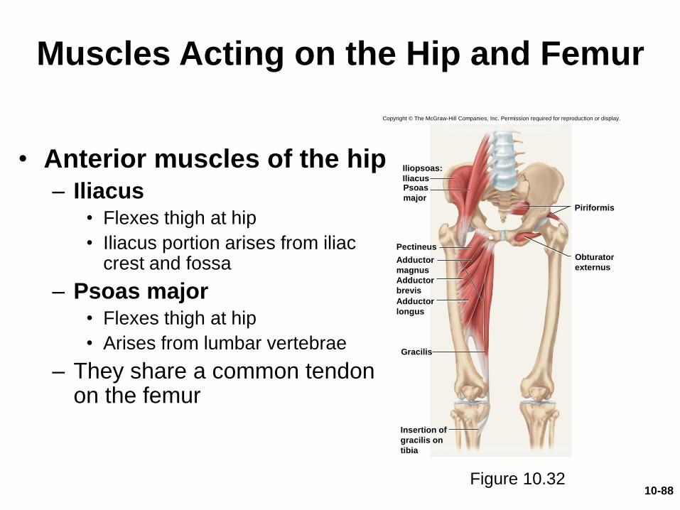
Muscles Acting on the Hip and Femur
• Anterior muscles of the hip– Iliacus
• Flexes thigh at hip
• Iliacus portion arises from iliac crest and fossa
– Psoas major• Flexes thigh at hip
• Arises from lumbar vertebrae
– They share a common tendon on the femur
Figure 10.32
Copyright © The McGraw-Hill Companies, Inc. Permission required for reproduction or display.
Iliopsoas:
IliacusPsoas
major
Adductor
longus
Adductor
brevis
Pectineus
Gracilis
Insertion of
gracilis on
tibia
Piriformis
Obturator
externusAdductor
magnus
10-88

Muscles Acting on the Hip and Femur
• Lateral and posterior muscles of the hip– Tensor fasciae latae
• Extends knee, laterally rotates knee
– Gluteus maximus• Forms mass of the buttock
• Prime hip extensor
• Provides most of lift when you climb stairs
– Gluteus medius andminimus
• Abduct and medially rotate thigh
Figure 10.3310-89
Muscles Acting on the Hip and
Femur
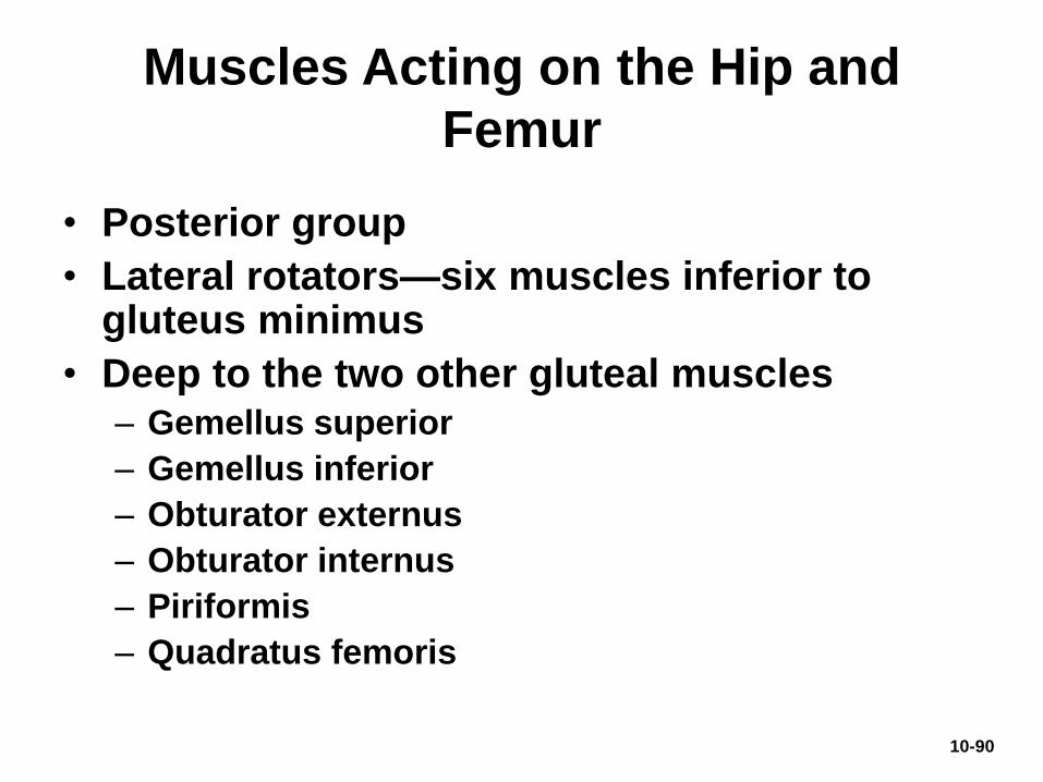
• Posterior group
• Lateral rotators—six muscles inferior to gluteus minimus
• Deep to the two other gluteal muscles– Gemellus superior
– Gemellus inferior
– Obturator externus
– Obturator internus
– Piriformis
– Quadratus femoris
10-90
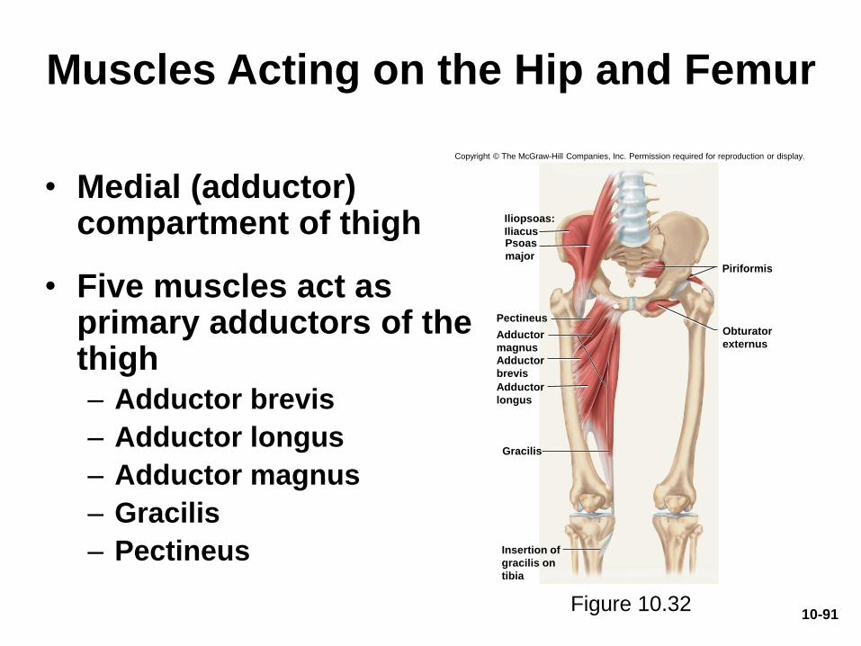
Muscles Acting on the Hip and Femur
• Medial (adductor) compartment of thigh
• Five muscles act as primary adductors of the thigh– Adductor brevis
– Adductor longus
– Adductor magnus
– Gracilis
– Pectineus
Figure 10.32
Copyright © The McGraw-Hill Companies, Inc. Permission required for reproduction or display.
Iliopsoas:
IliacusPsoas
major
Adductor
longus
Adductor
brevis
Pectineus
Gracilis
Insertion of
gracilis on
tibia
Piriformis
Obturator
externusAdductor
magnus
10-91
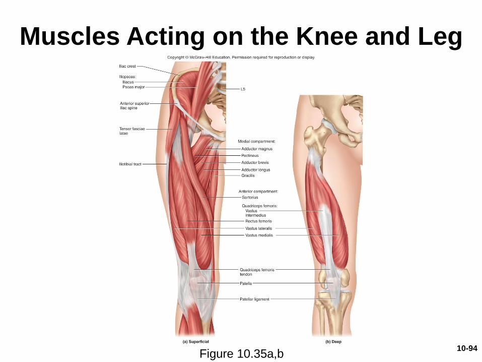
Muscles Acting on the Knee and Leg
• Anterior (extensor) compartment of the thigh
– Contains large quadriceps femoris muscle• Prime mover of knee extension
• Most powerful muscle in the body
• Has four heads—rectus femoris, vastus lateralis, vastus medialis, and vastus intermedius
– All converge on single quadriceps (patellar) tendon
– Extends to patella
– Then continues as patellar ligament
– Inserts on tibial tuberosity
– Sartorius: longest muscle in the body• “Tailor’s muscle”
10-92
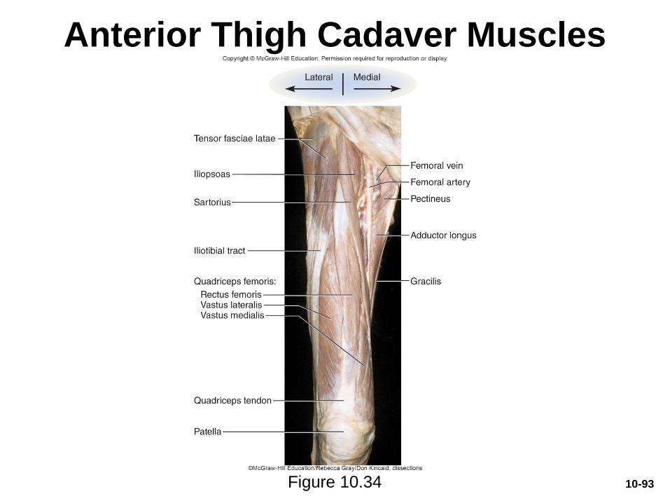
Anterior Thigh Cadaver Muscles
Figure 10.34 10-93
Muscles Acting on the Knee and Leg
Figure 10.35a,b10-94
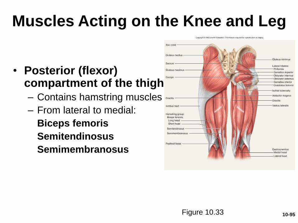
Muscles Acting on the Knee and Leg
• Posterior (flexor) compartment of the thigh– Contains hamstring muscles
– From lateral to medial:
Biceps femoris
Semitendinosus
Semimembranosus
Figure 10.33 10-95
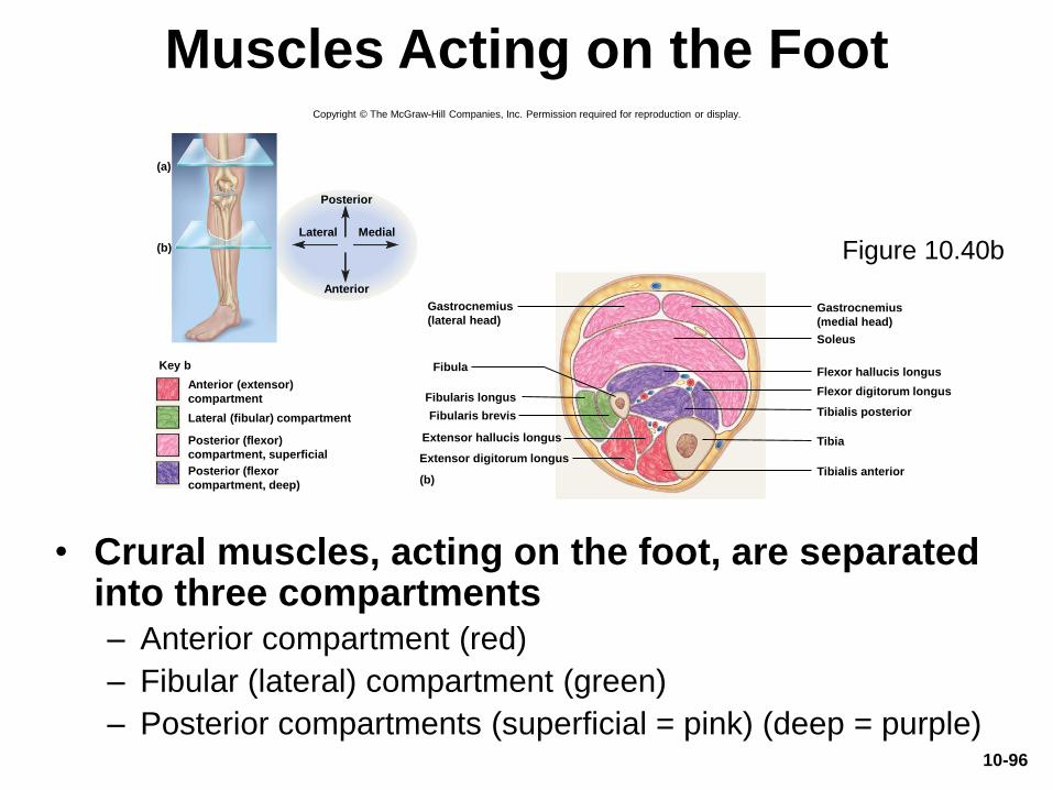
Muscles Acting on the Foot
• Crural muscles, acting on the foot, are separated into three compartments– Anterior compartment (red)
– Fibular (lateral) compartment (green)
– Posterior compartments (superficial = pink) (deep = purple)
Figure 10.40b
Copyright © The McGraw-Hill Companies, Inc. Permission required for reproduction or display.
(a)
(b)
Fibularis longus
Fibularis brevis
(b)
Gastrocnemius
(medial head)
Gastrocnemius
(lateral head)
Fibula
Extensor hallucis longus
Extensor digitorum longus
Soleus
Flexor hallucis longus
Tibialis posterior
Tibia
Tibialis anterior
Flexor digitorum longusAnterior (extensor)
compartment
Lateral (fibular) compartment
Posterior (flexor)
compartment, superficial
Posterior (flexor
compartment, deep)
Key b
Anterior
Posterior
Lateral Medial
10-96
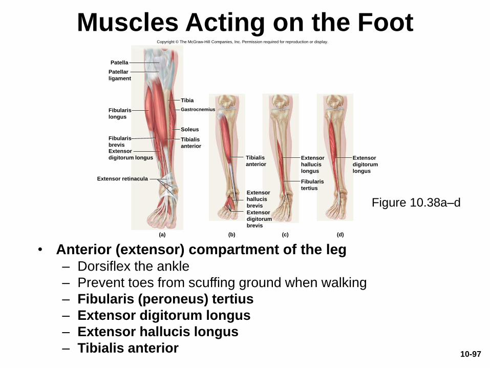
Muscles Acting on the Foot
• Anterior (extensor) compartment of the leg– Dorsiflex the ankle
– Prevent toes from scuffing ground when walking
– Fibularis (peroneus) tertius
– Extensor digitorum longus
– Extensor hallucis longus
– Tibialis anterior
Copyright © The McGraw-Hill Companies, Inc. Permission required for reproduction or display.
Tibialis
anterior
Extensor
digitorum
brevis
Extensor
hallucis
brevis
Fibularis
tertius
Extensor
digitorum
longus
Tibialis
anterior
Fibularis
longus
Extensor
digitorum longus
Fibularis
brevis
Patella
Patellar
ligament
Gastrocnemius
Soleus
Tibia
Extensor retinacula
Extensor
hallucis
longus
(a) (b) (c) (d)
Figure 10.38a–d
10-97
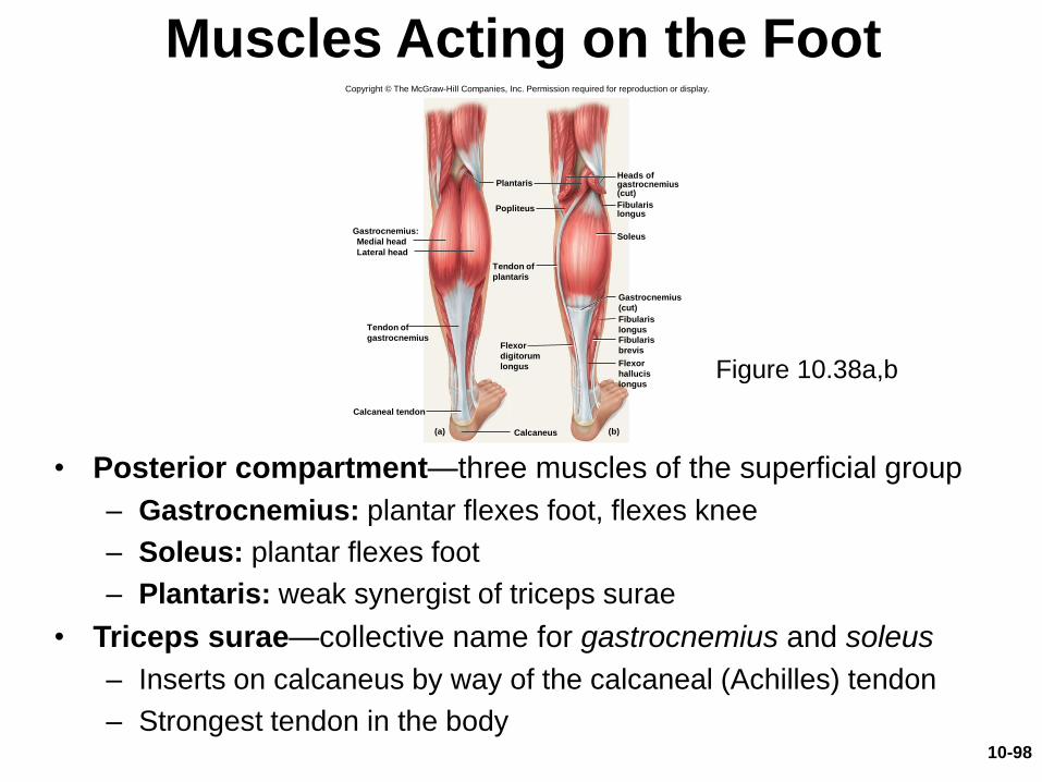
Muscles Acting on the Foot
• Posterior compartment—three muscles of the superficial group
– Gastrocnemius: plantar flexes foot, flexes knee
– Soleus: plantar flexes foot
– Plantaris: weak synergist of triceps surae
• Triceps surae—collective name for gastrocnemius and soleus
– Inserts on calcaneus by way of the calcaneal (Achilles) tendon
– Strongest tendon in the body
Copyright © The McGraw-Hill Companies, Inc. Permission required for reproduction or display.
Gastrocnemius:
Heads ofgastrocnemius(cut)
Calcaneal tendon
Calcaneus
Medial head
Lateral head
Popliteus
Flexor
digitorum
longus
Plantaris
Soleus
Flexor
hallucis
longus
Tendon of
plantaris
Fibularis
brevis
Fibularis
longus
Fibularislongus
Gastrocnemius
(cut)
Tendon of
gastrocnemius
(a) (b)
Figure 10.38a,b
10-98
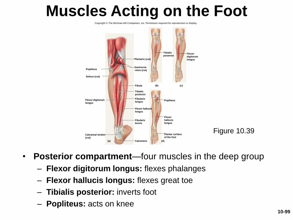
Muscles Acting on the Foot
• Posterior compartment—four muscles in the deep group
– Flexor digitorum longus: flexes phalanges
– Flexor hallucis longus: flexes great toe
– Tibialis posterior: inverts foot
– Popliteus: acts on knee
Copyright © The McGraw-Hill Companies, Inc. Permission required for reproduction or display.
Calcaneal tendon
(cut)
Calcaneus(a)
(b) (c)
(d)
Flexor digitorum
longus
Flexor
digitorum
longus
Flexor hallucis
longus
Flexor
hallucis
longus
Popliteus
Popliteus
Plantaris (cut)
Soleus (cut)
Fibula
Fibularis
longus
Fibularis
brevis
Gastrocne
-mius (cut)
Plantar surface
of the foot
Tibialis
posterior
Tibialis
posterior
Figure 10.39
10-99
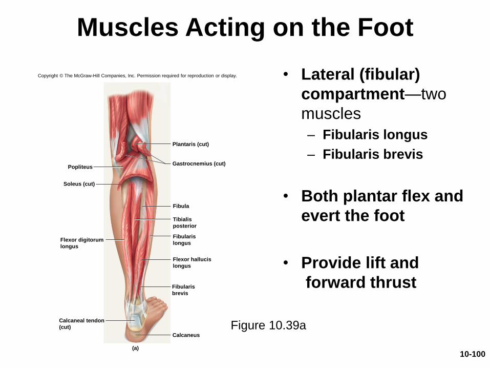
Muscles Acting on the Foot
• Lateral (fibular)
compartment—two
muscles
– Fibularis longus
– Fibularis brevis
• Both plantar flex and
evert the foot
• Provide lift and
forward thrust
Copyright © The McGraw-Hill Companies, Inc. Permission required for reproduction or display.
Calcaneal tendon
(cut)
Calcaneus
(a)
Flexor digitorum
longus
Flexor hallucis
longus
Popliteus
Plantaris (cut)
Soleus (cut)
Fibula
Fibularis
longus
Fibularis
brevis
Gastrocnemius (cut)
Tibialis
posterior
Figure 10.39a
10-100
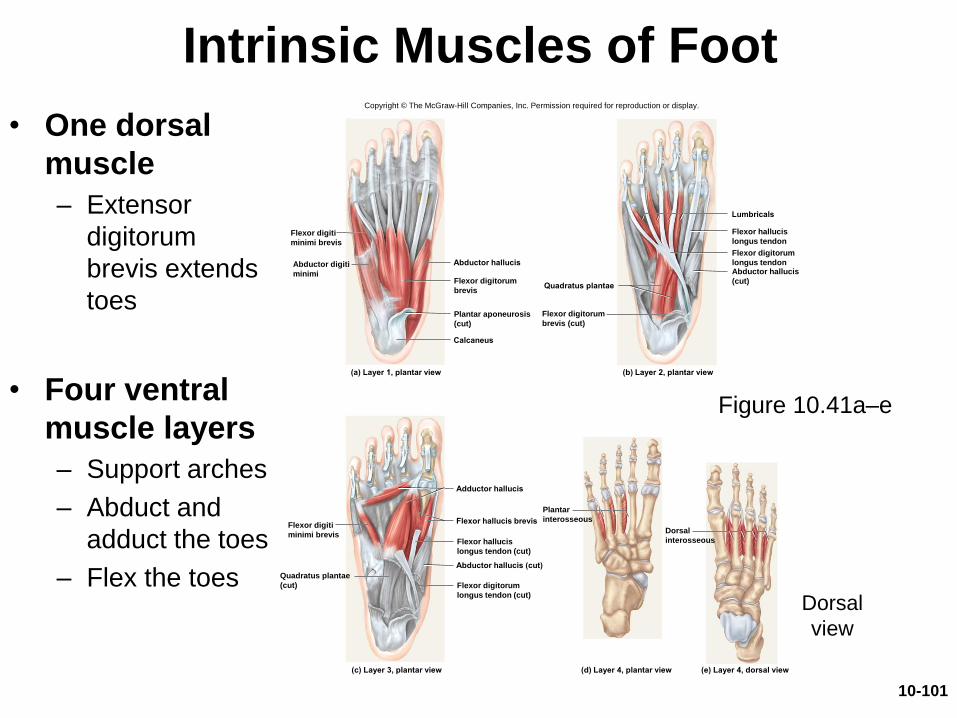
Intrinsic Muscles of Foot
• One dorsal
muscle
– Extensor
digitorum
brevis extends
toes
• Four ventral
muscle layers
– Support arches
– Abduct and
adduct the toes
– Flex the toes
Figure 10.41a–e
Dorsal
view
(a) Layer 1, plantar view (b) Layer 2, plantar view
Abductor hallucis
Calcaneus
Lumbricals
Quadratus plantae
(c) Layer 3, plantar view (d) Layer 4, plantar view (e) Layer 4, dorsal view
Adductor hallucis
Flexor hallucis brevis
Abductor hallucis (cut)
Flexor digiti
minimi brevis
Abductor digiti
minimiFlexor digitorum
brevis
Plantar aponeurosis
(cut)
Flexor digitorum
brevis (cut)
Flexor hallucis
longus tendon
Flexor digitorum
longus tendonAbductor hallucis
(cut)
Flexor hallucis
longus tendon (cut)
Flexor digitorum
longus tendon (cut)
Plantar
interosseous
Dorsal
interosseous
Copyright © The McGraw-Hill Companies, Inc. Permission required for reproduction or display.
Flexor digiti
minimi brevis
Quadratus plantae
(cut)
10-101
Common Athletic Injuries
• Muscles and tendons are vulnerable to sudden and intense stress
• Proper conditioning and warm-up needed
• Common injuries include:– Compartment syndrome
– Shin splints
– Pulled hamstrings
– Tennis elbow
– Pulled groin
– Rotator cuff injury
• Treat with rest, ice, compression, and elevation
• “No pain, no gain” is a dangerous misconception
10-102