chapter 11 218/martini ppt... · introduction •two major groups of appendicular muscles •the...
TRANSCRIPT
Lecture Presentation by
Steven Bassett
Southeast Community College
Chapter 11
The Muscular
System
Appendicular
Musculature
© 2015 Pearson Education, Inc.
Introduction
• Appendicular Musculature
• Appendicular muscles are responsible for:
• Stabilizing the pectoral and pelvic girdles
• Moving the upper and lower limbs
• Absorbing shocks and jolts as you walk, run, or
jump
• Aiding in strengthening the joint area
© 2015 Pearson Education, Inc.
Introduction
• Two Major Groups of Appendicular Muscles
• The muscles of the pectoral girdle and upper limbs
• The muscles of the pelvic girdle and lower limbs
© 2015 Pearson Education, Inc.
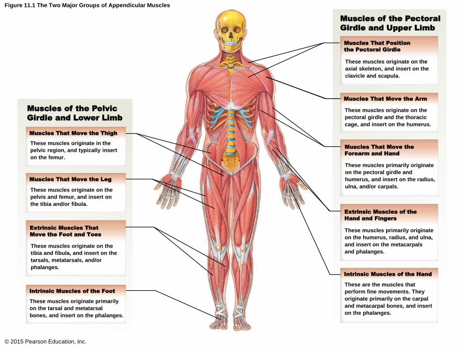
Figure 11.1 The Two Major Groups of Appendicular Muscles
© 2015 Pearson Education, Inc.
Muscles of the Pectoral
Girdle and Upper Limb
Muscles of the Pelvic
Girdle and Lower Limb
Muscles That Move the Thigh
Muscles That Move the Leg
Extrinsic Muscles That
Move the Foot and Toes
Intrinsic Muscles of the Foot
Muscles That Position
the Pectoral Girdle
Muscles That Move the Arm
Muscles That Move the
Forearm and Hand
Extrinsic Muscles of the
Hand and Fingers
Intrinsic Muscles of the Hand
These muscles originate on the
axial skeleton, and insert on the
clavicle and scapula.
These muscles originate on the
pectoral girdle and the thoracic
cage, and insert on the humerus.
These muscles primarily originate
on the pectoral girdle and
humerus, and insert on the radius,
ulna, and/or carpals.
These muscles primarily originate
on the humerus, radius, and ulna,
and insert on the metacarpals
and phalanges.
These are the muscles that
perform fine movements. They
originate primarily on the carpal
and metacarpal bones, and insert
on the phalanges.
These muscles originate in the
pelvic region, and typically insert
on the femur.
These muscles originate on the
pelvis and femur, and insert on
the tibia and/or fibula.
These muscles originate on the
tibia and fibula, and insert on the
tarsals, metatarsals, and/or
phalanges.
These muscles originate primarily
on the tarsal and metatarsal
bones, and insert on the phalanges.
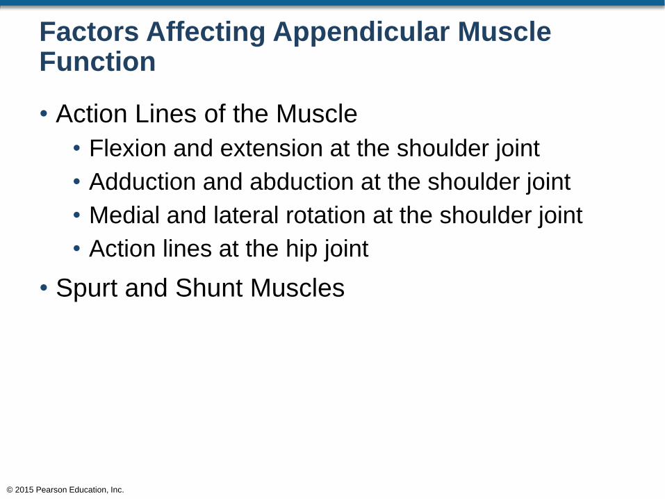
Factors Affecting Appendicular Muscle Function
• Action Lines of the Muscle
• Flexion and extension at the shoulder joint
• Adduction and abduction at the shoulder joint
• Medial and lateral rotation at the shoulder joint
• Action lines at the hip joint
• Spurt and Shunt Muscles
© 2015 Pearson Education, Inc.
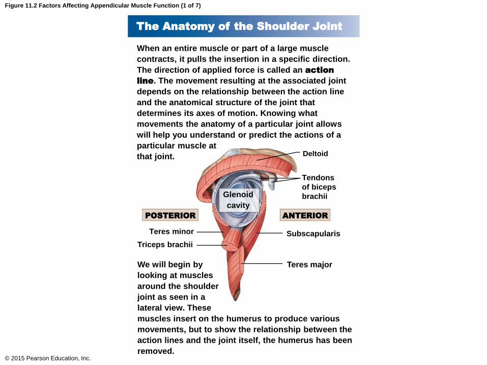
Figure 11.2 Factors Affecting Appendicular Muscle Function (1 of 7)
© 2015 Pearson Education, Inc.
The Anatomy of the Shoulder Joint
When an entire muscle or part of a large muscle
contracts, it pulls the insertion in a specific direction.
The direction of applied force is called an action
line. The movement resulting at the associated joint
depends on the relationship between the action line
and the anatomical structure of the joint that
determines its axes of motion. Knowing what
movements the anatomy of a particular joint allows
will help you understand or predict the actions of a
particular muscle at
that joint.
We will begin by
looking at muscles
around the shoulder
joint as seen in a
lateral view. These
muscles insert on the humerus to produce various
movements, but to show the relationship between the
action lines and the joint itself, the humerus has been
removed.
Deltoid
Tendons
of biceps
brachii
Subscapularis
Teres major
Teres minor
Triceps brachii
Glenoid
cavity
ANTERIORPOSTERIOR
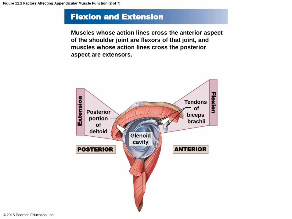
Figure 11.2 Factors Affecting Appendicular Muscle Function (2 of 7)
© 2015 Pearson Education, Inc.
Flexion and Extension
Muscles whose action lines cross the anterior aspect
of the shoulder joint are flexors of that joint, and
muscles whose action lines cross the posterior
aspect are extensors.
POSTERIOR ANTERIOR
Tendons
of
biceps
brachii
Posterior
portion
of
deltoidGlenoid
cavity
Extensio
nF
lexion
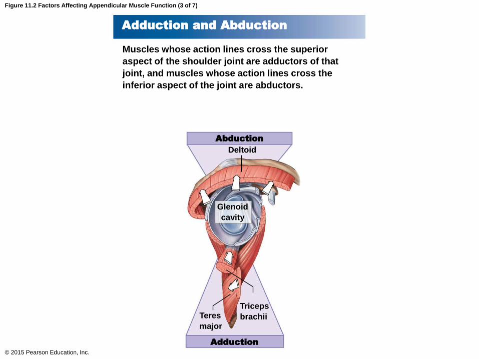
Figure 11.2 Factors Affecting Appendicular Muscle Function (3 of 7)
© 2015 Pearson Education, Inc.
Adduction and Abduction
Muscles whose action lines cross the superior
aspect of the shoulder joint are adductors of that
joint, and muscles whose action lines cross the
inferior aspect of the joint are abductors.
Abduction
Adduction
Deltoid
Glenoid
cavity
Triceps
brachiiTeres
major
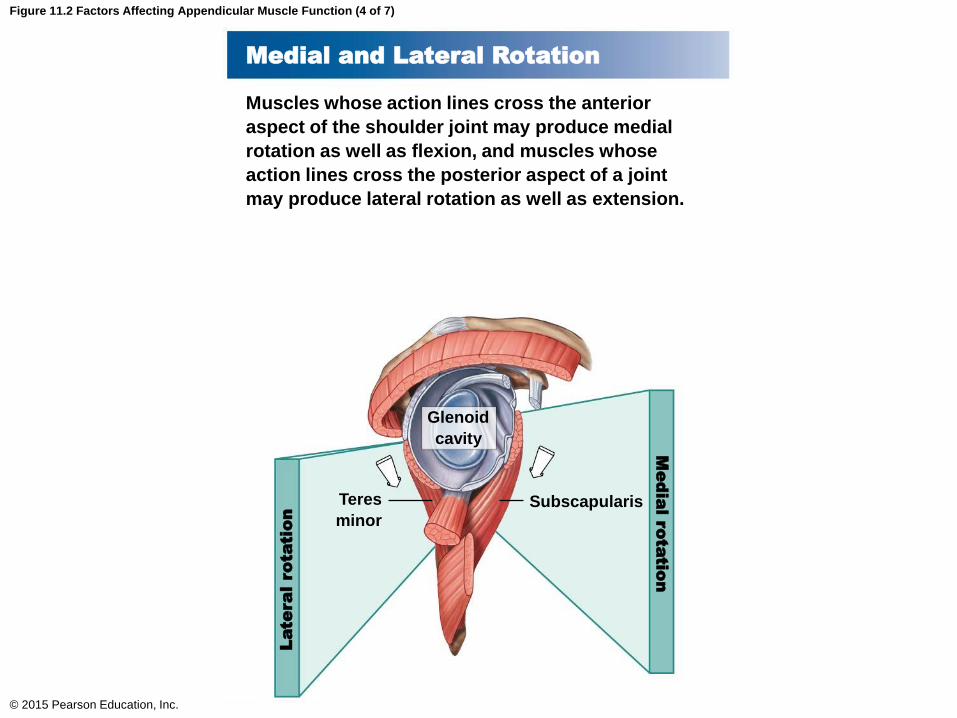
Figure 11.2 Factors Affecting Appendicular Muscle Function (4 of 7)
© 2015 Pearson Education, Inc.
Medial and Lateral Rotation
Muscles whose action lines cross the anterior
aspect of the shoulder joint may produce medial
rotation as well as flexion, and muscles whose
action lines cross the posterior aspect of a joint
may produce lateral rotation as well as extension.
Glenoid
cavity
SubscapularisTeres
minor
Medial rotatio
nLateral rotatio
n
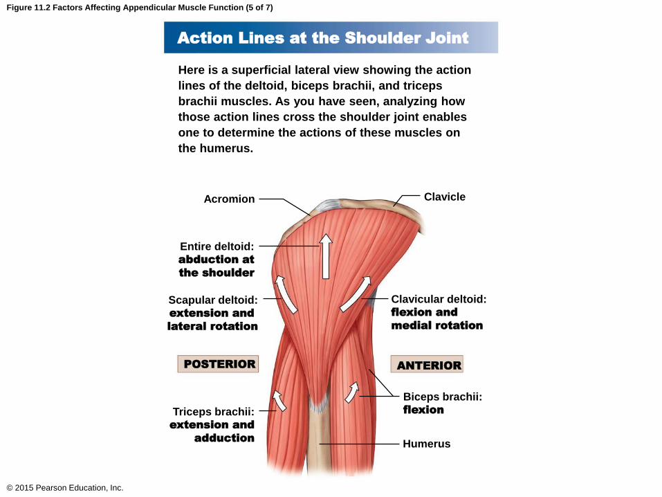
Figure 11.2 Factors Affecting Appendicular Muscle Function (5 of 7)
© 2015 Pearson Education, Inc.
Action Lines at the Shoulder Joint
Here is a superficial lateral view showing the action
lines of the deltoid, biceps brachii, and triceps
brachii muscles. As you have seen, analyzing how
those action lines cross the shoulder joint enables
one to determine the actions of these muscles on
the humerus.
Acromion Clavicle
Clavicular deltoid:flexion and
medial rotation
Entire deltoid:abduction at
the shoulder
Scapular deltoid:extension and
lateral rotation
Triceps brachii:extension and
adduction
Biceps brachii:flexion
Humerus
POSTERIOR ANTERIOR

Figure 11.2 Factors Affecting Appendicular Muscle Function (7 of 7)
© 2015 Pearson Education, Inc.
Action Lines at the Hip Joint
The hip joint, like the shoulder joint, is a multiaxial synovial joint that permits flexion/extension,
adduction/abduction, and medial/lateral rotation. Determining the action of a muscle on the hip
is identical to the process utilized for the shoulder, in that the action of a muscle on the hip is
determined by the structure of the joint and the location of the insertion of the muscle on the
femur (not shown) relative to the permitted axes of motion at the joint.
Adductor
magnus
Iliopsoas: flexion
Gluteus medius and
minimus: abduction
Obturator externus:
lateral rotation
Tensor fasciae latae:
medial rotation
Adductor longus:
adduction
Hamstring group:
extension and
lateral rotation
Action lines of the adductor magnus
Gluteal Group
ExtensionExtension and
abduction
Flexion,
abduction, and
medial rotation
Lateral Rotator GroupAdductor Group
Extension and
lateral rotationAdduction
ANTERIOR POSTERIOR
Gluteus
maximus
Gluteus medius
Gluteus minimus
Tensor
fasciae
latae
Acetabulum
Hamstring
groupAdductor
longus
Adductor magnus

Figure 11.2 Factors Affecting Appendicular Muscle Function (6 of 7)
© 2015 Pearson Education, Inc.
Spurt and Shunt Muscles
Determining the location of the insertion of a muscle
relative to the axis of the joint will provide additional
details about the functions of the muscle at that joint.
The primary action of a muscle whose insertion is
close to the joint will be the production of movement
at that joint. Such a muscle is termed a spurt
muscle, and spurt muscles are prime movers.
However, a muscle whose insertion is considerably
farther from the joint will generally help to stabilize
that joint in addition to producing motion at that
joint. This type of muscle, a synergist, is termed
a shunt muscle.
Biceps brachii:movement
and torque
(spurt muscle)Triceps brachii:movement
and torque
(spurt muscle)Brachioradialis:
stability and
movement
(shunt muscle)
Extensors
Flexors
Elbow joint
(monaxial)
Muscles of the Pectoral Girdle and Upper Limbs
• Muscles associated with the pectoral girdle and
upper limbs can be divided into four groups
• Muscles that position the pectoral girdle
• Muscles that move the arm
• Muscles that move the forearm and hand
• Muscles that move the hand and fingers
© 2015 Pearson Education, Inc.
Muscles of the Pectoral Girdle and Upper Limbs
• Muscles That Position the Pectoral Girdle
• These muscles also coordinate with the muscles
that move the arm
• Levator scapulae
• Elevates the scapula
• Pectoralis minor
• Protracts the shoulder
© 2015 Pearson Education, Inc.

Figure 11.6 Muscles That Position the Pectoral Girdle, Part II
© 2015 Pearson Education, Inc.
Muscles That
Position the
Pectoral Girdle
Levator scapulae
Trapezius
Subclavius
Pectoralis minor
Serratus anterior
Internal
intercostals
External
intercostals
T12
Short head
Long head
Biceps
brachii
Serratus anterior
Coracobrachialis
Pectoralis minor (cut)
Serratus
anterior
(insertion)
Serratus
anterior
(origin)
Pectoralis
minor
Trapezius
Pectoralis
major
Subclavius
Pectoralis
major (cut
and reflected)
Origin
Insertion
Muscles of the Pectoral Girdle and Upper Limbs
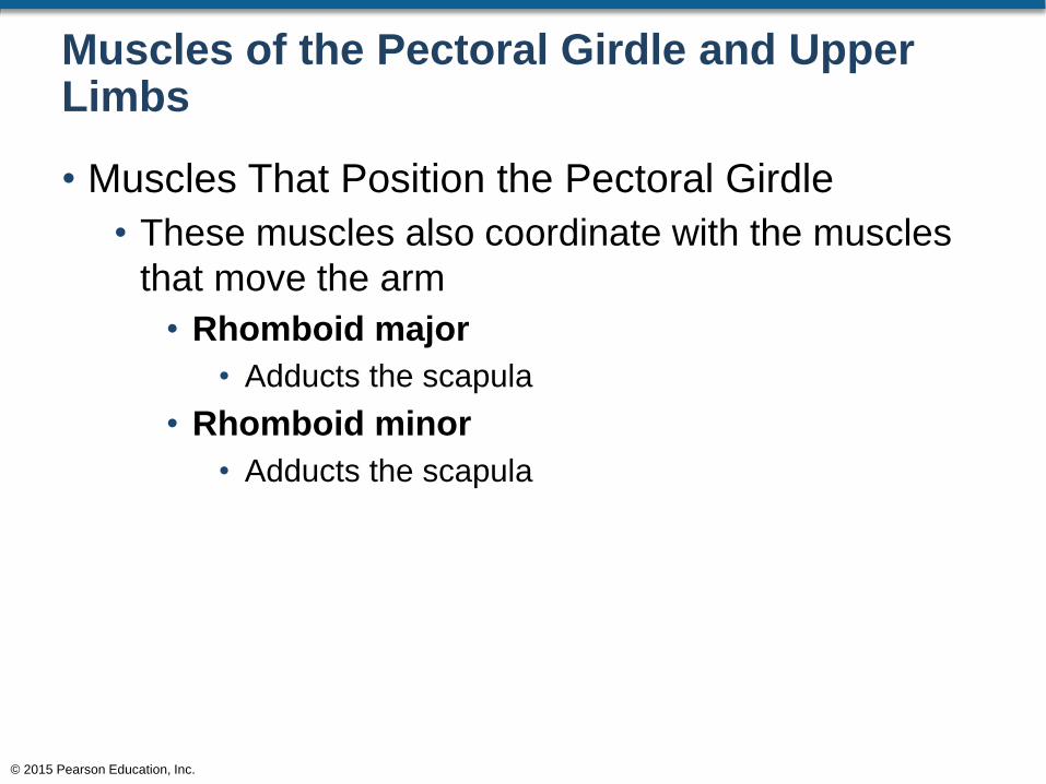
• Muscles That Position the Pectoral Girdle
• These muscles also coordinate with the muscles
that move the arm
• Rhomboid major
• Adducts the scapula
• Rhomboid minor
• Adducts the scapula
© 2015 Pearson Education, Inc.
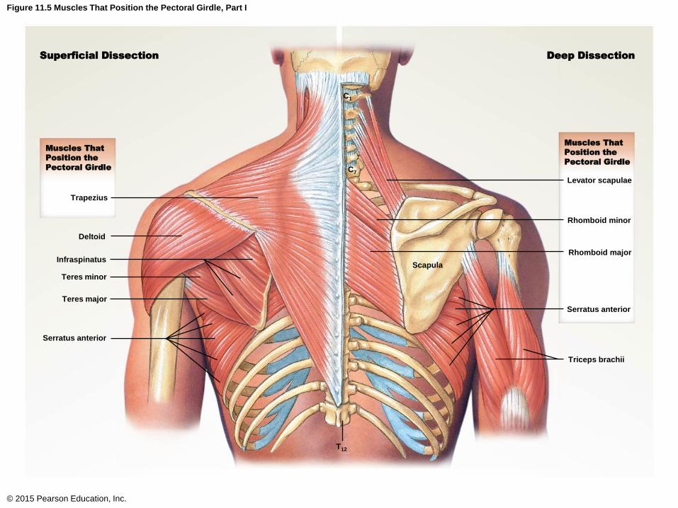
Figure 11.5 Muscles That Position the Pectoral Girdle, Part I
© 2015 Pearson Education, Inc.
Superficial Dissection Deep Dissection
Muscles That
Position the
Pectoral Girdle
Muscles That
Position the
Pectoral Girdle
Levator scapulae
Rhomboid minor
Rhomboid major
Serratus anterior
Triceps brachii
Scapula
C1
C7
Trapezius
Deltoid
Infraspinatus
Teres minor
Teres major
Serratus anterior
T12
Muscles of the Pectoral Girdle and Upper Limbs
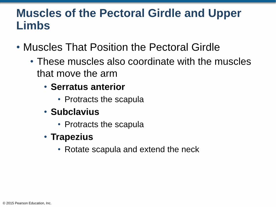
• Muscles That Position the Pectoral Girdle
• These muscles also coordinate with the muscles
that move the arm
• Serratus anterior
• Protracts the scapula
• Subclavius
• Protracts the scapula
• Trapezius
• Rotate scapula and extend the neck
© 2015 Pearson Education, Inc.
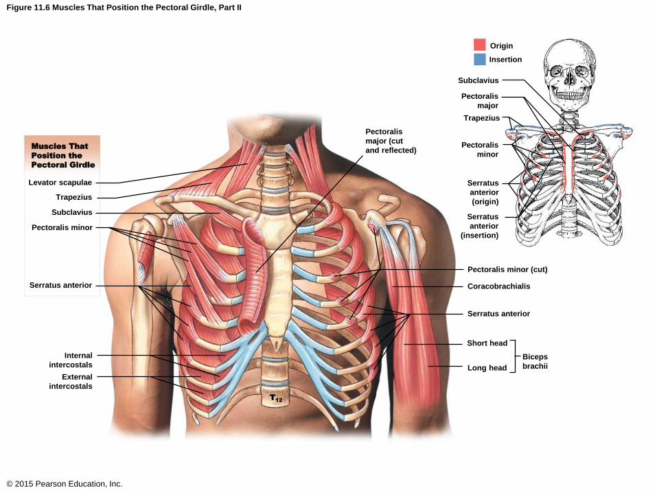
Figure 11.6 Muscles That Position the Pectoral Girdle, Part II
© 2015 Pearson Education, Inc.
Muscles That
Position the
Pectoral Girdle
Levator scapulae
Trapezius
Subclavius
Pectoralis minor
Serratus anterior
Internal
intercostals
External
intercostals
T12
Short head
Long head
Biceps
brachii
Serratus anterior
Coracobrachialis
Pectoralis minor (cut)
Serratus
anterior
(insertion)
Serratus
anterior
(origin)
Pectoralis
minor
Trapezius
Pectoralis
major
Subclavius
Pectoralis
major (cut
and reflected)
Origin
Insertion

Muscles of the Pectoral Girdleand Upper Limbs
© 2015 Pearson Education, Inc.
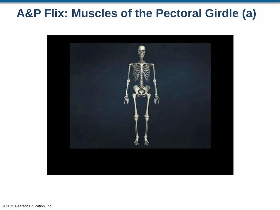
A&P Flix: Muscles of the Pectoral Girdle (a)
© 2015 Pearson Education, Inc.
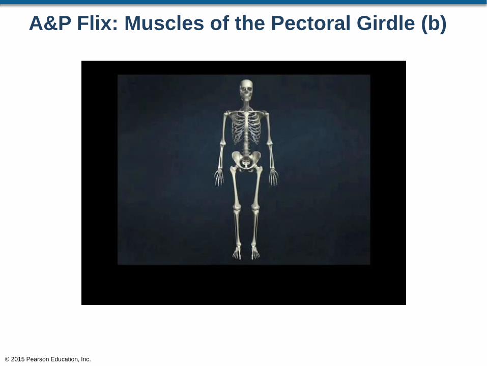
A&P Flix: Muscles of the Pectoral Girdle (b)
© 2015 Pearson Education, Inc.
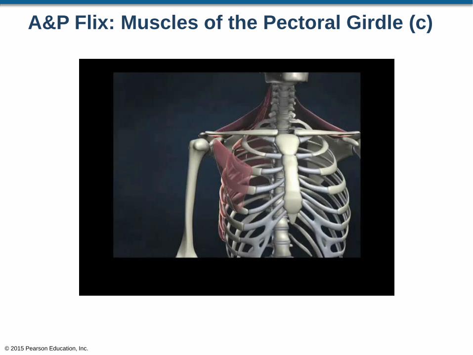
A&P Flix: Muscles of the Pectoral Girdle (c)
© 2015 Pearson Education, Inc.
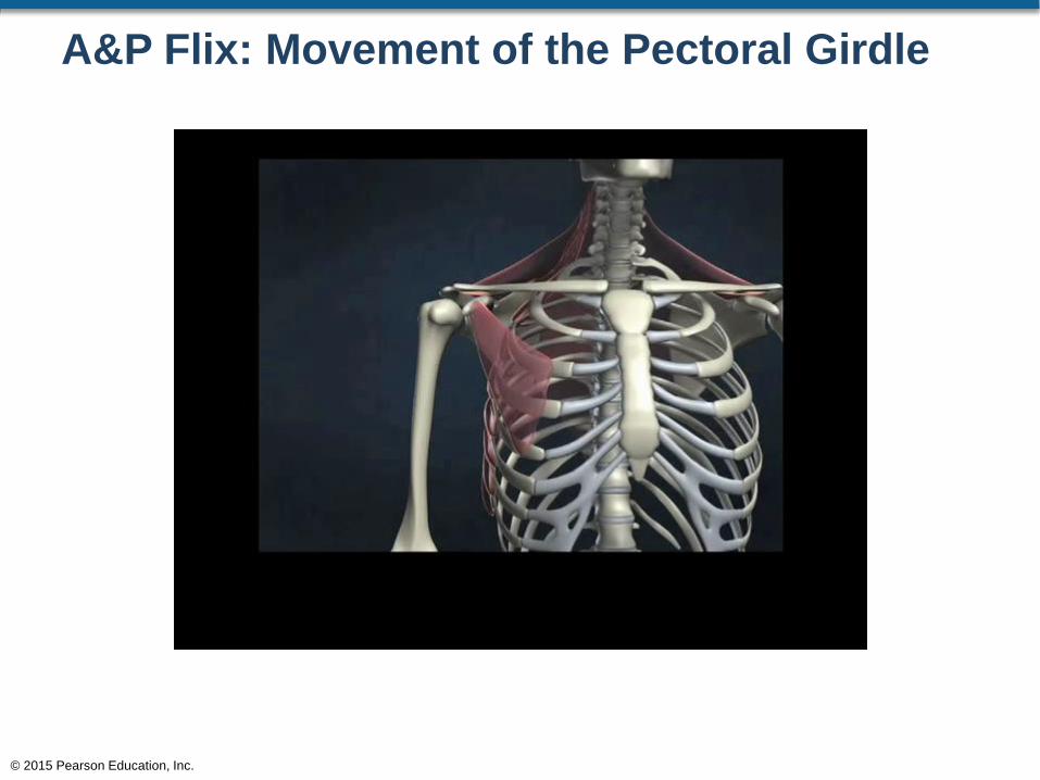
A&P Flix: Movement of the Pectoral Girdle
© 2015 Pearson Education, Inc.
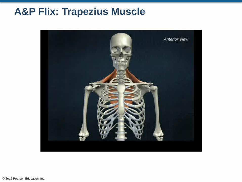
A&P Flix: Trapezius Muscle
© 2015 Pearson Education, Inc.
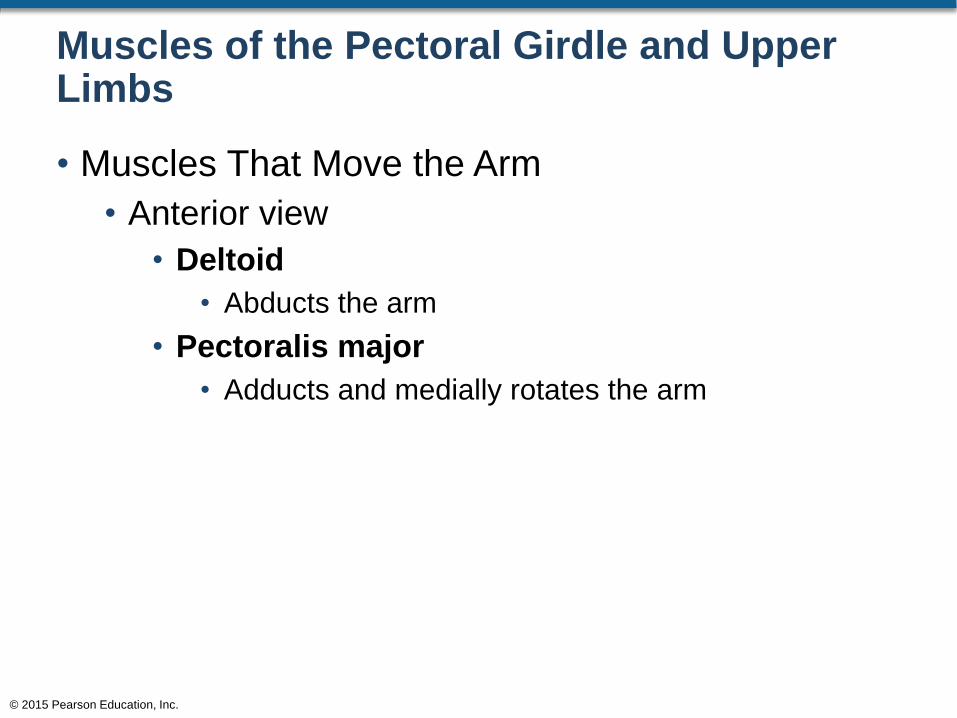
Muscles of the Pectoral Girdle and Upper Limbs
• Muscles That Move the Arm
• Anterior view
• Deltoid
• Abducts the arm
• Pectoralis major
• Adducts and medially rotates the arm
© 2015 Pearson Education, Inc.

Figure 11.7a Muscles That Move the Arm
© 2015 Pearson Education, Inc.
Superficial Dissection Deep Dissection
Muscles That
Move the Arm
Muscles That
Move the Arm
Ribs (cut)Clavicle
Deltoid
Pectoralis
major
Sternum
Subscapularis
Coracobrachialis
Teres major
Biceps brachii,
short head
Biceps brachii,
long head
Anterior view.
T12
a
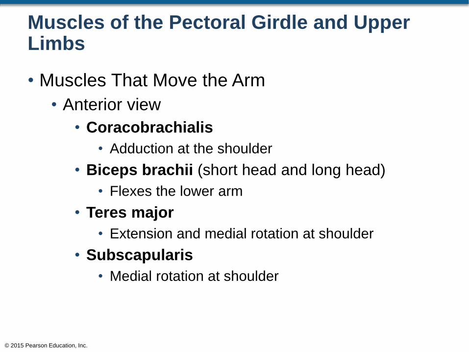
Muscles of the Pectoral Girdle and Upper Limbs
• Muscles That Move the Arm
• Anterior view
• Coracobrachialis
• Adduction at the shoulder
• Biceps brachii (short head and long head)
• Flexes the lower arm
• Teres major
• Extension and medial rotation at shoulder
• Subscapularis
• Medial rotation at shoulder
© 2015 Pearson Education, Inc.
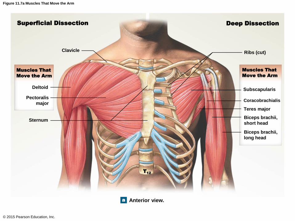
Figure 11.7a Muscles That Move the Arm
© 2015 Pearson Education, Inc.
Superficial Dissection Deep Dissection
Muscles That
Move the Arm
Muscles That
Move the Arm
Ribs (cut)Clavicle
Deltoid
Pectoralis
major
Sternum
Subscapularis
Coracobrachialis
Teres major
Biceps brachii,
short head
Biceps brachii,
long head
Anterior view.
T12
a
Muscles of the Pectoral Girdle and Upper Limbs
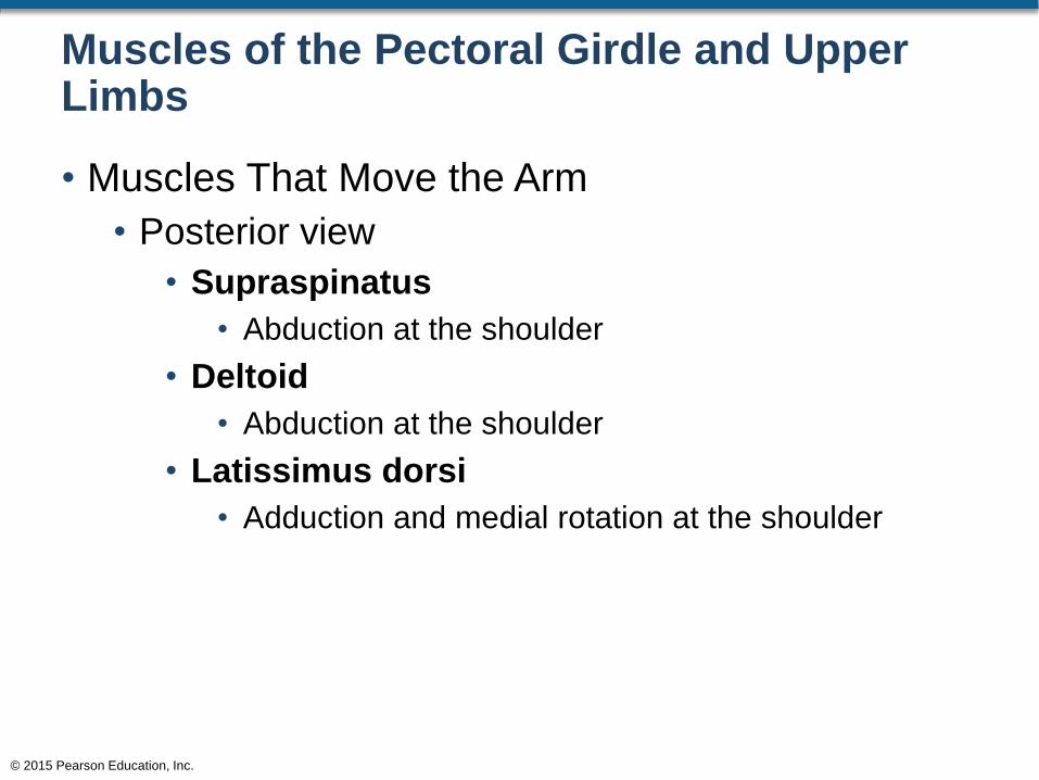
• Muscles That Move the Arm
• Posterior view
• Supraspinatus
• Abduction at the shoulder
• Deltoid
• Abduction at the shoulder
• Latissimus dorsi
• Adduction and medial rotation at the shoulder
© 2015 Pearson Education, Inc.
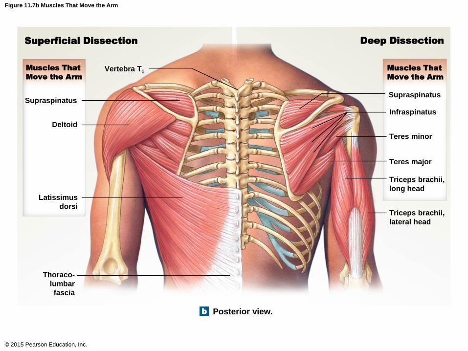
Figure 11.7b Muscles That Move the Arm
© 2015 Pearson Education, Inc.
Superficial Dissection Deep Dissection
Muscles That
Move the Arm
Muscles That
Move the Arm
b Posterior view.
Triceps brachii,
lateral head
Triceps brachii,
long head
Teres major
Teres minor
Infraspinatus
Supraspinatus
Vertebra T1
Supraspinatus
Deltoid
Latissimus
dorsi
Thoraco-
lumbar
fascia
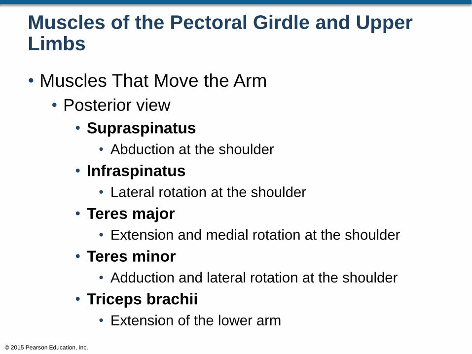
Muscles of the Pectoral Girdle and Upper Limbs
• Muscles That Move the Arm
• Posterior view
• Supraspinatus
• Abduction at the shoulder
• Infraspinatus
• Lateral rotation at the shoulder
• Teres major
• Extension and medial rotation at the shoulder
• Teres minor
• Adduction and lateral rotation at the shoulder
• Triceps brachii
• Extension of the lower arm
© 2015 Pearson Education, Inc.

Figure 11.7b Muscles That Move the Arm
© 2015 Pearson Education, Inc.
Superficial Dissection Deep Dissection
Muscles That
Move the Arm
Muscles That
Move the Arm
b Posterior view.
Triceps brachii,
lateral head
Triceps brachii,
long head
Teres major
Teres minor
Infraspinatus
Supraspinatus
Vertebra T1
Supraspinatus
Deltoid
Latissimus
dorsi
Thoraco-
lumbar
fascia
Muscles of the Pectoral Girdle and Upper Limbs
• Action Line of Muscles
• When a muscle contracts, it develops tension
• The direction the muscle moves upon developing
tension is known as the action line
© 2015 Pearson Education, Inc.
Muscles of the Pectoral Girdle and Upper Limbs
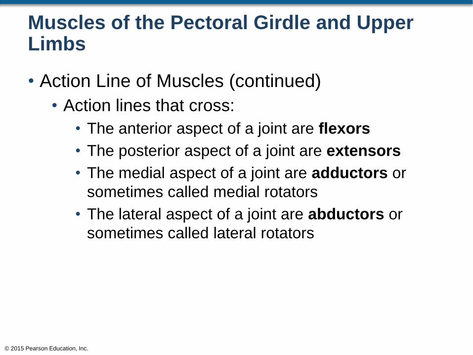
• Action Line of Muscles (continued)
• Action lines that cross:
• The anterior aspect of a joint are flexors
• The posterior aspect of a joint are extensors
• The medial aspect of a joint are adductors or
sometimes called medial rotators
• The lateral aspect of a joint are abductors or
sometimes called lateral rotators
© 2015 Pearson Education, Inc.

Figure 11.2 Factors Affecting Appendicular Muscle Function (5 of 7)
© 2015 Pearson Education, Inc.
Action Lines at the Shoulder Joint
Here is a superficial lateral view showing the action
lines of the deltoid, biceps brachii, and triceps
brachii muscles. As you have seen, analyzing how
those action lines cross the shoulder joint enables
one to determine the actions of these muscles on
the humerus.
Acromion Clavicle
Clavicular deltoid:flexion and
medial rotation
Entire deltoid:abduction at
the shoulder
Scapular deltoid:extension and
lateral rotation
Triceps brachii:extension and
adduction
Biceps brachii:flexion
Humerus
POSTERIOR ANTERIOR
Muscles That Move the Arm
© 2015 Pearson Education, Inc.
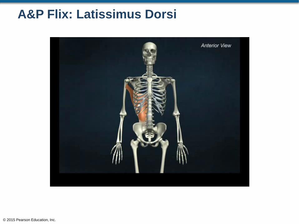
A&P Flix: Latissimus Dorsi
© 2015 Pearson Education, Inc.
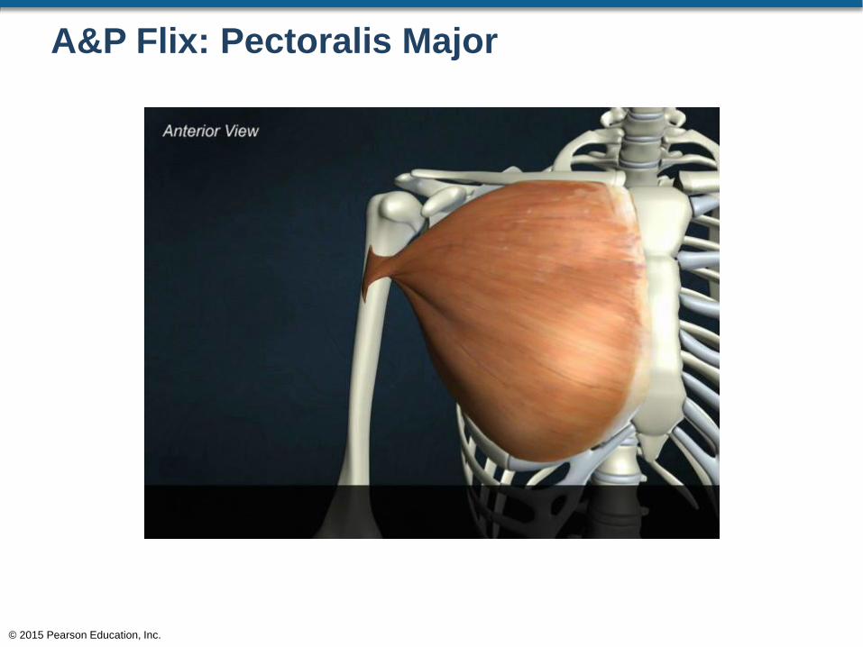
A&P Flix: Pectoralis Major
© 2015 Pearson Education, Inc.
Muscles of the Pectoral Girdle and Upper Limbs
• Muscles That Move the Forearm and Hand
• Most of these muscles originate on the humerus
and insert on the forearm and wrist
• Exceptions include:
• Long head of triceps brachii: originates on the
scapula and inserts on the olecranon
• Long head of biceps brachii: originates on the
scapula and inserts of the radial tuberosity of the
radius
© 2015 Pearson Education, Inc.
Muscles of the Pectoral Girdle and Upper Limbs
• Muscles That Move the Forearm and Hand
• Anterior view
• Muscles that move the forearm
• Biceps brachii
• Flexes at the elbow and shoulder
• Brachialis
• Flexes at the elbow
• Brachioradialis
• Flexes at the elbow
© 2015 Pearson Education, Inc.

Figure 11.8b Muscles That Move the Forearm and Hand, Part I
© 2015 Pearson Education, Inc.
Coracoid process
of scapula
Humerus
Coracobrachialis
Triceps brachii,
long head
Triceps brachii,
medial head
Pronator teres
Medial epicondyle
of humerus
FLEXORS AT
THE ELBOW
Biceps brachii,
long head
Biceps brachii,
short head
Brachialis
Brachioradialis
FLEXORS AT THE
WRIST
Flexor carpi radialis
Palmaris longus
Flexor carpi ulnaris
Flexor digitorum superficialis
Pronator quadratus
Palmar carpal ligament
Flexor retinaculum
Muscles That
Move the Hand
Muscles
That Move
the Forearm
Superficial muscles of the right
upper limb, anterior viewb
Muscles of the Pectoral Girdle and Upper Limbs
• Muscles That Move the Forearm and Hand
• Anterior view
• Muscles that move the hand
• Flexor carpi radialis
• Flexes and abducts the wrist
• Palmaris longus
• Flexes at the wrist
• Flexor carpi ulnaris
• Flexes and adducts the wrist
© 2015 Pearson Education, Inc.
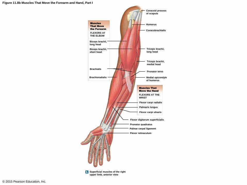
Figure 11.8b Muscles That Move the Forearm and Hand, Part I
© 2015 Pearson Education, Inc.
Coracoid process
of scapula
Humerus
Coracobrachialis
Triceps brachii,
long head
Triceps brachii,
medial head
Pronator teres
Medial epicondyle
of humerus
FLEXORS AT
THE ELBOW
Biceps brachii,
long head
Biceps brachii,
short head
Brachialis
Brachioradialis
FLEXORS AT THE
WRIST
Flexor carpi radialis
Palmaris longus
Flexor carpi ulnaris
Flexor digitorum superficialis
Pronator quadratus
Palmar carpal ligament
Flexor retinaculum
Muscles That
Move the Hand
Muscles
That Move
the Forearm
Superficial muscles of the right
upper limb, anterior viewb
Muscles of the Pectoral Girdle and Upper Limbs
• Muscles That Move the Forearm and Hand
• Posterior view
• Muscles that move the forearm
• Anconeus
• Extension at the elbow
• Triceps brachii
• Lateral head: extension at the elbow
• Long head: extension at the elbow and adduction at
the shoulder
• Medial head: extension at the elbow
© 2015 Pearson Education, Inc.

Figure 11.10b Muscles That Move the Forearm and Hand, Part II
© 2015 Pearson Education, Inc.
Infraglenoid
tubercle of
scapula
Olecranon of ulna
Brachioradialis
EXTENSORS AT THE
ELBOW
Triceps brachii,
lateral head
Triceps brachii,
long head
Anconeus
EXTENSORS AT THE
WRIST
Extensor carpi
radialis longus
Extensor carpi
ulnaris
Extensor carpi
radialis brevis
Abductor pollicis
longus
Extensor pollicis
brevis
Flexor carpi
ulnaris
Extensor
digitorum
Ulna
Radius
Extensor
retinaculum
A diagrammatic view of a
dissection of the superficial
muscles
b
Muscles That
Move the Hand
Muscles That
Move the Forearm
Muscles of the Pectoral Girdle and Upper Limbs
• Muscles That Move the Forearm and Hand
• Posterior view
• Muscles that move the hand
• Extensor carpi radialis longus
• Extension and abduction at wrist
• Extensor carpi ulnaris
• Extension and adduction at wrist
• Extensor carpi radialis brevis
• Extension and abduction at wrist
© 2015 Pearson Education, Inc.

Figure 11.10b Muscles That Move the Forearm and Hand, Part II
© 2015 Pearson Education, Inc.
Infraglenoid
tubercle of
scapula
Olecranon of ulna
Brachioradialis
EXTENSORS AT THE
ELBOW
Triceps brachii,
lateral head
Triceps brachii,
long head
Anconeus
EXTENSORS AT THE
WRIST
Extensor carpi
radialis longus
Extensor carpi
ulnaris
Extensor carpi
radialis brevis
Abductor pollicis
longus
Extensor pollicis
brevis
Flexor carpi
ulnaris
Extensor
digitorum
Ulna
Radius
Extensor
retinaculum
A diagrammatic view of a
dissection of the superficial
muscles
b
Muscles That
Move the Hand
Muscles That
Move the Forearm
Muscles That Move the Forearm and Hand
© 2015 Pearson Education, Inc.
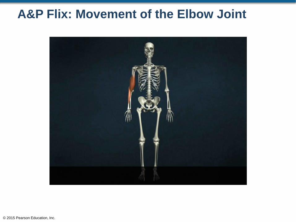
A&P Flix: Movement of the Elbow Joint
© 2015 Pearson Education, Inc.
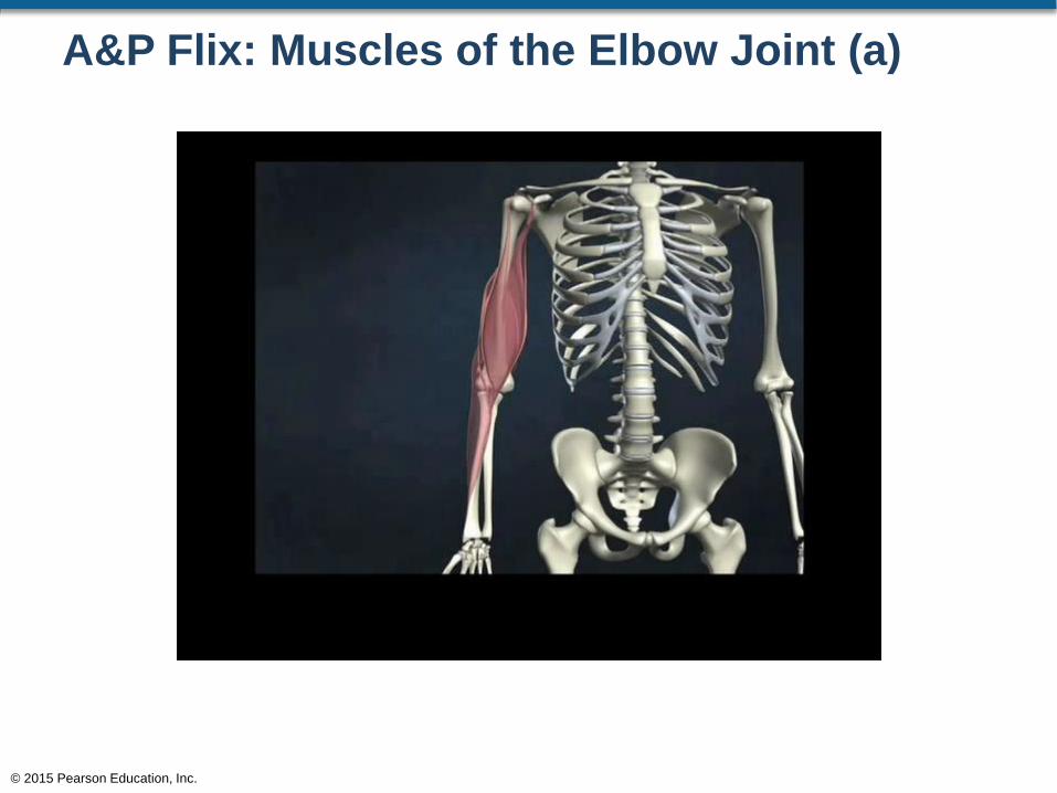
A&P Flix: Muscles of the Elbow Joint (a)
© 2015 Pearson Education, Inc.
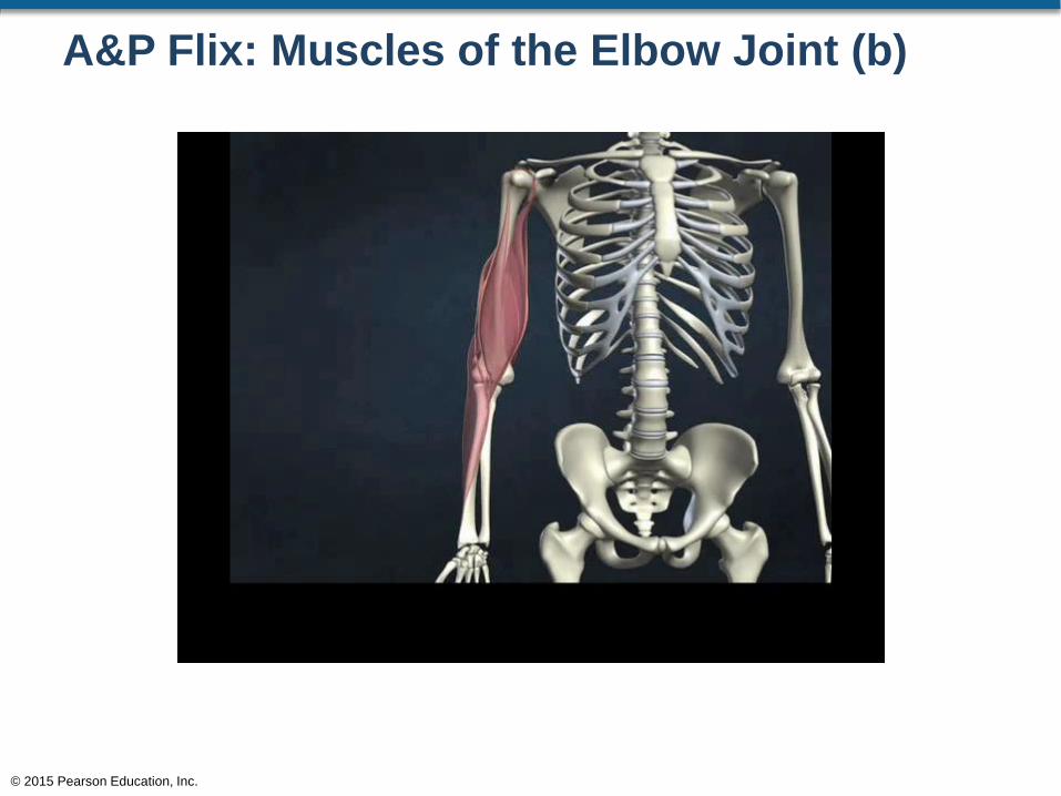
A&P Flix: Muscles of the Elbow Joint (b)
© 2015 Pearson Education, Inc.
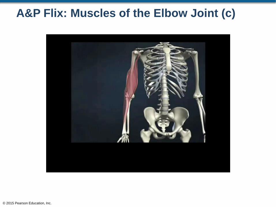
A&P Flix: Muscles of the Elbow Joint (c)
© 2015 Pearson Education, Inc.
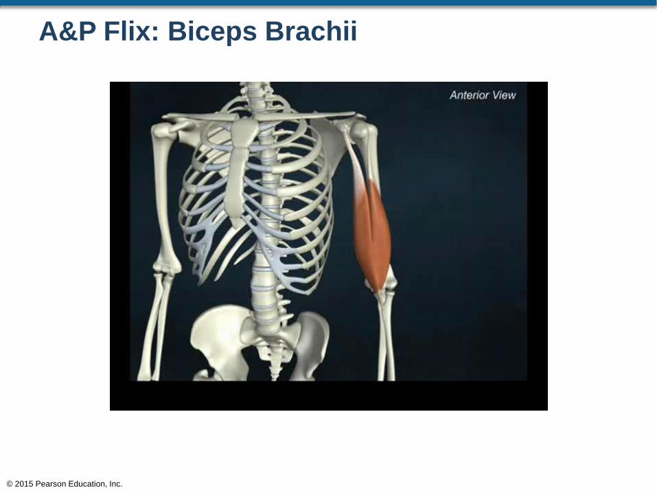
A&P Flix: Biceps Brachii
© 2015 Pearson Education, Inc.
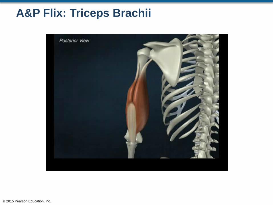
A&P Flix: Triceps Brachii
© 2015 Pearson Education, Inc.
Muscles of the Pectoral Girdle and Upper Limbs
• Muscles That Move the Hand and Fingers
• Extrinsic muscles of the hand
• Relatively large muscles that perform flexion and
extension at the joints of the fingers
• Provide strength and crude control of the hand and
fingers
• These muscles are found mostly in the forearm
© 2015 Pearson Education, Inc.
Muscles of the Pectoral Girdle and Upper Limbs
• Muscles That Move the Hand and Fingers
• Intrinsic muscles of the hand
• Smaller muscles that provide fine control of hand
• Responsible for flexion and extension, and
abduction and adduction of the fingers at the
metacarpophalangeal joints
• Responsible for opposition and reposition of the
thumb
• These muscles are found mostly in the palm or the
dorsum of the hand
© 2015 Pearson Education, Inc.
Muscles of the Pectoral Girdle and Upper Limbs
• Extrinsic Muscles of the Hand
• Anterior view
• Flexor digitorum superficialis
• Flexion at the wrist and some digits
• Flexor digitorum profundus
• Flexion at the wrist and some digits
• Flexor pollicis longus
• Flexion of the thumb
© 2015 Pearson Education, Inc.
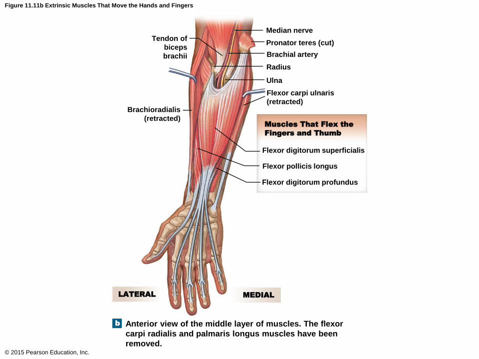
Figure 11.11b Extrinsic Muscles That Move the Hands and Fingers
© 2015 Pearson Education, Inc.
Median nerve
Pronator teres (cut)
Brachial artery
Radius
Ulna
Flexor carpi ulnaris
(retracted)
Tendon of
biceps
brachii
Brachioradialis
(retracted)
Flexor digitorum superficialis
Flexor pollicis longus
Flexor digitorum profundus
Muscles That Flex the
Fingers and Thumb
MEDIALLATERAL
Anterior view of the middle layer of muscles. The flexor
carpi radialis and palmaris longus muscles have been
removed.
b
Muscles of the Pectoral Girdle and Upper Limbs
• Extrinsic Muscles of the Hand (continued)
• Posterior view
• Abductor pollicis longus
• Abduction of thumb
• Extensor digitorum
• Extension of fingers and wrist
• Extensor pollicis brevis
• Extension of thumb, abducts the wrist
• Extensor digiti minimi
• Extension of little finger, extension of wrist
© 2015 Pearson Education, Inc.
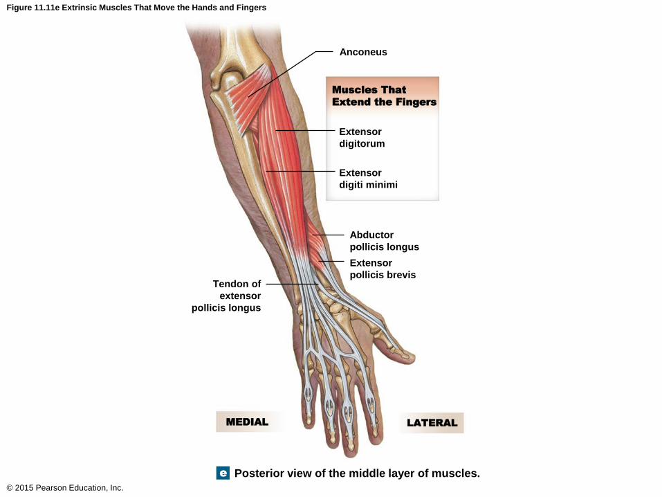
Figure 11.11e Extrinsic Muscles That Move the Hands and Fingers
© 2015 Pearson Education, Inc.
Anconeus
Extensor
digitorum
Extensor
digiti minimi
Muscles That
Extend the Fingers
Abductor
pollicis longus
Extensor
pollicis brevisTendon of
extensor
pollicis longus
MEDIAL LATERAL
Posterior view of the middle layer of muscles.e
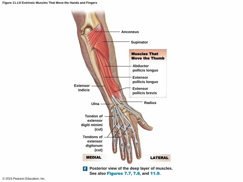
Figure 11.11f Extrinsic Muscles That Move the Hands and Fingers
© 2015 Pearson Education, Inc.
Anconeus
Supinator
Muscles That
Move the Thumb
Abductor
pollicis longus
Extensor
pollicis longus
Extensor
pollicis brevis
Radius
Extensor
indicis
Ulna
Tendon of
extensor
digiti minimi
(cut)
Tendons of
extensor
digitorum
(cut)
LATERALMEDIAL
Posterior view of the deep layer of muscles.
See also Figures 7.7, 7.8, and 11.9.
f
Muscles of the Pectoral Girdle and Upper Limbs
• Intrinsic Muscles of the Hand
• Adductor pollicis
• Adducts the thumb
• Flexor pollicis brevis
• Flexes and adducts thumb
• Opponens pollicis
• Opposition of the thumb
• Abductor pollicis brevis
• Abducts the thumb
© 2015 Pearson Education, Inc.
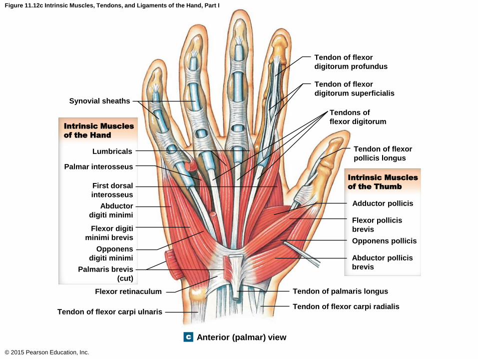
Figure 11.12c Intrinsic Muscles, Tendons, and Ligaments of the Hand, Part I
© 2015 Pearson Education, Inc.
Tendon of flexor
digitorum profundus
Tendon of flexor
digitorum superficialis
Tendons of
flexor digitorum
Tendon of flexor
pollicis longus
Intrinsic Muscles
of the Thumb
Adductor pollicis
Flexor pollicis
brevis
Opponens pollicis
Abductor pollicis
brevis
Tendon of palmaris longus
Tendon of flexor carpi radialis
Synovial sheaths
Intrinsic Muscles
of the Hand
Lumbricals
Palmar interosseus
First dorsal
interosseus
Abductor
digiti minimi
Flexor digiti
minimi brevis
Opponens
digiti minimi
Palmaris brevis
(cut)
Flexor retinaculum
Tendon of flexor carpi ulnaris
Anterior (palmar) viewc
Muscles of the Pectoral Girdle and Upper Limbs
• Intrinsic Muscles of the Hand
• Flexor digiti minimi brevis
• Flexes little finger
• Palmaris brevis
• Moves the skin on the medial side of the palm
• Abductor digiti minimi
• Abducts little finger
• Opponens digiti minimi
• Brings digits into opposition with the thumb
© 2015 Pearson Education, Inc.

Figure 11.12c Intrinsic Muscles, Tendons, and Ligaments of the Hand, Part I
© 2015 Pearson Education, Inc.
Tendon of flexor
digitorum profundus
Tendon of flexor
digitorum superficialis
Tendons of
flexor digitorum
Tendon of flexor
pollicis longus
Intrinsic Muscles
of the Thumb
Adductor pollicis
Flexor pollicis
brevis
Opponens pollicis
Abductor pollicis
brevis
Tendon of palmaris longus
Tendon of flexor carpi radialis
Synovial sheaths
Intrinsic Muscles
of the Hand
Lumbricals
Palmar interosseus
First dorsal
interosseus
Abductor
digiti minimi
Flexor digiti
minimi brevis
Opponens
digiti minimi
Palmaris brevis
(cut)
Flexor retinaculum
Tendon of flexor carpi ulnaris
Anterior (palmar) viewc
Muscles of the Pectoral Girdle and Upper Limbs
• Intrinsic Muscles of the Hand
• Lumbricals
• Flexion of the digits
• Dorsal interosseus
• Abduction of digits 2–4
• Palmar interosseus
• Adduction of digits 2, 4, and 5
© 2015 Pearson Education, Inc.

Figure 11.12c Intrinsic Muscles, Tendons, and Ligaments of the Hand, Part I
© 2015 Pearson Education, Inc.
Tendon of flexor
digitorum profundus
Tendon of flexor
digitorum superficialis
Tendons of
flexor digitorum
Tendon of flexor
pollicis longus
Intrinsic Muscles
of the Thumb
Adductor pollicis
Flexor pollicis
brevis
Opponens pollicis
Abductor pollicis
brevis
Tendon of palmaris longus
Tendon of flexor carpi radialis
Synovial sheaths
Intrinsic Muscles
of the Hand
Lumbricals
Palmar interosseus
First dorsal
interosseus
Abductor
digiti minimi
Flexor digiti
minimi brevis
Opponens
digiti minimi
Palmaris brevis
(cut)
Flexor retinaculum
Tendon of flexor carpi ulnaris
Anterior (palmar) viewc
Muscles That Move the Hand and Fingers
© 2015 Pearson Education, Inc.
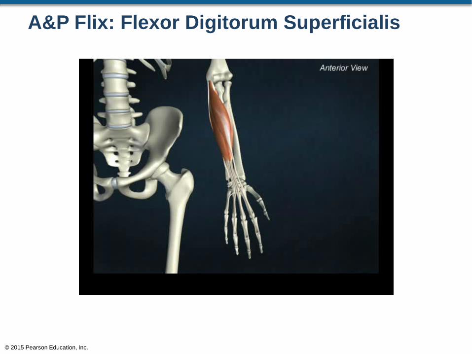
A&P Flix: Flexor Digitorum Superficialis
© 2015 Pearson Education, Inc.
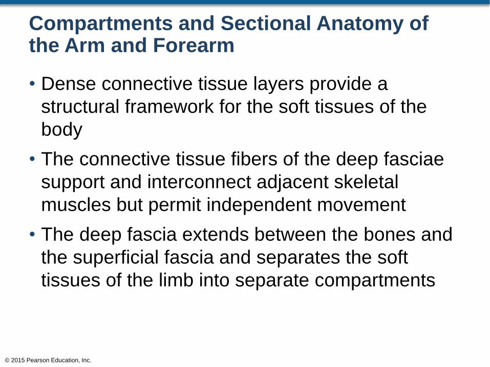
Compartments and Sectional Anatomy of the Arm and Forearm
• Dense connective tissue layers provide a
structural framework for the soft tissues of the
body
• The connective tissue fibers of the deep fasciae
support and interconnect adjacent skeletal
muscles but permit independent movement
• The deep fascia extends between the bones and
the superficial fascia and separates the soft
tissues of the limb into separate compartments
© 2015 Pearson Education, Inc.
Compartments and Sectional Anatomy of the Arm and Forearm
• Compartments of the Upper Limb
• The arm
• Anterior compartment (or flexor compartment)
• Posterior compartment (or extensor compartment)
• The forearm
• Anterior compartment (superficial and deep)
• Lateral compartment
• Posterior compartment
© 2015 Pearson Education, Inc.
Compartments and Sectional Anatomy of the Arm and Forearm
• Compartments of the Arm
• The anterior compartment of the arm
• Biceps brachii
• Brachialis
• Coracobrachialis
• The posterior compartment of the arm
• Triceps brachii
© 2015 Pearson Education, Inc.
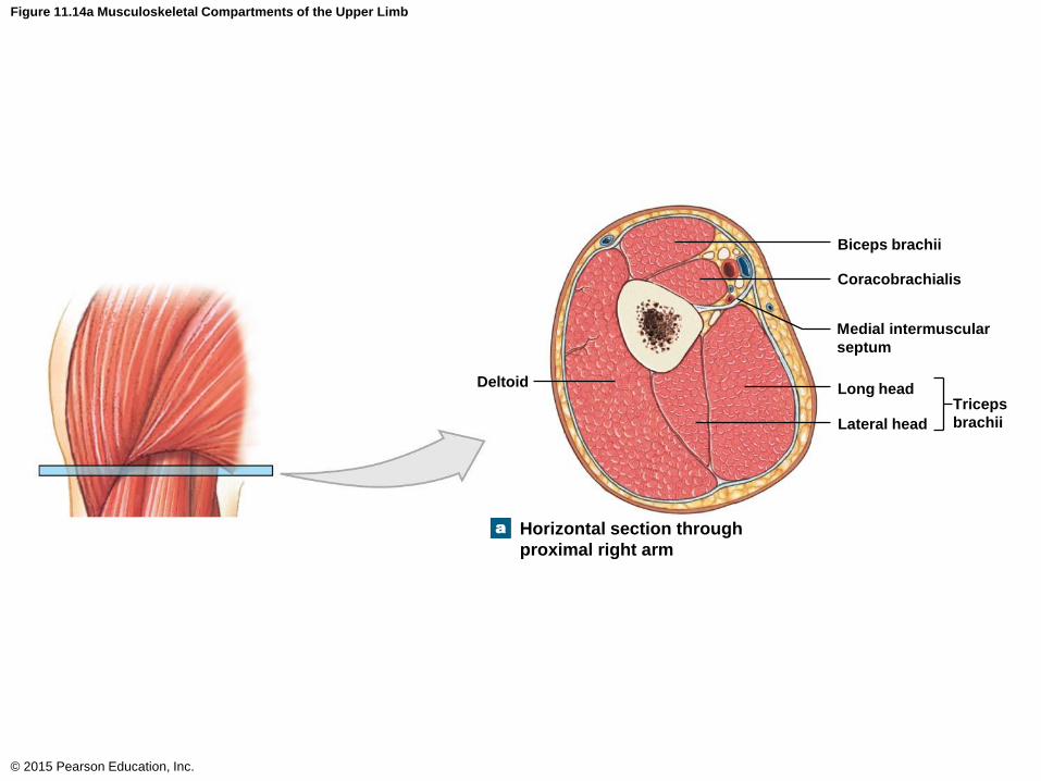
Figure 11.14a Musculoskeletal Compartments of the Upper Limb
© 2015 Pearson Education, Inc.
Biceps brachii
Coracobrachialis
Medial intermuscular
septum
Long head
Lateral head
Triceps
brachii
Deltoid
Horizontal section through
proximal right arm
a
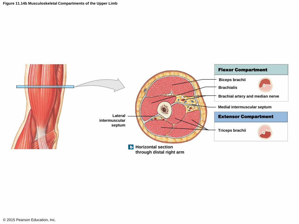
Figure 11.14b Musculoskeletal Compartments of the Upper Limb
© 2015 Pearson Education, Inc.
Flexor Compartment
Extensor Compartment
Biceps brachii
Brachialis
Brachial artery and median nerve
Triceps brachii
Lateral
intermuscular
septum
Medial intermuscular septum
Horizontal section
through distal right arm
b
Compartments and Sectional Anatomy of the Arm and Forearm
• Compartments of the Forearm
• The anterior superficial compartment
• Flexor carpi radialis
• Flexor carpi ulnaris
• Flexor digitorum superficialis
• Palmaris longus
• Pronator teres
• Flexor digitorum profundus
• Flexor pollicis longus
© 2015 Pearson Education, Inc.
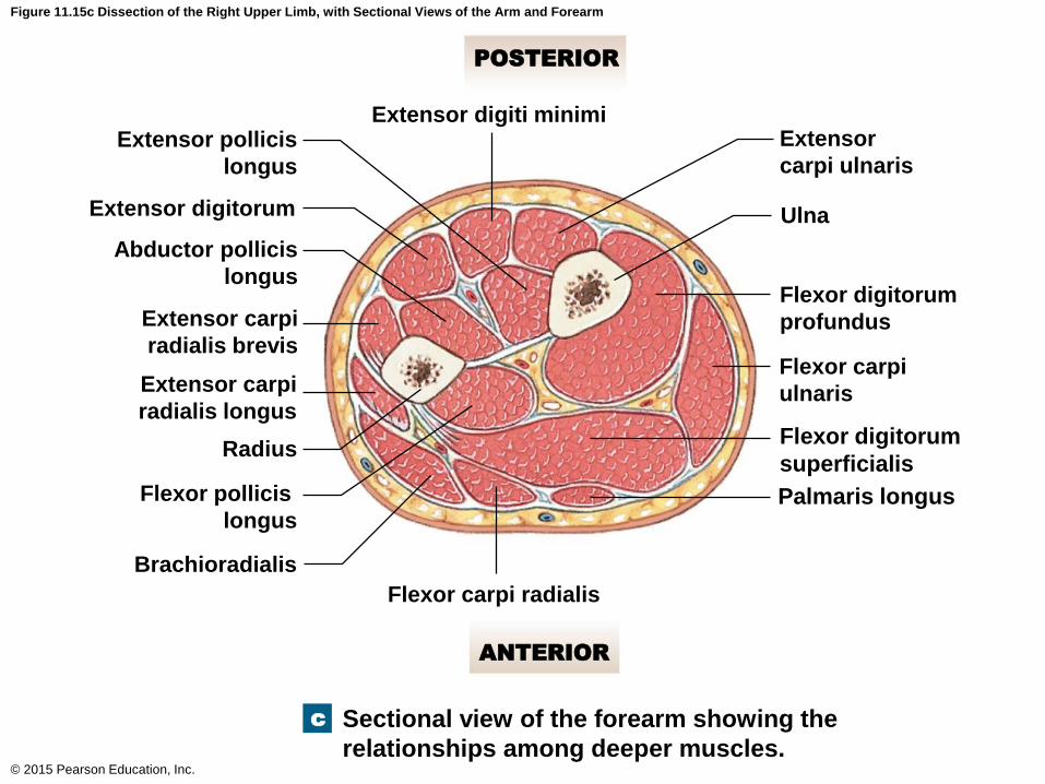
Figure 11.15c Dissection of the Right Upper Limb, with Sectional Views of the Arm and Forearm
© 2015 Pearson Education, Inc.
POSTERIOR
ANTERIOR
Extensor digiti minimiExtensor
carpi ulnaris
Ulna
Flexor digitorum
profundus
Flexor carpi
ulnaris
Flexor digitorum
superficialis
Palmaris longus
Flexor carpi radialis
Extensor pollicis
longus
Extensor digitorum
Abductor pollicis
longus
Extensor carpi
radialis brevis
Extensor carpi
radialis longus
Radius
Flexor pollicis
longus
Brachioradialis
Sectional view of the forearm showing the
relationships among deeper muscles.
c
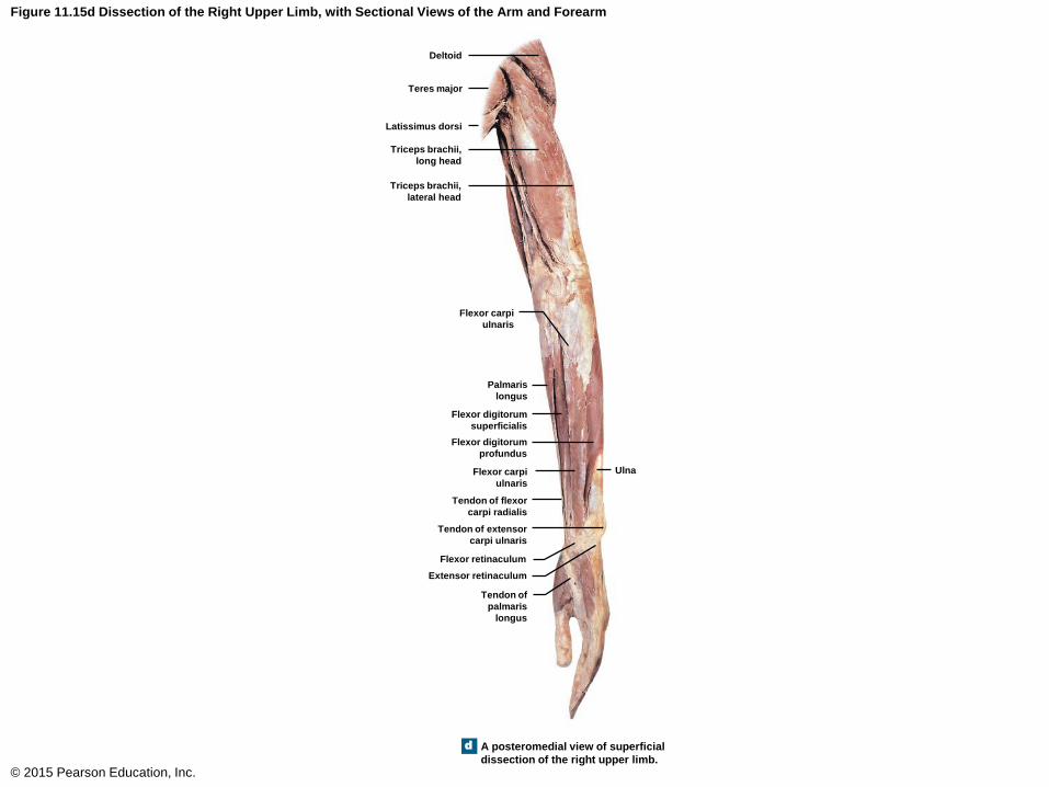
Figure 11.15d Dissection of the Right Upper Limb, with Sectional Views of the Arm and Forearm
© 2015 Pearson Education, Inc.
Deltoid
Teres major
Latissimus dorsi
Triceps brachii,
long head
Triceps brachii,
lateral head
Flexor carpi
ulnaris
Palmaris
longus
Flexor digitorum
superficialis
Ulna
Flexor digitorum
profundus
Flexor carpi
ulnaris
Tendon of flexor
carpi radialis
Tendon of extensor
carpi ulnaris
Flexor retinaculum
Extensor retinaculum
Tendon of
palmaris
longus
A posteromedial view of superficial
dissection of the right upper limb.
d
Compartments and Sectional Anatomy of the Arm and Forearm
• Compartments of the Forearm
• The anterior deep compartment
• Pronator quadratus
© 2015 Pearson Education, Inc.
Compartments and Sectional Anatomy of the Arm and Forearm
• Compartments of the Forearm
• The lateral compartment
• Brachioradialis
• Extensor carpi radialis brevis
• Extensor carpi radialis longus
© 2015 Pearson Education, Inc.
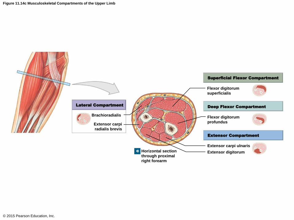
Figure 11.14c Musculoskeletal Compartments of the Upper Limb
© 2015 Pearson Education, Inc.
Superficial Flexor Compartment
Deep Flexor Compartment
Extensor Compartment
Lateral Compartment
Flexor digitorum
superficialis
Flexor digitorum
profundus
Extensor carpi ulnaris
Extensor digitorum
Brachioradialis
Extensor carpi
radialis brevis
Horizontal section
through proximal
right forearm
c
Compartments and Sectional Anatomy of the Arm and Forearm
• Compartments of the Forearm
• The posterior compartment
• Abductor pollicis longus
• Anconeus
• Extensor carpi ulnaris
• Extensor digitorum
• Extensor digiti minimi
• Extensor indicis
• Extensor pollicis brevis
• Extensor pollicis longus
• Supinator
© 2015 Pearson Education, Inc.

Figure 11.15c Dissection of the Right Upper Limb, with Sectional Views of the Arm and Forearm
© 2015 Pearson Education, Inc.
POSTERIOR
ANTERIOR
Extensor digiti minimiExtensor
carpi ulnaris
Ulna
Flexor digitorum
profundus
Flexor carpi
ulnaris
Flexor digitorum
superficialis
Palmaris longus
Flexor carpi radialis
Extensor pollicis
longus
Extensor digitorum
Abductor pollicis
longus
Extensor carpi
radialis brevis
Extensor carpi
radialis longus
Radius
Flexor pollicis
longus
Brachioradialis
Sectional view of the forearm showing the
relationships among deeper muscles.
c

Figure 11.15d Dissection of the Right Upper Limb, with Sectional Views of the Arm and Forearm
© 2015 Pearson Education, Inc.
Deltoid
Teres major
Latissimus dorsi
Triceps brachii,
long head
Triceps brachii,
lateral head
Flexor carpi
ulnaris
Palmaris
longus
Flexor digitorum
superficialis
Ulna
Flexor digitorum
profundus
Flexor carpi
ulnaris
Tendon of flexor
carpi radialis
Tendon of extensor
carpi ulnaris
Flexor retinaculum
Extensor retinaculum
Tendon of
palmaris
longus
A posteromedial view of superficial
dissection of the right upper limb.
d
Muscles of the Pelvic Girdle and Lower Limbs
• The muscles of the lower limbs are larger and
more powerful than those of the upper limbs.
• These muscles can be divided into three groups
• Muscles that move the thigh
• Muscles that move the leg
• Muscles that move the foot and toes
© 2015 Pearson Education, Inc.
Muscles of the Pelvic Girdle and Lower Limbs
• Muscles That Move the Thigh
• Originate on the pelvis; many are large and
powerful
• Four groups
• Gluteal group
• Lateral rotator group
• Adductor group
• Iliopsoas group
© 2015 Pearson Education, Inc.
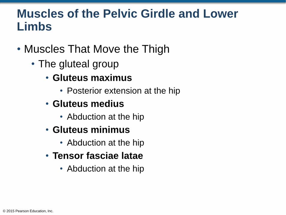
Muscles of the Pelvic Girdle and Lower Limbs
• Muscles That Move the Thigh
• The gluteal group
• Gluteus maximus
• Posterior extension at the hip
• Gluteus medius
• Abduction at the hip
• Gluteus minimus
• Abduction at the hip
• Tensor fasciae latae
• Abduction at the hip
© 2015 Pearson Education, Inc.
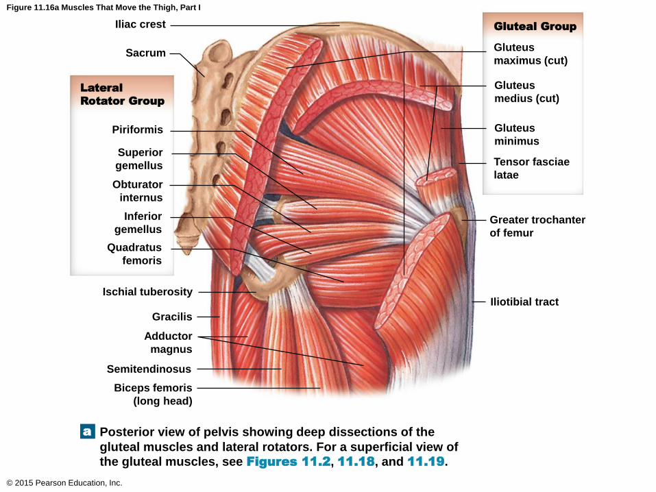
Figure 11.16a Muscles That Move the Thigh, Part I
© 2015 Pearson Education, Inc.
Gluteal Group
Lateral
Rotator Group
Iliac crest
Sacrum
Piriformis
Superior
gemellus
Obturator
internus
Inferior
gemellus
Quadratus
femoris
Ischial tuberosity
Gracilis
Adductor
magnus
Semitendinosus
Biceps femoris
(long head)
Gluteus
maximus (cut)
Gluteus
medius (cut)
Gluteus
minimus
Tensor fasciae
latae
Greater trochanter
of femur
Iliotibial tract
Posterior view of pelvis showing deep dissections of the
gluteal muscles and lateral rotators. For a superficial view of
the gluteal muscles, see Figures 11.2, 11.18, and 11.19.
a
Muscles of the Pelvic Girdle and Lower Limbs
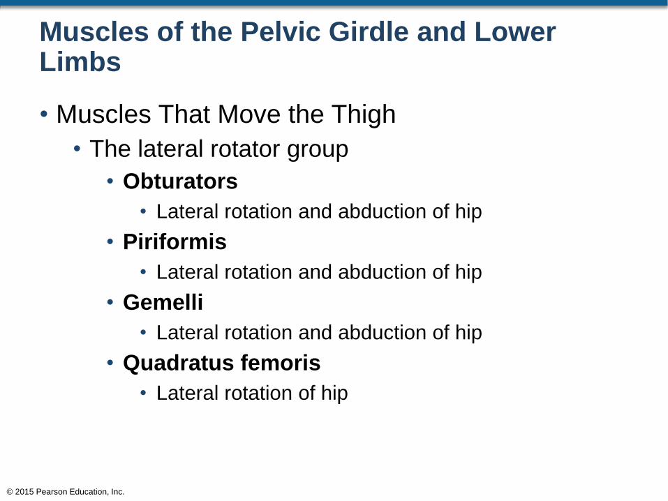
• Muscles That Move the Thigh
• The lateral rotator group
• Obturators
• Lateral rotation and abduction of hip
• Piriformis
• Lateral rotation and abduction of hip
• Gemelli
• Lateral rotation and abduction of hip
• Quadratus femoris
• Lateral rotation of hip
© 2015 Pearson Education, Inc.

Figure 11.16a Muscles That Move the Thigh, Part I
© 2015 Pearson Education, Inc.
Gluteal Group
Lateral
Rotator Group
Iliac crest
Sacrum
Piriformis
Superior
gemellus
Obturator
internus
Inferior
gemellus
Quadratus
femoris
Ischial tuberosity
Gracilis
Adductor
magnus
Semitendinosus
Biceps femoris
(long head)
Gluteus
maximus (cut)
Gluteus
medius (cut)
Gluteus
minimus
Tensor fasciae
latae
Greater trochanter
of femur
Iliotibial tract
Posterior view of pelvis showing deep dissections of the
gluteal muscles and lateral rotators. For a superficial view of
the gluteal muscles, see Figures 11.2, 11.18, and 11.19.
a
Muscles of the Pelvic Girdle and Lower Limbs
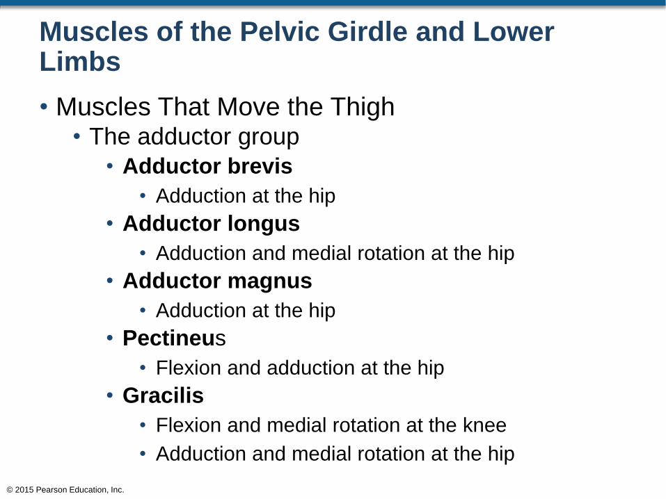
• Muscles That Move the Thigh • The adductor group
• Adductor brevis
• Adduction at the hip
• Adductor longus
• Adduction and medial rotation at the hip
• Adductor magnus
• Adduction at the hip
• Pectineus
• Flexion and adduction at the hip
• Gracilis
• Flexion and medial rotation at the knee
• Adduction and medial rotation at the hip
© 2015 Pearson Education, Inc.
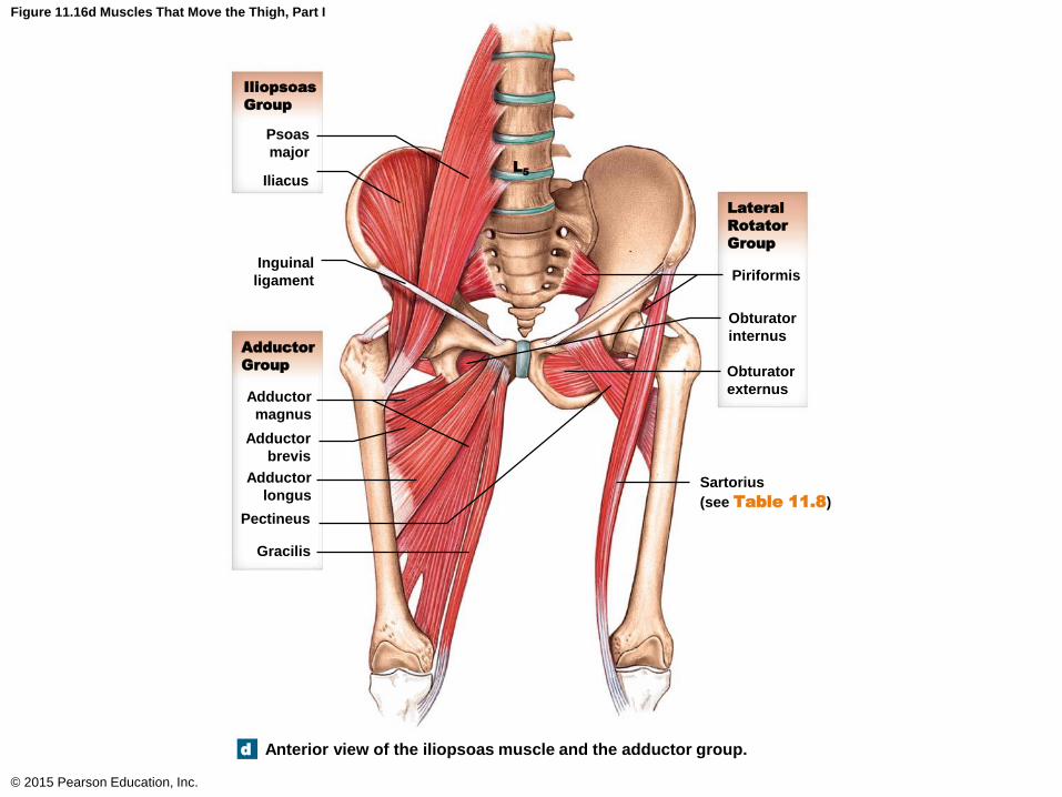
Figure 11.16d Muscles That Move the Thigh, Part I
© 2015 Pearson Education, Inc.
Lateral
Rotator
Group
Iliopsoas
Group
Adductor
Group
Psoas
major
Iliacus
Piriformis
Obturator
internus
Obturator
externus
Inguinal
ligament
L5
Adductor
magnus
Adductor
brevis
Adductor
longus
Pectineus
Gracilis
Sartorius
(see Table 11.8)
Anterior view of the iliopsoas muscle and the adductor group.d
Muscles of the Pelvic Girdle and Lower Limbs
• Muscles That Move the Thigh
• The iliopsoas group
• Iliacus
• Flexion at the hip
• Psoas major
• Flexion at the hip
© 2015 Pearson Education, Inc.

Figure 11.16d Muscles That Move the Thigh, Part I
© 2015 Pearson Education, Inc.
Lateral
Rotator
Group
Iliopsoas
Group
Adductor
Group
Psoas
major
Iliacus
Piriformis
Obturator
internus
Obturator
externus
Inguinal
ligament
L5
Adductor
magnus
Adductor
brevis
Adductor
longus
Pectineus
Gracilis
Sartorius
(see Table 11.8)
Anterior view of the iliopsoas muscle and the adductor group.d
Muscles That Move the Thigh
© 2015 Pearson Education, Inc.
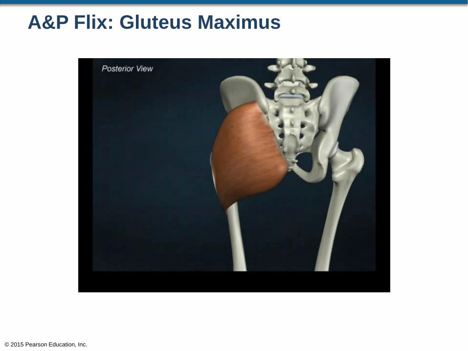
A&P Flix: Gluteus Maximus
© 2015 Pearson Education, Inc.
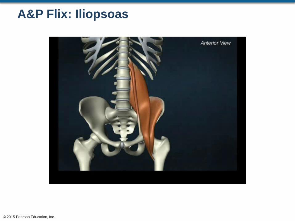
A&P Flix: Iliopsoas
© 2015 Pearson Education, Inc.
Muscles of the Pelvic Girdle and Lower Limbs
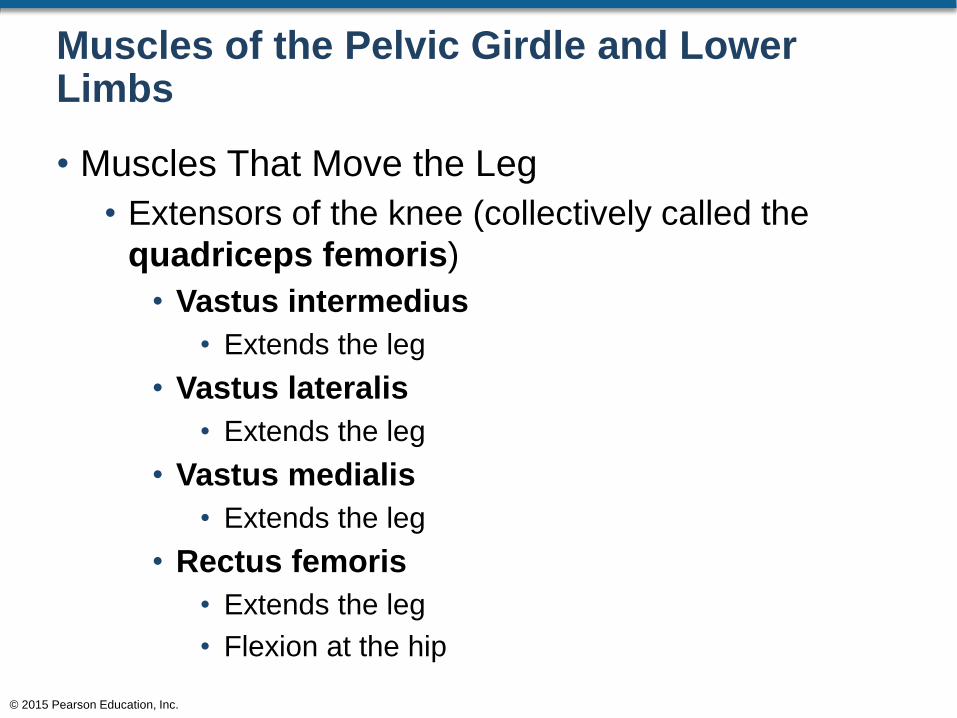
• Muscles That Move the Leg
• Extensors of the knee (collectively called the
quadriceps femoris)
• Vastus intermedius
• Extends the leg
• Vastus lateralis
• Extends the leg
• Vastus medialis
• Extends the leg
• Rectus femoris
• Extends the leg
• Flexion at the hip
© 2015 Pearson Education, Inc.
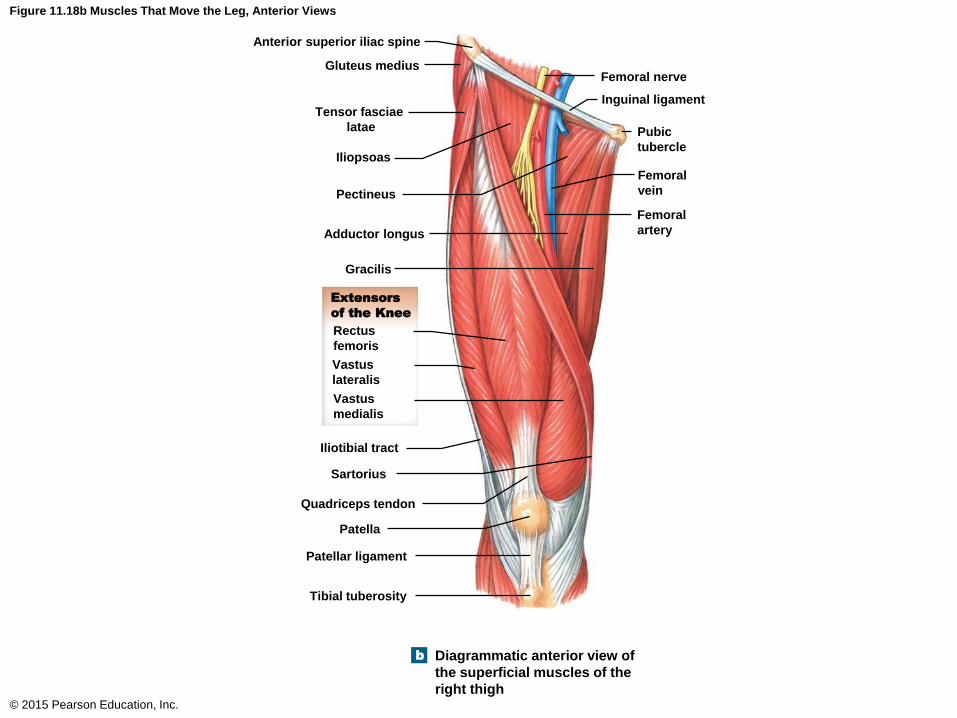
Figure 11.18b Muscles That Move the Leg, Anterior Views
© 2015 Pearson Education, Inc.
Femoral nerve
Inguinal ligament
Pubic
tubercle
Femoral
vein
Femoral
artery
Anterior superior iliac spine
Gluteus medius
Tensor fasciae
latae
Iliopsoas
Pectineus
Adductor longus
Gracilis
Extensors
of the Knee
Rectus
femoris
Vastus
lateralis
Vastus
medialis
Iliotibial tract
Sartorius
Quadriceps tendon
Patella
Patellar ligament
Tibial tuberosity
Diagrammatic anterior view of
the superficial muscles of the
right thigh
b
Muscles of the Pelvic Girdle and Lower Limbs
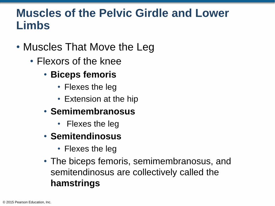
• Muscles That Move the Leg
• Flexors of the knee
• Biceps femoris
• Flexes the leg
• Extension at the hip
• Semimembranosus
• Flexes the leg
• Semitendinosus
• Flexes the leg
• The biceps femoris, semimembranosus, and
semitendinosus are collectively called the
hamstrings
© 2015 Pearson Education, Inc.

Figure 11.19b Muscles That Move the Leg, Posterior Views
© 2015 Pearson Education, Inc.
Adductor magnus
Gracilis
Iliotibial tract
Flexors of the Knee
Biceps femoris,
long head
Semitendinosus
Biceps femoris,
short head
Semimembranosus
Sartorius
Tibial nerve
Popliteal artery (red)
and vein (blue)
Medial head of
gastrocnemius
Lateral head of
gastrocnemius
Posterior view of superficial
muscles of the right thigh
b
Tensor fasciae
latae
Gluteus maximus
Gluteal aponeurosis
over gluteus medius
Iliac crest
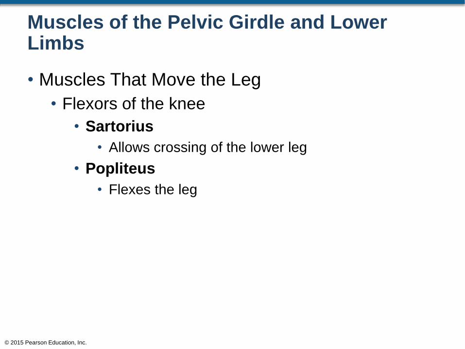
Muscles of the Pelvic Girdle and Lower Limbs
• Muscles That Move the Leg
• Flexors of the knee
• Sartorius
• Allows crossing of the lower leg
• Popliteus
• Flexes the leg
© 2015 Pearson Education, Inc.
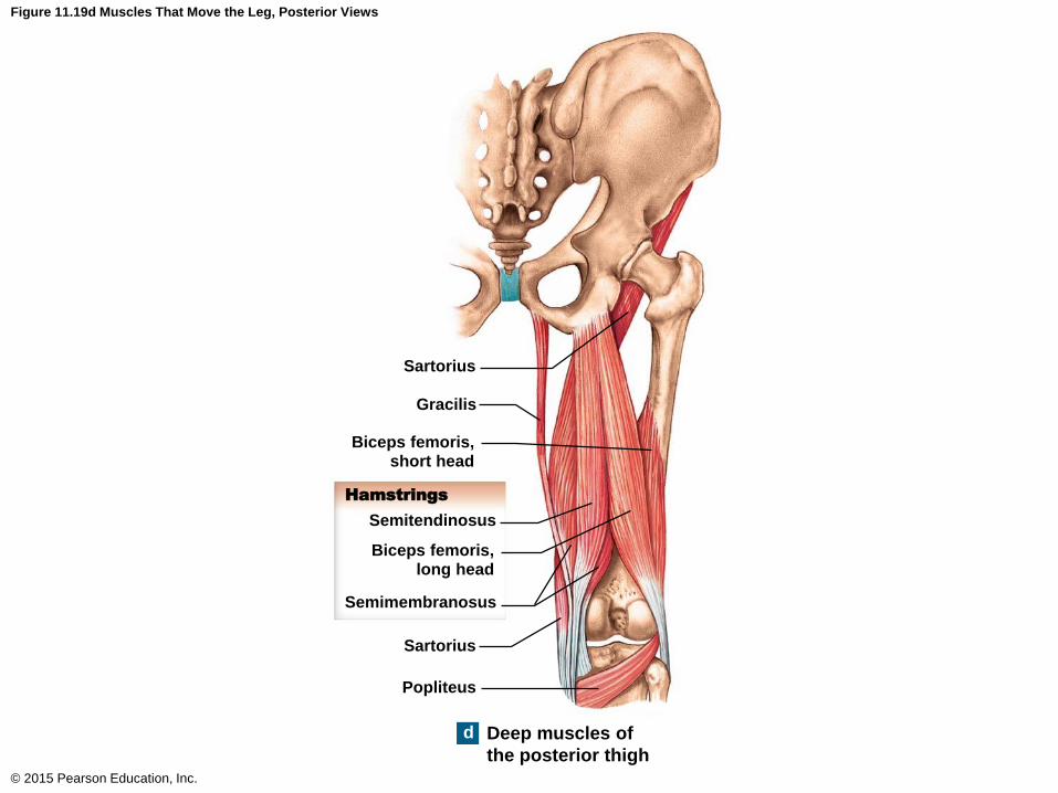
Figure 11.19d Muscles That Move the Leg, Posterior Views
© 2015 Pearson Education, Inc.
Sartorius
Gracilis
Biceps femoris,short head
Hamstrings
Semitendinosus
Biceps femoris,long head
Semimembranosus
Sartorius
Popliteus
Deep muscles of
the posterior thigh
d
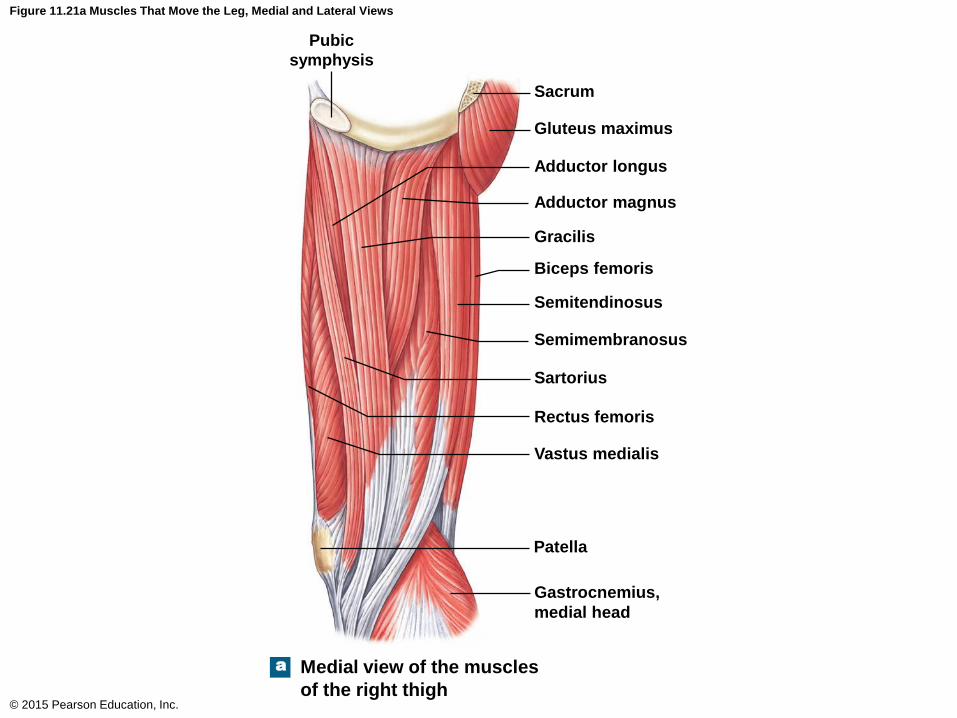
Figure 11.21a Muscles That Move the Leg, Medial and Lateral Views
© 2015 Pearson Education, Inc.
Pubic
symphysis
Sacrum
Gluteus maximus
Adductor longus
Adductor magnus
Gracilis
Biceps femoris
Semitendinosus
Semimembranosus
Sartorius
Rectus femoris
Vastus medialis
Patella
Gastrocnemius,
medial head
Medial view of the muscles
of the right thigh
a
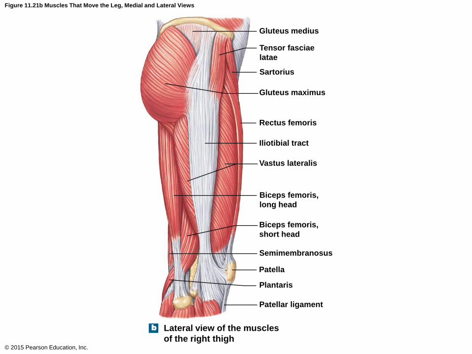
Figure 11.21b Muscles That Move the Leg, Medial and Lateral Views
© 2015 Pearson Education, Inc.
Gluteus medius
Tensor fasciae
latae
Sartorius
Gluteus maximus
Rectus femoris
Iliotibial tract
Vastus lateralis
Biceps femoris,
long head
Biceps femoris,
short head
Semimembranosus
Patella
Plantaris
Patellar ligament
Lateral view of the muscles
of the right thigh
b
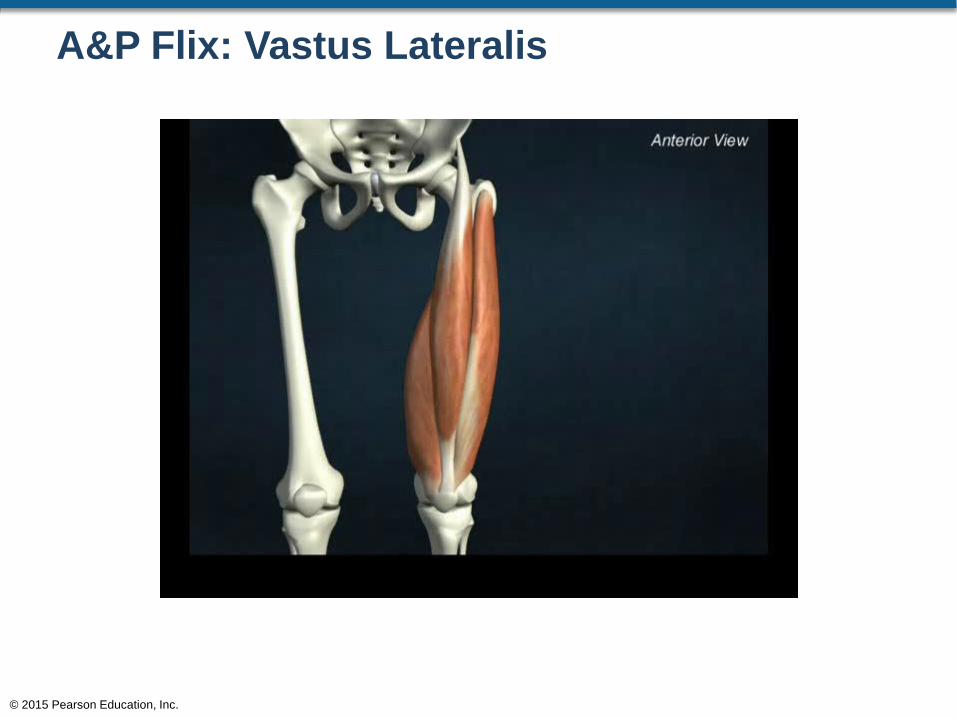
A&P Flix: Vastus Lateralis
© 2015 Pearson Education, Inc.
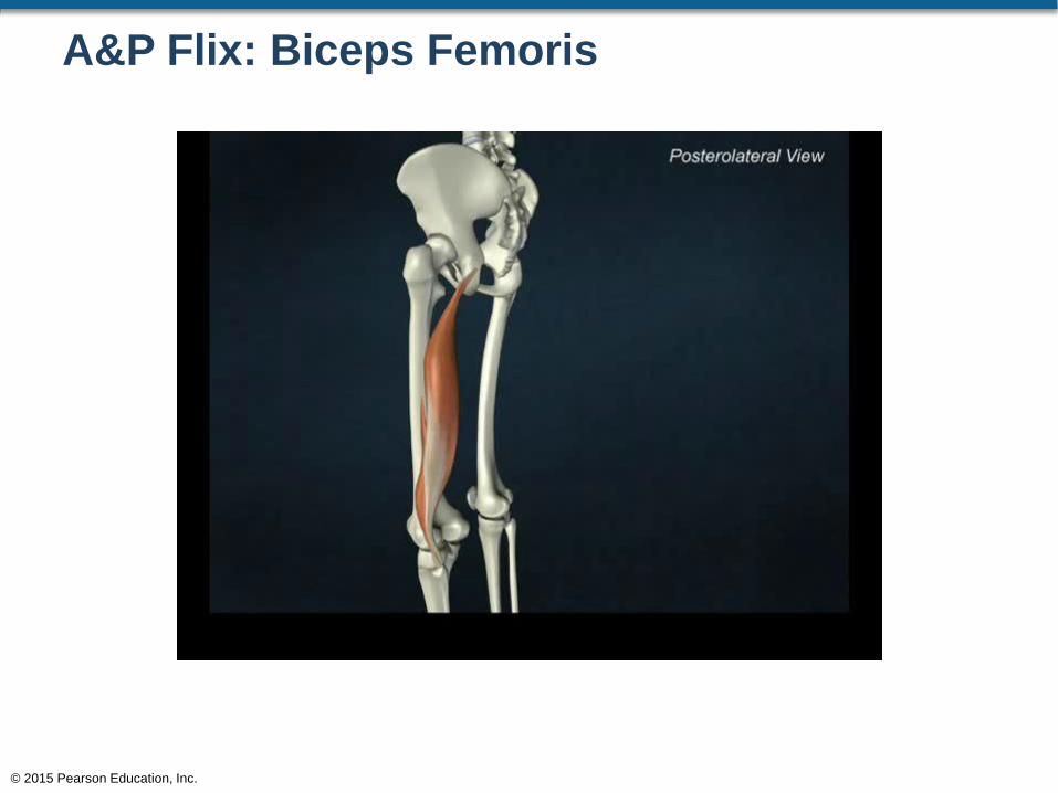
A&P Flix: Biceps Femoris
© 2015 Pearson Education, Inc.
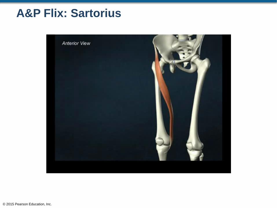
A&P Flix: Sartorius
© 2015 Pearson Education, Inc.
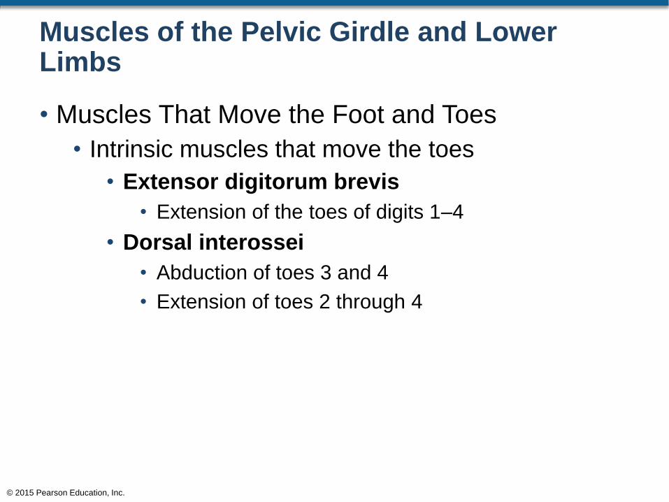
Muscles of the Pelvic Girdle and Lower Limbs
• Muscles That Move the Foot and Toes
• Extrinsic muscles of the foot and toes
• Muscles that originate on the distal end of the
femur or on the tibia or fibula but yet move the foot
and toes
• Intrinsic muscles of the toes
• Muscles that originate on some aspect of the foot
but yet move the toes
© 2015 Pearson Education, Inc.
Muscles of the Pelvic Girdle and Lower Limbs
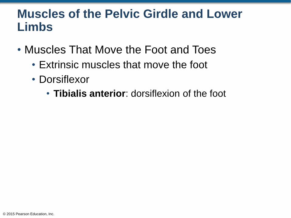
• Muscles That Move the Foot and Toes
• Extrinsic muscles that move the foot
• Dorsiflexor
• Tibialis anterior: dorsiflexion of the foot
© 2015 Pearson Education, Inc.

Figure 11.23a Extrinsic Muscles That Move the Foot and Toes, Anterior View
© 2015 Pearson Education, Inc.
Superficial Dissection Deep Dissection
Patella
Iliotibialtract
Patellarligament
Tibialtuberosity
Tibialisanterior
Fibularis longus
Dorsiflexors
Fibularis longus
Tibia
Digital
Extensors
Extensordigitorumlongus
Extensorhallucislongus
Fibula
Superior extensorretinaculum
Lateral malleolus
Inferior extensorretinaculum
Anterior views showing
superficial and deep muscles
of the right leg
a
Muscles of the Pelvic Girdle and Lower Limbs
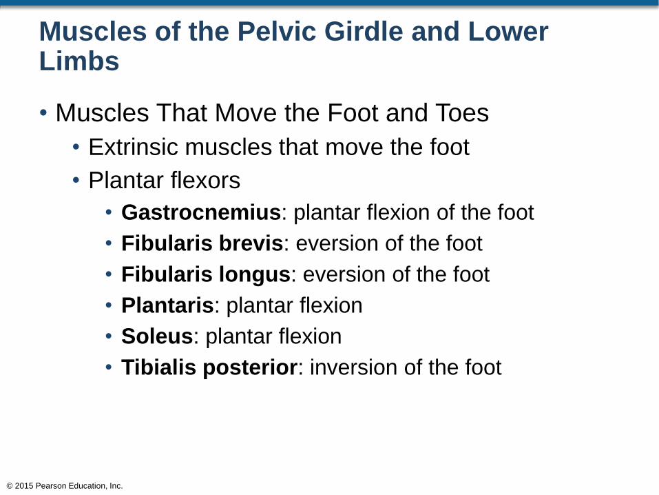
• Muscles That Move the Foot and Toes
• Extrinsic muscles that move the foot
• Plantar flexors
• Gastrocnemius: plantar flexion of the foot
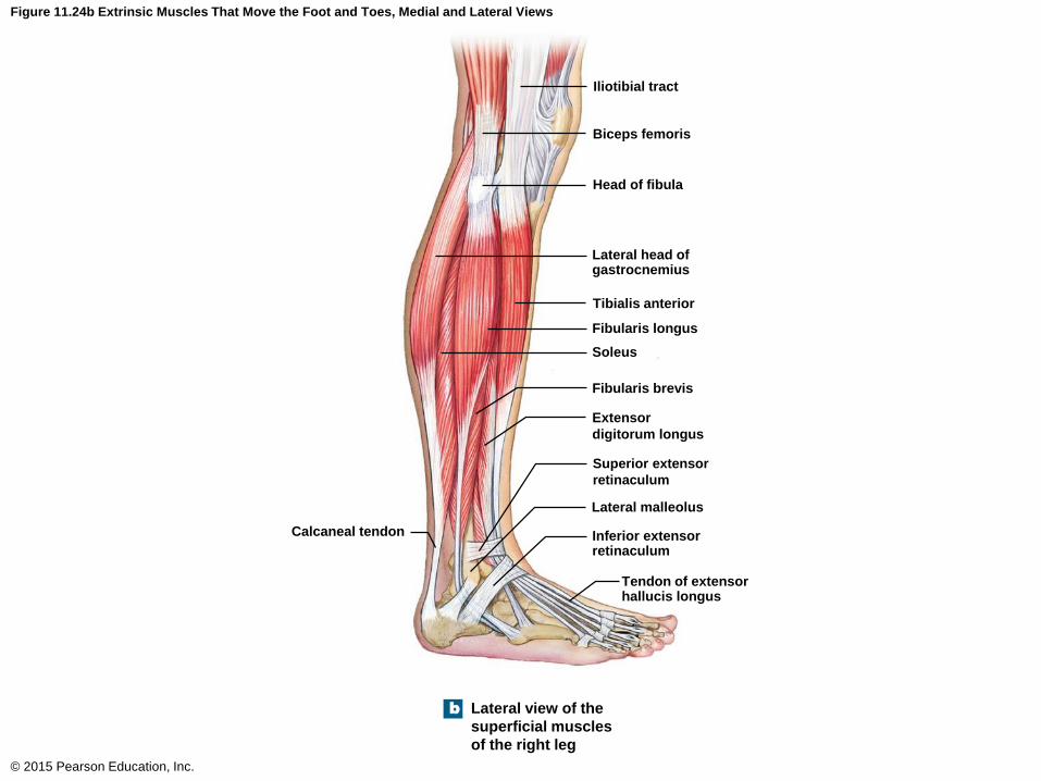
• Fibularis brevis: eversion of the foot
• Fibularis longus: eversion of the foot
• Plantaris: plantar flexion
• Soleus: plantar flexion
• Tibialis posterior: inversion of the foot
© 2015 Pearson Education, Inc.
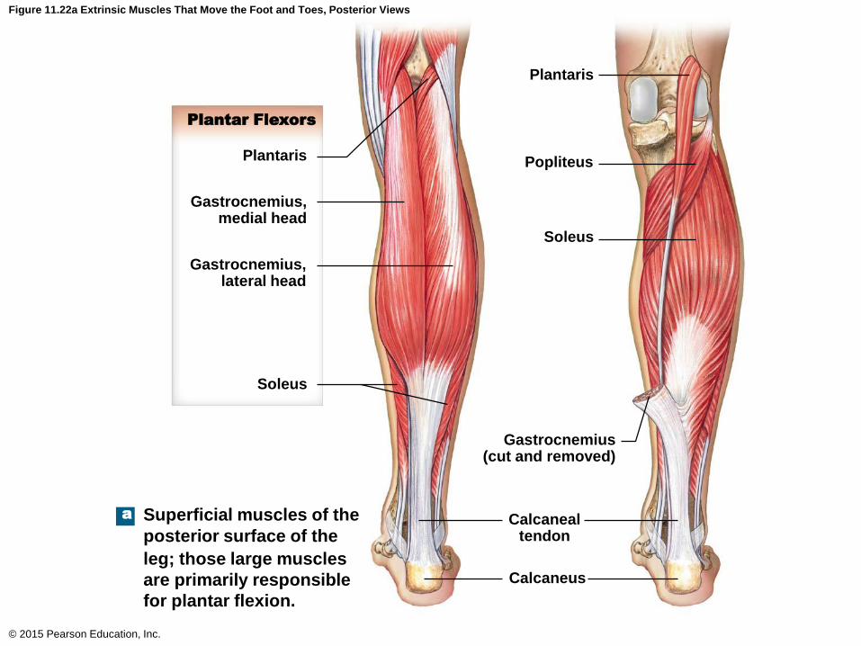
Figure 11.22a Extrinsic Muscles That Move the Foot and Toes, Posterior Views
© 2015 Pearson Education, Inc.
Plantaris
Plantar Flexors
Gastrocnemius,medial head
Gastrocnemius,lateral head
Soleus
Plantaris
Popliteus
Soleus
Gastrocnemius(cut and removed)
Calcanealtendon
Calcaneus
Superficial muscles of the
posterior surface of the
leg; those large muscles
are primarily responsible
for plantar flexion.
a
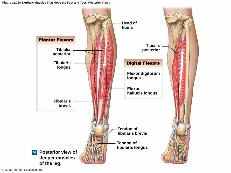
Figure 11.22c Extrinsic Muscles That Move the Foot and Toes, Posterior Views
© 2015 Pearson Education, Inc.
Head offibula
TibialisposteriorTibialia
posterior
Fibularislongus
Fibularisbrevis
Flexor digitorumlongus
Flexorhallucis longus
Tendon offibularis brevis
Tendon offibularis longus
Posterior view of
deeper muscles
of the leg.
c
Plantar Flexors
Digital Flexors
Iliotibial tract
Biceps femoris
Head of fibula
Lateral head ofgastrocnemius
Tibialis anterior
Fibularis longus
Soleus
Fibularis brevis
Extensor
digitorum longus
Superior extensor
retinaculum
Lateral malleolus
Inferior extensorretinaculum
Calcaneal tendon
Tendon of extensorhallucis longus
Lateral view of the
superficial muscles
of the right leg
b
Figure 11.24b Extrinsic Muscles That Move the Foot and Toes, Medial and Lateral Views
© 2015 Pearson Education, Inc.
Muscles of the Pelvic Girdle and Lower Limbs
• Muscles That Move the Foot and Toes
• Extrinsic muscles that move the toes
• Digital flexors and extensors
• Flexor digitorum longus: flexion of toes 2–5
• Flexor hallucis longus: flexion of the hallux
• Extensor digitorum longus: extension of toes 2–5
• Extensor hallucis longus: extension of the hallux
© 2015 Pearson Education, Inc.

Figure 11.22c Extrinsic Muscles That Move the Foot and Toes, Posterior Views
© 2015 Pearson Education, Inc.
Head offibula
TibialisposteriorTibialia
posterior
Fibularislongus
Fibularisbrevis
Flexor digitorumlongus
Flexorhallucis longus
Tendon offibularis brevis
Tendon offibularis longus
Posterior view of
deeper muscles
of the leg.
c
Plantar Flexors
Digital Flexors
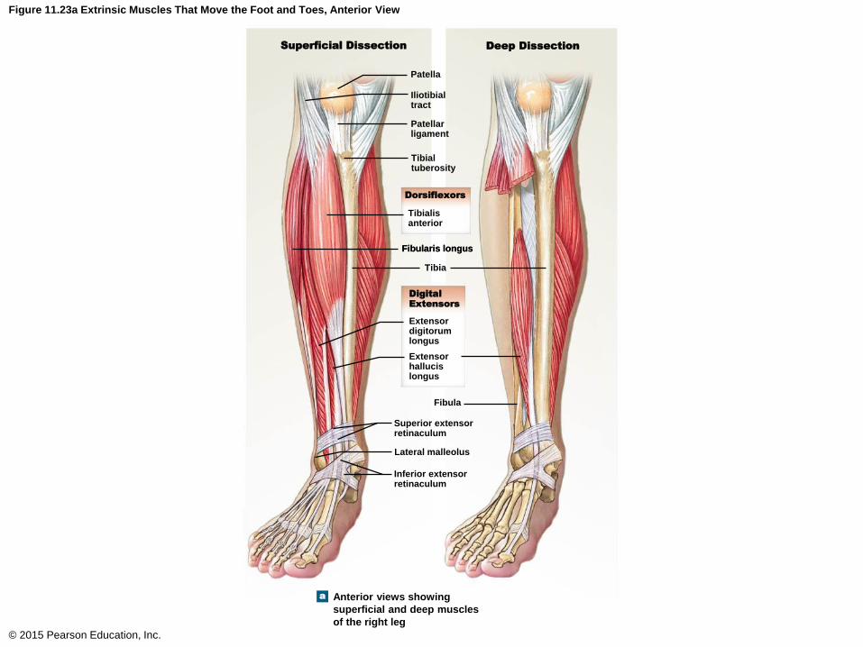
Figure 11.23a Extrinsic Muscles That Move the Foot and Toes, Anterior View
© 2015 Pearson Education, Inc.
Superficial Dissection Deep Dissection
Patella
Iliotibialtract
Patellarligament
Tibialtuberosity
Tibialisanterior
Fibularis longus
Dorsiflexors
Fibularis longus
Tibia
Digital
Extensors
Extensordigitorumlongus
Extensorhallucislongus
Fibula
Superior extensorretinaculum
Lateral malleolus
Inferior extensorretinaculum
Anterior views showing
superficial and deep muscles
of the right leg
a
Muscles of the Pelvic Girdle and Lower Limbs
• Muscles That Move the Foot and Toes
• Intrinsic muscles that move the toes
• Abductor hallucis: abduction of the hallux
• Flexor digitorum brevis: flexion of toes 2–5
• Abductor digiti minimi: abduction of the little toe
• Lumbricals: extension of toes 2–5
• Flexor hallucis brevis: flexion of the hallux
• Flexor digiti minimi brevis: flexion of the little toe
© 2015 Pearson Education, Inc.
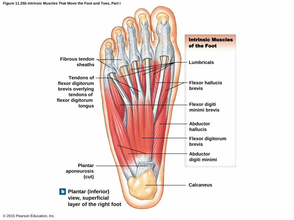
Figure 11.25b Intrinsic Muscles That Move the Foot and Toes, Part I
© 2015 Pearson Education, Inc.
Fibrous tendon
sheaths
Tendons of
flexor digitorum
brevis overlying
tendons of
flexor digitorum
longus
Lumbricals
Intrinsic Muscles
of the Foot
Flexor hallucis
brevis
Flexor digiti
minimi brevis
Abductor
hallucis
Flexor digitorum
brevis
Abductor
digiti minimi
Calcaneus
Plantar
aponeurosis
(cut)
Plantar (inferior)
view, superficial
layer of the right foot
b

Muscles of the Pelvic Girdle and Lower Limbs
• Muscles That Move the Foot and Toes
• Intrinsic muscles that move the toes
• Quadratus plantae: flexion of toes 2–5
• Flexor hallucis brevis: flexion of the hallux
• Adductor hallucis: adducts the hallux
• Plantar interossei: adduction of toes 3–5
© 2015 Pearson Education, Inc.
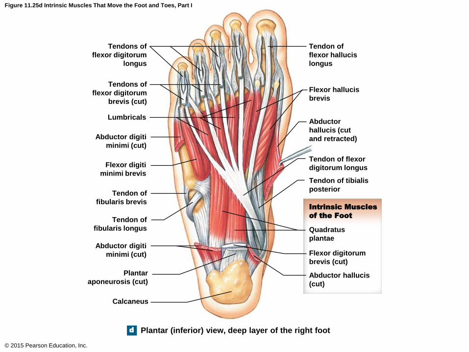
Figure 11.25d Intrinsic Muscles That Move the Foot and Toes, Part I
© 2015 Pearson Education, Inc.
Tendons of
flexor digitorum
longus
Tendons of
flexor digitorum
brevis (cut)
Lumbricals
Abductor digiti
minimi (cut)
Flexor digiti
minimi brevis
Tendon of
fibularis brevis
Tendon of
fibularis longus
Abductor digiti
minimi (cut)
Plantar
aponeurosis (cut)
Calcaneus
Tendon of
flexor hallucis
longus
Flexor hallucis
brevis
Abductor
hallucis (cut
and retracted)
Tendon of flexor
digitorum longus
Tendon of tibialis
posterior
Intrinsic Muscles
of the Foot
Quadratus
plantae
Flexor digitorum
brevis (cut)
Abductor hallucis
(cut)
d Plantar (inferior) view, deep layer of the right foot
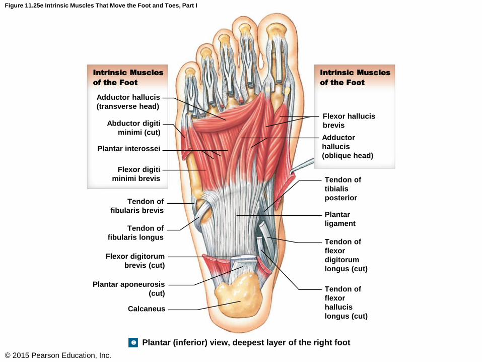
Figure 11.25e Intrinsic Muscles That Move the Foot and Toes, Part I
© 2015 Pearson Education, Inc.
Intrinsic Muscles
of the Foot
Adductor hallucis
(transverse head)
Abductor digiti
minimi (cut)
Plantar interossei
Flexor digiti
minimi brevis
Tendon of
fibularis brevis
Tendon of
fibularis longus
Flexor digitorum
brevis (cut)
Plantar aponeurosis
(cut)
Calcaneus
Intrinsic Muscles
of the Foot
Flexor hallucis
brevis
Adductor
hallucis
(oblique head)
Tendon of
tibialis
posterior
Plantar
ligament
Tendon of
flexor
digitorum
longus (cut)
Tendon of
flexor
hallucis
longus (cut)
e Plantar (inferior) view, deepest layer of the right foot
Muscles of the Pelvic Girdle and Lower Limbs
• Muscles That Move the Foot and Toes
• Intrinsic muscles that move the toes
• Quadratus plantae: flexion of toes 2–5
• Flexor hallucis brevis: flexion of the hallux
• Adductor hallucis: adducts the hallux
• Plantar interossei: adduction of toes 3–5
© 2015 Pearson Education, Inc.
Muscles of the Pelvic Girdle and Lower Limbs
• Muscles That Move the Foot and Toes
• Intrinsic muscles that move the toes
• Extensor digitorum brevis
• Extension of the toes of digits 1–4
• Dorsal interossei
• Abduction of toes 3 and 4
• Extension of toes 2 through 4
© 2015 Pearson Education, Inc.
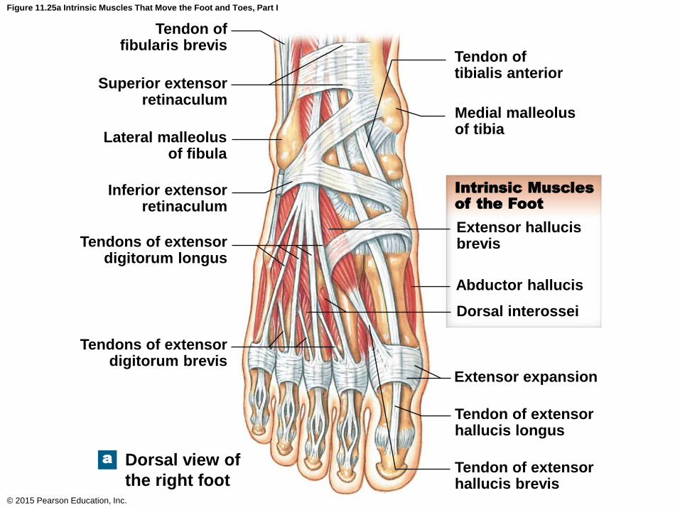
Figure 11.25a Intrinsic Muscles That Move the Foot and Toes, Part I
© 2015 Pearson Education, Inc.
Tendon offibularis brevis
Superior extensorretinaculum
Lateral malleolusof fibula
Inferior extensorretinaculum
Tendons of extensordigitorum longus
Tendons of extensordigitorum brevis
Tendon oftibialis anterior
Medial malleolusof tibia
Extensor hallucisbrevis
Abductor hallucis
Dorsal interossei
Intrinsic Muscles
of the Foot
Extensor expansion
Tendon of extensorhallucis longus
Tendon of extensorhallucis brevis
Dorsal view of
the right foot
a
Muscles That Move the Foot and Toes
© 2015 Pearson Education, Inc.
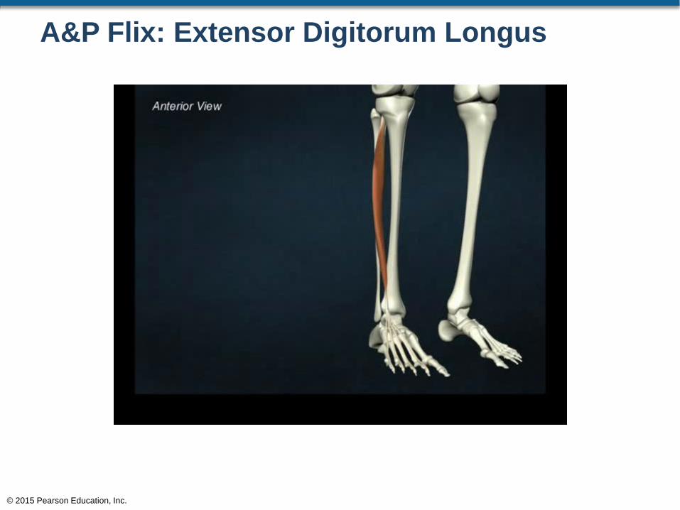
A&P Flix: Extensor Digitorum Longus
© 2015 Pearson Education, Inc.
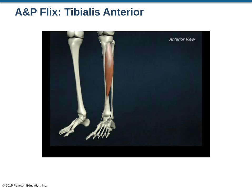
A&P Flix: Tibialis Anterior
© 2015 Pearson Education, Inc.
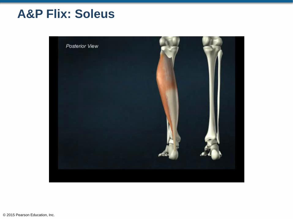
A&P Flix: Soleus
© 2015 Pearson Education, Inc.
Muscles of the Pelvic Girdle and Lower Limbs
• Action Line of Muscles
• When a muscle contracts, it develops tension
• The direction the muscle moves upon developing
tension is known as the action line
• The following figures show the action line of
muscles that move the thigh
© 2015 Pearson Education, Inc.
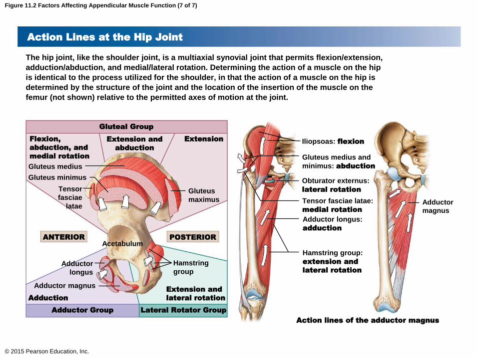
Figure 11.2 Factors Affecting Appendicular Muscle Function (7 of 7)
© 2015 Pearson Education, Inc.
Action Lines at the Hip Joint
The hip joint, like the shoulder joint, is a multiaxial synovial joint that permits flexion/extension,
adduction/abduction, and medial/lateral rotation. Determining the action of a muscle on the hip
is identical to the process utilized for the shoulder, in that the action of a muscle on the hip is
determined by the structure of the joint and the location of the insertion of the muscle on the
femur (not shown) relative to the permitted axes of motion at the joint.
Adductor
magnus
Iliopsoas: flexion
Gluteus medius and
minimus: abduction
Obturator externus:
lateral rotation
Tensor fasciae latae:
medial rotation
Adductor longus:
adduction
Hamstring group:
extension and
lateral rotation
Action lines of the adductor magnus
Gluteal Group
ExtensionExtension and
abduction
Flexion,
abduction, and
medial rotation
Lateral Rotator GroupAdductor Group
Extension and
lateral rotationAdduction
ANTERIOR POSTERIOR
Gluteus
maximus
Gluteus medius
Gluteus minimus
Tensor
fasciae
latae
Acetabulum
Hamstring
groupAdductor
longus
Adductor magnus
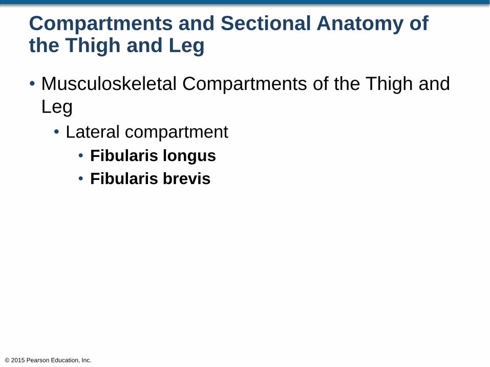
Compartments and Sectional Anatomy of the Thigh and Leg
• Musculoskeletal Compartments of the Thigh and
Leg
• Lateral compartment
• Fibularis longus
• Fibularis brevis
© 2015 Pearson Education, Inc.
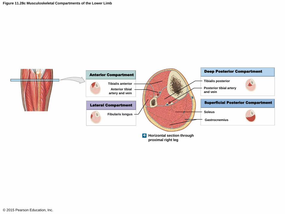
Figure 11.28c Musculoskeletal Compartments of the Lower Limb
© 2015 Pearson Education, Inc.
Tibialis anterior
Anterior tibial
artery and vein
Fibularis longus
Tibialis posterior
Posterior tibial artery
and vein
Soleus
Gastrocnemius
Deep Posterior Compartment
Superficial Posterior Compartment
Anterior Compartment
Lateral Compartment
c Horizontal section through
proximal right leg
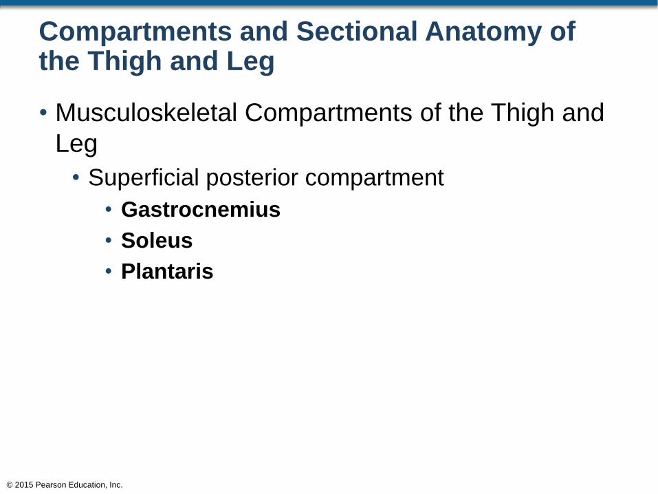
Compartments and Sectional Anatomy of the Thigh and Leg
• Musculoskeletal Compartments of the Thigh and
Leg
• Superficial posterior compartment
• Gastrocnemius
• Soleus
• Plantaris
© 2015 Pearson Education, Inc.

Figure 11.28c Musculoskeletal Compartments of the Lower Limb
© 2015 Pearson Education, Inc.
Tibialis anterior
Anterior tibial
artery and vein
Fibularis longus
Tibialis posterior
Posterior tibial artery
and vein
Soleus
Gastrocnemius
Deep Posterior Compartment
Superficial Posterior Compartment
Anterior Compartment
Lateral Compartment
c Horizontal section through
proximal right leg

Compartments and Sectional Anatomy of the Thigh and Leg
• Musculoskeletal Compartments of the Thigh and
Leg
• Anterior compartment
• Iliopsoas
• Quadriceps femoris
• Sartorius
• Extensor digitorum longus
• Extensor hallucis longus
• Fibularis tertius
• Tibialis anterior
© 2015 Pearson Education, Inc.
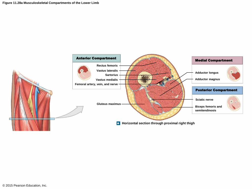
Figure 11.28a Musculoskeletal Compartments of the Lower Limb
© 2015 Pearson Education, Inc.
Anterior Compartment
Rectus femoris
Vastus lateralis
Sartorius
Vastus medialis
Femoral artery, vein, and nerve
Adductor longus
Adductor magnus
Sciatic nerve
Biceps femoris and
semitendinosis
Gluteus maximus
Medial Compartment
Posterior Compartment
Horizontal section through proximal right thigha
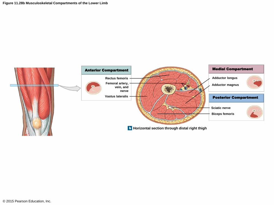
Figure 11.28b Musculoskeletal Compartments of the Lower Limb
© 2015 Pearson Education, Inc.
Anterior CompartmentMedial Compartment
Posterior Compartment
Rectus femoris
Femoral artery,
vein, and
nerve
Vastus lateralis
Adductor longus
Adductor magnus
Sciatic nerve
Biceps femoris
Horizontal section through distal right thighb

Figure 11.28c Musculoskeletal Compartments of the Lower Limb
© 2015 Pearson Education, Inc.
Tibialis anterior
Anterior tibial
artery and vein
Fibularis longus
Tibialis posterior
Posterior tibial artery
and vein
Soleus
Gastrocnemius
Deep Posterior Compartment
Superficial Posterior Compartment
Anterior Compartment
Lateral Compartment
c Horizontal section through
proximal right leg
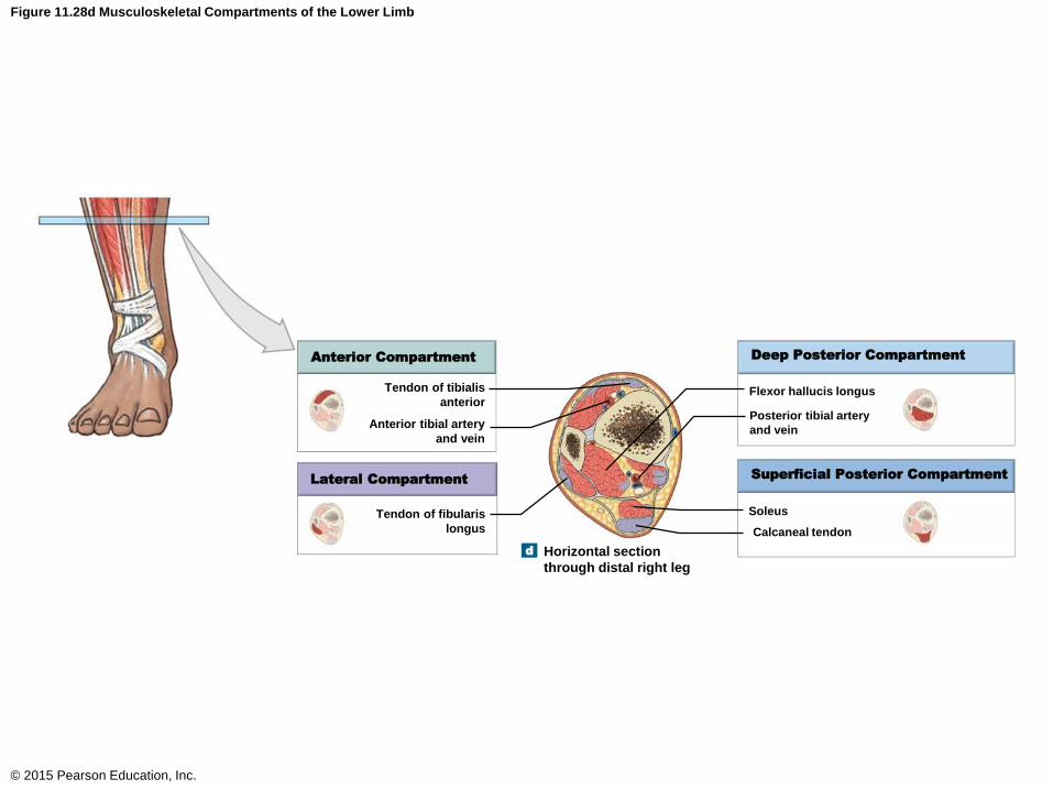
Figure 11.28d Musculoskeletal Compartments of the Lower Limb
© 2015 Pearson Education, Inc.
Anterior Compartment Deep Posterior Compartment
Superficial Posterior CompartmentLateral Compartment
Tendon of tibialis
anterior
Anterior tibial artery
and vein
Tendon of fibularis
longus
Flexor hallucis longus
Posterior tibial artery
and vein
Soleus
Calcaneal tendon
Horizontal section
through distal right leg
d
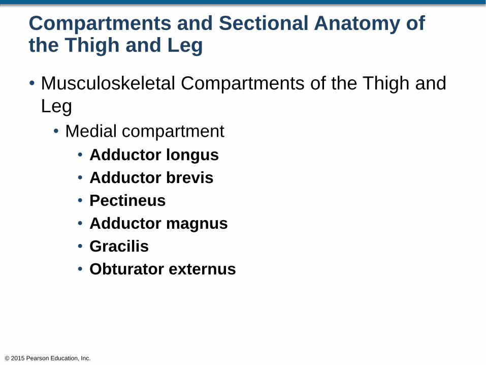
Compartments and Sectional Anatomy of the Thigh and Leg
• Musculoskeletal Compartments of the Thigh and
Leg
• Medial compartment
• Adductor longus
• Adductor brevis
• Pectineus
• Adductor magnus
• Gracilis
• Obturator externus
© 2015 Pearson Education, Inc.

Figure 11.28a Musculoskeletal Compartments of the Lower Limb
© 2015 Pearson Education, Inc.
Anterior Compartment
Rectus femoris
Vastus lateralis
Sartorius
Vastus medialis
Femoral artery, vein, and nerve
Adductor longus
Adductor magnus
Sciatic nerve
Biceps femoris and
semitendinosis
Gluteus maximus
Medial Compartment
Posterior Compartment
Horizontal section through proximal right thigha
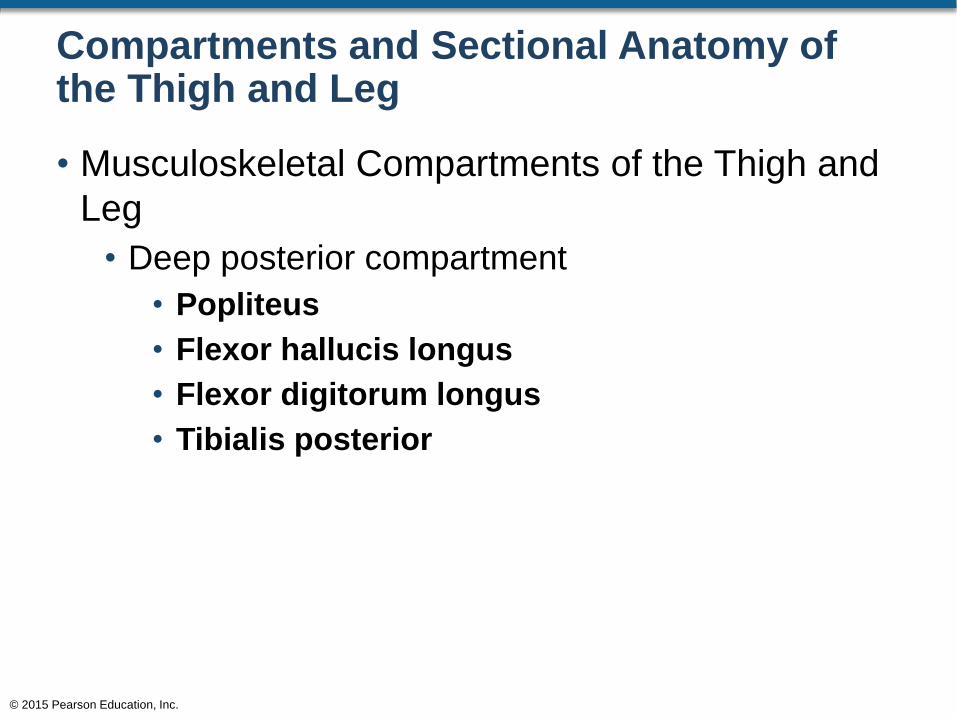
Compartments and Sectional Anatomy of the Thigh and Leg
• Musculoskeletal Compartments of the Thigh and
Leg
• Deep posterior compartment
• Popliteus
• Flexor hallucis longus
• Flexor digitorum longus
• Tibialis posterior
© 2015 Pearson Education, Inc.

Figure 11.28d Musculoskeletal Compartments of the Lower Limb
© 2015 Pearson Education, Inc.
Anterior Compartment Deep Posterior Compartment
Superficial Posterior CompartmentLateral Compartment
Tendon of tibialis
anterior
Anterior tibial artery
and vein
Tendon of fibularis
longus
Flexor hallucis longus
Posterior tibial artery
and vein
Soleus
Calcaneal tendon
Horizontal section
through distal right leg
d
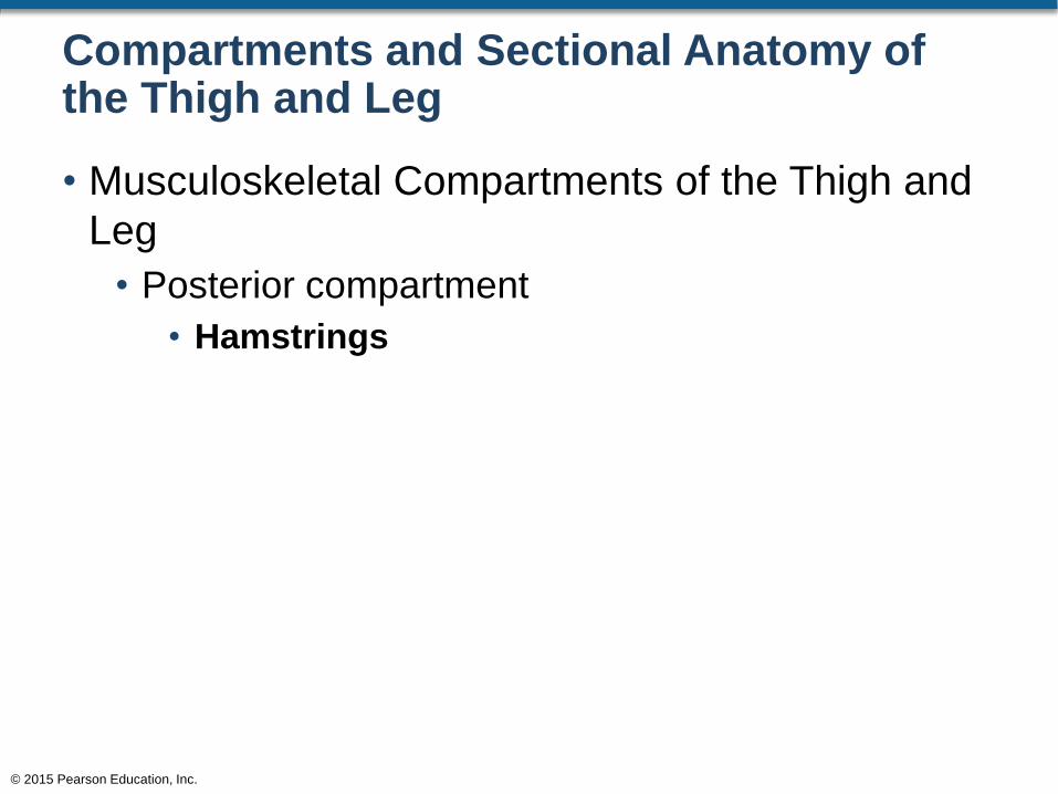
Compartments and Sectional Anatomy of the Thigh and Leg
• Musculoskeletal Compartments of the Thigh and
Leg
• Posterior compartment
• Hamstrings
© 2015 Pearson Education, Inc.