chapter 11 histology of nervous tissue j.f. thompson, ph.d
TRANSCRIPT
Chapter 11
Histology of Nervous Tissue
J.F. Thompson, Ph.D.

Histology of Nervous Tissue Despite the complexity of organization,
there are only two functional cell types neurons - excitable nerve cells that transmit
electrical signals neuroglia (glial) cells - support cells
Einstein’s brain was unusual in having more glial cells than most humans, not more neurons!
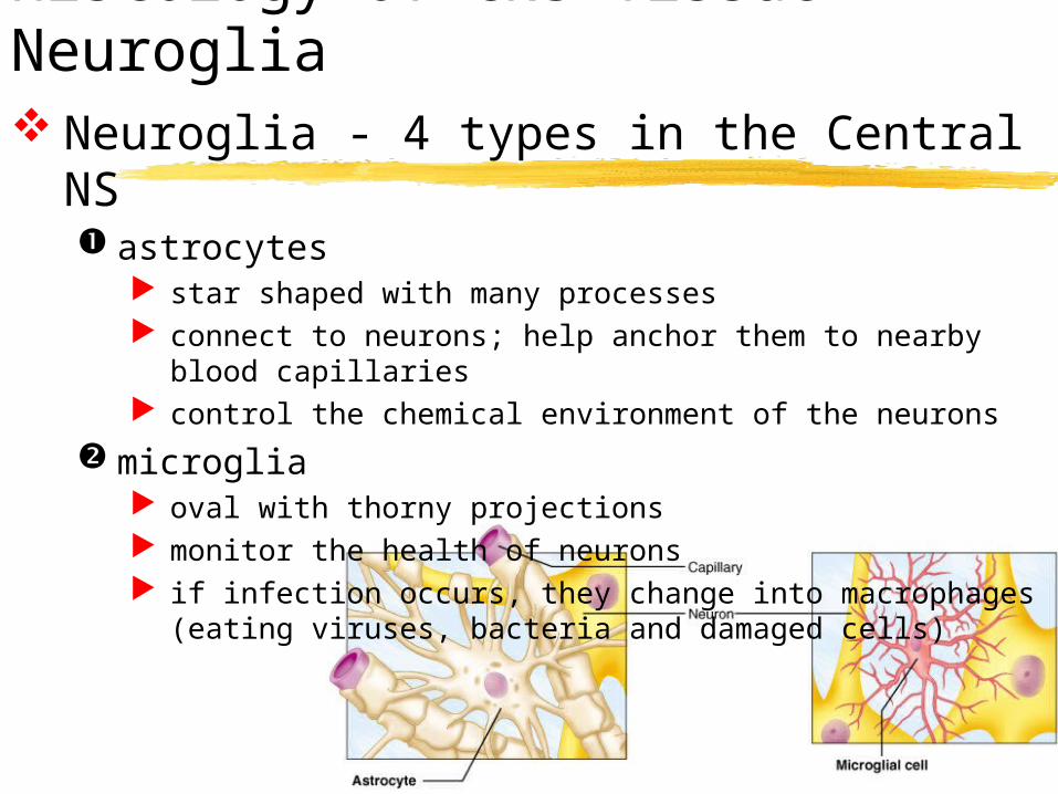
Histology of CNS Tissue - Neuroglia Neuroglia - 4 types in the Central NS
astrocytes star shaped with many processes connect to neurons; help anchor them to nearby blood
capillaries control the chemical environment of the neurons
microglia oval with thorny projections monitor the health of neurons if infection occurs, they change into macrophages (eating
viruses, bacteria and damaged cells)

Astrocytes and Microglial Cells
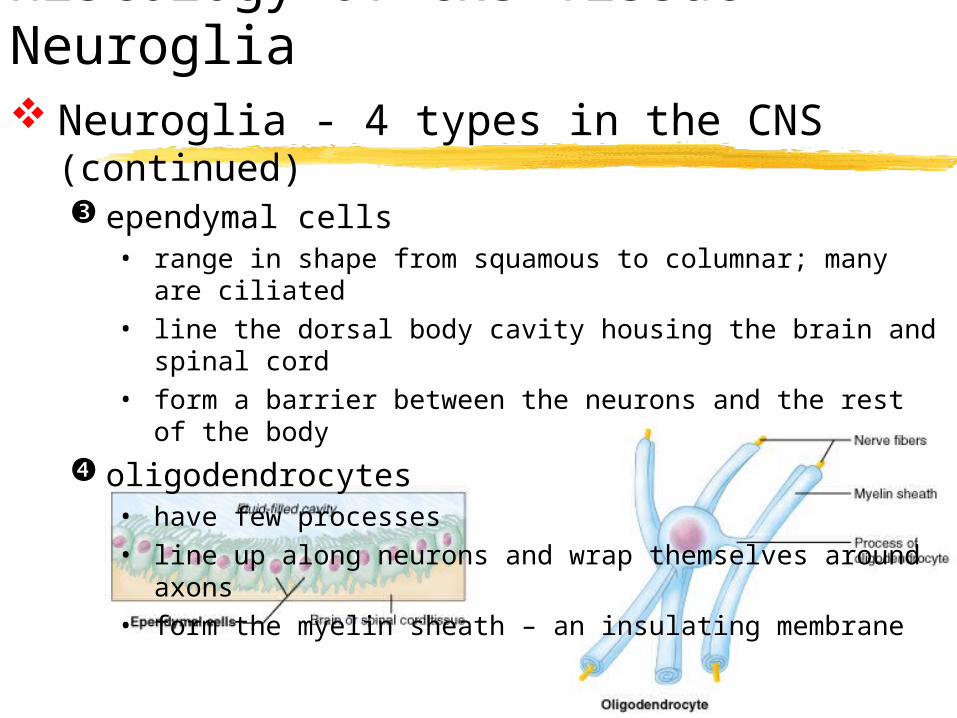
Histology of CNS Tissue - Neuroglia Neuroglia - 4 types in the CNS (continued)
ependymal cells • range in shape from squamous to columnar; many are
ciliated• line the dorsal body cavity housing the brain and spinal
cord• form a barrier between the neurons and the rest of the
body
oligodendrocytes• have few processes• line up along neurons and wrap themselves around axons• form the myelin sheath – an insulating membrane

Ependymal Cells and Oligodendrocytes

Histology of PNS Tissue - Neuroglia
Neuroglia - 2 types in the Peripheral NS
satellite cells surround neuron cell bodies in the periphery maintain the extracellular environment
neurolemmocytes (Schwann cells) surround axons/dendrites and form the myelin
sheath around larger nerve fibers in the periphery
similar to oligodendrocytes in function – insulators

Satellite Cells and Neurolemmocytes
Histology of CNS Tissue - Neurons Neurons - highly specialized cells which
conduct electrochemical signals (nerve impulses) extreme longevity – neurons live and
function normally for a lifetime amitotic
once mature, neurons lose the ability to divide damaged nervous tissue cannot regenerate
high metabolic rate need a large, constant supply of oxygen and
glucose can survive only a few minutes without oxygen

Neurons

Neuron Structure – Cell Body (Soma) Contains the usual cellular
organelles
Site of most cell metabolism
Receptive: membrane receptors initiate and transmit graded potentials (not action potentials) in response to incoming stimuli
Most neuron cell bodies are located within the CNS:Nuclei: Nuclei: clusters of neuron cell bodies in the CNS Ganglia: Ganglia: clusters of neuron cell bodies in the PNS

End CH 11Histology of Nervous Tissue