chapter 14: digestive system & body metabolism · two part system •part one: alimentary canal...
TRANSCRIPT

Chapter 14: Digestive System &
Body Metabolism Ingest Digest Absorb Defecate Rinse and Repeat
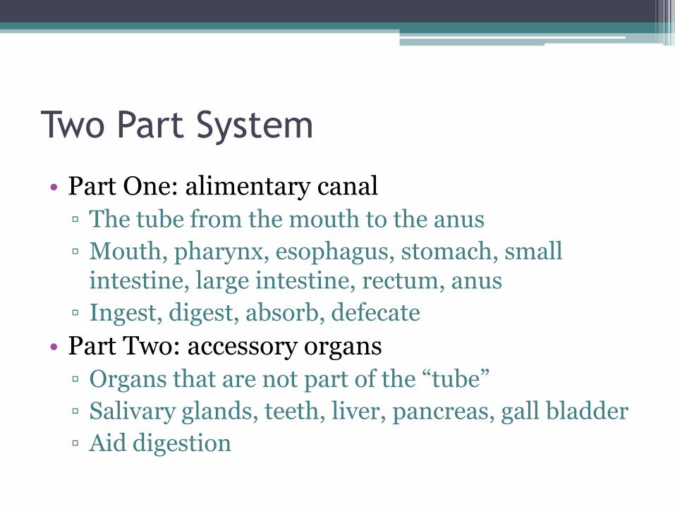
Two Part System
• Part One: alimentary canal
▫ The tube from the mouth to the anus
▫ Mouth, pharynx, esophagus, stomach, small intestine, large intestine, rectum, anus
▫ Ingest, digest, absorb, defecate
• Part Two: accessory organs
▫ Organs that are not part of the “tube”
▫ Salivary glands, teeth, liver, pancreas, gall bladder
▫ Aid digestion

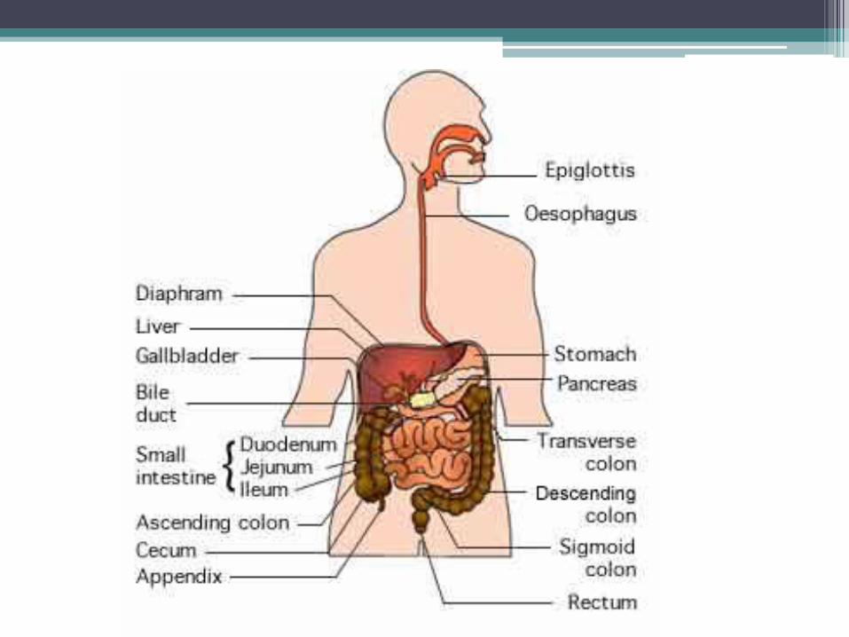
Alimentary Canal
AKA: Gastrointestinal (GI) Tract
• 9 meters long (30 feet) in cadaver
• Technically “outside” the body

Alimentary Canal Organs
Mouth (oral cavity)
• Mucous membrane lining
• Lips (labia), cheeks, hard and soft palates, uvula, vestibule (area cheek to teeth), oral cavity proper, tongue, lingual frenulum (lining under tongue), tonsils (palatine and lingual), papillae (taste buds)
• Chemical and mechanical digestion start here.
Alimentary Canal Organs
Mouth (oral cavity)
• Mechanical digestion: mastication (chewing)
• Chemical digestion: salivary amylase (enzyme that breaks down carbohydrates)
Alimentary Canal Organs
Pharynx
• Oropharynx to laryngopharynx in swallowing.
• Double walls of skeletal muscles, with one layer running parallel to the tube and one running perpendicular.
• This allows for the swallowing action of the pharynx
• No digestion here

Alimentary Canal Organs
Esophagus (AKA gullet)
• 10 inch (25 cm) tube from pharynx to stomach
• Conducting passage only; no digestion
• Made of four linings, or tunics, that extend through to the large intestine…


Alimentary Canal Organs
4 Tunics of the GI tract
• 1. mucosa: lines the lumen (inner cavity); mainly epithelia with some connective and smooth muscle; moist; some glands
• 2. submucosa: mainly soft connective tissue; holds blood and lymph vessels, lymph nodes, nerve plexus of autonomic system (control motion and secretions)

Alimentary Canal Organs
4 Tunics of the GI tract • 3. Muscularis externa: smooth muscle with inner
circular layer and outer longitudinal layer; performs peristalsis – the muscular contractions that move food down the tract
• 4. serosa (visceral peritoneum): outer lining; single layer of cells that produce serous fluid; turns into parietal peritoneum when it meets the abdominopelvic cavity; these are connected by the mesentary

Alimentary Canal Organs
Stomach • Left side of abdomen; 25 cm long; can hold 1
gallon of food Stomach regions: cardiac, body, pylorus 1. Cardiac region – most superior part of stomach • Cardioesophageal sphincter: food enters here • Fundus: expanded, superior portion of stomach 2. Body – midportion of stomach 3. Pylorus – funnel-shaped, most inferior portion • Pyloric sphincter (valve): chyme exits here
Alimentary Canal Organs
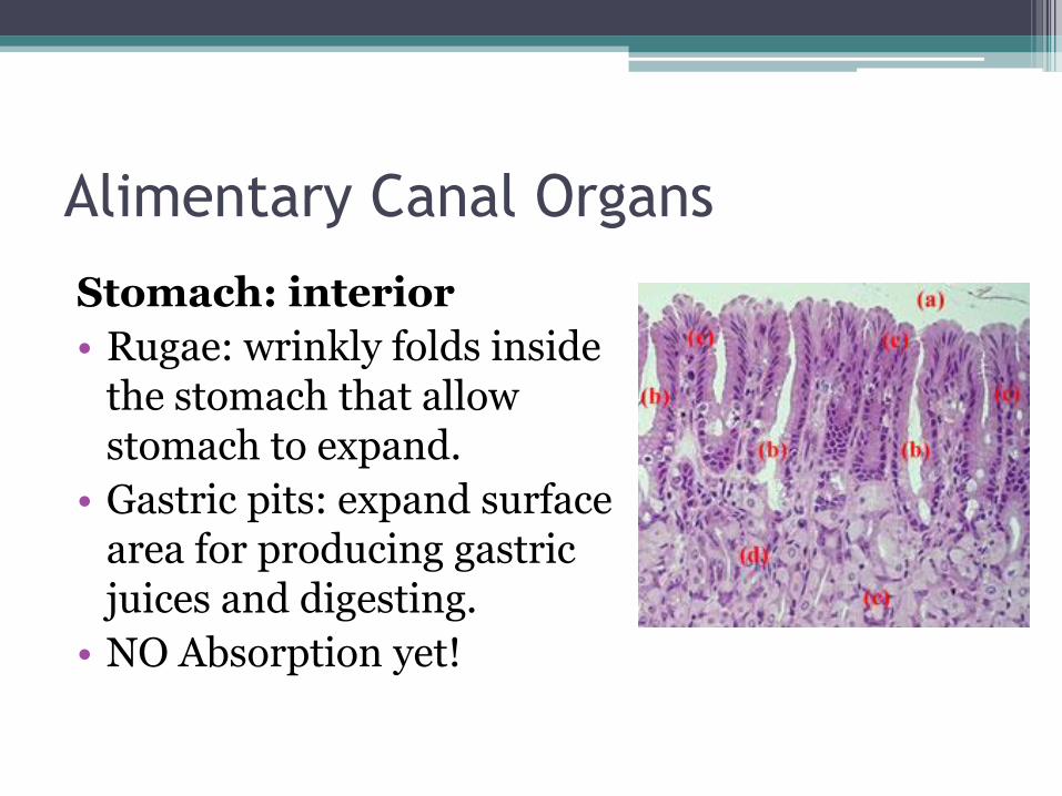
Stomach: interior
• Rugae: wrinkly folds inside the stomach that allow stomach to expand.
• Gastric pits: expand surface area for producing gastric juices and digesting.
• NO Absorption yet!
Alimentary Canal Organs
Stomach: digestion • Mechanical digestion via oblique muscle layer
that churns food • Chemical digestions via mucous and gastric juice
▫ Intrinsic factor: helps absorb vitamin B12 ▫ Pepsinogens: from chief cells, protein digesting
enzymes ▫ Hydrochloric acid: from parietal cells; enzyme
activator ▫ Gastrin: digestive hormones

Alimentary Canal Organs
Small Intestine
• Most digestion takes place here
• All nutrient absorption takes place here
• Longest part of alimentary canal; 2 – 4 meters
Regions of the small intestine:
• Duodenum: 25 cm long
• Jejunum 2.5 m long
• Ileum: 3.6 m long
• Ileocecal valve: connects small & large intestines
Alimentary Canal Organs
Small Intestine: digestion • Mechanical digestion: not much • Chemical digestions: tons! • Digestive enzymes are produced in: the
intestinal cells, pancreas and liver and are delivered to the duodenum.
• Pancreatic enzymes flow through the pancreatic duct
• Liver enzymes (bile) flow from the liver to the gall bladder then to the intestine via the bile duct

Alimentary Canal Organs
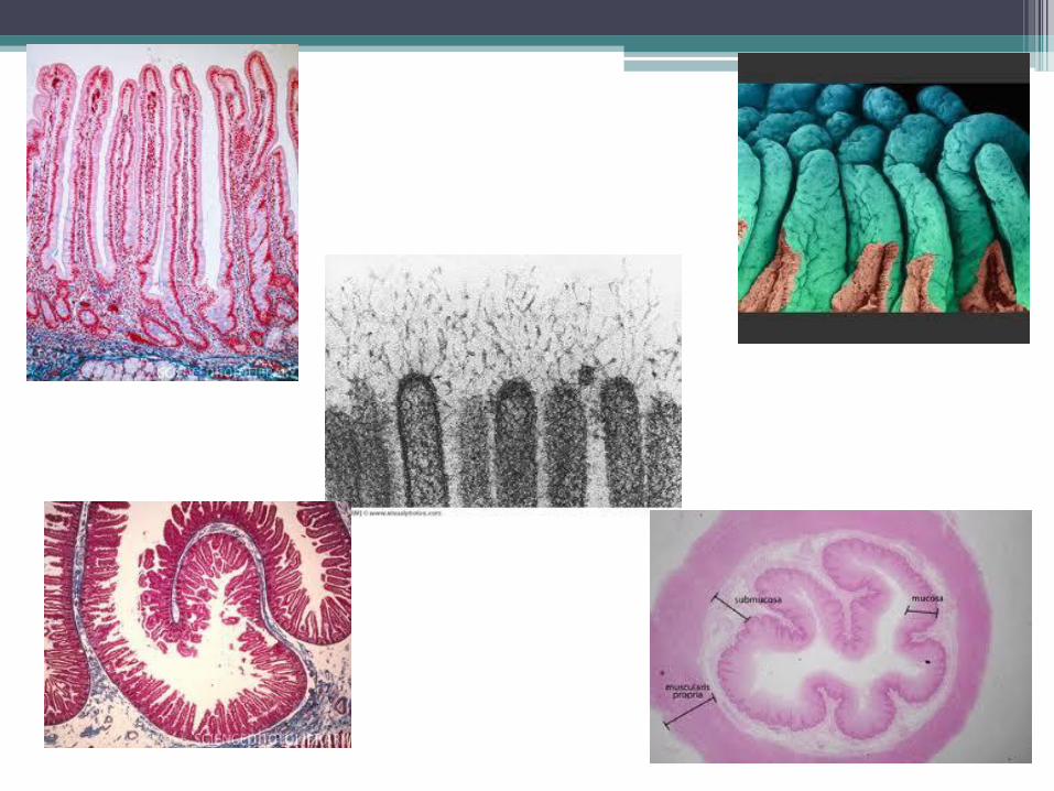
Small intestine: surface area • Three ways to increase surface area: 1. Microvilli: tiny projections on the surface of
the plasma membrane. 2. Villi: projections on the mucosal lining; filled
with blood and lymph capillaries; nutrients are absorbed at this level.
3. Plicae circulares (circular folds): deep folds in mucosa and submucosa (look like rugae of stomach).
Alimentary Canal Organs
Small intestine: surface area
• Plicae circulares do not act like rugae; they do not expand
• There are many of them at the beginning of the small intestine and decrease as you go towards the end of the tube.
• Payer’s patches increase as you go down the tube due to large numbers of bacteria in the undigested food.


Alimentary Canal Organs
Large intestine
• 1.5 meters long; from ileocecal valve to anus
• Major functions are to absorb water and eliminate feces (no real absorption of nutrients)
• No villi
• Lots and lots of goblet cells!
• They produce lots and lots of mucous for less friction.

Alimentary Canal Organs
Large intestine: areas
• subdivisions include: cecum, appendix, colon, rectum, anal canal
• Regions of the colon include: ascending colon, transverse colon, descending colon & sigmoid colon
Alimentary Canal Organs
The anus
• Open to the exterior
• The anal canal (pre-anus) has a double muscle control system for defication
▫ External sphincter: voluntary, skeletal muscle
▫ Internal sphincter: involuntary, smooth muscle

Accessory Organs
• Salivary Glands: three pair 1. Parotid glands: anterior to
the ears (huge) 2. Submadibular glands:
medium sized; lowest 3. Sublingual glands: smallest;
under tongue • All produce saliva which
contains: serous fluid, mucus, salivary amylase, lysozyme, antibodies (IgA)
Accessory Organs
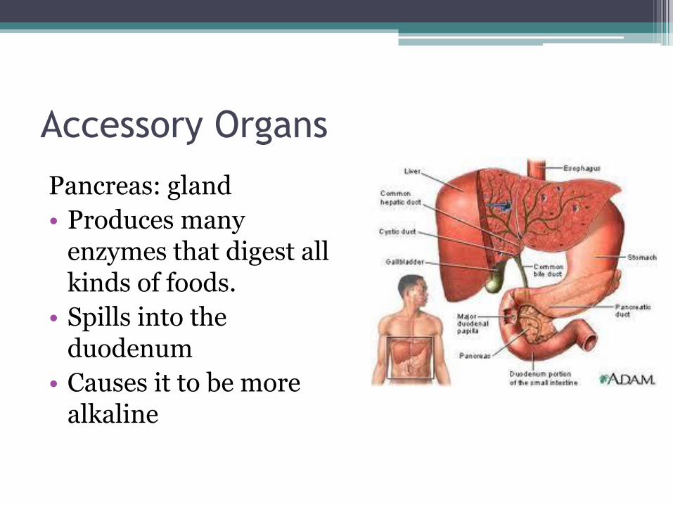
Pancreas: gland
• Produces many enzymes that digest all kinds of foods.
• Spills into the duodenum
• Causes it to be more alkaline
Accessory Organs
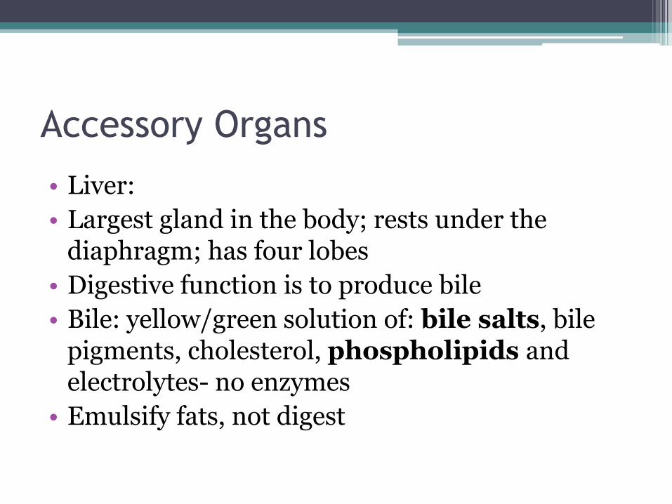
• Liver:
• Largest gland in the body; rests under the diaphragm; has four lobes
• Digestive function is to produce bile
• Bile: yellow/green solution of: bile salts, bile pigments, cholesterol, phospholipids and electrolytes- no enzymes
• Emulsify fats, not digest

Accessory Organs
• Gall bladder
• Green sac that holds bile
• Shoots the bile into the duodenum when needed
• Often is clogged and needs to be removed

Functions of the Digestive System
• 6 processes:
▫ Ingestion
▫ Propulsion
▫ Mechanical digestion
▫ Chemical digestion
▫ Absorption
▫ Defecation

Functions of the Digestive System
1. Ingestion – to bring food in through the mouth; NOT intravenously.
2. Propulsion – move food down the alimentary canal; swallowing, peristalsis, segmentation.
1. Swallowing – voluntary
2. Peristalsis – involuntary muscle contractions
3. Segmentation – involuntary; in small intestine only; moves food back and forth (more like mixing).

Functions of the Digestive System
3. Mechanical digestion: physical breakdown of food into smaller parts; increases surface area of food to enhance chemical digestion
Examples:
Mastication, churning in stomach, segmentation in small intestine

Functions of the Digestive System
4. Chemical digestion: chemical breakdown of food into nutrients for absorption into the blood stream.
Food nutrients:
• carbohydrates – monosaccharides like glucose; only carbs we digest are sucrose, lactose, maltose and starch
• Proteins – amino acids
• Lipids – fatty acids and glycerol

Functions of the Digestive System
5. Absorption: transporting nutrients from the alimentary canal to the blood stream or lymph; mainly in small intestine; a passive process
6. Defecation: elimination of undigested, unabsorbed substances as feces

Functions of the Digestive System
• Maintaining homeostasis within the lumen: 1. Stretch receptors 2. pH receptors 3. Breakdown product receptors
• Reflexes (activate or inhibit) 1. Glands secreting enzymes into lumen or
hormones into blood 2. Smooth muscles mix or propel food along the
tract

Functions of the Digestive System
Activities of the Mouth, Pharynx, & Esophagus • Ingestion • Mechanical digestion • Chemical digestion – saliva, carbohydrates • Food propulsion – deglutination (swallowing) and
peristalsis ▫ Swallowing – phase one – buccal phase: in the mouth,
voluntary, pushed by tongue into pharynx ▫ Swallowing – phase two – phyngeal-eophageal phase:
involuntary (vagus nerves), peristalsis takes over (no gravitational effect)

Functions of the Digestive System
Activities of the Stomach • Food breakdown • Gastric juice secretion is regulated by sight, smell,
and taste. • Gastrin (hormone): regulated by food and lower pH;
entices increased enzyme, mucus and HCl production
• Pepsinogen is activated by HCl to pepsin (protein digester)
• Rennin: milk protein digester (mainly in children) • Only absorption is aspirin and alcohol

Functions of the Digestive System
Activities of the Stomach
• Food propulsion
• Peristalsis moves 30 ml of chyme toward the pyloric sphincter
• Only 3 ml can squirt through at a time
• If the duodenum gets full, it closes the valve
• About 4-6 hours in the stomach
• Vomiting = reverse peristalsis

Functions of the Digestive System
Activities of the small intestine • Food breakdown and absorption • 3-6 hours • Brush border enzymes: break down sugars and proteins • Pancreatic juices: controlled by secretin and CCK
(hormones) 1. Pancreatic amylase – carbs 2. Protein digestion – trypsin, chymotrypsin,
carboxypeptidase 3. Lipases – lipids 4. Nucleases – nucleic acids 5. Bicarbonate – increase pH to 8

Functions of the Digestive System
Activities of the small intestine
• Liver and gall bladder: also controlled by secretin and CCK
• Secretin: increases bile production
• CCK (cholecystokinin): contracts the gall bladder
• Bile: emulsifies fat and allows the absorption of fat soluble vitamins (A, D, K)

Functions of the Digestive System
Activities of the small intestine
• Absorption of water and nutrients to the hepatic portal vein (lipids = passive; others = active)
• Food propulsion: peristalsis and segmentation

Functions of the Digestive System
Activities of the large intestine
(12 – 24 hours)
• Food breakdown: just by bacterial flora
• They can digest some cellulose that we can’t
• This produces methane and hydrogen sulfide flatulence. (about 500 ml of it/day)
• These bacteria also produce Vitamins K and some B
Functions of the Digestive System
Activities of the large intestine
• Absorption: water, vitamins K and B and some ions dissolved in the water
• That leaves us with feces!
• Feces contains: undigested food, mucus, millions of bacteria, and a little bit of water
Functions of the Digestive System
Activities of the large intestine
• Propulsion:
1. Peristalsis – limited here
2. Mass movements – long, slow, powerful contractions that only happen a few times per day (usually when eating)
▫ Fiber increases the strength of the mass movements and that actually softens the feces.

Functions of the Digestive System
Activity of the rectum and anus
• Defecation reflex in the rectum is stimulated when feces makes its way from the colon.
• Causes the sigmoid colon and rectum muscles to contract and anal sphincters to relax.
• Fast feces movements = diarrhea
• Slow feces movements = constipation