chapter 14 mechanisms of enzyme action biochemistry by reginald garrett and charles grisham
TRANSCRIPT

Chapter 14
Mechanisms of Enzyme Action
Biochemistry
by
Reginald Garrett and Charles Grisham

Essential QuestionAlthough the catalytic properties of enzymes
may seem almost magical, it is simply chemistry– the breaking and making of bonds– that give enzymes their prowess
What are the universal chemical principles that influence the mechanisms of enzymes
And allow us to understand their enormous catalytic power?

Outline of Chapter 141. What Are the Magnitudes of Enzyme-Induced
Rate Accelerations?2. What Role Does Transition-State Stabilization
Play in Enzyme Catalysis?3. How does Destabilization of ES Affect Enzyme
Catalysis?4. How Tightly Do Transition-State Analogs Bind
to the Active Site?5. What Are the Mechanisms of Catalysis?6. What Can Be Learned from Typical Enzyme
Mechanisms?

• Enzymes are powerful catalysts• The large rate accelerations of enzymes (107 to 1015)
correspond to large changes in the free energy of activation for the reaction
• All reactions pass through a transition state on the reaction pathway
• The active sites of enzymes bind the transition state of the reaction more tightly than the substrate
• By doing so, enzymes stabilize the transition state and lower the activation energy of the reaction
14.1 – What Are the Magnitudes of Enzyme-Induced Rated Accelerations?


H-O-H + Cl- H-O H Cl HO- + HCl Reactant Transition state Products
• Transition state (10-13sec) • Intermediate (10-13~10-3sec)
- -

14.2 – What Role Does Transition-State Stabilization Play in Enzyme Catalysis?
• The catalytic role of an enzyme is to reduce the energy barrier between substrate S and transition state
• Rate acceleration by an enzyme means that the energy barrier between ES and EX‡ must be smaller than the barrier between S and X ‡
• This means that the enzyme must stabilize the EX ‡ transition state more than it stabilizes ES→Enzymes bind the transition state structure more
tightly than the substrate

Figure 14.1 Enzymes catalyze reactions by lowering the activation energy. Here the free energy of activation for (a) the uncatalyzed reaction, Gu
‡, is larger than that for (b) the enzyme-catalyzed reaction, Ge
‡.
14.3 – How does Destabilization of ES Affect Enzyme Catalysis?
• The favorable interactions between the substrate and amino acid residues on the enzyme account for the intrinsic binding energy, Gb
• The intrinsic binding energy ensures the favorable formation of the ES complex
• If uncompensated, it makes the activation energy for the enzyme-catalyzed reaction unnecessarily large and wastes some of the catalytic power of the enzyme

Figure 14.2 The intrinsic binding energy of the enzyme-substrate (ES) complex (Gb ) is compensated to some extent by entropy loss due to the binding of E and S (TS) and by destabilization of ES (Gd) by strain, distortion, desolvation , and similar effects. If Gb were not compensated by TS and Gd, the formation of ES would follow the dashed line.
intrinsic binding energy
What Roles Do Entropy Loss and Destabilization of the ES Complex Play?
Raising the energy of ES raises the rate• For a given energy of EX‡, raising the
energy of ES will increase the catalyzed rate
• This is accomplished by 1. loss of entropy due to formation of ES2. destabilization of ES by
• structural strain & distortion• desolvation• electrostatic effects

Figure 14.3 (a) Catalysis does not occur if the ES complex and the transition state for the reaction are stabilized to equal extents. (b) Catalysis will occur if the transition state is stabilized to a greater extent than the ES complex (right). Entropy loss and destabilization of the ES complex Gd ensure that this will be the case.

Figure 14.4(a)
Formation of the ES complex results in a loss of entropy. Prior to binding, E and S are free to undergo translational and rotational motion. By comparison, the ES complex is a more highly ordered, low-entropy complex.

14.4 – How Tightly Do Transition-State Analogs Bind to the Active Site?
• Transition state is exists only for about 10 -13 sec, less than the time required for a bond vibration
• The nature of the elusive transition state can be explored using transition state analogs
• Transition state analogs are stable molecules, chemically and structurally similar to the transition state
• Transition-state analogs are only approximations of the transition state itself and will never bind as tightly as would be expected for the true transition state
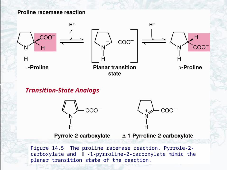
Figure 14.5 The proline racemase reaction. Pyrrole-2-carboxylate and -1-pyrroline-2-carboxylate mimic the planar transition state of the reaction.
Transition-State Analogs


Transition-State Analogs Make Our World Better
• Enzymes are often targets for drugs and other beneficial agents
• Transition state analogs often make ideal enzyme inhibitors (p452-453)– Enalapril and Aliskiren lower blood pressure– Statins lower serum cholesterol– Protease inhibitors are AIDS drugs– Juvenile hormone esterase is a pesticide target– Tamiflu is a viral neuraminidase inhibitor
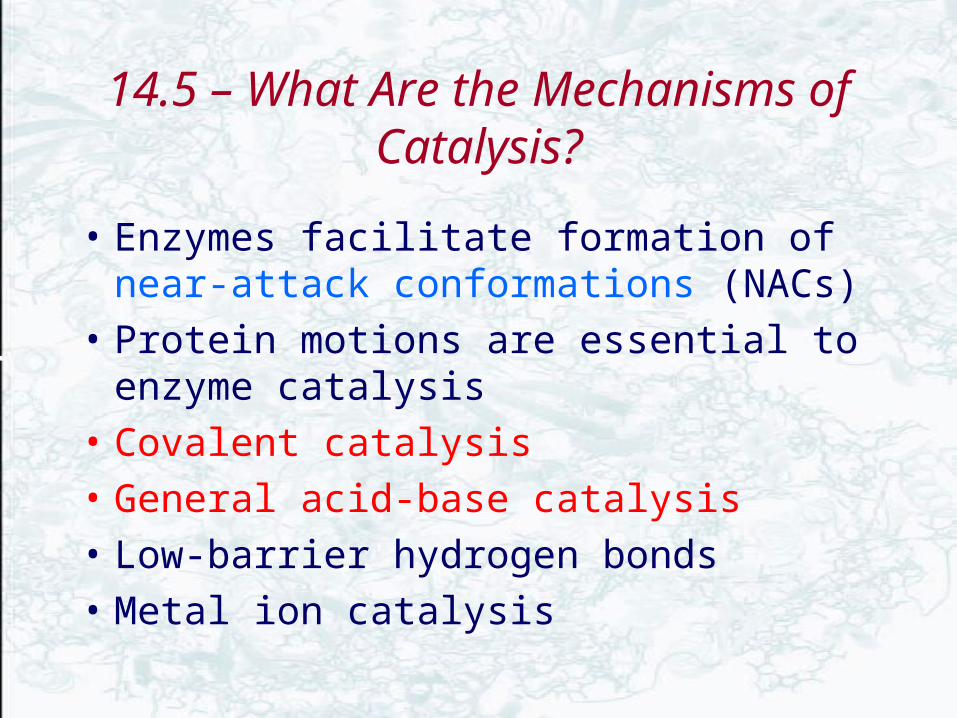
14.5 – What Are the Mechanisms of Catalysis?
• Enzymes facilitate formation of near-attack conformations (NACs)
• Protein motions are essential to enzyme catalysis
• Covalent catalysis
• General acid-base catalysis
• Low-barrier hydrogen bonds
• Metal ion catalysis

Enzymes facilitate formation of near-attack conformations
• X-ray crystal structure studies and computer modeling have shown that the reacting atoms and catalytic groups are precisely positioned for their roles
• This preorganization of active site allow it to select and stabilize conformations of substrate(s) in which the reacting atoms are in van der Waals contact and at an angle resembling the bond to be formed in the transition state
• such arrangements have been termed near-attack conformations (NACs)
• NACs are precursors to reaction transition states

• In the absence of an enzyme, potential reactant molecules adopt a NAC only about 0.0001% of the time
• On the other hand, NACs have been shown to form in enzyme active sites from 1% to 70% of the time
Figure 14.7 NACs are characterized as having reacting atoms within 3.2 Å and an approach angle of ±15° of the bonding angle in the transition state.
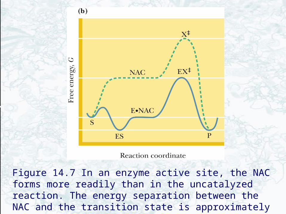
Figure 14.7 In an enzyme active site, the NAC forms more readily than in the uncatalyzed reaction. The energy separation between the NAC and the transition state is approximately the same in the presence and absence of the enzyme.

Figure 14.8 The active site of liver alcohol dehydrogenase – a near-attack complex.
The side-chain oxygen of ser48 approaches within 1.8 A of the hydroxyl hydrogen of the substrate,Benzyl alcohol

Protein motions are essential to enzyme catalysis• Proteins are constantly moving (p173; table 6.2)– bonds
vibrate, side chains bend and rotate, backbone loops wiggle and sway, and whole domains move as a unit
• Enzymes depend on such motions to provoke and direct catalytic events
• Protein motions support catalysis in several ways: Active site conformation changes can– Assist substrate binding– Bring catalytic groups into position around a
substrate– Induce formation of NACs– Assist in bond making and bond breaking– Facilitate conversion of substrate to product

Human Cyclophilin A
Arg55Lys82Leu98Ser99Ala101Gln102Ala105Gly109

14.5 – What Are the Mechanisms of Catalysis?
Covalent catalysis• Some enzyme reactions derive much of their
rate acceleration from the formation of covalent bonds between enzyme and substrate
BX + Y BY + X
BX + Enz E:B + X + Y Enz + BY
• Most enzymes that carry out covalent catalysis have ping-pong kinetic mechanisms

• The side chains of amino acids in proteins offer a variety of nucleophilic centers for catalysis, including amines, carboxylate, aryl and alkyl hydroxyls, imidazoles, and thiol groups
• These groups are readily attack electrophilic centers of substrates, forming covalently bonded enzyme-substrate intermediate

Figure 14.11 Examples of covalent bond formation between enzyme and substrate. In each case, a nucleophilic center (X:) on an enzyme attacks an electrophilic center on a substrate.
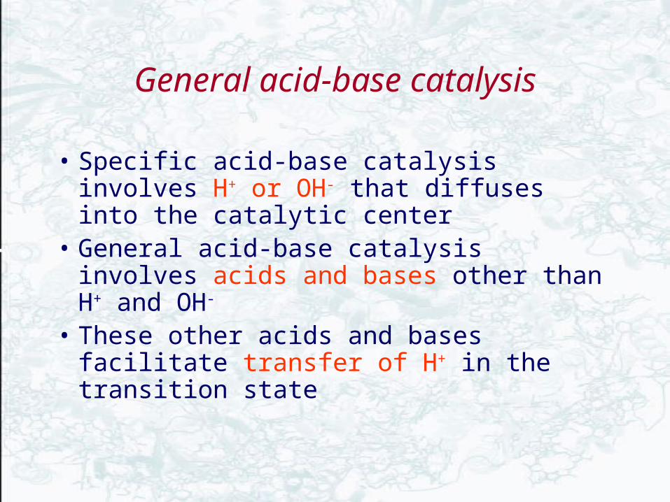
General acid-base catalysis
• Specific acid-base catalysis involves H+ or OH- that diffuses into the catalytic center
• General acid-base catalysis involves acids and bases other than H+ and OH-
• These other acids and bases facilitate transfer of H+ in the transition state
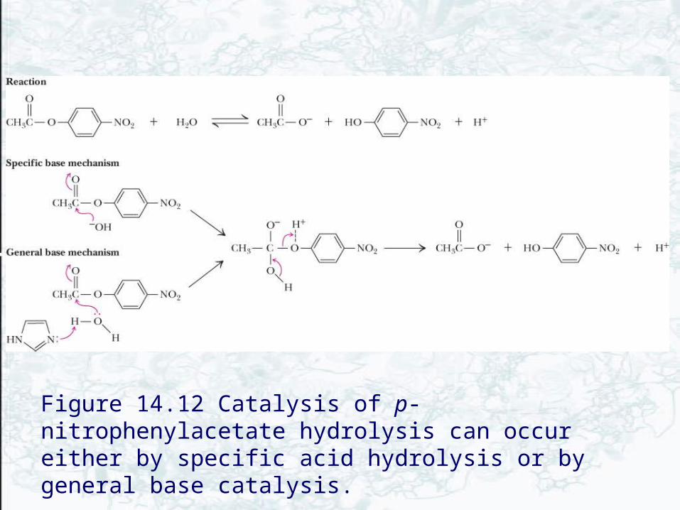
Figure 14.12 Catalysis of p-nitrophenylacetate hydrolysis can occur either by specific acid hydrolysis or by general base catalysis.

Low-Barrier Hydrogen Bonds
• The typical H-bond strength is 10-30 kJ/mol, and the O-O separation is typically 0.28 nm
• As distance between heteroatoms becomes smaller (<0.25 nm), H bonds become stronger
• Stabilization energies of LBHB may approach 60 kJ/mol in solution
• pKa values of the two electronegative atoms must be similar
• Energy released in forming an LBHB can assist catalysis

Low-barrier hydrogen bond (LBHB)• A weak H-bond may become an LBHB in the transition state
for the reaction• The energy released in forming the LBHB is used to help the
reaction to lowering the activation barrier for the reaction
0.1 nm 0.18nm1 order 0.07
0.24 nm (LBHB)0.5 order
Quantum mechanical tunneling

14.5 – What Are the Mechanisms of Catalysis?
Metal ion catalysis
• Many enzymes require metal ions for maximal activity (metalloenzymes)
1. Stabilizing the increased electron density or negative charge
2. Provide a powerful nucleophile at neutral pH
M2+ + NucH M2+(NucH) M2+(NucH) + H+
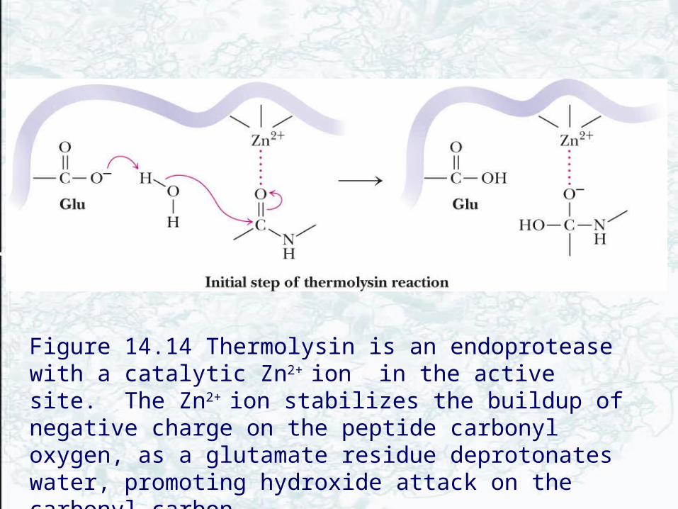
Figure 14.14 Thermolysin is an endoprotease with a catalytic Zn2+
ion in the active site. The Zn2+ ion stabilizes the buildup of negative charge on the peptide carbonyl oxygen, as a glutamate residue deprotonates water, promoting hydroxide attack on the carbonyl carbon.
14.6 – What Can Be Learned from Typical Enzyme Mechanisms?
• Serine proteases and aspartic proteases are good examples– Serine proteases employ a covalent and general acid-
base catalysis– Aspartic proteases only use general acid-base
catalysis
• Chorismate mutase use the formation of a NAC to carry out its reaction
The Serine Proteases• Serine proteases are a class of proteolytic
enzymes whose catalytic mechanism is based on an active-site serine residue
• The family includes trypsin, chymotrypsin, elastase, thrombin, subtilisin, plasmin, TPA, etc..
• The first three of these are digestive enzymes and are synthesized in the pancreas and secreted into the digestive tract as inactive proenzymes, or zymogens
• Trypsin, chymotrypsin, and elastase all carry out the same reaction—the cleavage of a peptide chain (see Table 5.2)
The Serine Proteases• These three enzymes all have similar sequences
(fig 14.15) and 3-D structures (fig 14.16) • A "catalytic triad" at the active site (fig 14.17) • Ser is part of a "catalytic triad" of Ser, His, Asp • Enzymologists agree, however, to number them
always as His-57, Asp-102, Ser-195• The active site is actually a depression on the
surface of the enzyme, with a pocket that the enzyme uses to identify the residue for which it is specific (fig 14.18)


Figure 14.15Comparison of the amino acid sequences of chymotrypsinogen, trypsinogen, and elastase. Each circle represents one amino acid. Numbering is based on the sequence of chymotrypsinogen. Filled circles indicate residues that are identical in all three proteins. Disulfide bonds are indicated in yellow. The positions of the three catalytically important active-site residues (His57, Asp102, and Ser195) are indicated.
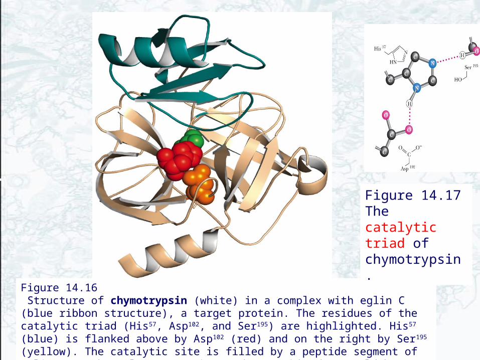
Figure 14.16 Structure of chymotrypsin (white) in a complex with eglin C (blue ribbon structure), a target protein. The residues of the catalytic triad (His57, Asp102, and Ser195) are highlighted. His57 (blue) is flanked above by Asp102 (red) and on the right by Ser195 (yellow). The catalytic site is filled by a peptide segment of eglin. Note how close Ser195 is to the peptide that would be cleaved in a chymotrypsin reaction.
Figure 14.17The catalytic triad of chymotrypsin .

Figure 14.18 The substrate-binding pockets of trypsin, chymotrypsin, and elastase.

Serine Protease Mechanism
A mixture of covalent and general acid-base catalysis
• Asp-102 functions only to orient His-57 • His-57 acts as a general acid and base • Ser-195 forms a covalent bond with peptide
to be cleaved • Covalent bond formation turns a trigonal C
into a tetrahedral C • The tetrahedral oxyanion intermediate is
stabilized by N-Hs of Gly-193 and Ser-195
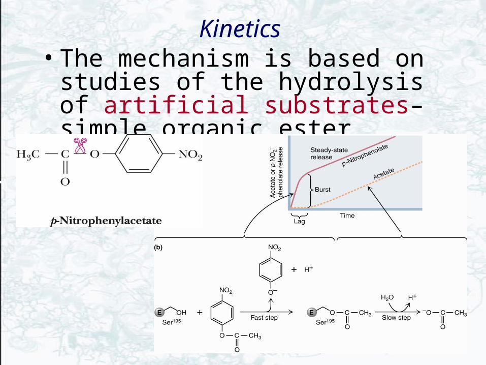
Kinetics • The mechanism is based on studies of
the hydrolysis of artificial substrates– simple organic ester
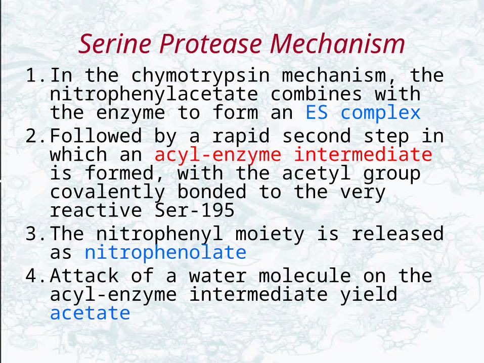
Serine Protease Mechanism1. In the chymotrypsin mechanism, the
nitrophenylacetate combines with the enzyme to form an ES complex
2. Followed by a rapid second step in which an acyl-enzyme intermediate is formed, with the acetyl group covalently bonded to the very reactive Ser-195
3. The nitrophenyl moiety is released as nitrophenolate
4. Attack of a water molecule on the acyl-enzyme intermediate yield acetate

Figure 14.23A detailed mechanism for the chymotrypsin reaction. Note the low-barrier hydrogen bond (LBHB) in (c) and (g).
Tetrahedral oxyanion transition state
Tetrahedral oxyanion transition state
ES complex
Acyl-enzyme intermediate Acyl-enzyme-H2O intermediate

The Serine Protease Mechanism in Detail
Figure 14.21 The chymotrypsin mechanism: binding of a model substrate.

The Serine Protease Mechanism in Detail
Figure 14.21 The chymotrypsin mechanism: the formation of the covalent ES complex involves general base catalysis by His57

The Serine Protease Mechanism in Detail
Figure 14.21 The chymotrypsin mechanism: His57 stabilized by a LBHB.
1st tetrahedral intermediate

The Serine Protease Mechanism in Detail
Figure 14.21 The chymotrypsin mechanism: collapse of the tetrahedral intermediate releases the first product.
The Serine Protease Mechanism in Detail
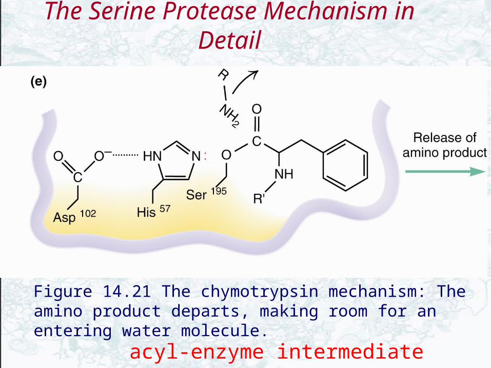
Figure 14.21 The chymotrypsin mechanism: The amino product departs, making room for an entering water molecule.
acyl-enzyme intermediate

The Serine Protease Mechanism in Detail
Figure 14.21 The chymotrypsin mechanism: Nucleophilic attack by water is facilitated by His57, acting as a general base.
acyl-enzyme-H2O complex

The Serine Protease Mechanism in Detail
Figure 14.21 The chymotrypsin mechanism: Collapse of the tetrahedral intermediate cleaves the covalent intermediate, releasing the second product.
2nd tetrahedral intermediate
The Serine Protease Mechanism in Detail
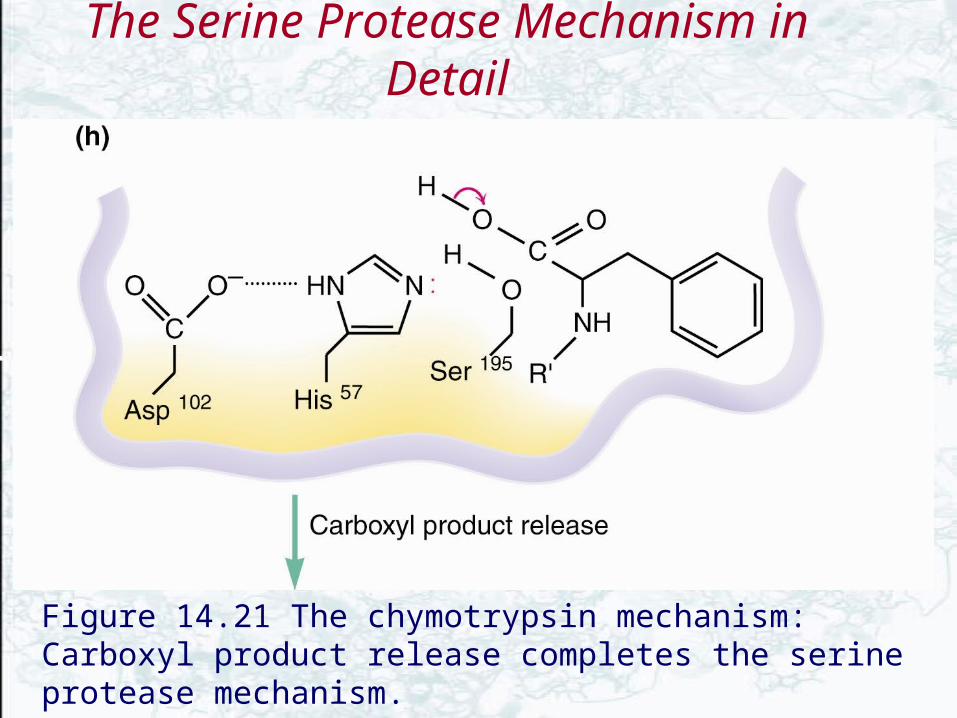
Figure 14.21 The chymotrypsin mechanism: Carboxyl product release completes the serine protease mechanism.
The Serine Protease Mechanism in Detail
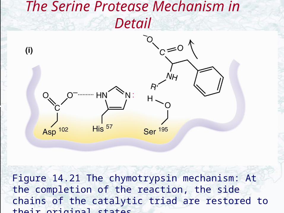
Figure 14.21 The chymotrypsin mechanism: At the completion of the reaction, the side chains of the catalytic triad are restored to their original states.

Transition-State Stabilization in the Serine Proteases
• The chymotrypsin mechanism involves two tetrahedral oxyanion intermediates
• These intermediates are stabilized by a pair of amide groups that is termed the “oxyanion hole”
• The amide N-H groups of Ser195 and Gly193 provide primary stabilization of the tetrahedral oxyanion

The “oxyanion hole”
The oxyanion hole of chymotrypsin stabilizes the tetrahedral oxyanion intermediate seen in the mechanism of Figure 14.21.

The Aspartic Proteases• These enzymes are active at acidic pH• possesses two Asp residues at the active site
and two Asps work together as general acid-base catalysts

The Aspartic Proteases
• Most aspartic proteases have a tertiary structure consisting of two lobes (N-terminal and C-terminal) with approximate two-fold symmetry (fig 14.22b, pepsin)
• HIV-1 protease is a homodimer (fig 14.22a)

Aspartic Protease Mechanism
• pH dependence (fig 14.23)
• The aspartate carboxyl groups functioned alternately as general acid and general base– Deprotonated Asp acts as general base, accepting a
proton from HOH, forming OH- in the transition state
– Other Asp (general acid) donates a proton, facilitating formation of tetrahedral intermediate

Figure 14.23pH-rate profiles for (a) pepsin and (b) HIV protease. (Adapted fro Denburg,J., et at., 1968. The effect of PH on the rates of hydrolysis of three acylated dipeptiedes by pepsin.

A Mechanism for the Aspartic Proteases
Figure 14.24 Mechanism for the aspartic proteases. LBHBs play a role in states E, ES, ET’, EQ’, and EP’Q.

HIV-1 ProteaseA novel aspartic protease
• HIV-1 protease cleaves the polyprotein products of the HIV genome, producing several proteins necessary for viral growth and cellular infection
• HIV-1 protease is a homodimer - more genetically economical for the virus
• Active site is two-fold symmetric • Two aspartate residues, Asp-25 and Asp-25’

Figure 14.26HIV mRNA provides the genetic information for synthesis of a polyprotein . Proteolytic cleavage of this polyprotein by HIV protease produces the individual proteins required for viral growth and cellular infection.

Figure 14.27(left) HIV-1 protease complexed with the inhibitor Crixivan (red) made by Merck. The flaps (residues 46-55 from each subunit) covering the active site are shown in green and the active site aspartate residues involved in catalysis are shown in white. (right) The close-up of the active site shows the interaction of Crixivan with the carboxyl groups of the essential aspartate residues.
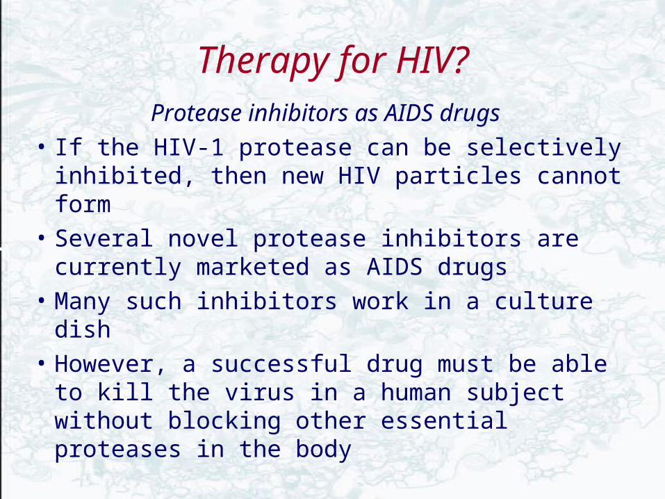
Therapy for HIV?
Protease inhibitors as AIDS drugs
• If the HIV-1 protease can be selectively inhibited, then new HIV particles cannot form
• Several novel protease inhibitors are currently marketed as AIDS drugs
• Many such inhibitors work in a culture dish
• However, a successful drug must be able to kill the virus in a human subject without blocking other essential proteases in the body

Protease inhibitor drugs used by AIDS Patients

Chorismate Mutase: A Model for Understanding Catalytic Power and Efficiency
• Direct comparison of enzyme-catalyzed reactions and their uncatalyzed counterparts is difficult
• Chorismate mutase acts in the biosynthesis of phenylalanine and tyrosine in microorganisms and plants
• It involves a single substrate and catalyzes a concerted intramolecular rearrangement of chorismate to prephenate
• One C-O bond is broken and one C-C bond is formed


The chorismate mutase reaction (and its uncatalyzed counterpart) occur via chair states
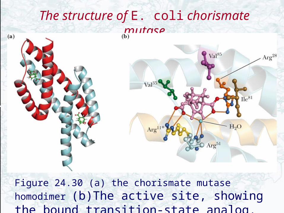
The structure of E. coli chorismate mutase
Figure 24.30 (a) the chorismate mutase homodimer (b)The active site, showing the bound transition-state analog.
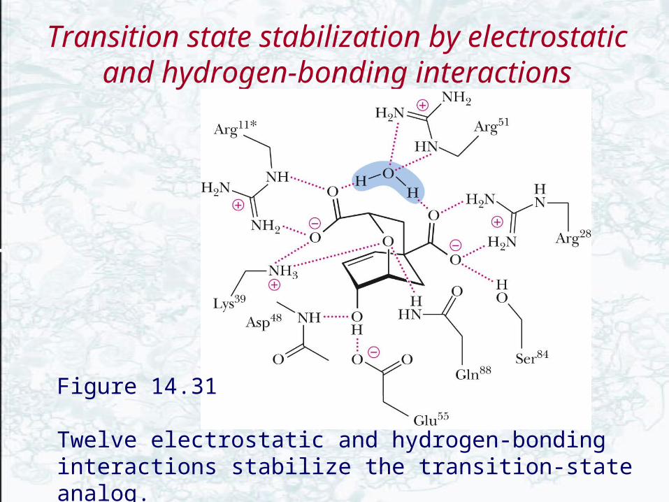
Transition state stabilization by electrostatic and hydrogen-bonding interactions
Figure 14.31
Twelve electrostatic and hydrogen-bonding interactions stabilize the transition-state analog.
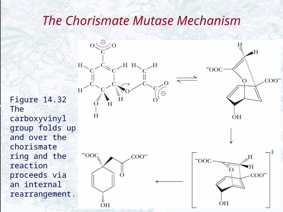
The Chorismate Mutase Mechanism
Figure 14.32 The carboxyvinyl group folds up and over the chorismate ring and the reaction proceeds via an internal rearrangement.
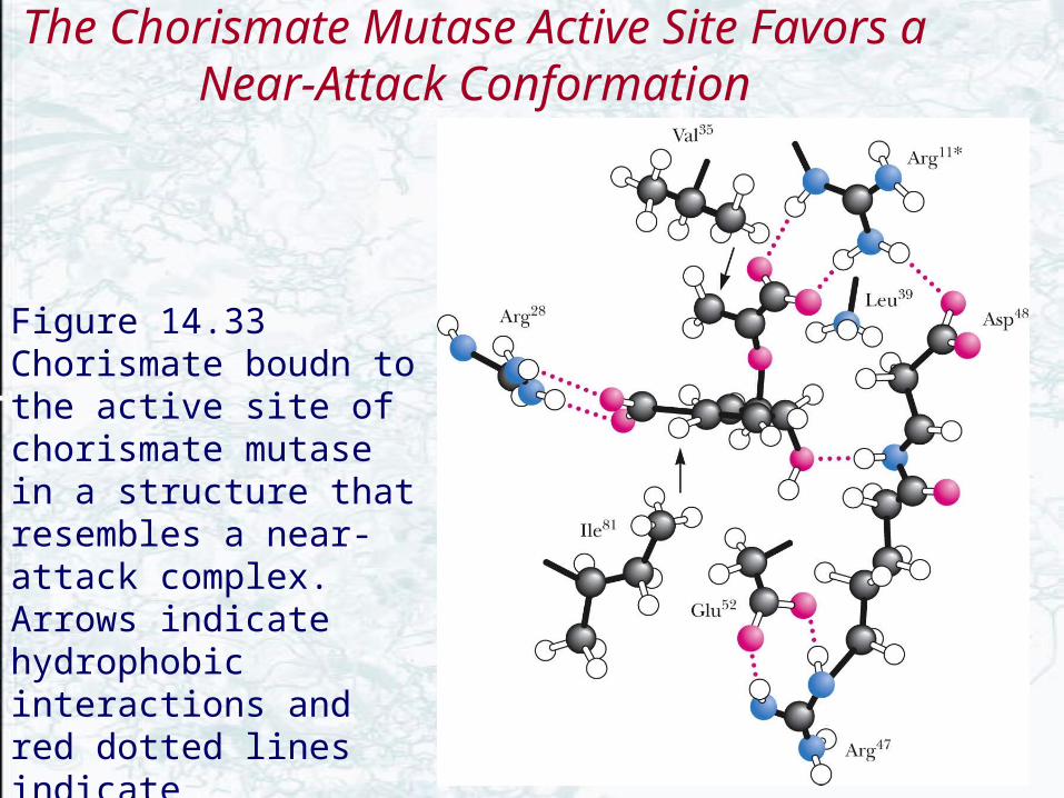
The Chorismate Mutase Active Site Favors a Near-Attack Conformation
Figure 14.33 Chorismate boudn to the active site of chorismate mutase in a structure that resembles a near-attack complex. Arrows indicate hydrophobic interactions and red dotted lines indicate electrostatic interactions.
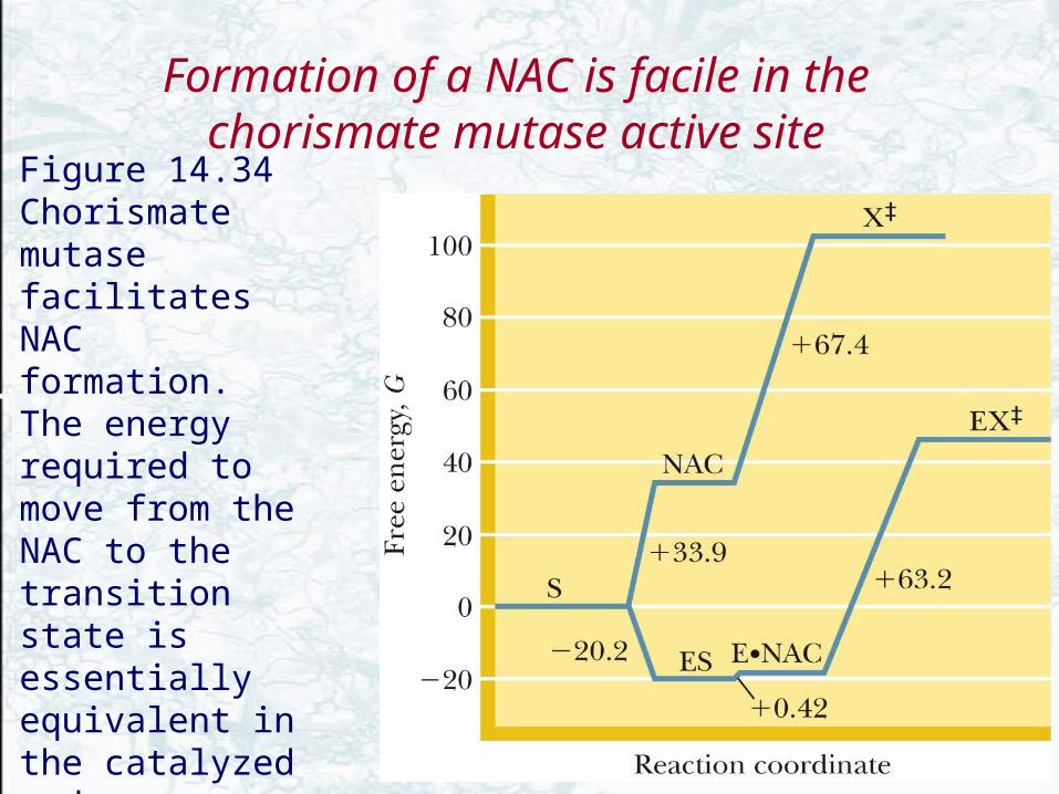
Formation of a NAC is facile in the chorismate mutase active site
Figure 14.34 Chorismate mutase facilitates NAC formation. The energy required to move from the NAC to the transition state is essentially equivalent in the catalyzed and uncatalyzed reactions.