chapter 141 – monitoring and staging human...
TRANSCRIPT

Keenan, S., & Hirshkowitz, M. (2011). Monitoring and staging human sleep. In M.H. Kryger, T. Roth, & W.C. Dement (Eds.), Principles and practice of sleep medicine, 5th edition, (pp 1602-1609). St. Louis: Elsevier Saunders.
Chapter 141 – Monitoring and Staging Human Sleep Sharon Keenan, Max Hirshkowitz Abstract
Polysomnography involves recording a wide assortment of bioparameters while a person sleeps. The electroencephalogram, electrooculogram, and skeletal muscle electromyogram can be summarized according to specific scoring criteria as sleep stages N1, N2, N3, and R (previously called stage 1, 2, 3, 4, and REM). Scoring criteria depend upon EEG bandwidth activity (delta, theta, alpha, and beta), EEG events (vertex sharp waves, sleep spindles, and K complexes), eye movement activity (slow and rapid eye movements), and the level of muscle tone. Stage N3 is characterized by high-voltage slow-wave activity. Stage N2 contains sleep spindles and K complexes. Stage N1 has low-voltage, mixed-frequency background, possibly slow eye movements, and vertex sharp waves. If rapid eye movements accompany the low-voltage, mixed-frequency EEG and skeletal muscle tone is low, rapid eye movement (REM or R) sleep is present. Central nervous system arousals also can occur from sleep, either spontaneously or resulting from pathophysiology. Quantitative analysis of sleep stages and CNS arousals provide evidence for, contribute to the definition of, and index the severity of some sleep disorders. Similarly, these measures can provide objective outcome measures for assessing therapeutic interventions. This chapter summarizes recording, digital processing, and scoring techniques used for evaluating brain activity during human sleep and its disturbance by CNS arousals.
History
From a behavioral perspective, immobility and reduced environmental responsiveness characterize human sleep. This state stands in contrast to purposeful (presumably) activities and provides the basis for dichotomizing observable living existence as either sleep or wakefulness. Furthermore, sleep and wakefulness cycle in a lawful, orderly fashion. Some rhythms are seasonal, some are daily (circadian), and some occur more than once a day. Changes in sleep cycle duration and composition in response to reduced sleep testify to sleep–wake cycle autoregulation, with a dynamic tension providing overall system homeostasis. Once techniques were developed to transcend observation, electroencephalography (EEG) revealed a complex array of brain activities clustered in a manner strongly suggesting multiple sleep states.
All scientific inquiry begins with observation and description. From there it proceeds to classification based ultimately upon measurement. Thus, when Loomis and colleagues[1] electroencephalographically recorded their first continuous all-night studies, they faced the daunting task of devising a system to describe sleep patterns in normal healthy human subjects. Thus sleep staging was born. In the original studies, amplified activity derived from electrodes that were placed on the scalp's surface at several loci produced ink tracings on paper wrapped around a slowly rotating cylinder. An enormous 8-ft “drum polygraph” enabled all-night sleep recording. One electrode was located near the eye and undoubtedly detected eye movement. However, rapid eye movement (REM) sleep remained unrecognized until Aserinsky published, in part, his University of Chicago doctoral study results 17 years later.[2] Aserinsky actually christened them “jerky eye movements” (JEMs) and in the first paper referred to the phenomenon as periodic ocular motility.
Perhaps it was the quirkiness of the original commercially available polygraph systems (e.g., Ofner, Beckman, and Grass) with their tendency to polarize electrodes, problematic rechargeable car battery–like systems, and aperiodic (and difficult to predict) recording interference artifacts. Or perhaps it was Loomis's silence on the matter of eye movements during sleep. In either case, Aserinsky's pilot work reportedly met with considerable skepticism. Ultimately, however, REM sleep's discovery, and particularly its correlation with dreaming, altered the course of sleep research for decades.[2a] The near-exclusive

focus on REM sleep, to the point that all other sleep states were considered simply non-REM (NREM), overshadowed substantial findings (and likely impeded progress) in other sleep research arenas (e.g., neuroendocrinology, physiology, and medicine). The spotlight on REM sleep made electrooculographic (EOG) recording de rigueur when performing sleep studies.
Meanwhile, in Lyon, France, Michel Jouvet noted postural difference during sleep in cats.[3] These differences correlated with sleep state and reduced skeletal electromyographic (EMG) activity. REM sleep (and, by association, dreaming) coincided with marked hypotonia in descending alpha and gamma motor neurons. The hypotonia induced functional paralysis that was quickly ascribed the purpose of keeping the sleeper from enacting dreamed activities. This sleep state–related EMG alteration added the final compulsory recording component to the procedure now known as polysomnography (PSG).
Polysomnography, in addition to brainwave, eye movement, and muscle tone recording, can also assess respiratory, cardiac, and limb movement activity (discussed in detail in other chapters in this volume and elsewhere[3a]). PSG in its simplest form (including EEG, EOG, and EMG), however, provides the basic information requisite for classifying sleep state and examining sleep processes.
Electrode Placement and Application
To make EEG, EOG, and EMG recordings, electrodes are placed on the scalp and skin surfaces. The site must be cleaned and properly prepared to assure good contact and limit electrical impedance to 5000 ohms or less. Scalp electrodes can be affixed with collodion or with electrode paste. Facial electrodes can be applied with double-sided adhesive electrode collars and paper tape. Although prescribed sites for electrode application have changed over the years, the system used to identify location remains the EEG society's international 10-20 system. In this system, the intersection of lines drawn from the left to right preauricular point, with the midpoint along the scalp between the nasion and inion, serves to landmark the vertex, designated Cz. Other loci can be found by measuring 10% and 20% downward along longitudinal and lateral surfaces. Specific locations are designated with a letter indicating the brain area below the electrode (e.g., C for central lobe, O for occipital lobe, F for frontal lobe) and a number ascribing specific points (odd numbers for the left side, even numbers for the right, and z for midline). EEG electrode placements should be precise; consequently, appropriate measurement techniques must be applied to ensure accuracy. Additionally, EEG amplifiers require calibration at the beginning and end of PSG recording to allow actual waveform amplitude measurements.
The classic and amazingly long-lived standardized technique (i.e., the manual produced by the ad hoc committee chaired by Rechtshaffen and Kales) requires a single monopolar central-lobe scalp EEG electrode referenced to a contralateral mastoid electrode (either C3-M2 or C4-M1). This single-channel brainwave recording, when paired with right and left eye EOGs and submentalis EMG, sufficiently reveals brain, eye, and muscle activity for classifying sleep stages.[4] As polysomnography evolved from a psychophysiologic research method to a clinical procedure, an occipital lead has supplemented centrally derived EEG to provide better visualization of waveforms needed to determine sleep onset and central nervous system (CNS) arousals.[5],[6]
EOG recording capitalizes on the eyes’ cornea–retina potential difference. Strong positive corneal potential fields affect electrodes placed near the eyes’ right and left outer canthi. The recording traces the response to this positive charge moving toward or away from the recording site. Each electrode is referenced to a neutral site, typically over the mastoid behind the ear. Thus, lateral eye movements produce out-of-phase tracings for right and left EOG tracings as the cornea moves toward one electrode and away from the other (provided that two channels are dedicated to tracing eye movements). This arrangement makes eye movements easily differentiable from in-phase frontal lobe EEG activity that is also present when recording from these sites. To discern vertical eye movements, we place the right-side EOG electrode 1 cm above the outer cantus and the left-side electrode 1 cm below (or vice versa). An alternative recording montage devised to enhance vertical eye movement detection entails lowering both recording sites to 1 cm below the outer canthi and referencing each to the middle of the forehead (Fpz).

Skeletal muscle activity level is estimated from a pair of electrodes arranged to record submentalis EMG. An electrode placed midline but 1 cm above the mandible's inferior edge is referenced to another placed 2 cm below and 2 cm to the right (or left). As a precaution, a backup electrode is also attached at the laterally homologous site of the reference electrode. The resulting submentalis EMG recording serves qualitatively (because it is uncalibratable) to provide an overall estimate for muscle activity level.
The American Academy of Sleep Medicine (AASM) has published a standardized manual for conducting clinical polysomnography in their accredited sleep disorders centers.[7] This AASM standards manual makes recommendations for recording, scoring, and summarizing sleep stages, CNS arousals, breathing, various kinds of movement, and electrocardiographic activity. By bringing instructional guidelines for a range of techniques into a single volume, the AASM manual will strongly influence practice, particularly in North America. Researchers, however, should not feel constrained by these clinical guidelines. New discoveries and future techniques need to continue unshackled by even a de facto standard clinical practice cookbook.
AASM specifies recording frontal, central, and occipital monopolar EEG from F4, C4, and O2. The contralateral mastoid (M1) serves as the theoretically neutral reference. Electrodes are placed at F3, C3, and O1 sites (and referenced to M2) to provide redundancy for backup when needed. The AASM manual sanctions the use of midline bipolar recordings for frontal and occipital EEG; however, the AASM frequently asked questions (FAQ) states that frontal bipolar derivations are not appropriate for measuring frontal EEG activity. The FAQ also states that EEG amplitudes can be measured from the C4-M1 derivation. The AASM manual recommends using mastoid-referenced EOG with separate channels for E2 and E1, but it also approves a forehead-referenced alternative montage. Submentalis EMG is recorded in the traditional manner.
Digital Recording Requirements
The first time a polysomnographic signal was digitized, whether it originated from analogue or digital amplifying circuits, an entirely new set of factors required consideration. The two most important questions to resolve involved specifying amplitude and temporal resolution. Selection of voltage per digital unit (bit) and sampling rate likely had more to do with computer hardware limitations than conceptual considerations. Amazingly, no standard was established for digital polysomnography until publication of the AASM standards manual.
The AASM standards manual specifies minimum 12-bit representation for amplitude, providing 4096 units
to represent a 2.5-volt regulated current (IREG) range, or its equivalent (Video 141-1 ). In this manner, even the smallest signals, exceeding the level of electrical noise, can be detected. Temporal resolution during recording depends on sampling rate and ultimately must allow accurate waveform reconstruction, provide enough data to potentially overcome frequency aliasing, and be appropriate for high- and low-pass digital filter settings. One size does not fit all: The minimum temporal resolution needed during data acquisition to meet these requirements varies for different bioelectrical signals (Table 141-1).
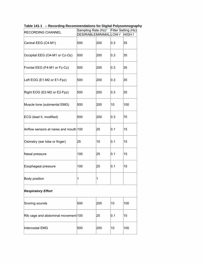
Table 141-1 -- Recording Recommendations for Digital Polysomnography
RECORDING CHANNEL Sampling Rate (Hz)* Filter Setting (Hz)DESIRABLE MINIMAL LOW f HIGH f
Central EEG (C4-M1) 500 200 0.3 35
Occipital EEG (O4-M1 or Cz-Oz) 500 200 0.3 35
Frontal EEG (F4-M1 or Fz-Cz) 500 200 0.3 35
Left EOG (E1-M2 or E1-Fpz) 500 200 0.3 35
Right EOG (E2-M2 or E2-Fpz) 500 200 0.3 35
Muscle tone (submental EMG) 500 200 10 100
ECG (lead II, modified) 500 200 0.3 70
Airflow sensors at nares and mouth 100 25 0.1 15
Oximetry (ear lobe or finger) 25 10 0.1 15
Nasal pressure 100 25 0.1 15
Esophageal pressure 100 25 0.1 15
Body position 1 1
Respiratory Effort
Snoring sounds 500 200 10 100
Rib cage and abdominal movement 100 25 0.1 15
Intercostal EMG 500 200 10 100
E1, left eye; E2, right eye; ECG, electrocardiogram; EEG, electroencephalogram; EMG, electromyogram; EOG, electrooculogram; f (frequency), Fpz, frontal pole; M, mastoid.
* Higher sampling rates increase file storage requirements but provide increased temporal resolution. The tradeoff between fidelity and practicality is a matter of debate.
Additional digital specifications involve data selection, display pagination, and calibration. Recorded channels must be selectable, and their calibration must be displayable. The viewable data should provide user-selectable time-frame compression and expansion (ranging from 5 seconds to an entire night shown on a page). Display screens definition should be at least 1600 × 1200 pixels. Digital polysomnographs should provide the capability to view data as it appeared when it was first recorded and when staging or when each event was marked and classified manually. Accompanying video at a minimum of one frame per second should be synchronized with the polysomnographic display.
Eeg Bandwidths, Waveforms, and Other Activity Bandwidths
One approach to differentiating EEG involves separating activity into dominant frequency bandwidths. Delta activity includes brain waves with a frequency less than 4 Hz. Sleep-related delta waves occurring at the low end of the frequency spectrum are called slow waves. Slow waves have high amplitude (≥75 mV) and low frequency (≤2 Hz). Theta activity includes 4- to 7-Hz waves prominent in central and temporal leads. Alpha activity consists of an occipitally prominent 8- to 13-Hz rhythm, and beta waves include the low-amplitude waves at even higher frequencies.
Waveforms
In addition to ongoing EEG activity oscillating predominantly within one or another of the specific bandwidths, distinct transient waveform events occur. These include vertex sharp waves, K complexes, sleep spindles, and saw-tooth theta waves. Vertex sharp waves are sharply contoured, negative-going (upward, as per EEG polarity convention) waves that stand out from the background activity. As the name implies, they appear prominently in EEGs derived from electrodes placed near Cz.
The K-complex begins much like a vertex sharp wave but is immediately followed by a large, usually much slower, positive component. Overall, the K-complex is usually clearest in central and frontal regions and has a duration criterion of 0.5 seconds or more. A sleep spindle is a readily apparent 0.5-second (or longer) burst of 12- to 14-Hz activity generated by the thalamus and thalamocortical pathways. The name derives from its spindlelike shape. A sawtooth wave is a variant of theta activity, with each wave also containing a notch, making it sawtooth-shaped.
Other nonpathologic sleep-related waveforms exist (e.g., benign epileptiform transients of sleep [BETS], sensory motor rhythm [SMR], Wicket rhythm [mu rhythm], and positive occipital sharp transients of sleep [POSTS]. These normal variants do not occur consistently during polysomnography.
Activity Patterns
Sleep EEG also contains dynamic activity patterns not captured by sleep staging schema or identification of individual waveforms. The cyclic alternating pattern (CAP) includes waveform bursts (usually high-amplitude slow, sharp, or polymorphic waves) separated by quiescent periods.[8] The pattern's burst component sometimes includes transient alpha bandwidth components that qualify as CNS arousals and

thus can index sleep disturbance. However, a CAP occurring without frank arousals is thought to signify more subtle sleep instability.
Sleep Staging Rules and Central Nervous System Arousals
For more than 40 years, the standardized technique described in A Manual of Standardized Terminology, Techniques and Scoring System for Sleep Stages of Human Subjects[4] has provided the unifying methodology for human sleep research. This standardized manual combines elements from various systems that had evolved and provides adequate detail to achieve general use. However, to a large extent, its enormous success stems from the consensus it attained from the multinational, multidiscipline stakeholders composing its development committee. That is, when the committee members returned to their respective laboratories, they used the techniques and taught them to scientists and clinicians in training.
Staging, as a summarizing technique, necessarily must define a period over which the summary applies. The standardized manual endorsed 20- and 30-second time domains. This flexibility deferred to extant technology; that is, generally available polygraph machine paper chart drive speeds. Over time, the 30-second epoch won out because it provided enough detail to see waveforms (EEG standards dictate minimal paper speed of 10 mm/sec to ensure ability to discern individual EEG waveforms); at 10 mm/sec, one epoch fit on a standard 30-cm wide paper fan-fold polygraph page; and one 1000-page box of polygraph paper would hold a complete recording (or two if one also recorded on the back).

Sleep Sta
Wakefulneof alpha Edetermina
Figthe Easso(Fropre
aging Rules
ess (stage W)EEG activity ination of sleep
ure 141-1 StagEEG and EOG. Aociated with relaxom Butkov N.ss].)
W) in a relaxedn 50% or moronset.
ge wake (W), eyeAlpha activity is mxed wakefulness. Atlas of clini
d subject with re of the epoc
es closed. This emost prominent i
s. ical polysomn
eyes closed ch (Fig. 141-1
example demonsin the occipital ch
nography, 2nd
is differentiat). Poorly defi
strates a classic whannel. The chin
d ed. Medford
ted from sleepned alpha EE
wake pattern, win EMG displays n
d, Ore: Synap
p by the preseEG complicate
th alpha rhythm normal muscle to
pse Media; [in
ence es
in one
n

Stage 1 wbackgrounblinking, a(Fig. 141-activity slo
Figrelatrema(Fropre
winds up beingnd EEG devoabsence of sa-2). Stage 1 sowing, and slo
ure 141-2 Stagtively low voltageains tonic, althouom Butkov N.ss].)
g largely definoid of sleep spaccadic eye mleep may, buow eye move
ge 1 sleep (N1). e mixed-frequencugh it can attenu. Atlas of clini
ned by excluspindles and Kmovements, at does not ne
ements.
The onset of N1cy EEG with a prate slightly with sical polysomn
sion; that is, itK-complexes, and alpha actiecessarily, inc
is identified by trominence of thesleep onset.
nography, 2nd
t is a low-voltaminimal slowvity less than
clude vertex s
the disappearanceta activity in the
d ed. Medford
age, mixed-frw-wave activityn 50% of the esharp waves,
ce of alpha rhythrange of 4 to 7 H
d, Ore: Synap
requency y, cessation oepoch duratiobackground
hm, replaced by Hz. The chin EM
pse Media; [in
of on
G
n

Stage 2 cmixed-freq
Figa ba(Fropre
haracteristicsquency backg
ure 141-3 Stagackground of mixom Butkov N.ss].)
s include sleeground EEG a
ge 2 sleep (N2). xed-frequency EE. Atlas of clini
p spindles anand minimal (
Stage N2 is idenEG. The chin EMical polysomn
nd K-complex(<20% of the
ntified by the presMG displays normnography, 2nd
xes (Fig. 141-3epoch) slow-
sence of K-compmal muscle tone, d ed. Medford
3) occurring o-wave activity
plexes and/or sleas expected dur
d, Ore: Synap
on a low-voltay.
eep spindles agaring NREM sleep
pse Media; [in
age,
ainst p. n

Slow-wavderivationis scored when dura
Figepocscor(Fropre
ve sleep (stagn) with a 75-mwhen the duration reaches
ure 141-4 Slowch. By the Rechtred as N3. om Butkov N.ss].)
ges 3 and 4 slmV or greater ration of slow s 50% or more
w-wave sleep (Ntchaffen and Kale
. Atlas of clini
leep) containsamplitude enwaves comp
e.
3). In this exampes criteria, this e
ical polysomn
s delta EEG aduring for 20%
poses 20% to
ple, high-amplitudpoch is scored a
nography, 2nd
activity (record% or more of 50% of the e
de slow waves oas stage 4. By the
d ed. Medford
ded from a man epoch (Fi
epoch and sta
occupy greater the revised AASM
d, Ore: Synap
monopolar cenig. 141-4). Sta
age 4 is score
han 50% of the criteria, this epo
pse Media; [in
ntral age 3 d
och is
n

REM sleefrequencylow-voltagfalling betfalling beffeatures bresumptioon the supstate disti
Figidenlow-(Fropre
In 2007, tsummarizcombiningrevising te3 and 4, a
ep is scored wy EEG in assoge, mixed-freqtween epochsfore or after (abut lack rapid on of K-complpposition thatnct from wake
ure 141-5 REMntified by the pres-chin EMG. om Butkov N.ss].)
he AASM stazed in Table 1g stages 3 anerminology (Rand W for wak
when saccadicociation with aquency EEG s of REM sleeand contiguoueye movemeexes or sleept REM sleep refulness and
M sleep. During Rsence of rapid ey
. Atlas of clini
ndards manu41-2. Essentd 4 sleep and
R for REM slekefulness); an
c eye movema very low levand continuin
ep (with eye mus with) clearents are scorep spindles occrepresents a NREM sleep
REM sleep, chin ye movements in
ical polysomn
ual provided retially, changesd applying amep, N1 for NRnd simplifying
ents occur duvel of submenng low-level smovements) ar REM sleep ted as REM slecurs. These spersistent cen
p.
muscle tone dron combination wit
nography, 2nd
evised criterias include stan
mplitude criterREM stage 1,g smoothing ru
uring epochs ntalis EMG acubmentalis E
are also scorethat have comeep until an asmoothing rulentral nervous
ops to the lowestth relatively low-v
d ed. Medford
a for scoring sndardizing epria for slow wa N2 for NREMules. Some c
with low-voltactivity (Fig. 14EMG (without ed as REM slemparable EEGarousal, EMGles gloss over system (CNS
t level of the recovoltage mixed-fre
d, Ore: Synap
sleep stages.poch length ataves to frontaM stage 2, N3hanges are c
age, mixed-41-5). Epochseye movemeeep. Epochs
G and EMG G level increasr minor transitS) organizatio
ording. REM sleeequency EEG an
pse Media; [in
. Changes aret 30 seconds;al EEG activity3 for NREM scontroversial.[
with ents)
se, or tions onal
ep is nd
n
e y; tages 9-14]

Table 141-2 -- Comparison of Traditional and AASM (2007) Sleep Stage Scoring Systems
PARAMETER R&K CLASSIFICATION CRITERIA AASM CLASSIFICATION CRITERIA
Epoch length 15 or 30 seconds, user's choice 30 seconds, mandated
Stage nomenclature
Wakefulness, stage 1 sleep, stage 2 sleep, stage 3 sleep, stage 4 sleep, REM sleep, movement time
Stages W, N1, N2, N3, and R
Wakefulness EEG alpha activity for ≥50% of an epoch Same
Slow-wave sleep
EEG slow-wave activity for ≥50% of the epoch for stage 4 sleep or ≥20% of the epoch for stage 3 sleep
Same, except that stages 3 and 4 are combined to N3
Stage 2 sleep Sleep spindles or K-complexes; EEG slow-wave activity for <20% of the epoch
Same
Stage 1 sleep Low-voltage, mixed-frequency activity; possibly vertex sharp waves; possibly slow eye movements; no sleep spindles or K-complexes; EEG alpha activity for <50% of the epoch
Same
REM sleep Low-voltage, mixed-frequency EEG activity; very low submental EMG activity; possibly saw tooth EEG theta activity; at least one unequivocal rapid eye movement
Same
Movement time
Polysomnographic activity obscured to the point of not being readable for more than 50% of the epoch; the preceding epoch is scored as stage 1, 2, 3, 4, or REM sleep
This epoch classification is eliminated
Smoothing rules
When an epoch is classified as a particular stage but is surrounded by epochs lacking unique features (e.g., a sleep spindle, slow-rolling eye movements, or CNS arousal) and would otherwise have been scored as stage 1 sleep, the classified epoch scoring is generalized to the surrounding epochs (but only for 3 minutes). These smoothing rules apply to stage 2 and REM sleep.
Same, except that there is no 3-minute limit to the generalization
AASM, American Academy of Sleep Medicine; CNS, central nervous system; EEG, electroencephalogram; EMG, electromyogram; R&K, Rechtchaffen and Kales; REM, rapid eye movement.

Central N
Sleep stagover a 30-the need tunder thesecond (oconsidereactivity. Tmust increrepresentmembersendorsed explanato
FigappeseenEMGincregeneREMchanEMGM1 c(Co
Summa
The sleepyoung adu50%, N3 awakefulneapproximaepisodes.sleep sespredomina
Nervous Sys
ging fails to re-second time to appreciateauspices of t
or longer) EEGed biomarkerso qualify as aease in subms the minimu). Events of sthis scoring t
ory notes.
ure 141-6 Arouears near the cen reflected in theG channel in assease in EMG acteralized presenta
M sleep: low-voltannel shows an inG artifact on the channel, are conourtesy of Max
arizing Nor
p stage patterults, stage R accounts for 1ess might accately 90 minu These episosion containsates in the firs
stem Arousa
epresent briedomain. Incrsleep fragme
the AmericanG frequency is for CNS actian arousal, 10entalis EMG m duration thhorter duratiotechnique and
usals from NREMnter of the epoch
e other EEG chanociation with thistivity is noted on ation of EMG artage, mixed-frequ
ncreased tone, anEEG channels a
nsistent with a pox Hirshkowitz
rmal Sleep
rn across the accounts for 12.5% to 20%
count for 5% tutes after sleeodes increase s less REM slest third of the
l Scoring
f CNS arousareasing clinicaentation; cons Sleep Disordncreases to tivation. The a0 seconds of sleads for at leat could be re
on likely also d simplified th
M(A) and REM(Bh. The distributionnels but with (exs event. It is comthe EMG and almifact throughout uency EEG, rapidnd the EEG backfter the short bur
ossible transition z, PhD, DABS
night can be approximatel
%, and N1 accto 10% of the ep onset, after
in duration aeep than the night. Age-m
als because ital application sequently, a sders Associatheta, alpha, o
arousals mostsleep must preast 1 secondeliably scoredhave clinical
he original 11
B) sleep. A, A parn of the electricaxpected) decreasmon to see K-comost simultaneothe EEG and EOd eye movementkground activity irst. These data, eto wake from RE
SM).
represented dy 20% to 25%counts for thetime in bed. S
r which it reocas the night pr
second half. matched sleep
t summarizesof polysomno
scoring systemtion (later to bor beta (but nt often entail erecede the evd (Fig. 141-6)d by visual inssignificance. rules to a sin
roxysmal burst oal field changes ased amplitude. T
omplex activity evously in the E2-MOG channels. Prits, and very low Eis low voltage anespecially the buEM sleep
diagrammatic% of total sleee remainder. IStage R typicccurs every 9rogresses; theBy contrast, s
p bears great
s EEG, EOG, ographic techm for arousalbecome the Aot to sleep spemergent occvent. In REM ). The 3-seconspection (amoThe AASM s
ngle statemen
of high-amplitudeassociated with tThere is little or nvoked by auditor
M1 channel. This or to the event thEMG. After the e
nd fast. There is aurst of alpha activ
cally (Fig. 141ep time, stagen normal slee
cally does not90 to 120 minuerefore, the fislow-wave acsimilarity in m
and EMG acthnique heightes was develo
AASM). Abruppindles) are cipital EEG alsleep, activitynd duration ong the task ftandards man
nt with two
e slow activity this event can beno change in the ry stimuli. B, An is followed by a here is evidence event, the EMG a continuation ofvity seen in the O
1-7). In healthe N2 accountsepers, t appear until utes in distincrst half of the
ctivity (stage Nmen and wom
tivity ened
oped[6] pt 3-
pha y
force nual
e
brief of
f O2-
hy s for
ct e N3)
men;

however, quantitativ
Fig
Table 141PARAME
AASM Re
Lights out
Lights on
Total slee
Total reco
Sleep late
REM slee
Wake afteonset
women mighvely summari
ure 141-7 Norm
1-3 -- ParamTER N
ecommended
t clock time L
clock time L
ep time T
ording time T
ency S
ep latency R
er sleep W
t have slightlyzed, and Tab
mal sleep histog
meters DeriveNOTATION
d Parameters
L-out
L-on
TST
TRT
SLAT
RLAT
WASO
y better preseble 141-3 prov
ram illustrating s
ed from SleeEXPLAN
s
The clockhimself o
The clock
Minutes s
Elapsed
Elapsed (in minute
Elapsed
Minutes s
erved stage Nvides definitio
sleep macroarchi
p Staging anATION
k time (in hh:mor herself to fa
k time (in hh:m
scored as sta
time from L-o
time from L-oes)
time in minute
scored as sta
N3 with advanons for commo
itecture (stages)
nd CNS Arou
mm) that the all asleep
mm) that the
age N1, N2, N
out to L-on (in
out to first epo
es from SLAT
age W from fir
ncing age. Sleonly used par
for a young adu
usal Scoring
subject was i
subject was a
N3, or R
n minutes)
och of stage N
T to first epoc
rst sleep epoc
eep can be rameters.
lt.
instructed to a
awakened
N1, N2, N3, o
ch of stage R
ch to L-on
allow
or R

PARAMETER NOTATION EXPLANATION
Sleep efficiency SEI TST as a percentage of TRT
Time in each stage MW, M1, M2, M3, MR
Minutes scored as W, N1, N2, N3, and R (individually)
Sleep stage percentages
P1, P2, P3, PR Time scored as N1, N2, N3, and REM as a percentage of TST (individually)
Number of CNS arousals
NArsls The number of CNS arousals
CNS arousal CNS AI The number of CNS arousals scored per hour of TST
Other Useful Parameters
Latency to persistent sleep
LTPS Elapsed time (in minutes) from L-out to first of 10 consecutive minutes of sleep
Latency to unequivocal sleep
LUS
Elapsed time (in minutes) from L-out to first epoch of N2, N3, or R or to three consecutive (or more) epochs of N1 If N1 is followed by an epoch of N2, N3, or R, LUS is calculated from L-out to the first epoch of N1
Sleep-period time SPT Minutes from first to last epoch scored as N1, N2, N3, or R
Number of REM episodes
NREME Number of stage R occurrences
Number of awakenings
NWake Number of stage W occurrences
Wake index WI Number of awakenings per hour of TST
Sleep fragmentation index
SFI Number of awakenings and CNS arousals per hour of TST
Number of stage NShifts Number of stage transitions during TRT

PARAMETER NOTATION EXPLANATION shifts
Stage shift index SSI Number of stage transitions per hour of TRT
Latency to arising LTA Duration of final stage W if it was ongoing when L-on occurred
CNS, central nervous system; REM, rapid eye movement [sleep].
Ambiguous Sleep Stages and Sleep Quality
Sleep stage scoring was developed to summarize EEG, EOG, and EMG correlates of normal sleep. Under normal circumstances, particular events cluster the vast majority of the time. By contrast, this tight coupling tends to loosen when patients rebound from sleep deprivation; sustain brain injury; are afflicted with sleep, medical, neurologic, psychiatric, or sleep disorders; or ingest psychoactive substances. The resulting intrusion, translocation, or migration of specific EEG, EOG, or EMG activity, characteristic of one stage into another, produces ambiguous epochs that are difficult to classify according to the usual scoring rules. This departure from normal processes can provide qualitative evidence of an underlying sleep dysfunction.
Perhaps the most common ambiguities accompany pharmacotherapy. Gamma-aminobutyric acid A (GABAA) and benzodiazepine receptor agonists generally increase spindle activity in the EEG. These pharmacologically induced spindles typically are of higher frequency (16 to 18 Hz), often of longer duration, occur more frequently (higher density), and can appear not only in N2 but also in other stages of sleep and even in wakefulness.
Another commonly noted drug effect involves serotonin agonist augmentation of eye movement activity. In some persons, rapid eye movements occur at sleep onset, in stage N2, and stage N3, making the scoring of REM sleep a challenge. The phenomenon is so common that many sleep specialists refer to it as Prozac eyes (referring to fluoxetine, the prototypical selective serotonin reuptake inhibitor).
Another serotonin agonist–provoked sleep alteration involves elevated muscle activity during REM sleep. In some cases, these medications produce a loss of atonia, permitting attempted dream enactments (i.e., iatrogenic REM Sleep behavior disorder [RBD]). Individual PSG epochs during these events do not meet usual stage classification criteria. Similar REM sleep ambiguities occur in idiopathic, Parkinson's disease–related, and posttraumatic stress disorder–related RBD.
Patients suffering from neurodegenerative diseases or brain insult can manifest an overall erosion of EEG sleep events. This includes reduced sleep spindles, K-complexes, and slow-wave activity. We also sometimes observe this in patients with sleep apnea, heart failure, and metabolic disorders. The resulting nearly featureless sleep EEG can be difficult to score according to normal staging rules. By contrast, another very different scoring problem can occur in patients with severely fragmented sleep produced by obstructive apnea. In these patients, a continual cycle of falling asleep, airway collapse, struggle to breathe, awakening, and falling asleep occurs. Thus, the patient remains in a transition state that does not fit well into any sleep stage category. It was once proposed that this pattern be scored as t-sleep.
In some persons, copious EEG alpha activity permeates ongoing background activity. In sleep states marked by low-amplitude, mixed-frequency activity, alpha bursts meeting criteria for CNS arousal can be scored as such (alpha intrusion). However, when slow waves characterize the dominant ongoing

background EEG activity and the alpha coincides with delta, arousals are not scored. This alpha-delta sleep sometimes accompanies pain syndromes, but it appears to lack specificity. A related phenomenon, also ascribed to pain, involves K-complex bursts followed by EEG alpha activity. Many sleep specialists consider K-alpha a variety of the CAP.
Clinical Pearl
Sleep staging and CNS arousal scoring provide important clinical information about sleep-related brain process. Ultimately, persons who awaken sleepy or unrefreshed or who have difficulty initiating or maintaining sleep can be assayed for sleep integrity, quantity, and quality using polysomnography. Human sleep is a brain process. Pathophysiologies such as increased airway resistance and leg movements produce CNS arousals that fragment and destroy the fabric of sleep. Disorders often alter sleep patterns and overall architecture. Treatments may promote return to normal. Quantitative analysis through staging and arousal scoring objectively documents sleep disruption and provides a severity index for sleep disorders.

References
1. Loomis AL, Harvey N, Hobart GA: Cerebral states during sleep, as studied by human brain potentials. J Exp Psychol 1937; 21:127-144.
2. Aserinsky E, Kleitman N: Regularly occurring periods of eye motility, and concomitant phenomena, during sleep. Science 1953; 118:273-274.
2a. Dement W, Kleitman N: Cyclic variations in EEG during sleep and their relation to eye movements, body motility, and dreaming. Electroencephalogr Clin Neurophysiology 1957; 9:673-690.
3. Jouvet M: Neurophysiology of the states of sleep. Physiol Rev 1967; 47:117-177.
3a. Hirshkowitz M, Kryger MH: Diagnostic methods. In: Kryger MH, ed. Atlas of clinical sleep medicine, Philadelphia: Elsevier; 2010.
4. Rechtschaffen A, Kales A: A manual of standardized, techniques and scoring system for sleep stages in human subjects. Washington DC, US Government Printing Office, 1968. NIH Publication No. 204
5. Littner MR, Kushida C, Wise M, Davila DGAASM Standards of Practice Committee, et al: Practice parameters for clinical use of the multiple sleep latency test and maintenance of wakefulness test. An American Academy of Sleep Medicine Report. Sleep 2005; 28:113-121.
6. Bonnet M, Carley D, Carskadon M, Easton P, et al: EEG arousals: scoring rules and examples. ASDA report. Sleep 1992; 15:173-184.
7. Iber C, Ancoli-Israel S, Chesson A: Quan SF for the American Academy of Sleep Medicine. The AASM manual for the scoring of sleep and associated events: rules, terminology and technical specifications. 1st ed. Westchester, Ill, American Academy of Sleep Medicine, 2007.
8. Terzano MG, Parrino L, Smerieri A, Chervin R, et al: Atlas, rules, and recording technique for scoring of cyclic alternating pattern (CAP) in human sleep. Sleep Med 2002; 3:187-199.
9. Moser D, Anderer P, Gruber G, et al: Sleep classification according to AASM and Rechtschaffen & Kales: effects on sleep scoring parameters. Sleep 2009; 32:139-149.
10. Danker-Hopfe H, Anderer P, Zeitlhofer J, et al: Interrater reliability for sleep scoring according to the Rechtschaffen & Kales and the new AASM standard. J Sleep Res 2009; 18:74-84.
11. Parrino L, Ferri R, Zucconi M, Fanfulla F: Commentary from the Italian Association of Sleep Medicine on the AASM manual for the scoring of sleep and associated events: for debate and discussion. Sleep Med 2009; 10:799-808.
12. Grigg-Damberger MM: The AASM scoring manual: a critical appraisal. Curr Opin Pulm Med 2009; 15:540-549.
13. Novelli L, Ferri R, Bruni O: Sleep classification according to AASM and Rechtschaffen and Kales: effects on sleep scoring parameters of children and adolescents. J Sleep Res 2010; 19:238-247.
14. Miano S, Paolino MC, Castaldo R, Villa MP: Visual scoring of sleep: A comparison between the Rechtschaffen and Kales criteria and the American Academy of Sleep Medicine criteria in a pediatric population with obstructive sleep apnea syndrome. Clin Neurophysiol 2010; 121:39-42.
