chapter 15 eye and ocular adnexa, auditory systems · palpated, and otosclerosis appeared to be...
TRANSCRIPT

CPT ® copyright 2012 American Medical Association. All rights reserved. www.aapc.com 15.1
Eye and Ocular Adnexa, Auditory Systems C h a p t e r
15
Case 1Anesthesia: Laryngeal mask anesthesia.
Preoperative diagnosis: Retinal detachment, right eye.
Postoperative diagnosis: Retinal detachment, right eye.
Procedure: Scleral buckle, cryoretinopexy, drainage of subretinal fluid, C3F8 gas in the right eye.
Procedure: After the patient had received adequate laryngeal mask anesthesia, he was prepped and draped in usual sterile fashion. A wire lid speculum was placed in the right eye.
A limbal peritomy was done for 360 degrees using 0.12 forceps and Westcott scissors. Each of the intramuscular quadrants was dissected using Aebli scissors. The muscles were isolated using a Gass muscle hook with an 0 silk suture attached to it. The patient had an inspection of the intramuscular quadrants and there was no evidence of any anomalous vortex veins or thin sclera. The patient had an examination of the retina using an indirect ophthalmoscope and he was noted to have 3 tears in the temporal and inferotemporal quadrant and 2 tears in the superior temporal quadrant. These were treated with cryoretinopexy. Most posterior edge of each of the tears was marked with a scleral marker followed by a surgical marking pen. The patient had 5-0 nylon sutures placed in each of the 4 intramuscular quadrants. The 2 temporal sutures were placed with the anterior bite at about the muscle insertion, the posterior bite 9 mm posterior to this. In the nasal quadrants the anterior bite was 3 mm posterior to the muscle insertion and the posterior bite was 3 mm posterior to this. A 240 band was placed 360 degrees around the eye and a 277 element from approximately the 5-1 o’clock position. The patient had another examination of the retina and was noted to have a moderate amount of subretinal fluid, so a drainage sclerotomy site was created at approximately the 9:30 o’clock position incising the sclera until the choroid was visible. The choroid was then punctured with a #30-gauge needle. A moderate amount of subretinal fluid was drained from the subretinal space. The eye became relatively soft and 0.35 ml of C3FS gas was injected into the vitreous cavity 3.5 mm posterior to the limbus. The superior temporal and inferior temporal and superior nasal sutures were tied down over the scleral buckle. The 240 band was tightened up and excessive scleral buckling material was removed from the eye. The inferior nasal suture was tied down over the scleral buckle and all knots were rotated posteriorly. The eye was reexamined. The optic nerve was noted to be nicely perfused. The tears were supported on the scleral buckle. There was a small amount of residual subretinal fluid. The patient received posterior sub-Tenon Marcaine for postoperative pain control. The 0 silk sutures were removed from the eye. The conjunctiva was closed with #6-0 plain gut suture. The patient received subconjunctival Ancef and dexamethasone. The patient was patched with atropine and Maxitrol ointment.
2.3.
4.
1. The postoperative diagnosis is used for coding.
2. Exam reveals the location of the tears.
3. Cryoretinopexy is the use of intense cold to close the tear in the retina.
4. A sclerotomy is performed to drain subretinal fluid.
5. Sclera buckling is performed.
1.
5.
15.2 2013 Medical Coding Training: CPC Practical Application Workbook—Instructor CPT ® copyright 2012 American Medical Association. All rights reserved.
Eye and Ocular Adnexa, Auditory Systems Chapter 15
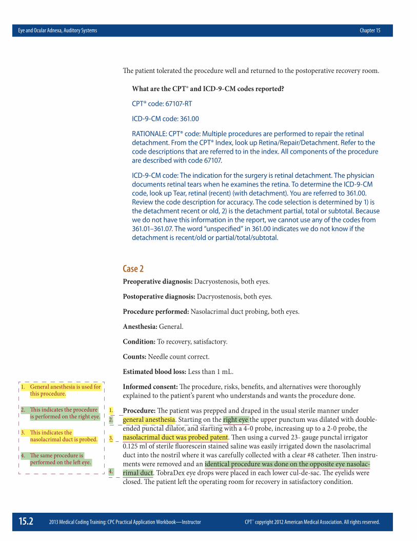
The patient tolerated the procedure well and returned to the postoperative recovery room.
What are the CPT® and ICD-9-CM codes reported?
CPT® code: 67107-RT
ICD-9-CM code: 361.00
RATIONALE: CPT® code: Multiple procedures are performed to repair the retinal detachment. From the CPT® Index, look up Retina/Repair/Detachment. Refer to the code descriptions that are referred to in the index. All components of the procedure are described with code 67107.
ICD-9-CM code: The indication for the surgery is retinal detachment. The physician documents retinal tears when he examines the retina. To determine the ICD-9-CM code, look up Tear, retinal (recent) (with detachment). You are referred to 361.00. Review the code description for accuracy. The code selection is determined by 1) is the detachment recent or old, 2) is the detachment partial, total or subtotal. Because we do not have this information in the report, we cannot use any of the codes from 361.01–361.07. The word “unspecified” in 361.00 indicates we do not know if the detachment is recent/old or partial/total/subtotal.
Case 2Preoperative diagnosis: Dacryostenosis, both eyes.
Postoperative diagnosis: Dacryostenosis, both eyes.
Procedure performed: Nasolacrimal duct probing, both eyes.
Anesthesia: General.
Condition: To recovery, satisfactory.
Counts: Needle count correct.
Estimated blood loss: Less than 1 mL.
Informed consent: The procedure, risks, benefits, and alternatives were thoroughly explained to the patient’s parent who understands and wants the procedure done.
Procedure: The patient was prepped and draped in the usual sterile manner under general anesthesia. Starting on the right eye the upper punctum was dilated with double-ended punctal dilator, and starting with a 4-0 probe, increasing up to a 2-0 probe, the nasolacrimal duct was probed patent. Then using a curved 23- gauge punctal irrigator 0.125 ml of sterile fluorescein stained saline was easily irrigated down the nasolacrimal duct into the nostril where it was carefully collected with a clear #8 catheter. Then instru-ments were removed and an identical procedure was done on the opposite eye nasolac-rimal duct. TobraDex eye drops were placed in each lower cul-de-sac. The eyelids were closed. The patient left the operating room for recovery in satisfactory condition.
1. General anesthesia is used for this procedure.
2. This indicates the procedure is performed on the right eye.
3. This indicates the nasolacrimal duct is probed.
4. The same procedure is performed on the left eye.
2.
3.
4.
1.

CPT ® copyright 2012 American Medical Association. All rights reserved. www.aapc.com 15.3
Chapter 15 Eye and Ocular Adnexa, Auditory Systems
What are the CPT® and ICD-9-CM codes reported?
CPT® codes: 68811-50 or 68811-RT, 68811-LT
ICD-9-CM code: 375.56
RATIONALE: CPT® codes: During this encounter, the provider probes the nasolacriminal duct on the right and left eye. When this procedure is performed using general anesthesia, the proper code selection is 68811. There is a parenthetical note to append modifier 50 if the procedure is performed bilaterally.
ICD-9-CM code: The patient is diagnosed with dacryostenosis. From ICD-9-CM Index to Diseases, look up dacryostenosis. There is no indication the condition is congen-ital. The correct code is 375.56. Verify the code accuracy in the Tabular List.
Case 3Preoperative diagnosis: Bilateral protruding ears.
Postoperative diagnosis: Bilateral protruding ears.
Procedure: Bilateral otoplasty.
Anesthesia: General.
Estimated blood loss: Minimal.
Complications: None.
Procedure is as follows: The patient was placed in the supine position. She was prepped and draped in the usual sterile fashion. Measurements were taken from the helix to the mastoid at the superior, mid, and inferior portions and they were within 1 to 2 mm of the same bilaterally and were approximately 17 mm superior, 24 mm middle, and 25 mm inferior. The right ear was begun first. A curved incision was made just anterior to the sulcus of the posterior ear. This was done with a 15-blade scalpel. Electrocautery was used for hemostasis and further dissection. An iris scissors was used to dissect the soft tissues off of the mastoid region and the posterior ear. The concha was shut back and sutured in place with clear 4-0 nylon suture and in a horizontal mattress pattern. Three tacking sutures were used. This brought the ear back approximately 2 to 3 mm. However, greater correction was needed and, therefore, Mustarde’ sutures were placed.
The mid and superior portions of the antihelical fold were placed. These were spaced widely on either side of the helical fold. They were then sutured in place, tacking the fold more acutely to a point that was deemed acceptable and held in that position. So in this, a margin of skin was excised along the posterior ear and closure of the wound was performed with 5-0 chromic suture. Prior to closure, full hemostasis had been obtained with electrocautery. Both ears were done in the exact same fashion; therefore, only one is dictated in detail.
The patient was then checked very carefully for symmetry. Postoperative measurements were approximately 14 mm superior, 15 mm mid, and 16 mm lower.
1. Procedure is performed on the right ear.
2. An incision is made.
3. The concha which is the external part of the ear is sutured in place.
4. This is a suturing technique used to perform otoplasty.
5. There are a total of three portions of the external ear that are repaired in this otoplasty.
6. This indicates that a bilateral procedure is performed.
1.
2.
3.
4.
5.
6.

15.4 2013 Medical Coding Training: CPC Practical Application Workbook—Instructor CPT ® copyright 2012 American Medical Association. All rights reserved.
Eye and Ocular Adnexa, Auditory Systems Chapter 15
What are the CPT® and ICD-9-CM codes reported?
CPT® code: 69300-50
ICD-9-CM code: 744.29
RATIONALE: CPT® code: In this case an otoplasty is performed with is a surgical fixa-tion of the external ear. The patient has protruding ears which are being corrected. From the CPT® Index, look up Otoplasty. You are referred to 69300. The code description matches the procedure performed. There is a parenthethical note that states if performed bilaterally to append modifier 50.
ICD-9-CM code: To determine the diagnosis code, look up Protrusion/ear, congen-ital. You are referred to 744.29 which reports a congenital condition. The cause of protruding ears develops at birth and the appearance of the protrusion can be seen later in life.
Case 4Preoperative diagnosis: Right otosclerosis.
Postoperative diagnosis: Right otosclerosis.
Type of procedure: Right stapedectomy.
Anesthesia: General endotracheal.
Findings: There was otosclerosis on the anterior footplate of the stapes with preoperative conductive hearing loss in the right ear.
Description of procedure: The patient was taken to the OR and placed in the supine posi-tion. Following induction of general endotracheal anesthesia, the head was turned to the left and the right ear was prepped and draped in the usual fashion. Then 1% Xylocaine with 1:100,000 epinephrine was infiltrated in the skin along the posterior ear canal wall and the skin over the tragus.
After a short waiting time, an incision was made over the tragus, and a piece of posterior tragal perichondrium was harvested for a graft and set aside to dry. A speculum was then placed in the canal. The canal was quite large. An incision was made along the posterior canal wall, and a tympanomeatal flap was elevated and laid forward to include the fibrous annulus without perforation. The middle ear was inspected. The ossicular chain was palpated, and otosclerosis appeared to be fixing the stapes. The chorda tympani nerve was very carefully preserved and not manipulated and was kept moist throughout the procedure. No curetting of bone was necessary in order to access the footplate. A control hole was made in the footplate with a straight pick. The incudostapedial joint was separate with an IS joint knife. The stapedius tendon was severed, and the superstructure of the stapes was fractured over the promontory and removed. The footplate was then picked out with a 45-degree pick, completely removing all fragments. Great care was taken not to suction in the vestibule. The distance between the incus and the oval window was then
1. Location of otosclerosis.
2. A graft of ear cartilage is obtained for the procedure.
2.
1.

CPT ® copyright 2012 American Medical Association. All rights reserved. www.aapc.com 15.5
Chapter 15 Eye and Ocular Adnexa, Auditory Systems
measured. The tragal perichondrial graft was then taken and laid over the oval window with complete coverage. A 3.75 Shea platinum Teflon cup piston was then chosen. The platinum wires were opened and the shaft was placed down against the graft and into the oval window niche. The cup was placed under the long process of the incus by gently lifting the incus, and the platinum wires were snugly crimped around the long process of the incus. An excellent round window reflex was achieved upon palpation of the ossicular chain at this point.
Small dry pressed Gelfoam pledgets were then placed around the shaft of the prosthesis and over the graft. The tympanomeatal flap was replaced. The lateral surface of the drum was covered with Gelfoam, and the canal was filled with antibiotic ointment. The inci-sion over the tragus was closed with running, interlocking 5-0 plain, fast-absorbing gut. A cotton ball was placed in the canal, and the patient was awakened, extubated, and returned to recovery in satisfactory condition. He will be discharged when fully awake and will return to my office in two weeks. He will avoid strenuous activity, keep the ear dry, keep a clean cotton ball in the ear, apply antibiotic ointment to the tragal incision, avoid driving while dizzy, and he was given prescriptions for Lorcet Plus, Keflex, and Xanax.
What are the CPT® and ICD-9-CM codes reported?
CPT® codes: 69660-RT, 21235-51
ICD-9-CM codes: 387.8, 389.05
RATIONALE: CPT® codes: In this case the stapes is removed and replaced with a pros-thesis. A cartilage graft is also placed. From the CPT® Index, look up stapedectomy. You are referred to 69660–69662. When you review the code descriptions, there are two options. 69661 includes footplate drill out. The footplate was picked out. Some-times, the footplate bone is too thick for a laser or pick to be effective. When the footplate bone is too thick, a drill is required (footplate drillout). This is not the case. 69660 is the correct code. Modifier 50 is appended to the procedure code to report the procedure is performed on the right ear. To report the harvesting of the carti-lage graft, use 21235. Modifier 51 is appended to the lesser value RVU code when multiple procedures are performed.
ICD-9-CM codes: The indications for the surgery are otosclerosis and conductive hearing loss. From the ICD-9-CM Index to Diseases, look up /Otosclerosis. You are referred to 387.9 which is an unspecified code. From the note we know that the otosclerosis is located on the anterior footplate of the stapes which is a specified type that does not have a specific code. The more appropriate code in this case is 387.8. Next look up Loss/hearing/conductive/unilateral. You are referred to 389.05. Verify the code description in the Tabular List for accuracy.
3. The stapes is removed.
4. The graft is placed.
5. A prosthesis is used.
6. Ossicular continuity is achieved.
6.
3.
4. 5.

15.6 2013 Medical Coding Training: CPC Practical Application Workbook—Instructor CPT ® copyright 2012 American Medical Association. All rights reserved.
Eye and Ocular Adnexa, Auditory Systems Chapter 15
Case 5Preoperative Diagnosis: Serous otitis media with effusion and adenoidal hypertrophy.
Postoperative Diagnosis: Serous otitis media with effusion, and adenoidal hypertrophy.
Name of Procedure: Bilateral ventilation tube placement, Donaldson-Activent type, Adenoidectomy.
Anesthesia: General
Estimated Blood Loss: Less than 5 mL.
Findings: Patient s a 1 ½ -year-old white male with a history of the above noted diagnosis. Operative findings included bilateral thickened drums. He had a right serous effusion. The left was aerated for the most part. He had an intact palate and a 3-4 + adenoid pad.
Technique: Patient was brought into the operative suite and comfortably positioned on the table. General mask anesthesia was induced. Appropriate drapes were placed. Atten-tion was turned to the right ear. The external canal was cleaned of cerumen and irrigated with alcohol. A radial incision was made in the right tympanic membrane. Middle ear was evacuated of effusion and Donaldson-Activent tube was followed by Ciprodex otic drops. The same procedure was performed on the contralateral side. The bed was turned 30° m clockwise fashion. The Crowe-Davis mouth gag was inserted and suspended. The palate was palpated and felt to be intact. The soft palate was elevated and under direct nasopharyngoscopy the adenoid was removed with powered adenoidectomy blade taking care to avoid injury to the eustachian tube orifice. The base was cauterized with Bovie suction cautery and a pack was placed. After several minutes the packs were removed. The nasopharynx and oral cavity was irrigated and suctioned free of debris. The stomach was evacuated with orogastric tube. Reevaluation showed no further active bleeding. Further drapes and instruments were removed. The patient was returned to the care of Anes-thesia, allowed to awaken, extubated and transported in stable condition to the recovery room having tolerated the procedure well.
What are the CPT® and ICD-9-CM codes reported?
CPT® codes: 42830, 69436-50-51
ICD-9-CM codes: 474.12, 381.4
RATIONALE: CPT® codes: Two procedures were performed for this case. The first procedure reported is for the removal of the adenoids. In the Index, look up Adenoids/Excision referring you to codes 42830-42836. Code 42830 is the correct code to report due to the age of the patient. A secondary adenoidectomy was not performed since documentation does not indicate that adenoid tissue had grown back since an initial adenoidectomy. The second procedure reported is the insertion of the ventilating tubes. An incision is made in the tympanum to insert the tubes. In the Index, look up Tympanostomy referring you to codes 69433-69436. The difference between the two codes is what type of anesthesia was used to perform the procedure. General Anesthesia was used which makes code 69436 the correct one to report. The procedure is performed on both the left and right ear
1. Two diagnoses to report.
2. General anesthesia is used.
3. Age of the patient.
4. Indication of which ear the tube will be placed.
5. Tympanostomy.
6. Placement of the ventilating tube.
7. Placement of tube performed on the left side.
8. Adenoidectomy.
1.
2.
3.
4.5.
7. 6.
8.

CPT ® copyright 2012 American Medical Association. All rights reserved. www.aapc.com 15.7
Chapter 15 Eye and Ocular Adnexa, Auditory Systems
which requires appending modifier 50. Some payers may prefer modifier LT and RT instead of modifier 50. In that case, the codes are 69436-RT and 69436-LT. For exam purposes, follow CPT® guidelines and append modifier 50. Modifier 51 is appended to indicate more than one procedure was performed.
ICD-9-CM codes: Two diagnosis codes are reported. The first code is for the adenoid hypertrophy. In the Index to Diseases look for Hypertrophy, hypertrophic/adenoids (infectional) referring you to code 474.12. The second diagnosis code is for the serous otitis media with effusion. This means the patient has fluid in the middle ear that cannot drain. This is supported in the documentation in which the note states: Middle ear was evacuated of effusion. In the Index to Diseases, look for Otitis/with effusion/serous referring you to code 381.4. Verify codes in the Tabular List.
Case 6Preoperative diagnosis: Tympanic membrane perforation, conductive hearing loss in the right ear.
Postoperative diagnosis: Tympanic membrane perforation, conductive hearing loss in the right ear.
Name of procedure: Right tympanoplasty via the postauricular approach.
Anesthesia: General.
Estimated blood loss: Less than 20 ml.
Complications: None.
Specimens: None.
Indications: This is a 9-year-old white female with the above diagnoses and now presents for surgical intervention.
Intraoperative findings: Intraoperative findings revealed tympanosclerosis posteriorly with a central eardrum perforation of approximately 30% of the surface of the eardrum. There was no cholesteatoma. The ossicular chain is intact.
Description of operattve procedure: Under satisfactory general anesthesia the patient was given preoperative intravenous antibiotic. The right ear was prepared and draped in the usual sterile fashion. A postauricular incision was made and the temporalis fascia graft was harvested. The posterior ear canal skin was elevated and tympanomeatal flap was developed. The Rosen needle was used to freshen the edge of the perforation. Gelfoam was placed in the middle ear space. The graft was cut into the appropriate size and laid medial to the remnant of the tympanic membrane anteriorly, posteriorly, inferiorly and superiorly. Antibiotic ointment and Gelfoam were placed in the ear canal. Closure of the wound was done in layers with 4-0 Vicryl for the subcutaneous tissue and 4-0 Prolene for skin. Pressure dressing was placed around the right ear. The patient tolerated the proce-dure well.
1. Code the postoperative diagnosis. Two codes are required to fully describe the patient’s diagnosis.
2. The findings include additional diagnosis information.
3. This is the approach. Postauricular incision is made behind the auricle of the ear. A graft is harvested from the temporalis fascia to repair the perforated tympanic membrane.
4. The graft is cut to size and placed.
1.
2.
3.
4.

15.8 2013 Medical Coding Training: CPC Practical Application Workbook—Instructor CPT ® copyright 2012 American Medical Association. All rights reserved.
Eye and Ocular Adnexa, Auditory Systems Chapter 15
What are the CPT® and ICD-9-CM codes reported?
CPT® code: 69620-RT
ICD-9-CM code: 384.21, 389.05, 385.00
RATIONALE: CPT® code: In this case, the patient has a perforated tympanic membrane which requires repair. The physician uses a graft which is obtained from the temporalis fascia. The surgery is performed on the tympanic membrane only. The physician does not perform a procedure on the middle ear or a mastoidectomy. In the CPT® Index, look up Tympanoplasty. All the code references include other surgical procedures. There is an instruction to “see myringoplasty”. Myring/o means tympanic membrane and plasty means surgical fixation. You are referred to 69620. The description includes “confined to drum head and donor area” which describes our procedure. 69610 is not correct because a patch (which is made of paper) was not placed, a graft was placed. All other tympanoplasty codes include additional surgical procedures which is not appropriate in this case. Modifier RT is appended to identify the procedure was performed on the right ear.
ICD-9-CM code: The indication for the surgery is tympanic membrane perforation and conductive hearing loss. In the findings section, the physician documents the perforation is center and the patient also has tympanosclerosis. From the ICD-9-CM Index to Diseases, look up Perforation/tympanum/central. You are referred to 384.21. From the report we know the hearing loss is on the right side which means it is unilateral. For the next diagnosis code, look up Loss/hearing/conductive/unilateral. You are referred to 389.05. For the last diagnosis, look up Tympanosclerosis. There is no indication if the tympanic membrane is the only structure involved. The code selected is 385.00. Verify all code descriptions in the Tabular List for accuracy.

CPT ® copyright 2012 American Medical Association. All rights reserved. www.aapc.com 15.9
Chapter 15 Eye and Ocular Adnexa, Auditory Systems
Case 7
Operative Report Preoperative diagnosis: Foreign body, right external ear canal.
Anesthetic: General. TIME BEGAN: 1015 TIME ENDED: 1035
Postoperative diagnosis: Foreign body, right external ear canal.
Pathology Specimen: None.
Operation: Removal of foreign body using the microscope.
Date of procedure: 05/12/xx TIME BEGAN: 1021 TIME ENDED: 1022
Description of operation: Under general anesthesia with the microscope in place, a pearly white plastic ball was seen virtually obstructing the entire ear canal. Gently with a curette, this was teased out of the ear canal atraumatically. The ear canal and eardrum were perfectly intact.
The patient tolerated the procedure well and was returned to the recovery room in satis-factory condition.
What are the CPT® and ICD-9-CM codes reported?
CPT® code: 69205-RT
ICD-9-CM code: 931
RATIONALE: CPT® code: When coding removal of a foreign body in the external auditory canal, there are two choices. 69200 reports the removal without anesthesia and 69205 reports with anesthesia. In this case, anesthesia is used which makes 69205 the correct answer. Modifier RT is appended to report the procedure was performed on the right ear.
ICD-9-CM code: To locate the diagnosis code, look up Foreign body/entering through orifice/auditory canal. You are referred to 931. Verify the code description in the Tabular List for accuracy.
1. The postoperative diagnosis is used for coding.
2. General anesthesia is used.
3. The foreign body is removed.
1.
2.
3.

15.10 2013 Medical Coding Training: CPC Practical Application Workbook—Instructor CPT ® copyright 2012 American Medical Association. All rights reserved.
Eye and Ocular Adnexa, Auditory Systems Chapter 15
Case 8Preoperative diagnosis: Left lower eyelid basal cell carcinoma
Postoperative diagnosis: Left lower eyelid basal cell carcinoma
Operation: Excision of left lower eyelid basal cell carcinoma with flaps and full thickness skin graft and tarsorrhaphy.
Indication for surgery: The patient is a very pleasant female who complains of a one year history of a left lower eyelid lesion and this was recently biopsied and found to be basal cell carcinoma. She was advised that she would benefit from a complete excision of the left lower eyelid lesion. She is aware of the risks of residual tumor, infection, bleeding, scar-ring and possible need for further surgery. All questions have been answered prior to the day of surgery. She consents to the surgery.
Operative procedure: The patient was placed on the operating room table in the supine position and an intrave-nous line was established by hospital staff prior to sedation and analgesia. Throughout the entire case the patient received monitored anesthesia care. The patient’s entire face was prepped and draped in the usual sterile fashion with a Betadine solution and topical tetra-caine and corneal protective shields were placed over both corneas. A surgical marking pen was used to mark the tumor. 3 mm markings were obtained around the tumor. The tumor was noted to encompass approximately 1/3 of the left lower eyelid. A wedge resec-tion was performed and this was marked and 2% Xylocaine with 1:100,000 epinephrine, 0.5% Marcaine with 1:100,000 epinephrine was infiltrated around the lesion. This was excised with a #15 blade. This was sent for intraoperative fresh frozen sections. Intraop-erative fresh frozen sections revealed persistent basal cell carcinoma at the medial margin. Another 2 mm of margin was discarded and a revised left lower eyelid medial margin was sent for permanent sections. The area could not be closed primarily thus a tarsocon-junctival advancement flap was advanced from the left upper eyelid to fill the defect. This was sutured in place with multiple 5-0 Vicryl sutures. The anterior lamella defect of skin was closed by harvesting a full-thickness skin graft from the left upper eyelid and placing it in the left lower eyelid defect. This was sutured in place with multiple interrupted 5-0 chromic gut sutures. The eyelids were sutured shut both on the medial aspect of the Hughes flap as well as the lateral aspect of the Hughes flap with a 4-0 silk suture. A pres-sure dressing and TobraDex ointment were applied. The patient tolerated the procedure well and was transported back to the recovery area in excellent condition
What are the CPT® and ICD-9-CM codes reported?
CPT® codes: 67966-E2, 67971-51-E2, 67875-51-LT
ICD-9-CM code: 173.11
RATIONALE: CPT® codes: In this case an excision of a basal cell carcinoma is performed. More than 1/3 of the lower eyelid is excised. A full thickness graft as well as a flap (adjacent tissue transfer) is required for the closures. From the CPT® Index, look up Excision/Lesion/Eyelid. Refer to the codes referenced in the index. Under code 67840 there is a parenthetical note which states “For excision and repair of eyelid by reconstructive surgery, see 67961, 67966.” 67961 is the code
1. The postoperative diagnosis is used for coding.
2. Listed procedure.
3. MAC anesthesia used.
4. 3 mm margin is excised in addition to the lesion.
5. The size of the lesion is 1/3 of the left lower eyelid.
6. An additional 2 mm is excised.
7. A flap is used to close the defect.
8. A FTSK from the upper eyelid is used to repair the defect of the lower eyelid.
9. A tarsorrhaphy is performed.
1.
2.
3.
4.5.
7.
6.
9.
8.

CPT ® copyright 2012 American Medical Association. All rights reserved. www.aapc.com 15.11
Chapter 15 Eye and Ocular Adnexa, Auditory Systems
for an excision and repair of a full thickness flap involving up to one-fourth of the lid margin. In this case the excision is larger. Code 67966 reports the excision and reconstruction with a flap or an excision over one-fourth of the lid margin which is one of the correct codes for this case. A full thickness skin graft from one eyelid to another is also performed. In the CPT® Index, look up Reconstruction/Eyelid. Refer to the codes referenced in the index. Code 67971 reports the full thickness skin graft from one eyelid to another which is performed in this scenario. A tarsorrhaphy is performed. When looked up in the CPT® Index you are referred to 67875. Review the code description for accuracy. When multiple procedures are performed, they are sequenced in order from the most labor intensive (highest RVUs) to the lowest. In this case, the proper sequence is 67966, 67971, 67875. The procedure codes 67966 and 67971 are performed on the left lower eyelid which is reported with modi-fier E2. Code 67875 is reported with an LT modifier to indicate the sutures placed in the upper and lower eyelids to shut the left eye. When multiple procedures are performed, modifier 51 is appended to the procedure codes that are listed after the first listed CPT® code.
ICD-9-CM code: To determine the ICD-9 code, look up carcinoma/basal cell. There is guidance to “see also neoplasm/skin/malignant”. From the Neoplasm Table, look at skin/eyelid/basal cell carcinoma in the malignant column. You are referred to 173.11. Refer to the Tabular List to verify the code accuracy.

15.12 2013 Medical Coding Training: CPC Practical Application Workbook—Instructor CPT ® copyright 2012 American Medical Association. All rights reserved.
Eye and Ocular Adnexa, Auditory Systems Chapter 15
Case 9Preoperative diagnosis:
1. Phacomorphic cataract, right eye.
Postoperative diagnosis:Cataract, right eye.
Procedure:1. Complex phacoemulsification with manual stretch of the iris, right eye.
2. Peripheral iridectomy, right eye.
Anesthesia: Topical.
Indications: The patient was seen in the Ophthalmology office with a complaint of decreased vision in the right eye and was diagnosed with a cataract, right eye. The patient was symptomatic and therefore given the option of cataract surgery for improved vision or observation. The details of the procedure were discussed at length as well as the poten-tial risks which include but are not limited to permanent decrease of vision from infec-tion, inflammation, bleeding, retinal detachment and need for reoperation. The patient understood the above and desired to proceed with cataract surgery.
Description of procedure: The patient received dilating drops and anesthesia in the preoperative area and was later brought into the operating room. The patient was sedated by the anesthesia staff. The patient was then prepped and draped in the usual sterile manner. The microscope was focused onto the right eye and the speculum was inserted to separate the eyelids. The tip of the 2.8 mm keratome blade was used at the 6:00 o’clock position to create the paracentesis that after which Amvisc plus was injected into the ante-rior chamber to create a deep anterior chamber. The same blade was used at 1:00 o’clock to create the main clear corneal wound into the anterior chamber. A two hand technique using iris expansion devices was used to expand the size of the pupil. The instruments were used at the sites directly opposite of one another to stretch the iris. They were then rotated 180 degrees to stretch the iris in that new meridian. The cystotome needle on the balanced salt solution syringe was used to initially create the capsulorrhexis flap and the capsulorrhexis forceps were used to create the continuous capsulorrhexis tear. A flat tip hydrodissection cannula on the balanced salt solution syringe was used to hydrodissect and hydrodelineate the lens. The phacoemulsification unit was used to remove the nucleus and irrigation and aspiration was used to remove the residual cortex. The bag was inflated with Amvisc plus and a lens of 27.5 diopter model SI40MB was injected into the bag and then dialed into place. The Amvisc plus was removed with irrigation and aspiration mode. The anterior chamber was then inflated to the appropriate firmness using balanced salt solution. After the globe was inflated to the appropriate firmness, 0.1 cc of Vancomycin was injected into the anterior chamber. The wounds were checked for leakage and none was found. The globe was checked for appropriate firmness and found to be desirable. The speculum was disinserted and the patient was brought into the postoperative area where postoperative instructions for surgical eye care were given, including the use of topical eye drops and the need for subsequent follow-up.
1. The postoperative diagnosis is used for coding.
2. Topical anesthesia is used.
3. The procedure begins in the right eye.
4. This describes the approach.
5. Manual iris expansion.
6. A capsulorrhexis tear is created.
7. Phacoemulsification is used to break up the lens so it can be removed.
8. An intraocular lens is inserted.
1.
2.
3.
4.5.
7.
6.
8.

CPT ® copyright 2012 American Medical Association. All rights reserved. www.aapc.com 15.13
Chapter 15 Eye and Ocular Adnexa, Auditory Systems
What are the CPT® and ICD-9-CM codes reported?
CPT® code: 66982-RT
ICD-9-CM code: 366.9
RATIONALE: CPT® code: This case is coded as an extracapsular cataract removal because phacoemulsification is performed. The physician also uses devices to expand the iris and a capsulorrhexis tear is created making this a complex procedure. In the CPT® Index, look for Removal/Cataract/with Replacement/Extracapsular and you are referred to 66982, 66984. The procedures performed are reported with 66982. Modifier RT is appended to report the procedure was performed on the right eye.
ICD-9-CM code: The patient is diagnosed with a cataract. From the Index to Diseases, look up Cataract. There are no subterms that pertain to this case. You are referred to 366.9.

15.14 2013 Medical Coding Training: CPC Practical Application Workbook—Instructor CPT ® copyright 2012 American Medical Association. All rights reserved.
Eye and Ocular Adnexa, Auditory Systems Chapter 15
Case 10IV Sedation and Local
Preoperative diagnosis: Cataract of the Left Eye
Postoperative diagnosis: Cataract of the Left Eye
Procedure performed: Cataract Extraction, Foldable Posterior Chamber Intraocular Lens of the Left Eye
Procedure: The patient was brought to the Operating Room and placed on the operating table in the supine position. An intravenous line was started in the patient’s left arm. After appropriate sedation, a left O’Brien and left retrobulbar block were administered, which consisted of a 50/60 mixture of 0.75% Bupivacaine and 2% lidocaine. The Honan balloon was then placed over the operative eye. While the surgeon scrubbed for 5 minutes the patient was prepped and draped in the usual sterile fashion including instillation of 5% Betadine solution to the left cornea and cul-de-sac, which was irrigated with balanced salt solution and the use an eyelid drape. A limbal incision was performed with the super sharp blade. Provisc was injected into the anterior chamber. A capsulotomy was performed with a cystitome and Utrata forceps such that it was 6 mm and oval in shape. Hydrodissection was performed with balanced salt solution. The nucleus was removed using the phacoemulsification mode of the Alcon 20,000 Legacy Series System by divide and conquer technique under Viscoat control. The cortex was removed using the irriga-tion aspiration mode. The anterior chamber was then filled with Proviso and the AcrySof foldable posterior chamber intraocular lens was then inserted into the capsular bag and rotated into position such that the optic was well centered. The Proviso was removed using the irrigation and aspiration mode. Miochol was injected to constrict the pupil. The wound was checked and deemed to be watertight. A collagen shield soaked in Ciloxan and Pred Forte was applied. The standard postoperative patch and shield were placed and the patient was transferred to the Recovery Room in stable condition.
What are the CPT® and ICD-9-CM codes reported?
CPT® code: 66984-LT
ICD-9-CM code: 366.9
RATIONALE: CPT® code: This is an extracapsular cataract removal. Phacoemulsifica-tion is performed to remove the cataract. The IOL is inserted into the capsular bag. In the CPT® Index, look for Removal/Cataract/with Replacement/Extracapsular and you are referred to 66982, 66984. This procedure is reported with Modifier LT is appended to report the procedure is performed on the left eye.
ICD-9-CM code: The indication of the surgery is cataract. In the Index to Diseases, look up Cataract. You are referred to 366.9. There is no additional information provided to select a more specific diagnosis code.
1. IV Sedation and local anesthesia is used.
2. The postoperative diagnosis is used for coding.
3. Indicates the type of IOL.
4. The surgeon performs a block and sedation which is included in the surgical package.
5. The left eye is prepped for the surgery.
6. An incision is made.
7. The physician performs a capsulotomy.
8. Phacoemulsification is performed.
9. The intraocular lens is inserted in the capsular bag.
1.
2.
3.
4.
5.
7.
6.
9.
8.