chapter 15: respiration copyright © the mcgraw-hill companies, inc. permission required for...
TRANSCRIPT
Chapter 15: Respiration
Copyright © The McGraw-Hill Companies, Inc. Permission required for reproduction or display.

The respiratory tract

The Respiratory Tract• Air is cleansed, warmed, and moistened as it
passes the cilia and mucus in the nostrils and nasal cavity.
• In the nose, the hairs and the cilia act as a screening device.
• In the trachea, the cilia beat upward, carrying dust and mucus into the pharynx.
• Exhaled air carries out heat and moisture.

The path of air
The Nose• The two nasal cavities are divided by a septum.
• They contain olfactory cells, receive tear ducts from eyes, and communicate with sinuses.
• The nasal cavities empty into the nasopharynx.
• Auditory tubes lead from the middle ears to the nasopharynx.
The Pharynx• The pharynx (throat) is a passageway from the
nasal cavities to oral cavities and to the larynx.
• The pharynx contains the tonsils; the respiratory tract assists the immune system in maintaining homeostasis.
• The pharynx takes air from the nose to the larynx and takes food from the oral cavity to the esophagus.

The Larynx• The larynx is a cartilaginous structure lying
between the pharynx and the trachea.• The larynx houses the vocal cords.• A flap of tissue called the epiglottis covers the
glottis, an opening to the larynx. • In young men, rapid growth of the larynx and
vocal cords changes the voice.

Placement of the vocal cords
The Trachea• The trachea, supported by C-shaped
cartilaginous rings, is lined by ciliated cells, which sweep impurities up toward the pharynx.
• Smoking destroys the cilia.• The trachea takes air to the bronchial tree. • Blockage of the trachea requires an operation
called a tracheostomy to form an opening.

Cilia in the trachea
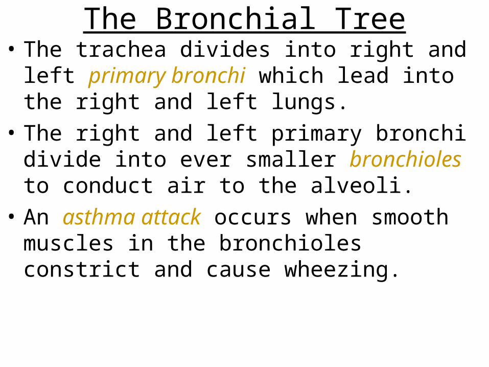
The Bronchial Tree• The trachea divides into right and left primary
bronchi which lead into the right and left lungs.
• The right and left primary bronchi divide into ever smaller bronchioles to conduct air to the alveoli.
• An asthma attack occurs when smooth muscles in the bronchioles constrict and cause wheezing.

The Bronchial Tree

The Lungs• Lungs are paired, cone-shaped organs.
• Lungs are functionally composed of tiny air-sacs called aveoli
• The right lung has three lobes, and the left lung has two lobes, allowing for the space occupied by the heart.
• The lungs are bounded by the ribs and diaphragm.

The Alveoli• Alveoli are the tiny air sacs of the lungs made
up of squamous epithelium and surrounded by blood capillaries.
• Alveoli function in gas exchange, oxygen diffusing into the bloodstream and carbon dioxide diffusing out.
• Infant respiratory distress syndrome occurs in premature infants where underdeveloped lungs lack surfactant (thin film of lipoprotein) and collapse.

Gas exchange in the lungs

Inspiration and Expiration• There is a continuous column of air from the
pharynx to the alveoli, and the lungs lie within the sealed-off thoracic cavity.
• The thoracic cavity is bounded by the rib cage and diaphragm.
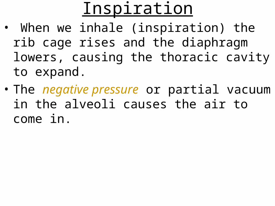
Inspiration• When we inhale (inspiration) the rib cage rises
and the diaphragm lowers, causing the thoracic cavity to expand.
• The negative pressure or partial vacuum in the alveoli causes the air to come in.
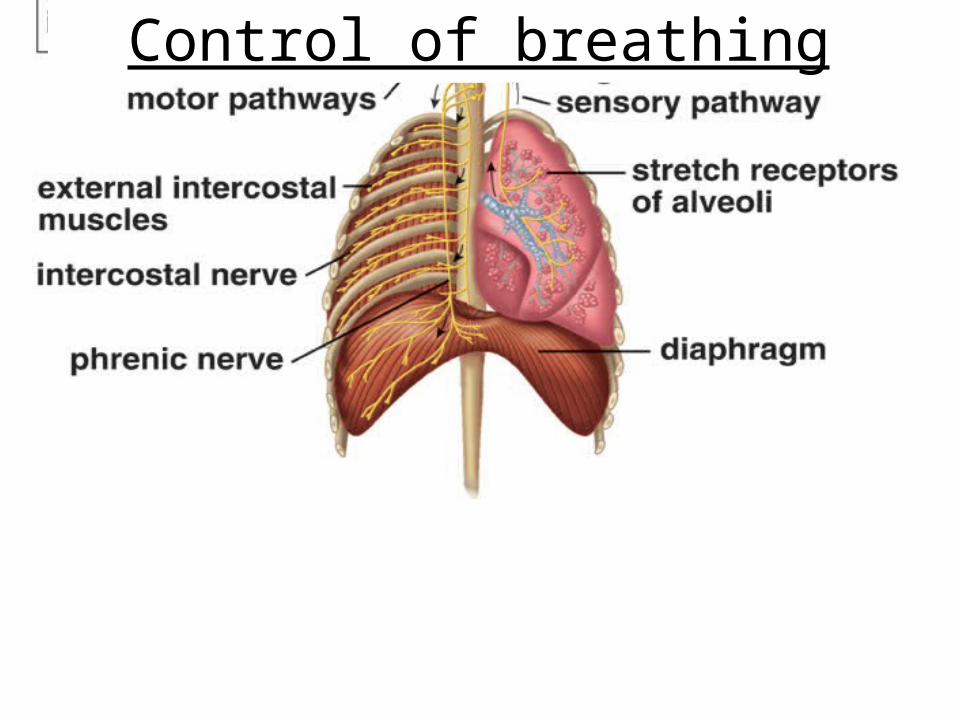
Control of breathing
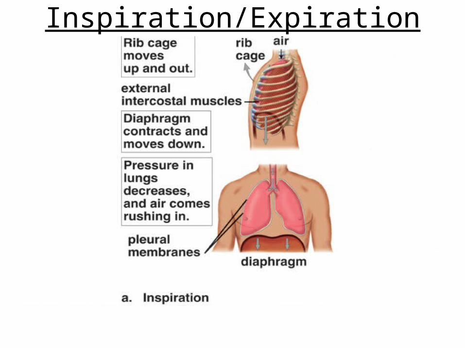
Inspiration/Expiration
Expiration• When we exhale (expiration), the rib cage
lowers and diaphragm to resume dome shape.
• Expiration is passive, while inspiration is active.

Internal Respiration• Internal respiration is the diffusion of O2 from systemic
capillaries into tissues and CO2 from tissue fluid into systemic capillaries through hemoglobin in Red Blood Cells.
• Oxyhemoglobin gives up O2, which diffuses out of the blood and into the tissues because the level of O2 in tissues is lower than that of the blood.
• CO2 diffuses from tissue cells into the blood, it enters red blood cells where a small amount is taken up by hemoglobin

• All this “internal respiration” occurs at the capillaries (single-cell thickness allows for complete diffusion)
• Blood leaving capillaries (which become “venules” and then “veins” is a dark maroon color because red blood cells contain reduced hemoglobin.
Internal Respiration
External and internal respiration
External and internal respiration

Sites of upper respiratory infections

Sinusitis• Sinusitis is infection of the cranial sinuses
within the facial skeleton that drain into nasal cavities.
• It occurs when nasal congestion blocks the sinus openings and is relieved when drainage is restored.
• Pain and tenderness over the lower forehead and cheeks, and toothache, accompany this condition.
Otitis Media• Otitis media is bacterial infection of the middle
ear.
• Children suffer when a nasal infection spreads to the middle ear by way of the auditory tube and antibiotics are usually used to clear the infection.
• Sometimes drainage tubes (called tympanostomy tubes) are inserted into the eardrums of children with recurrent infections.
•

• Tonsillitis• Tonsillitis is infection of tonsils and recurrent
infections that make breathing or swallowing difficult may be relieved by a tonsillectomy.
• Laryngitis• Laryngitis is an infection of the larynx and usually
results in a loss of voice.
• Persistent hoarseness is a warning sign of cancer.

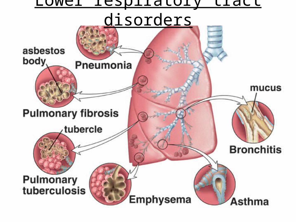
Lower Respiratory Tract Disorders• Lower respiratory infections include:
1) acute bronchitis, an infection of primary and secondary bronchi;
2) pneumonia involving a bacterial or viral infection of the lungs; and
3) pulmonary tuberculosis (infection caused by tubercle bacillus).

Restrictive Pulmonary Disorders• In restrictive pulmonary disorders, vital capacity
is reduced because the lungs have lost their elasticity due to inhaled particles such as silica, coal dust, or asbestos.
• Fibrous connective tissue builds in the lungs in pulmonary fibrosis, caused by exposure to inhaled particles, including those of fiberglass.
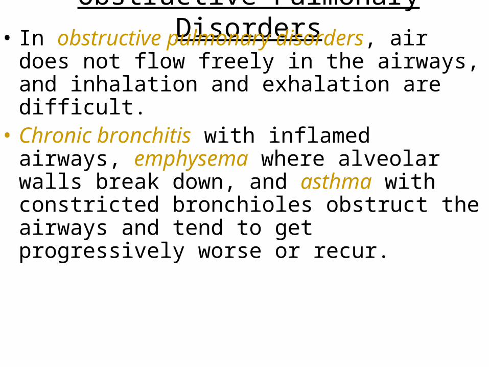
Obstructive Pulmonary Disorders• In obstructive pulmonary disorders, air does not
flow freely in the airways, and inhalation and exhalation are difficult.
• Chronic bronchitis with inflamed airways, emphysema where alveolar walls break down, and asthma with constricted bronchioles obstruct the airways and tend to get progressively worse or recur.
Lower respiratory tract disorders

Lung Cancer• Lung cancer follows this sequence of events:
thickening of airway cells, loss of cilia on the lining, cells with atypical nuclei, tumor development, and finally metastasis.
• Removal of a lobe or lung, called pneumonectomy, may remove the cancer.
• Smoking, whether active or passive, is a major cause of lung cancer.

Normal lung versus cancerous lung

• During inspiration, the pressure in the lungs decreases and air comes rushing in; during expiration, increased pressure in the thoracic cavity causes air to leave the lungs.
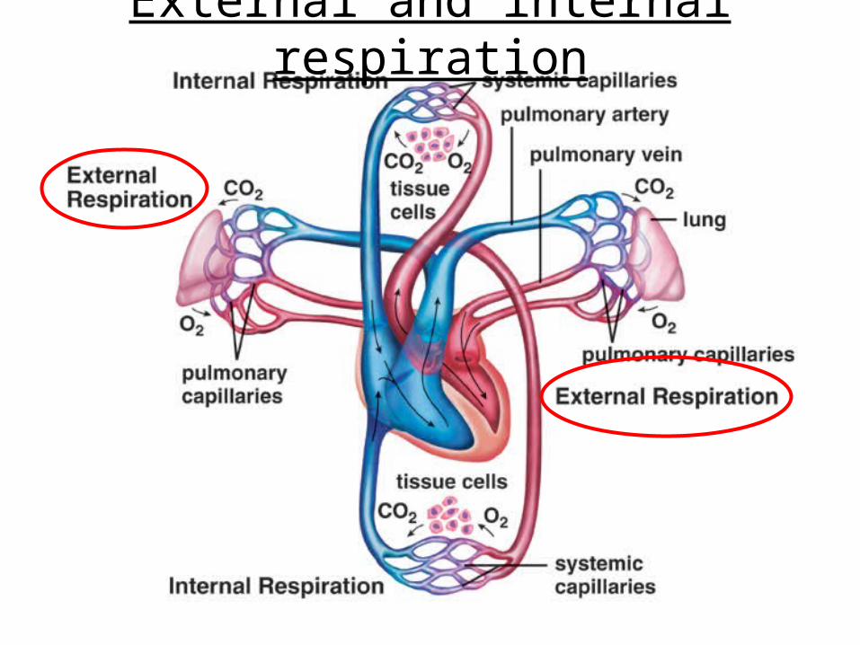
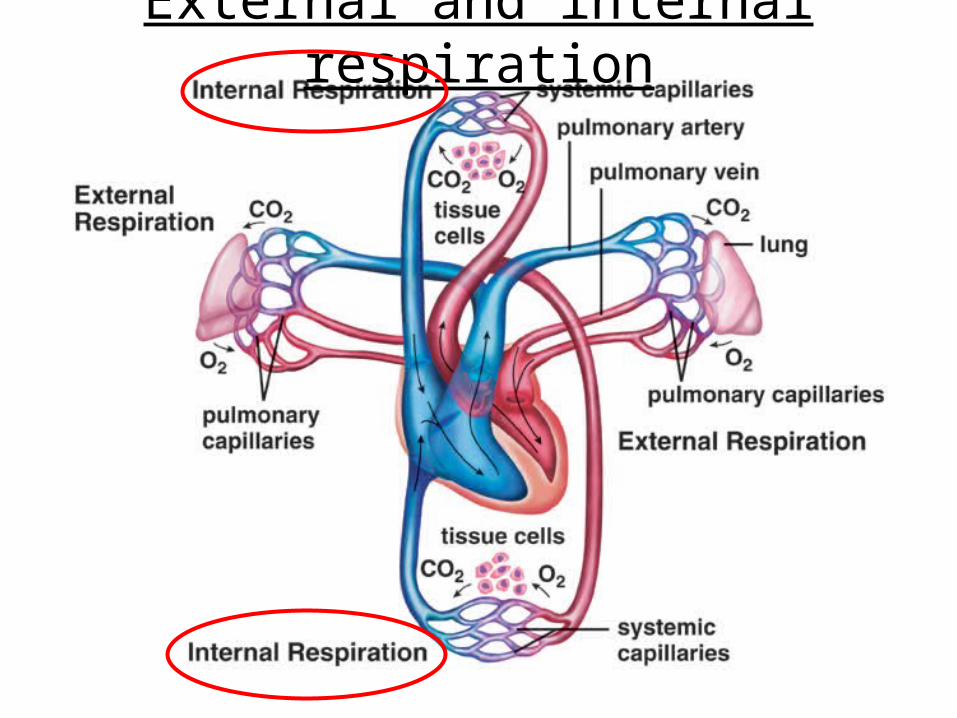
• External respiration occurs in the lungs where oxygen diffuses into the blood and carbon dioxide diffuses out of the blood.
• Internal respiration occurs in the tissues where oxygen diffuses out of the blood into tissue cells and carbon dioxide diffuses into the blood.

• The respiratory pigment hemoglobin transports oxygen from the lungs to the tissues and aids in the transport of carbon dioxide from the tissues to the lungs.
• The respiratory tract is especially subject to disease because it is exposed to infectious agents; also, cigarette smoking contributes to two major lung disorders—emphysema and cancer.
Chapter 17: Nervous System
Copyright © The McGraw-Hill Companies, Inc. Permission required for reproduction or display.

Nervous Tissue
• The nervous system is divided into a central nervous system (CNS), consisting of the brain and spinal cord, and a peripheral nervous system (PNS), consisting of nerves carrying sensory and motor information between the CNS and muscles and glands.
• Both systems have two types of cells: neurons that transmit impulses.

Organization of the nervous system

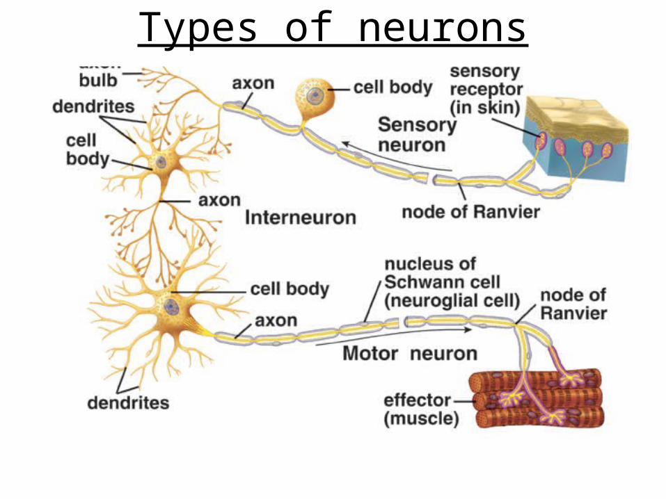
Neuron Structure• Neurons are composed of dendrites that receive
signals, a cell body with a nucleus, and an axon that conducts a nerve impulse away.
• Sensory neurons take information from sensory receptors to the CNS.
• Interneurons occur within the CNS and integrate input.
• Motor neurons take information from the CNS to muscles or glands.
Types of neurons

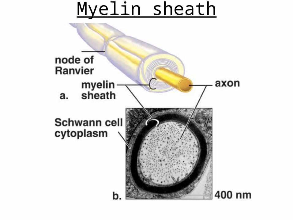
Myelin Sheath• Long axons are covered by a protective myelin
sheath formed by another type of cell. This sheath acts like insulation on a wire, increasing the speed of transmission.
• The sheath contains lipid myelin which gives nerve fibers their white, glistening appearance.
• Multiple sclerosis is a disease of the myelin sheath.
Myelin sheath

The Nerve Impulse• The nervous system uses the nerve impulse to
convey information.
• The nature of a nerve impulse has been studied by using excised axons and a voltmeter.
• Voltage (in millivolts, mV) measures the electrical potential difference between the inside and outside of the axon.

Resting Potential• When an axon is not conducting a nerve
impulse, the inside of an axon is negative (-65mV) compared to the outside; this is the resting potential.
• A sodium-potassium pump in the membrane actively transports Na+ out of the axon and K+ into the axon to establish resting potential.
• The membrane is more permeable to K+ and much of the resting potential is due to the excess of K+ outside of the neuron.

Resting potential

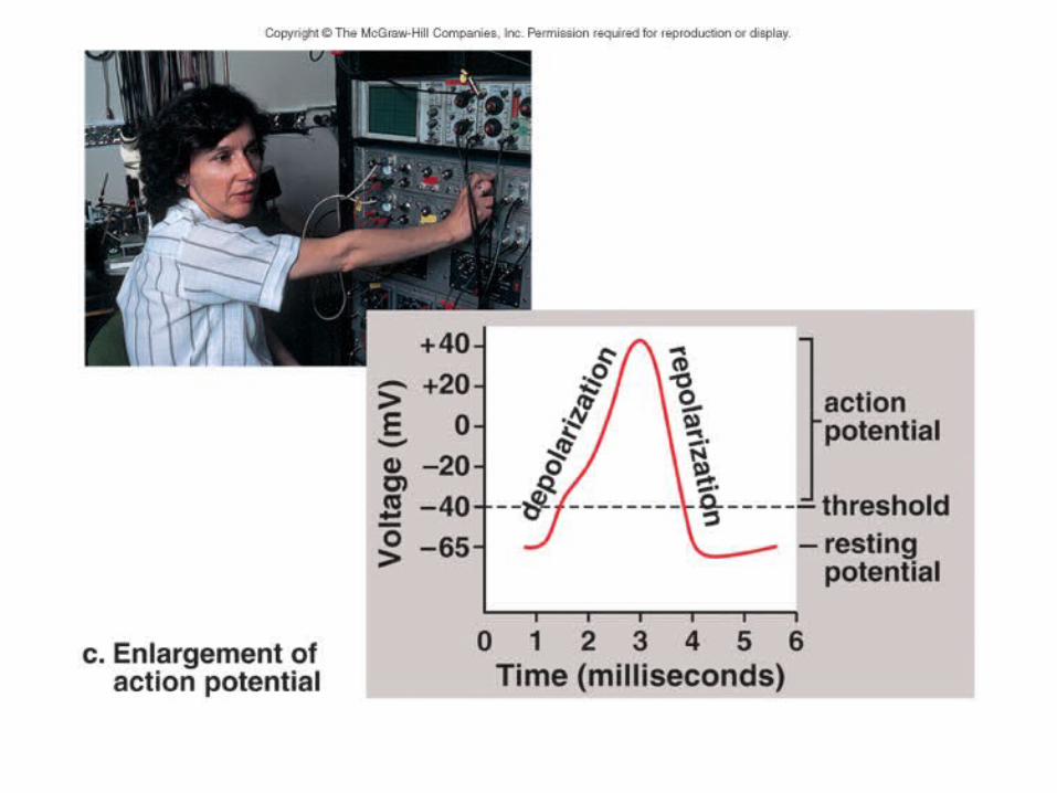
Action Potential• An action potential is a rapid change in
polarity as the nerve impulse occurs. • The action potential occurs if a stimulus
causes the membrane to depolarize past threshold.
• An intense stimulus causes many firings (reaching action potential) in an axon; a weak stimulus may cause only a few.
• The action potential requires two types of gated channel proteins: one each for Na+ and K+.

• Sodium Gates Open• The gates of sodium channels open first
and Na+ flows into the axon. • The membrane potential depolarizes to
+40 MV.
• Potassium Gates Open• The gates of potassium channels open
next and K+ flows to the outside of the axon.
• The membrane potential repolarizes to –65 MV.

Action potential

Propagation of an Action Potential
• The action potential travels the length of an axon, with each portion of the axon undergoing depolarization then repolarization.
• A refractory period ensures that the action potential will not move backwards.
• In myelinated fibers, the action potential only occurs at the nodes of Ranvier.
• This “jumping” from node-to-node is called saltatory conduction.

Transmission Across a Synapse• The tip of an axon forms an axon bulb that
is close to a dendrite or cell body of another neuron; this region of close proximity is called the synapse.
• Transmission of a nerve impulse takes place when a neurotransmitter molecule stored in synaptic vesicles in the axon bulb is released into a synaptic cleft between the axon and the receiving neuron.

• When a nerve impulse reaches an axon bulb, gated channels for calcium open and Ca2+ flow into the bulb.
• This sudden rise in Ca2+ causes synaptic vesicles to move and merge with the presynaptic membrane, releasing their neurotransmitter molecules into the cleft.
• The binding of the neurotransmitter to receptors in the postsynaptic membrane causes either excitation or inhibition.

Synapse structure and function
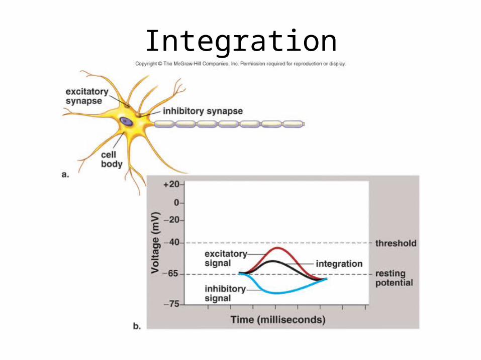
Synaptic Integration
• Many synapses per single neuron is not uncommon.
• Excitatory signals have a depolarizing effect, and inhibitory signals have a hyperpolarizing effect on the post- synaptic membrane.
• Integration is the summing up of these excitatory and inhibitory signals.
Integration

Neurotransmitter Molecules• Out of 25, two well-known
neurotransmitters are acetylcholine (ACh) and norepinephrine (NE).
• Neurotranmitters that have done their job are removed from the cleft; the enzyme acetylcholinesterase (AChE) breaks down acetylcholine.
• Neurotransmitter molecules are removed from the cleft by enzymatic breakdown or by reabsorption, thus preventing continuous stimulation or inhibition.

The Central Nervous System
• The central nervous system (CNS) consists of the spinal cord and brain.
• Both are protected by bone, wrapped in protective membranes called meninges, and surrounded and cushioned with cerebrospinal fluid that is produced in the ventricles of the brain.

• The ventricles are interconnecting cavities that produce and serve as a reservoir for cerebrospinal fluid.
• The CNS receives and integrates sensory input and formulates motor output.
• Gray matter contains cell bodies and short, nonmyelinated fibers; white matter contains myelinated axons that run in tracts.

Organization of the nervous system
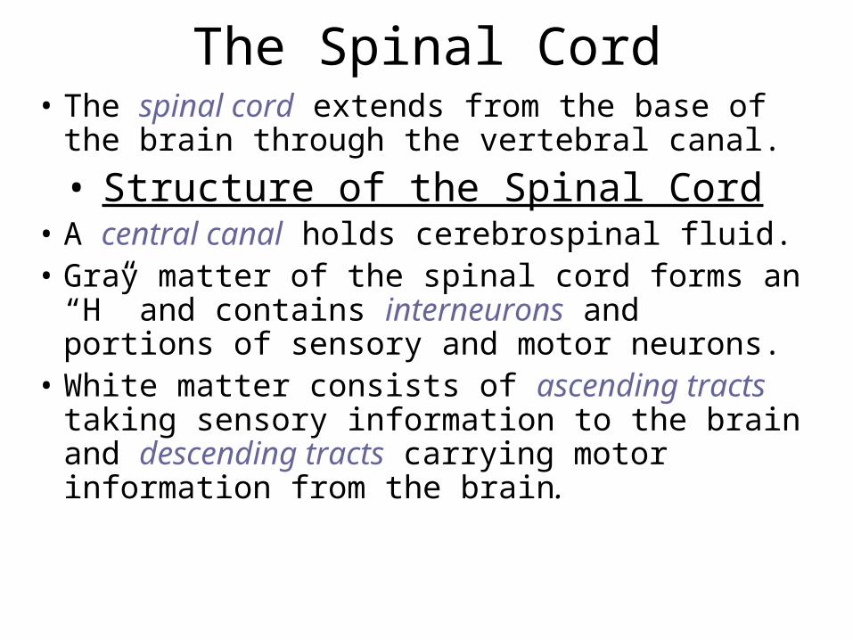
The Spinal Cord• The spinal cord extends from the base of
the brain through the vertebral canal.
• Structure of the Spinal Cord• A central canal holds cerebrospinal fluid. • Gray matter of the spinal cord forms an “H”
and contains interneurons and portions of sensory and motor neurons.
• White matter consists of ascending tracts taking sensory information to the brain and descending tracts carrying motor information from the brain.

Spinal cord

Functions of the Spinal Cord• The spinal cord is the center for many
reflex arcs. • It also sends sensory information to the
brain and receives motor output from the brain, extending communication from the brain to the peripheral nerves for both control of voluntary skeletal muscles and involuntary internal organs.
• Severing the spinal cord produces paralysis.

The Brain
• The brain has four cavities called ventricles.
• The cerebrum has two lateral ventricles, the diencephalon has the third ventricle, and the brain stem and cerebellum have the fourth ventricle.
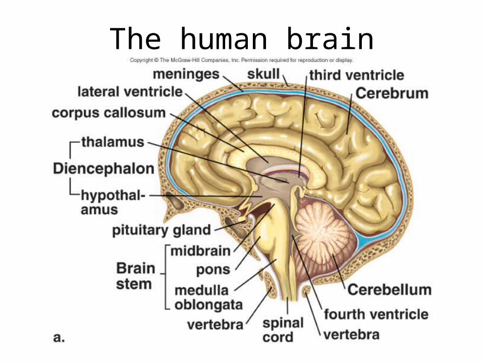
The human brain

The Cerebrum• The cerebrum or telencephalon has two
cerebral hemispheres connected by the corpus callosum.
• Learning, memory, language and speech take place in the cerebrum.
• Sulci divide each hemisphere into lobes including the frontal, parietal, occipital, and temporal lobes.

Cerebral hemispheres

The Cerebral Cortex• The cerebral cortex is a thin, highly
convoluted outer layer of gray matter covering both hemispheres.
• The primary motor area is in the frontal lobe; this commands skeletal muscle.
• The primary somatosensory area is dorsal to the central sulcus or groove.
• The primary visual area is at the back occipital lobe.
• The temporal lobe has the primary auditory area.
• The parietal lobe provides taste sensation. • All have adjacent association areas that
integrate signals; the prefrontal area is an important association area for appropriate behavior.
• White matter consists mostly of long myelinated axons forming tracts; these cross over so the left side of the brain handles right side information.
• Basal nuclei are masses of gray matter deep within the white matter integrate motor commands.

The lobes of a cerebral hemisphere

The Diencephalon• The hypothalamus and thalamus are in the
diencephalon that encircles the third ventricle.
• The hypothalamus controls homeostasis and the pituitary gland, and the thalamus receives all sensory input except smell and integrates it and sends it to the cerebrum.
• The pineal gland is also located here and secretes melatonin that may regulate our daily rhythms.
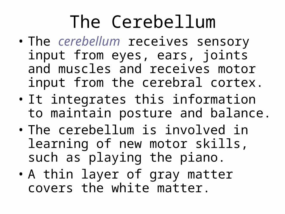
The Cerebellum• The cerebellum receives sensory input
from eyes, ears, joints and muscles and receives motor input from the cerebral cortex.
• It integrates this information to maintain posture and balance.
• The cerebellum is involved in learning of new motor skills, such as playing the piano.
• A thin layer of gray matter covers the white matter.
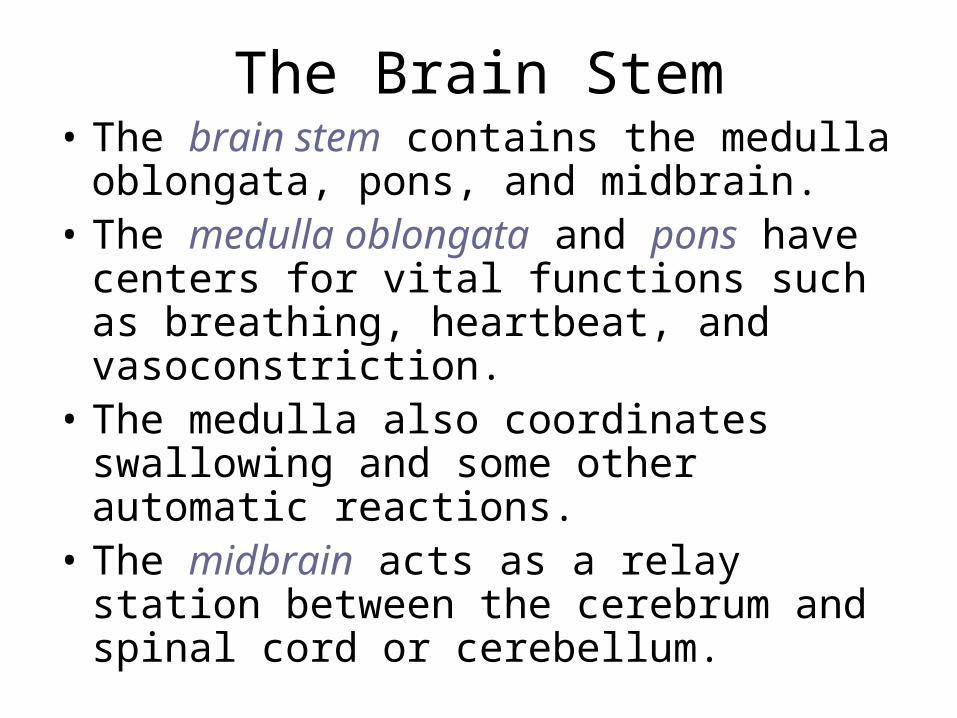
The Brain Stem• The brain stem contains the medulla
oblongata, pons, and midbrain. • The medulla oblongata and pons have
centers for vital functions such as breathing, heartbeat, and vasoconstriction.
• The medulla also coordinates swallowing and some other automatic reactions.
• The midbrain acts as a relay station between the cerebrum and spinal cord or cerebellum.

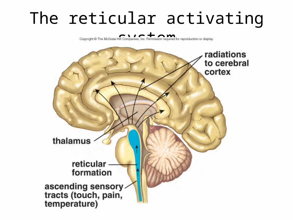
The Reticular Formation
• The reticular formation is a complex network of nuclei and fibers that extend the length of the brain stem.
• One portion of the reticular formation, called the reticular activating system, arouses the cerebrum via the thalamus causing alertness.
• An inactive reticular activating system results in sleep.
The reticular activating system

The Limbic System and Higher Mental Functions
• Limbic System • The limbic system is involved in our
emotions and higher mental functions.• The limbic system is a complex network of
tracts and nuclei involving cerebral lobes, basal nuclei and the diencephalon.
• Two structures, the hippocampus and amygdala are essential for learning and memory.

The limbic system
Higher Mental Functions
• Animal research, MRI, and PET scans allow researchers to study the functioning of the brain.
• Memory and Learning
• Memory is the ability to hold a thought in mind or recall events from the past.
• Learning takes place when we retain and utilize past memories.

• Short-term memory involves activity in the prefrontal area.
• Long-term memory includes semantic memory (numbers, words, etc.) and episodic memory (persons, events, etc.).
• Skill memory involves ability to ride a bike, for example, and involves all motor areas of the cerebrum below the level of consciousness.
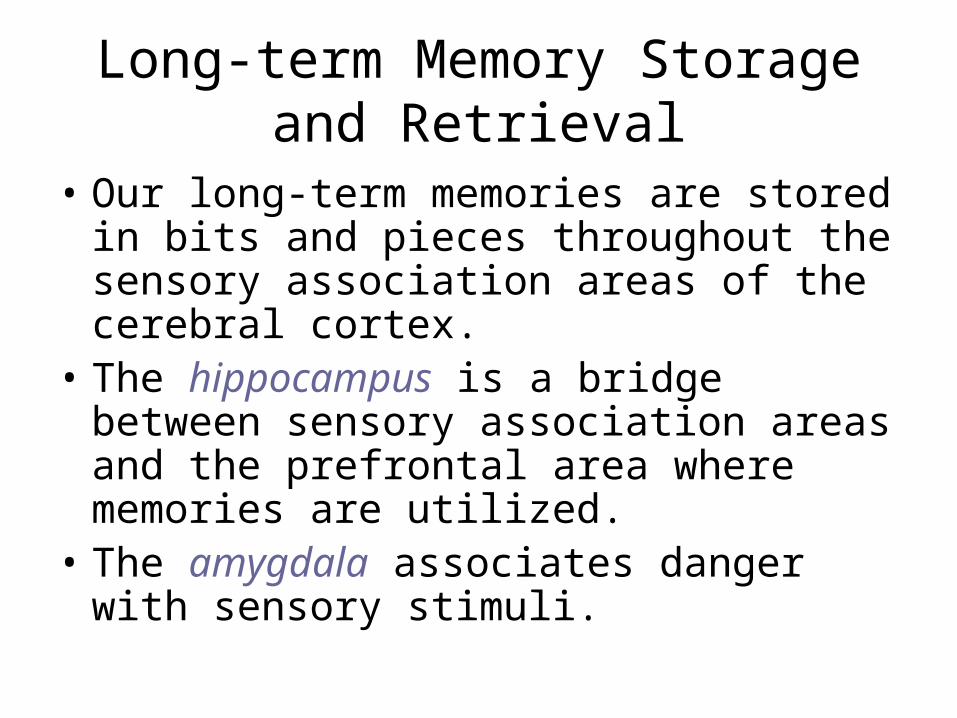
Long-term Memory Storage and Retrieval
• Our long-term memories are stored in bits and pieces throughout the sensory association areas of the cerebral cortex.
• The hippocampus is a bridge between sensory association areas and the prefrontal area where memories are utilized.
• The amygdala associates danger with sensory stimuli.
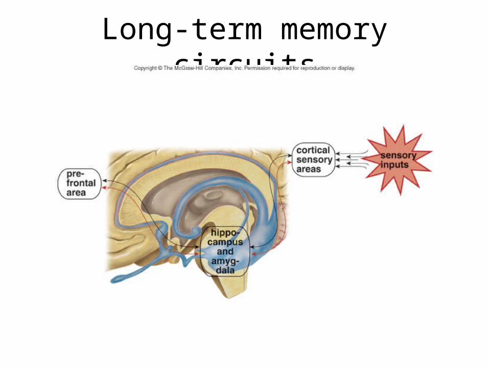
Long-term memory circuits
Long-Term Potentiation• Long-term potentiation is increased
response at synapses within the hippocampus and is essential to long-term memory.
• However, a postsynaptic neuron in the hippocampus can become too excited and then die.
• Excitotoxicity, a form of cell death, is due to the neurotransmitter glutamate rushing in too quickly.
Language and Speech
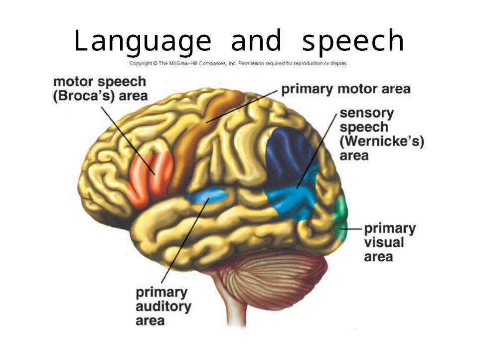
• Language and speech are dependent upon Broca’s area (a motor speech area) and Wernicke’s area (a sensory speech area) that are involved in communication.
• These two areas are located only in the left hemisphere; the left hemisphere functions in language in general and not just in speech.
Language and speech

The Peripheral Nervous System
• The peripheral nervous system (PNS) contains nerves (bundles of axons) and ganglia (cell bodies).
• Sensory nerves carry information to the CNS, motor nerves carry information away, and mixed nerves have both types of fibers.
• Humans have 12 pairs of cranial nerves and 31 pairs of spinal nerves.

Nerve structure

Cranial nerves

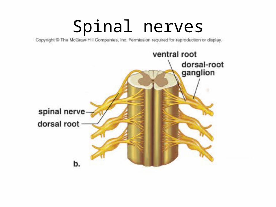
• The dorsal root of a spinal nerve contains sensory fibers that conduct sensory impulses from sensory receptors toward the spinal cord.
• Dorsal root ganglia near the spinal cord contain the cell bodies of sensory neurons.
• The ventral root of a spinal nerve contains motor fibers that conduct impulses away from the spinal cord to effectors.
Spinal nerves

Somatic System
• The somatic system serves the skin, skeletal muscles, and tendons.
• The brain is always involved in voluntary muscle actions but somatic system reflexes are automatic and may not require involvement of the brain.
The Reflex Arc• Involuntary reflexes allow us to respond
rapidly to external stimuli.• In reflexes, sensory receptors generate
nerve impulses carried to interneurons in the spinal cord.
• Next, interneurons signal motor neurons which conduct nerve impulses to a skeletal muscle that contracts, giving the response to the stimulus.
• Pain is not felt until the brain receives nerve impulses.

A spinal nerve reflex arc
Autonomic System• The autonomic system of the PNS
regulates the activity of cardiac and smooth muscle and glands.
• The system is divided into sympathetic and parasympathetic divisions that:
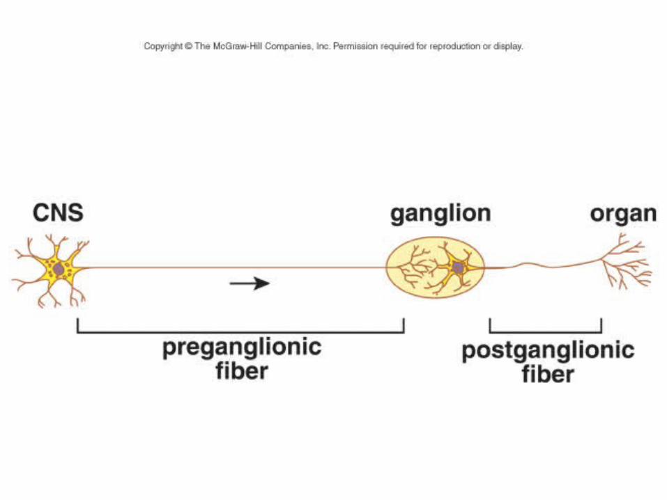
1) Function automatically and involuntarily;2) Innervate all internal organs; and3) Use two neurons and one ganglion.

Sympathetic Division
• The sympathetic division is associated with responses that occur during times of stress, including “fight or flight” reactions.
• The postganglionic axon releases mainly norepinephrine which acts similar to adrenaline, the hormone from the adrenal medulla.

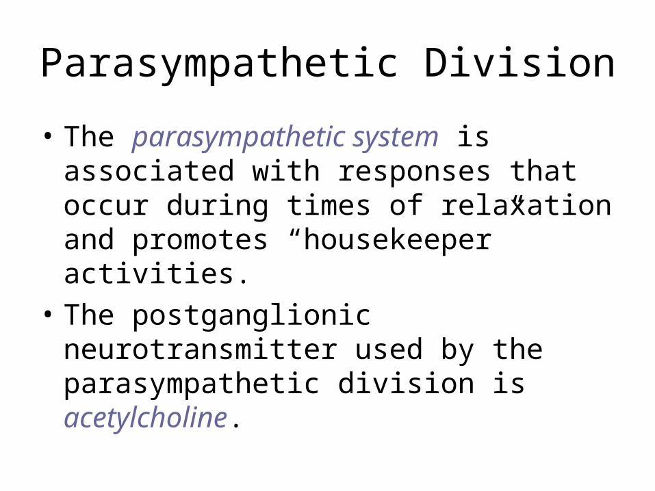
Parasympathetic Division
• The parasympathetic system is associated with responses that occur during times of relaxation and promotes “housekeeper” activities.
• The postganglionic neurotransmitter used by the parasympathetic division is acetylcholine.

Autonomic nervous system
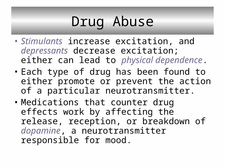
Drug Abuse• Stimulants increase excitation, and
depressants decrease excitation; either can lead to physical dependence.
• Each type of drug has been found to either promote or prevent the action of a particular neurotransmitter.
• Medications that counter drug effects work by affecting the release, reception, or breakdown of dopamine, a neurotransmitter responsible for mood.

Drug actions at a synapse

Drug use

Alcohol• Alcohol may affect the inhibiting transmitter
GABA or glutamate, an excitatory neurotransmitter.
• Alcohol is primarily metabolized in liver and heavy doses can cause liver scar tissue and cirrhosis.
• Alcohol is an energy source but it lacks nutrients needed for health.
• Cirrhosis of the liver and fetal alcohol syndrome are serious conditions associated with alcohol intake.
Nicotine
• Nicotine is an alkaloid derived from tobacco.
• In the CNS, nicotine causes neurons to release dopamine; in the PNS, nicotine mimics the activity of acetylcholine and increases heart rate, blood pressure, and digestive tract mobility.
• Nicotine induces both physiological and psychological dependence.

Cocaine• Cocaine is an alkaloid derived from the
shrub Erythroxylum cocoa, often sold as potent extract termed “crack.”
• Cocaine prevents uptake of dopamine by the presynaptic membrane, is highly likely to cause physical dependence, and requires higher doses to overcome tolerance.
• This makes overdosing is a real possibility; overdosing can cause seizures and cardiac arrest.
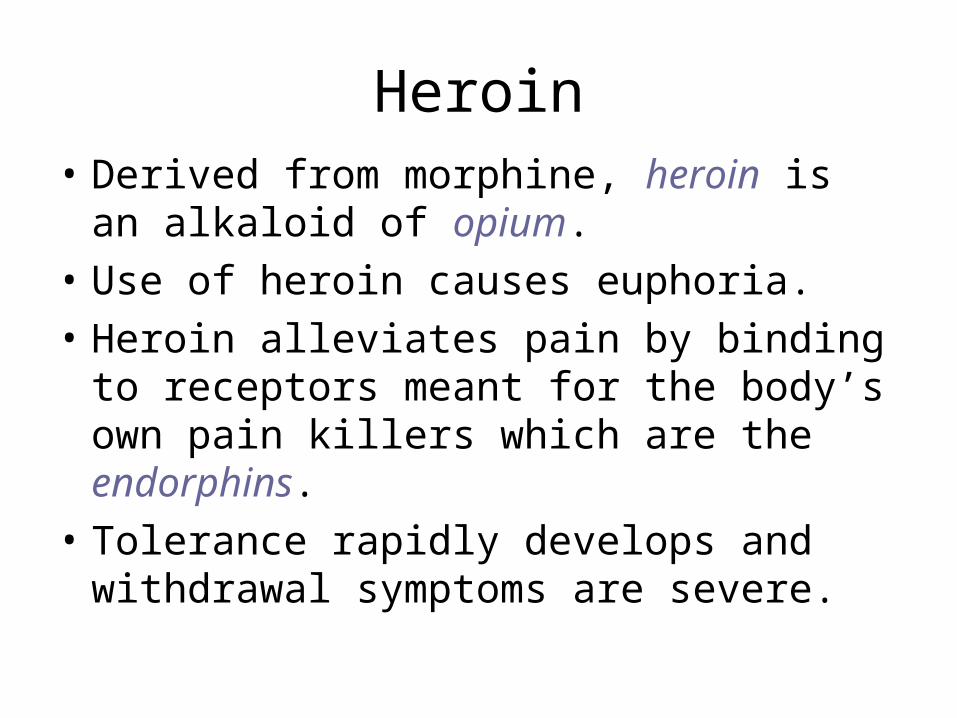
Heroin• Derived from morphine, heroin is an
alkaloid of opium.
• Use of heroin causes euphoria.
• Heroin alleviates pain by binding to receptors meant for the body’s own pain killers which are the endorphins.
• Tolerance rapidly develops and withdrawal symptoms are severe.

Marijuana
• Marijuana is obtained from the plant Cannabis sativa that contains a resin rich in THC (tetrahydrocannabinol).
• Effects include psychosis and delirium and regular use can lead to dependence.
• Long-term marijuana use may lead to brain impairment, and a fetal cannabis syndrome has been reported.
Chapter Summary• The nervous system consists of two types
of cells: neurons and mesoglia.• Neurons are specialized to carry nerve
impulses.• A nerve impulse is an electrochemical
change that travels along the length of a neuron fiber.
• Transmission of signals between neurons is dependent on neurotransmitter molecules.

• The central nervous system is made up of the spinal cord and the brain.
• The parts of the brain are specialized for particular functions.
• The cerebral cortex contains motor areas, sensory areas, and association areas that are in communication with each other.
• The cerebellum is responsible for maintaining posture; the brainstem houses reflexes for homeostasis.
• The reticular formation contains fibers that arouse the brain when active and account for sleep when they are inactive.
• The limbic system contains specialized areas that are involved in higher mental functions and emotional responses.
• Long-term memory depends upon association areas that are in contact with the limbic system.
• There are particular areas in the left hemisphere that are involved in language and speech.
• The peripheral nervous system contains nerves that conduct nerve impulses toward and away from the central nervous system.
• The autonomic nervous system has sympathetic and parasympathetic divisions with counteracting activities.
• Use of psychoactive drugs such as alcohol, nicotine, marijuana, cocaine, and heroin is detrimental to the body.

Ex 22: Annelida (segmented worms)
Copyright © The McGraw-Hill Companies, Inc. Permission required for reproduction or display.
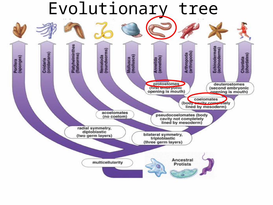
Evolutionary tree

Annelids
• Annelids are segmented both externally, and internally by partitions called septa.
• Annelids have a hydrostatic skeleton, and partitioning of the coelom permits each body segment to move independently.
• The tube-within-a-tube body plan allows the digestive tract to have specialized organs.
• Annelids have an extensive closed circulatory system with blood vessels that run the length of the body and branch to every segment.
• The brain is connected to a ventral solid nerve cord with ganglia in each segment.
• The excretory system has nephridia in each segment.
• A nephridium is a tubule that collects wastes and excretes through an opening in the body wall.
Marine Worms
• Polychaetes are marine worms with paddlelike parapodia at the side of each segment.
• Some polychaetes are sessile tube worms.
• A clam worm is a predaceous marine worm with a defined head region.
• During breeding seasons, some worms form sex organs in special segments and shed these segment during breeding.

Polychaete diversity
Earthworms• Earthworms are oligochaetes having few
setae per segment. • Most scavenge for food in the soil and the
moist body wall functions in gas exchange. • When muscles contract in each segment,
setae anchor in the soil, and aid locomotion. • Five “hearts” pump blood and a branch
blood vessel reaches each segment. • These worms are hermaphroditic.

• Segmentation in earthworms is evidenced by:
• Body rings
• Coelom divided by septa
• Setae on most segments
• Ganglia and lateral nerves in each segment
• Nephridia in most segments
• Branch blood vessels in each segment

Earthworm, Lumbricus

Leeches
• Most leeches are fluid feeders that attach themselves to open wounds using suckers.
• Bloodsuckers, such as the medicinal leech, can cut through tissue.
• An anticoagulant (hirudin) in their saliva keeps blood from clotting.

Ex 23: Arthropods!
Copyright © The McGraw-Hill Companies, Inc. Permission required for reproduction or display.

Evolutionary tree

Arthropods• Arthropods are the most varied and
numerous of animals.
• The success of arthropods is largely attributable to a flexible exoskeleton, jointed appendages, and specialization of body regions.
• Three body regions – head, thorax, and abdomen – with specialized appendages in each region, and a well-developed nervous system characterize this group.

Arthropod diversity
Crustaceans• Crustaceans are largely marine and have
a head that bears compound eyes, two pair of antennae, and specialized mouth parts.
• Five pairs of walking legs include a first pair of pinching claws.
• In the crayfish, head and thorax are fused into a cephalothorax which is covered on the top and sides by carapace.
• The abdominal segments have swimmerets.

• The crayfish has an open circulatory system in which the heart pumps blood into a hemocoel consisting of sinuses where the hemolymph flows about the organs.
• Respiration takes place by gills under the hard carapace, and there is a ventral solid nerve cord.
• Sexes are separate in the crayfish.
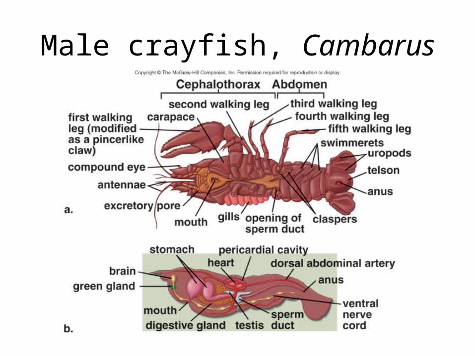
Male crayfish, Cambarus
Insects• The head of an insect usually bears a pair
of antennae, compound eyes, and simple eyes.
• The thorax bears three pairs of legs and up to two pairs of wings, and the abdomen contains most of the internal organs.
• The insect exoskeleton is lighter and contains less chitin than that of many other arthropods.

Insect diversity

• Grasshoppers are examples of insects adapted to a terrestrial life; they respire by tracheae and have wings that allow them to evade enemies; the third pair of legs is suitable for jumping.
• There is a tympanum for the reception of sound waves and a male penis for passing sperm to the female without desiccation.

• Malpighian tubules function in excretion in grasshopper.
• Grasshoppers undergo gradual metamorphosis from nymph to adult.
• Butterflies undergo complete metamorphosis, changing from larva to pupa to adult.

Female grasshopper

Arachnids
• The arachnids include terrestrial spiders, scorpions, ticks, and mites.
• The cephalothorax bears six pairs of appendages: the chelicerae and the pedipalps, and four pairs of walking legs.
• Scorpions are the oldest terrestrial arthropods.
• Ticks and mites are parasitic.
• Spiders are well-adapted to life on land and have Malphigian tubules – they secrete uric acid, helping to conserve water.
• Spiders spin silk used in various ways. • Where spiders spin webs, the type of web
is a feature that demonstrates the evolutionary relationship among spiders.
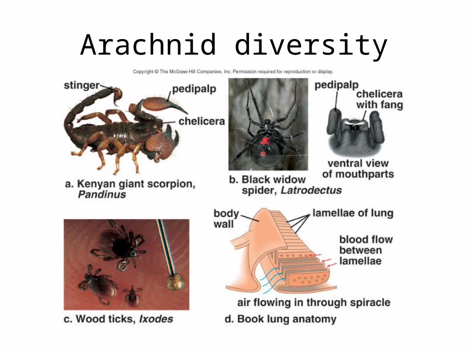
Arachnid diversity