chapter 16 the molecular basis of inheritance. but first… bessbugs! (bess beetles)
TRANSCRIPT
Chapter 16
The Molecular Basis of Inheritance
But first… Bessbugs! (Bess beetles)
Overview: Life’s Operating Instructions
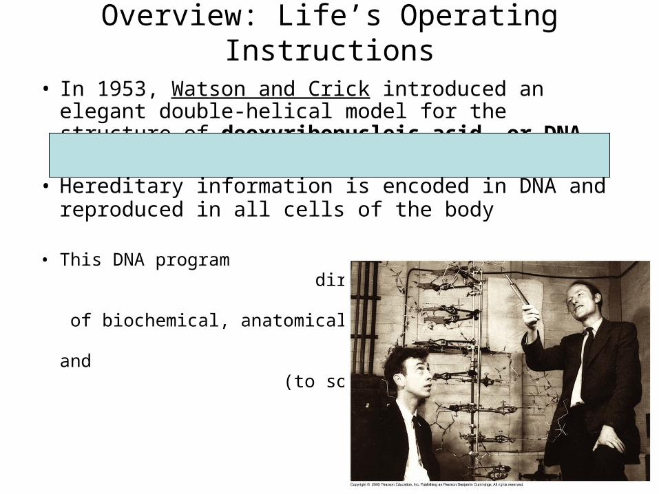
• In 1953, Watson and Crick introduced an elegant double-helical model for the structure of deoxyribonucleic acid, or DNA
• Hereditary information is encoded in DNA and reproduced in all cells of the body
• This DNA program directs the development of biochemical, anatomical, physiological, and (to some extent) behavioral traits

Each nucleotide
consists of a:
1. pentose sugar
2. nitrogenous base
3. phosphate group
Nucleic acid
1869: Friedrich Miescherisolated DNA from pus!
DNA and RNA are nucleic acids.

Sugar–phosphatebackbone
Nitrogenous bases
Thymine (T)
Adenine (A)
Cytosine (C)
Guanine (G)
Nitrogenous base
Phosphate
DNA nucleotide
Sugar(deoxyribose)
3 end
5 end
What is the genetic material?
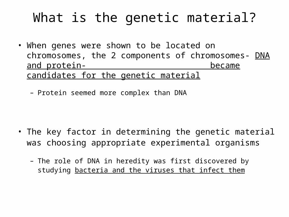
• When genes were shown to be located on chromosomes, the 2 components of chromosomes- DNA and protein- became candidates for the genetic material
– Protein seemed more complex than DNA
• The key factor in determining the genetic material was choosing appropriate experimental organisms
– The role of DNA in heredity was first discovered by studying bacteria and the viruses that infect them

Evidence That DNA Can Transform BacteriaDiscovery of genetic role of DNA began w/ research by Frederick Griffith in 1928,who worked with two strains of a bacterium, Streptococcus pneumoniae1. pathogenic “S” (smooth) strain2. harmless “R” (rough) strain
When he mixed heat-killed remains of the pathogenic strain with living cells of the harmless strain, some living cells became pathogenic
He called this phenomenon transformation,
now defined as a change in genotype and phenotype due to assimilation of foreign DNA

Evidence That Viral DNA Can Program Cells
• In 1944, Oswald Avery, Maclyn McCarty, and Colin MacLeod announced that the transforming substance was DNA
– Avery-MacLeod-McCarty experiment
• More evidence for DNA as the genetic material came from studies of a virus that infects bacteria
• Such viruses, called bacteriophages (or phages),
are widely used in molecular genetics research
Phage
DNA
Bacterial cell
Radioactive protein
Radioactive DNA
Batch 1: radioactive sulfur (35S)
Batch 2: radioactive phosphorus (32P)
radioactive sulfurlabels protein
radioactive phosphorus labels DNA
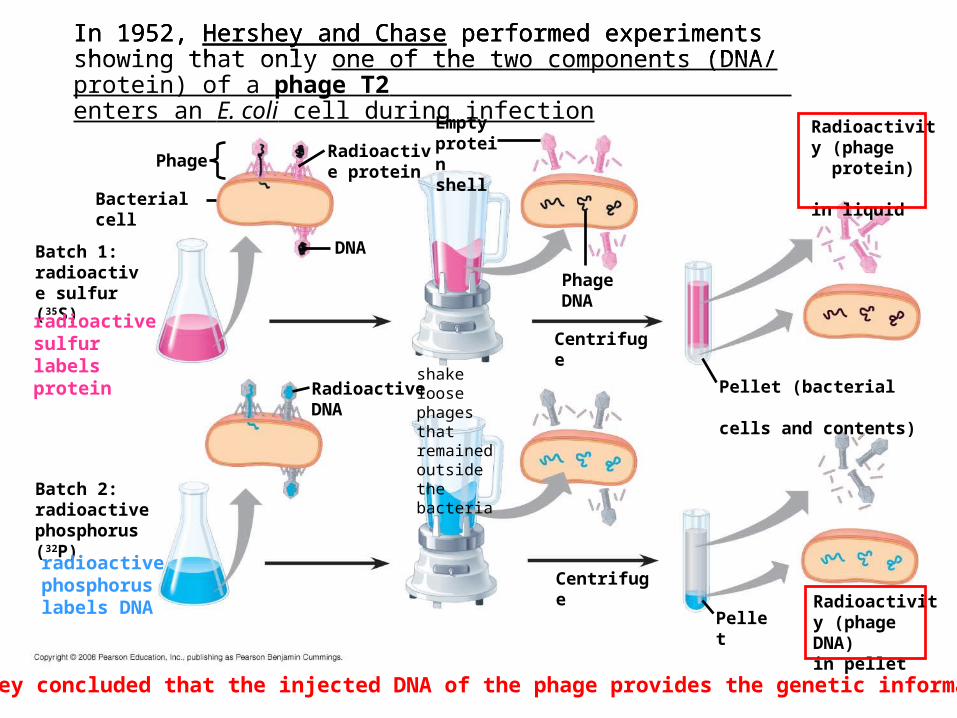
In 1952, Hershey and Chase performed experiments showing that only one of the two components (DNA/ protein) of a phage T2 enters an E. coli cell during infection
Phage
DNA
Bacterial cell
Radioactive protein
Radioactive DNA
Batch 1: radioactive sulfur (35S)
Batch 2: radioactive phosphorus (32P)
Empty protein shell
Phage DNA
radioactive sulfurlabels protein
radioactive phosphorus labels DNA
In 1952, Hershey and Chase performed experiments showing that only one of the two components (DNA/ protein) of a phage T2 enters an E. coli cell during infection
shake loose phages that remained outside the bacteria
Phage
DNA
Bacterial cell
Radioactive protein
Radioactive DNA
Batch 1: radioactive sulfur (35S)
Batch 2: radioactive phosphorus (32P)
Empty protein shell
Phage DNA
Centrifuge
Centrifuge
Pellet
Pellet (bacterial cells and contents)
Radioactivity (phage protein) in liquid
Radioactivity (phage DNA) in pellet
radioactive sulfurlabels protein
radioactive phosphorus labels DNA
In 1952, Hershey and Chase performed experiments showing that only one of the two components (DNA/ protein) of a phage T2 enters an E. coli cell during infection
They concluded that the injected DNA of the phage provides the genetic information
shake loose phages that remained outside the bacteria

Building a Structural Model of DNAClue #1
Erwin Chargaff
In 1950, reported that DNA composition varies from one species to the next
• This evidence of diversity made DNA a more credible candidate for the genetic material
Chargaff’s rules state that in any species there is an equal number of A and T bases, and an equal number of G and C bases
A TG C

Building a Structural Model of DNAClue #2
• Wilkins and Franklin were using a technique called
X-ray crystallography to study molecular structure
• Franklin produced a picture of the DNA molecule using this technique
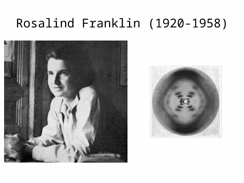
Rosalind Franklin (1920-1958)
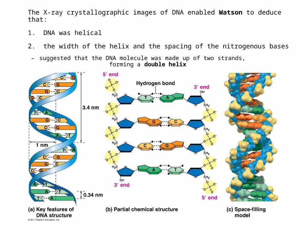
The X-ray crystallographic images of DNA enabled Watson to deduce that:
1. DNA was helical
2. the width of the helix and the spacing of the nitrogenous bases
– suggested that the DNA molecule was made up of two strands, forming a double helix

• At first, Watson and Crick thought the bases paired like with like (A with A, and so on), but such pairings did not result in a uniform width
• Instead, pairing a purine with a pyrimidine resulted in a uniform width consistent with the X-ray
Adenine (A) Thymine (T)
Guanine (G) Cytosine (C)
Sugar
Sugar
Sugar
Sugar
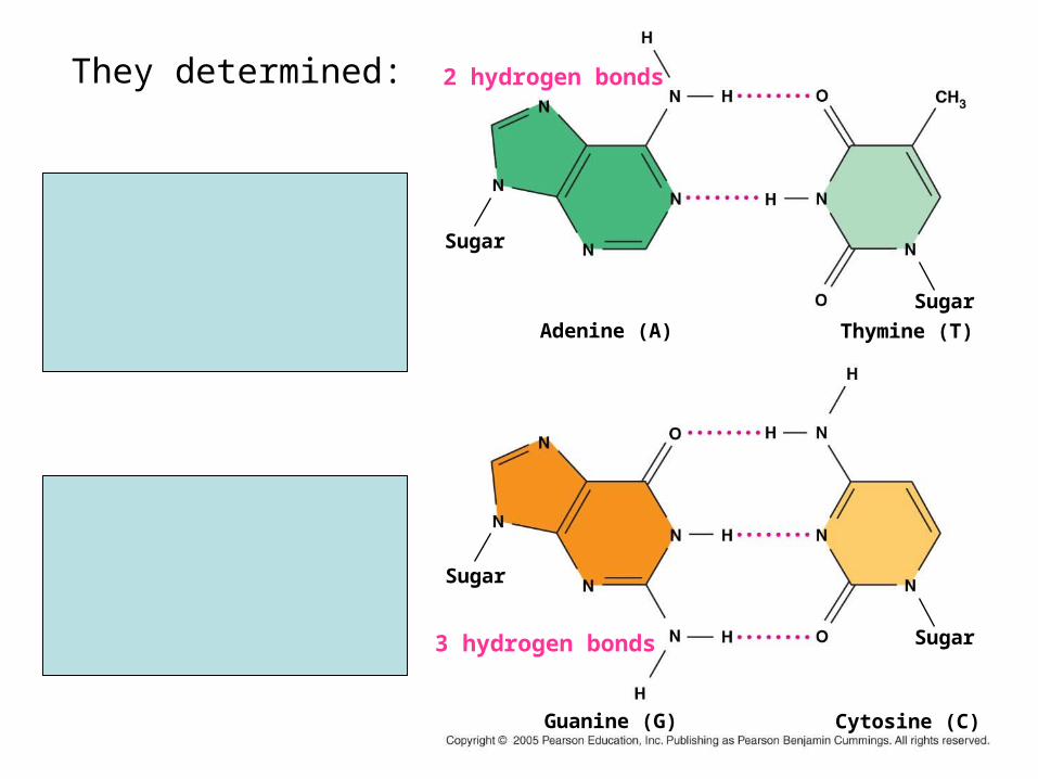
They determined:
adenine (A) paired only with thymine (T)
guanine (G) paired only with cytosine (C)
2 hydrogen bonds
3 hydrogen bonds


DNA Replication• Watson and Crick noted that the specific base pairing
suggested a possible copying mechanism for genetic material
• Since the two strands of DNA are complementary, each strand acts as a template for building a new strand in replication
• In DNA replication, the parent molecule unwinds, and 2 new daughter strands are built based on base-pairing rules
The parent molecule hastwo complementarystrands of DNA. Each baseis paired by hydrogenbonding with its specificpartner, A with T and Gwith C.
A T
C G
T A
A T
G C
The parent molecule hastwo complementarystrands of DNA. Each baseis paired by hydrogenbonding with its specificpartner, A with T and Gwith C.
A T
C G
T A
A T
G C G
A T
C G
T A
A T
C
The first step in replicationis separation of the twoDNA strands.
The parent molecule hastwo complementarystrands of DNA. Each baseis paired by hydrogenbonding with its specificpartner, A with T and Gwith C.
The first step in replicationis separation of the twoDNA strands.
Each parental strand nowserves as a template thatdetermines the order ofnucleotides along a new,complementary strand.
A T
C G
T A
A T
G C G
A T
C G
T A
A T
G
A
C
T
A
C
T
G
A
T
C
A
C
T
A
G
T
G
A
T
C
The parent molecule hastwo complementarystrands of DNA. Each baseis paired by hydrogenbonding with its specificpartner, A with T and Gwith C.
The first step in replicationis separation of the twoDNA strands.
Each parental strand nowserves as a template thatdetermines the order ofnucleotides along a new,complementary strand.
The nucleotides areconnected to form thesugar-phosphate back-bones of the new strands.Each “daughter” DNAmolecule consists of oneparental strand of onenew strand.
A T
C G
T A
A T
G C G
A T
C G
T A
A T
G
A
C
T
A
C
T
G
A
T
C
A
C
T
A
G
T
G
A
T
C G
A
C
T
A
T
G
A
T
C G
A
C
T
A
T
G
A
T
C

April 23, 2012

• Updated syllabus
• Homework

Ch 16 so far
• The history– Griffith (1928)– Avery-MacLeod-McCarty (1944)– Hershey and Chase (1952)– Francis and Crick (and Franklin) (1953)– Meselson and Stahl (1958)
• DNA Replication
• Chromosome molecular structure
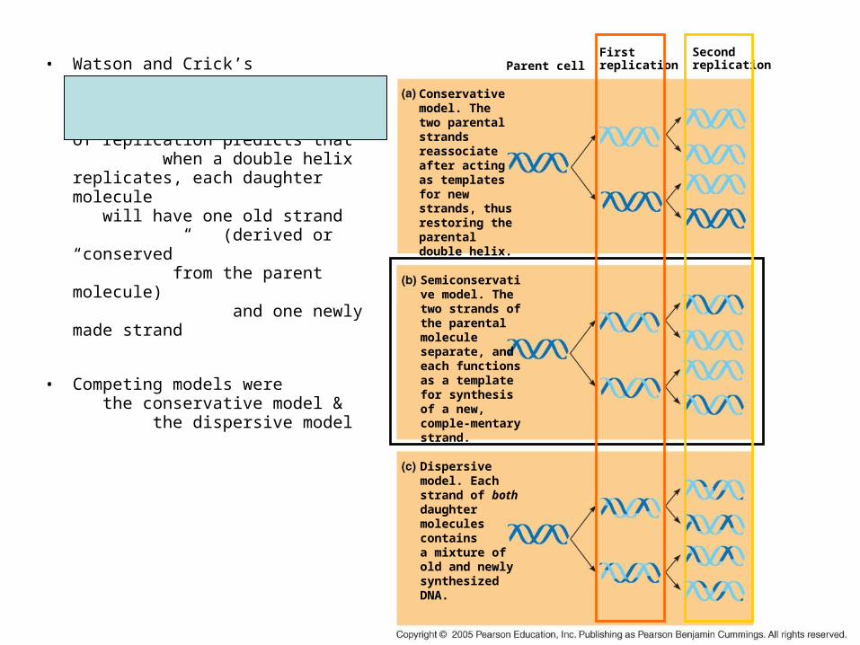
• Watson and Crick’s
semiconservative
model
of replication predicts that when a double helix replicates, each daughter molecule will have one old strand (derived or “conserved” from the parent molecule) and one newly made strand
• Competing models were the conservative model & the dispersive model
Conservative model. The two parental strands reassociate after acting as templates for new strands, thus restoring the parental double helix.
Semiconservative model. The two strands of the parental moleculeseparate, and each functions as a template for synthesis of a new, comple-mentary strand.
Dispersive model. Each strand of both daughter molecules contains a mixture of old and newly synthesized DNA.
Parent cellFirstreplication
Secondreplication

Bacteriacultured in mediumcontaining15N
DNA samplecentrifugedafter 20 min(after firstreplication)
DNA samplecentrifugedafter 40 min(after secondreplication)
Bacteriatransferred tomediumcontaining14N
Lessdense
Moredense
Conservativemodel
First replication
Semiconservativemodel
Second replication
Dispersivemodel
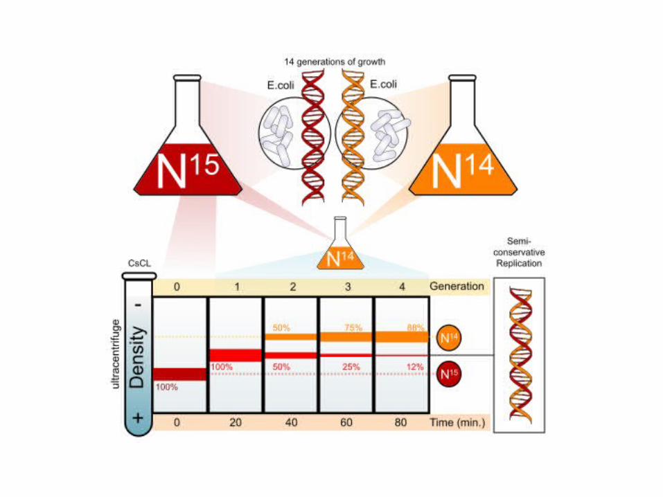
Meselson & Stahl- To test the 3 models they labeled the nucleotides of the old strands with a heavy isotope of nitrogen, while any new nucleotides were labeled with a lighter isotope
The first replication produced a band of hybrid DNA, eliminating the conservative model
A second replication produced both light and hybrid DNA, eliminating the dispersive model and supporting the semiconservative model
hybrid
light (14N)
hybrid
hybrid
light (14N)
heavy (15N) heavy (15N)
light (14N)
heavy (15N) light (14N)
hybrid hybrid
light (14N)
mostly light (14N)
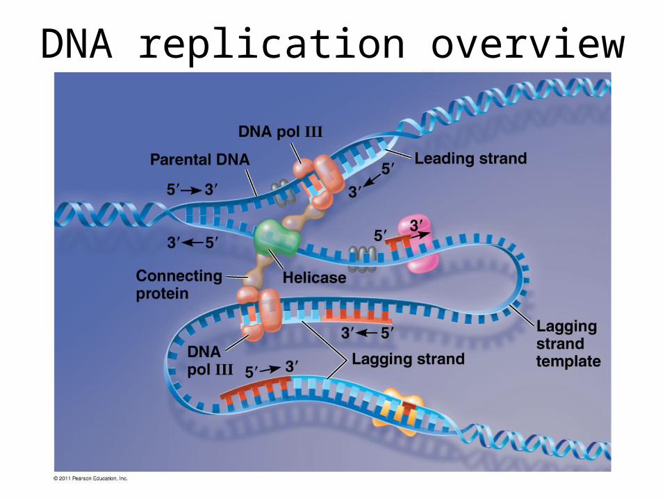
DNA replication overview
Origin of replication Parental (template) strand
Daughter (new) strand
Replication fork
Replication bubble
Two daughter DNA molecules
(a) Origins of replication in E. coli
Origin of replication Double-stranded DNA molecule
Parental (template) strandDaughter (new) strand
Bubble Replication fork
Two daughter DNA molecules
(b) Origins of replication in eukaryotes
0.5 µm
0.25 µm
Double-strandedDNA molecule
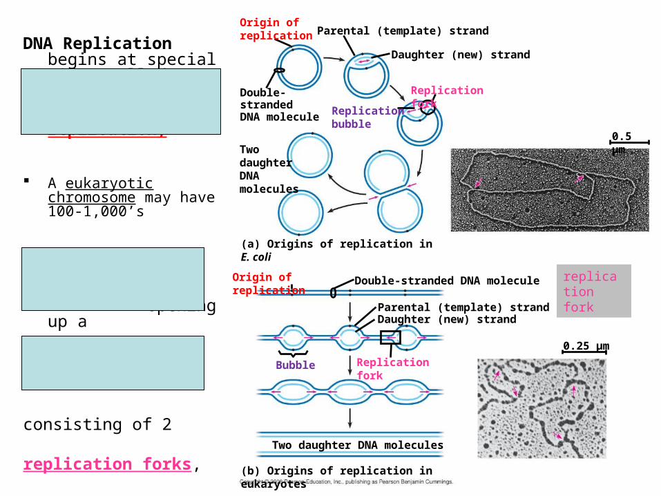
DNA Replication begins at special sites called
origins of replication,
A eukaryotic chromosome may have 100-1,000’s
The two DNA strands are separated, opening up a
replication “bubble”
consisting of 2
replication forks,
Y-shaped regions where new DNA strands are elongating at each end
replication fork

Topoisomerase
Helicase
Single-strand binding proteins RNA primer
(5-10 nucleotides)
55
5 3
3
3
• Helicase-
untwists the double helix and separates the template DNA strands at the replication fork
• Single-strand binding protein-
binds to and stabilizes single-stranded DNA until it can be used as a template
• Topoisomerase-
corrects “overwinding” ahead of replication forks by breaking, swiveling, & rejoining DNA strands
Primase-
synthesizes the initial nucleotide strand, a short RNA primer
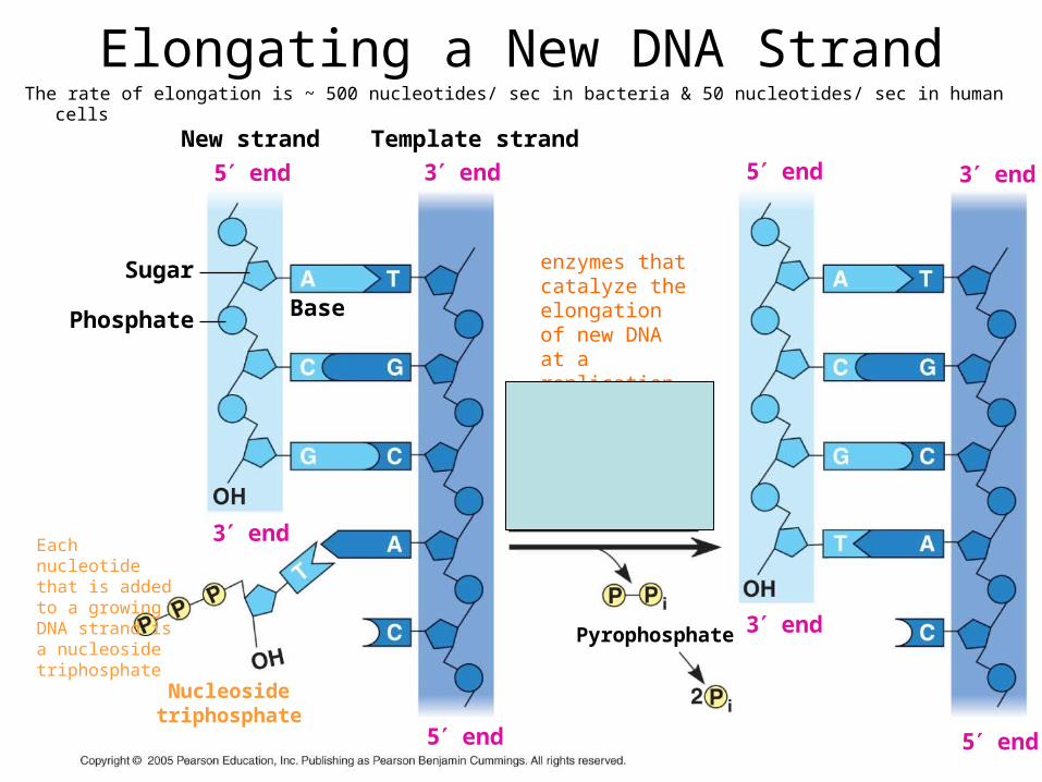
Elongating a New DNA StrandThe rate of elongation is ~ 500 nucleotides/ sec in bacteria & 50 nucleotides/ sec in human cells
New strand
5 end
Phosphate Base
Sugar
Template strand
3 end 5 end 3 end
5 end
3 end
5 end
3 end
Nucleosidetriphosphate
DNA polymerase
Pyrophosphate
enzymes that catalyze the elongation of new DNA at a replication fork
Each nucleotide that is added to a growing DNA strand is a nucleoside triphosphate

Antiparallel Elongation
• The antiparallel structure of the double helix (two strands oriented in opposite directions) affects replication
• DNA polymerases add nucleotides only to the free 3end of a growing strand; therefore, a new DNA strand can elongate only in the 5to3direction
Leading strand
Overview
Origin of replicationLagging strand
Leading strandLagging strand
Primer
Overall directions of replication
Origin of replication
RNA primer
“Sliding clamp”
DNA poll IIIParental DNA
5
3
3
3
3
5
5
5
5
5
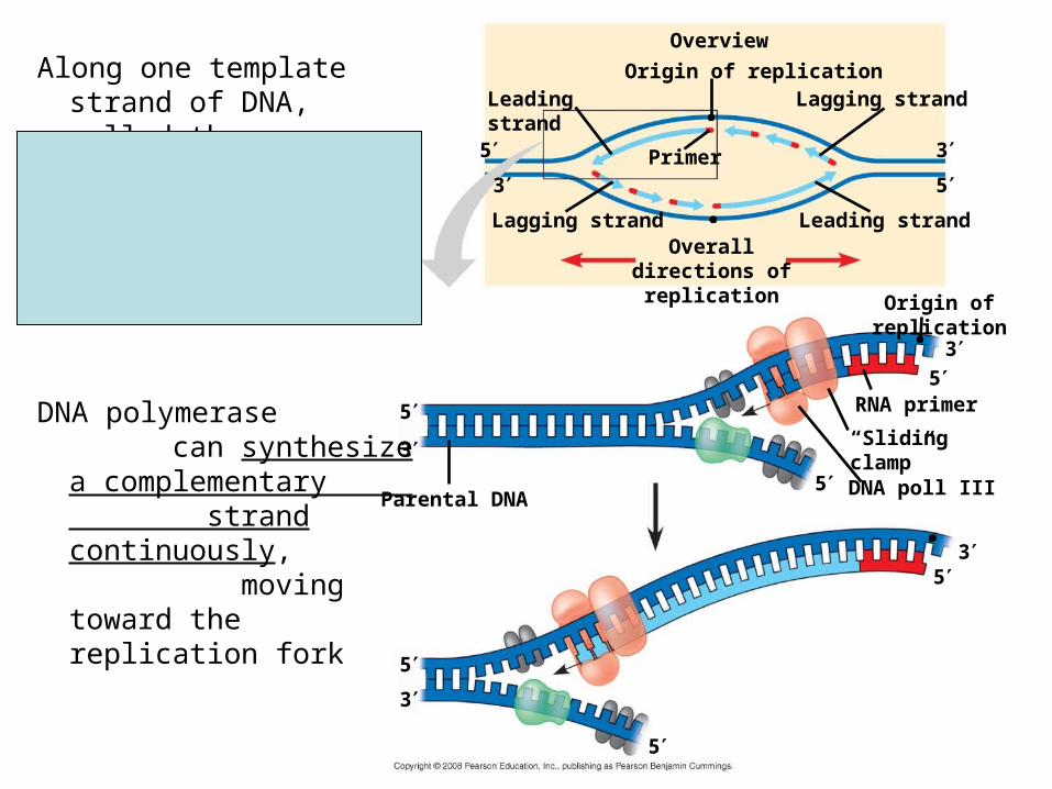
Along one template strand of DNA, called the leading strand,
DNA polymerase can synthesize a complementary strand continuously, moving toward the replication fork
5
5
3
3
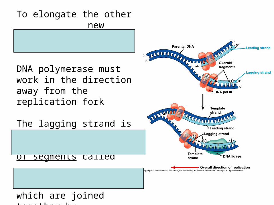
To elongate the other new strand, called the lagging strand,
DNA polymerase must work in the direction away from the replication fork
The lagging strand is synthesized as a series of segments called
Okazaki fragments,
which are joined together by DNA ligase

Synthesis of the lagging strand
3 5
35Templatestrand
Overall direction of replication
Primase joins RNA nucleotides into a primer.
13
33 5
5
5
35Templatestrand
RNA primer
Overall direction of replication
DNA pol III adds DNA nucleotides to the primer, forming an Okazaki fragment.
2
Primase joins RNA nucleotides into a primer.
13
3
335
3 5
5
5
5
35Templatestrand
RNA primer
Okazakifragment
Overall direction of replication
After reaching the next RNA primer (not shown), DNA pol III falls off.
3
DNA pol III adds DNA nucleotides to the primer, forming an Okazaki fragment.
2
Primase joins RNA nucleotides into a primer.
13
3
3
35 3
5
35
3 5
5
5
5
35Templatestrand
RNA primer
Okazakifragment
Overall direction of replication
After the second fragment is primed, DNA pol III adds DNAnucleotides until it reaches the first primer and falls off.
4
After reaching the next RNA primer (not shown), DNA pol III falls off.
3
DNA pol III adds DNA nucleotides to the primer, forming an Okazaki fragment.
2
Primase joins RNA nucleotides into a primer.
13
3
3
35
35 3
5
35
35
3 5
5
5
5
35Templatestrand
RNA primer
Okazakifragment
Overall direction of replication
DNA pol 1 replaces the RNA with DNA, adding to the 3 end of fragment 2.
5
After the second fragment is primed, DNA pol III adds DNAnucleotides until it reaches the first primer and falls off.
4
After reaching the next RNA primer (not shown), DNA pol III falls off.
3
DNA pol III adds DNA nucleotides to the primer, forming an Okazaki fragment.
2
Primase joins RNA nucleotides into a primer.
13
3
3
35
35
35
35
35
35
35
3 5
5
5
5
35Templatestrand
RNA primer
Okazakifragment
Overall direction of replication
DNA ligase forms a bond between the newest DNAand the adjacent DNA of fragment 1.
6 The lagging strand in this region is nowcomplete.
7
DNA pol 1 replaces the RNA with DNA, adding to the 3 end of fragment 2.
5
After the second fragment is primed, DNA pol III adds DNAnucleotides until it reaches the first primer and falls off.
4
After reaching the next RNA primer (not shown), DNA pol III falls off.
3
DNA pol III adds DNA nucleotides to the primer, forming an Okazaki fragment.
2
Primase joins RNA nucleotides into a primer.
1

REVIEW

April 25, 2012
Self Description

Announcements
• Homework due tomorrow (Apr 26)
• Old exams
• Exam III to hand back at the end of class
OverviewOrigin of replication
Leading strand
Leading strand
Lagging strand
Lagging strandOverall directions
of replication
Leading strand
Lagging strand
Helicase
Parental DNA
DNA pol III
Primer Primase
DNA ligase
DNA pol III
DNA pol I
Single-strand binding protein
5
3
5
5
5
5
3
3
3
313 2
4
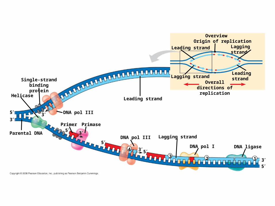
The DNA Replication Machine
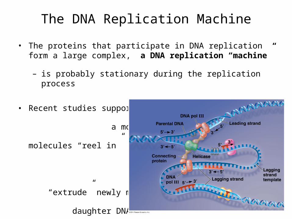
• The proteins that participate in DNA replication form a large complex, a DNA replication “machine”
– is probably stationary during the replication process
• Recent studies support a model in which DNA polymerase molecules “reel in” parental DNA and “extrude” newly made daughter DNA molecules
Proofreading & Repairing DNA
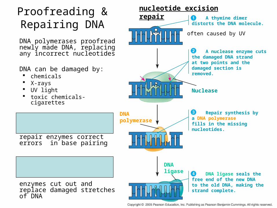
DNA polymerases proofread newly made DNA, replacing any incorrect nucleotides
DNA can be damaged by: chemicals X-rays UV light toxic chemicals- cigarettes
mismatch repair of DNA-
repair enzymes correct errors in base pairing
nucleotide excision repair-
enzymes cut out and replace damaged stretches of DNA
DNA ligase
DNA polymerase
DNA ligase seals thefree end of the new DNAto the old DNA, making thestrand complete.
Repair synthesis bya DNA polymerasefills in the missingnucleotides.
A nuclease enzyme cutsthe damaged DNA strandat two points and the damaged section isremoved.
Nuclease
A thymine dimerdistorts the DNA molecule.
often caused by UV
nucleotide excision repair
Replicating the Ends of
DNA Molecules
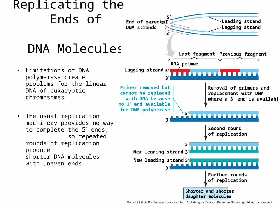
• Limitations of DNA polymerase create problems for the linear DNA of eukaryotic chromosomes
• The usual replication machinery provides no way to complete the 5 ends, so repeated rounds of replication produce shorter DNA molecules with uneven ends
End of parentalDNA strands
5
3
Lagging strand 5
3
Last fragment
RNA primer
Leading strandLagging strand
Previous fragment
Primer removed butcannot be replacedwith DNA becauseno 3 end available
for DNA polymerase5
3
Removal of primers andreplacement with DNAwhere a 3 end is available
Second roundof replication
5
3
5
3Further roundsof replication
New leading strand
New leading strand
Shorter and shorterdaughter molecules
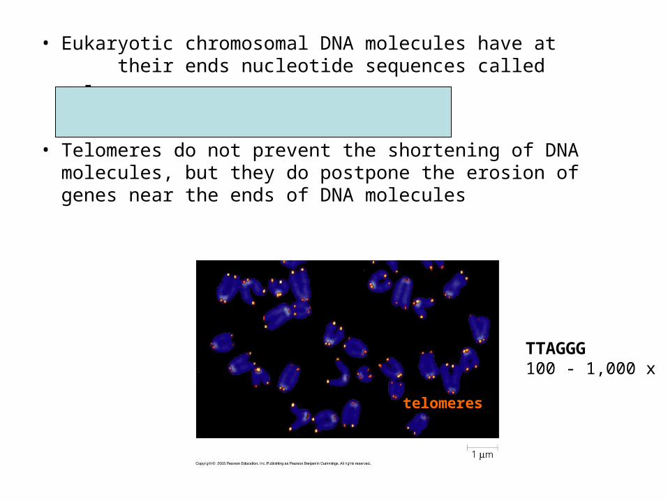
• Eukaryotic chromosomal DNA molecules have at their ends nucleotide sequences called
telomeres
• Telomeres do not prevent the shortening of DNA molecules, but they do postpone the erosion of genes near the ends of DNA molecules
telomeres
TTAGGG100 - 1,000 x
• If chromosomes of germ cells became shorter in every cell cycle, essential genes would eventually be missing from gametes they produce
• An enzyme called telomerase
catalyzes the lengthening of telomeres in germ cells
• There is evidence of telomerase activity in cancer cells, which may allow cancer cells to persist
• It has been proposed that the shortening of telomeres is connected to aging
– The shortening of telomeres might protect cells from cancerous growth by limiting the number of cell divisions

Chromosome structure
• The bacterial chromosome is a double-stranded, circular DNA molecule associated with a small amount of protein
• Eukaryotic chromosomes have linear DNA molecules associated with a large amount of protein
• Chromatin is a complex of DNA and protein, and is found in the nucleus of eukaryotic cells
• Histones are proteins that are responsible for the first level of DNA packing in chromatin
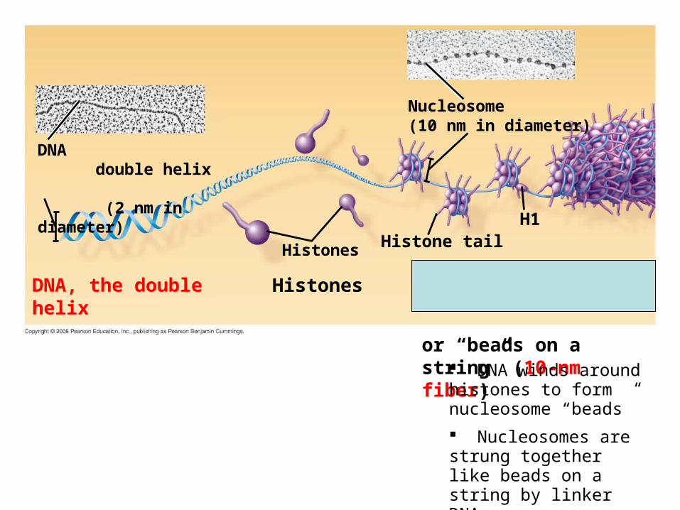
DNA double helix (2 nm in diameter)
Nucleosome(10 nm in diameter)
Histones Histone tailH1
DNA, the double helix Histones Nucleosomes,
or “beads on a string” (10-nm fiber) DNA winds around
histones to form nucleosome “beads”
Nucleosomes are strung together like beads on a string by linker DNA
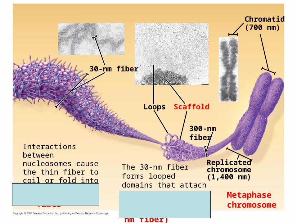
30-nm fiber
Chromatid (700 nm)
Loops Scaffold
300-nm fiber
Replicated chromosome (1,400 nm)
30-nm fiberLooped domains (300-nm fiber)
Metaphase chromosome
Interactions between nucleosomes cause the thin fiber to coil or fold into this thicker fiber
The 30-nm fiber forms looped domains that attach to proteins

• Most chromatin is loosely packed in the nucleus during interphase and condenses prior to mitosis
• euchromatin- loosely packed chromatin
• heterochromatin- highly condensed chromatin
• during interphase a few regions of chromatin (centromeres and telomeres)
• Dense packing of the heterochromatin makes it difficult for the cell to express genetic information coded in these regions
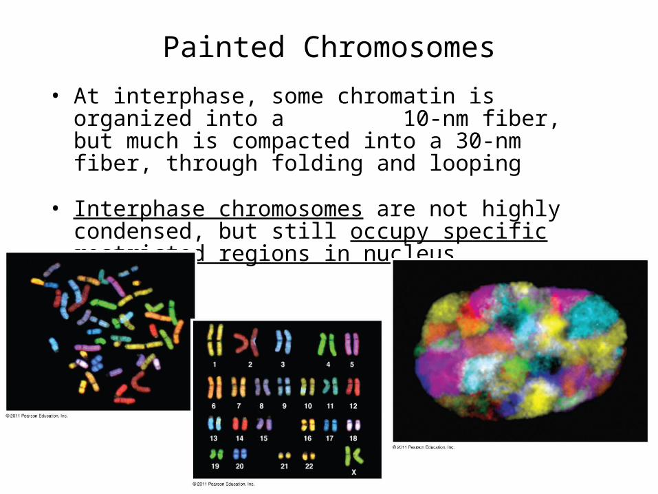
Painted Chromosomes
• At interphase, some chromatin is organized into a 10-nm fiber, but much is compacted into a 30-nm fiber, through folding and looping
• Interphase chromosomes are not highly condensed, but still occupy specific restricted regions in nucleus