chapter 16: the special...
TRANSCRIPT

Chapter 16:
The Special Senses
1. Describe the structures of the
Special Senses
2. Explain the pathways of
sound in the ear and light in
the eye
3. Identify, describe, and discuss
the receptors and neural
pathways involved in each of
the five special senses
Chapter objectives:
1. Taste
2. Smell
3. Sight
4. Hearing
5. Touch (Chapt 14)
Chemical Senses
Developed by
John Gallagher, MS, DVM

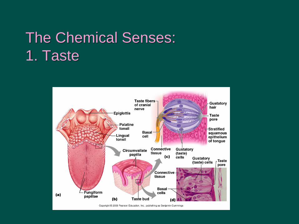
The Chemical Senses:
1. Taste
Chemoreceptors in Taste Buds
– Mostly in papillae on the tongue
Circumvallate, fungiform
Each has groups of gustatory
cells
Sweet, Sour, Salt, Bitter, Umami
CN VII and IX to medulla
oblongata
The Chemical Senses:
1. Taste
The Chemical Senses:
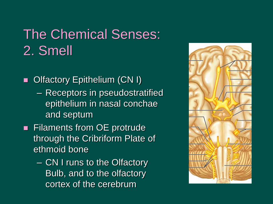
2. Smell
Olfactory Epithelium (CN I)
– Receptors in pseudostratified
epithelium in nasal conchae
and septum
Filaments from OE protrude
through the Cribriform Plate of
ethmoid bone
– CN I runs to the Olfactory
Bulb, and to the olfactory
cortex of the cerebrum

Factoids:
Most dominant sense
70% of the body’s receptors
are in the eyes
40% of cortex dedicated to
visual processing
Most metabolically active
tissue
Medical careers:
Optician
Optometrist
Ophthalmologist
Vision (Eye and Accessories)

Dissected View

Palpebrae = Eyelids
Continuation of skin
Eyelashes
Tarsal Plate of Hyaline C.
Tarsal (Meibomian) glands on
inner margin of lid – The oily portion of the tear film
– Swollen gland = chalazion
Conjunctiva (= mucous membrane) – Palpebral or Bulbar
– Over cornea very thin (5-7 cells
thick)
– Conjunctivitis (pink eye) Chalazion
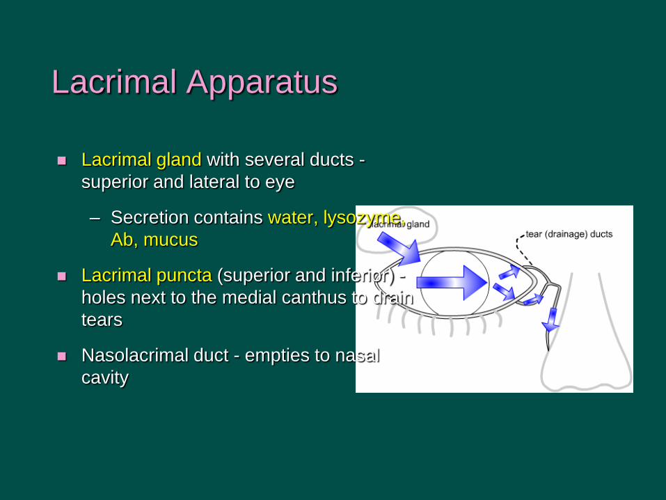
Lacrimal Apparatus
Lacrimal gland with several ducts -
superior and lateral to eye
– Secretion contains water, lysozyme,
Ab, mucus
Lacrimal puncta (superior and inferior) -
holes next to the medial canthus to drain
tears
Nasolacrimal duct - empties to nasal
cavity

Extrinsic Eye Muscles (review)
4 rectus muscles
– Lateral (CN XI)
– medial, superior,
inferior (CN III)
2 obliques
– Superior (CN IV)
– Inferior (CN III)
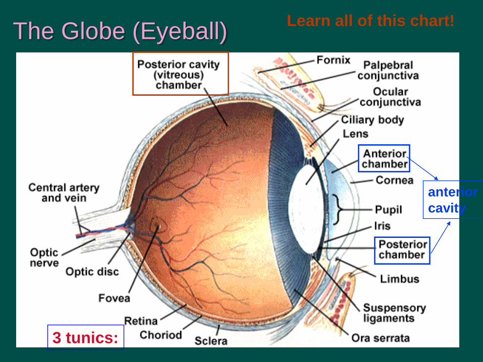
The Globe (Eyeball)
anterior
cavity
3 tunics:
Learn all of this chart!

The Three Layers (tunics):
1. Fibrous Tunic (tough outer layer)
1. sclera - white part of fibrous tunic
2. cornea - transparent anterior part
1. Avascular: nutrition via diffusion
2. pain receptors
3. Layer of noncornified stratified squamous epithelium
3. limbus - boundary between the above
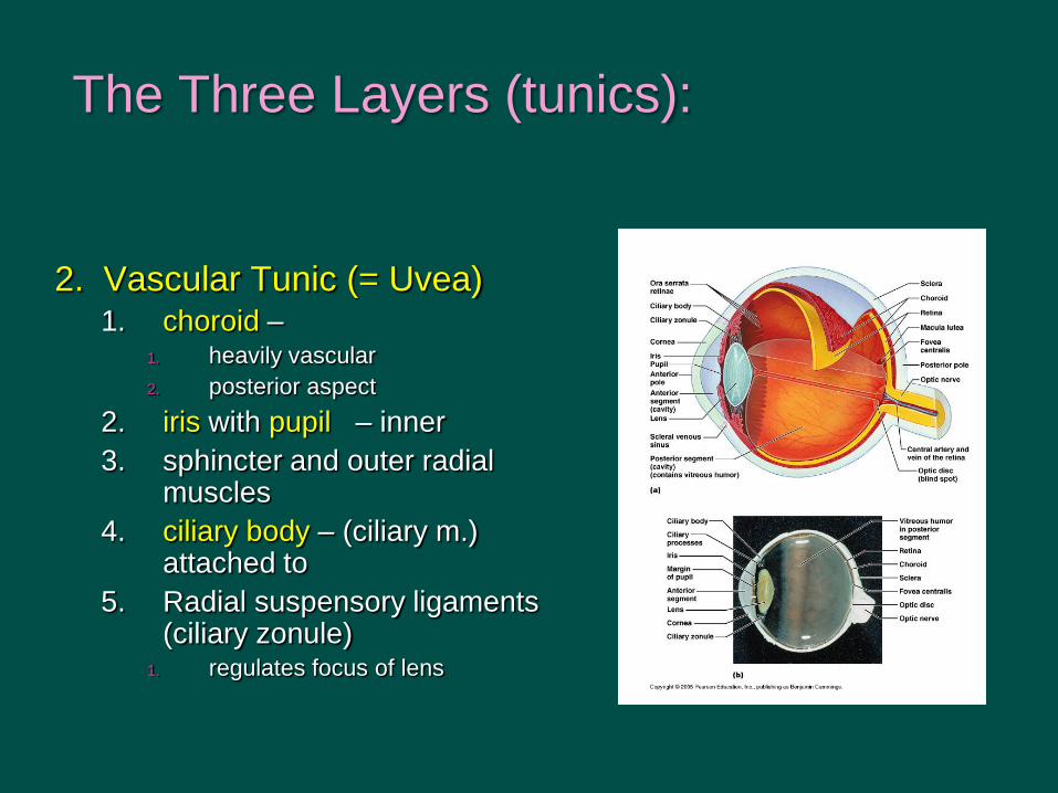
The Three Layers (tunics):
2. Vascular Tunic (= Uvea) 1. choroid –
1. heavily vascular
2. posterior aspect
2. iris with pupil – inner
3. sphincter and outer radial muscles
4. ciliary body – (ciliary m.) attached to
5. Radial suspensory ligaments (ciliary zonule)
1. regulates focus of lens

The Three Layers (tunics):
3) The Sensory Tunic AKA Nervous Tunic, retina
Outer layer pigmented - inner layer photoreceptors 106
a) rods - black/white vision, dim light
b) cones - color vision, intense light
Bipolar cells - synapse with rods and cones
Ganglion cells - synapse with bipolar cells
Ora serrata - anterior edge of retina
Macula lutea – fovea centralis - all cones, best vision
Optic disc – blind spot, where optic nerve exits eye
Optic nerve (CN II)

Retina

Retina
•Photoreceptors
•Infolding membranes contain
photopigments
•Rods
•Most numerous
•Non-acute vision
•Cones
•Concentrated in macula
•Color vision –red, green blue
Fig 16.10

Eye Fundus:
Age Related Macular Degeneration
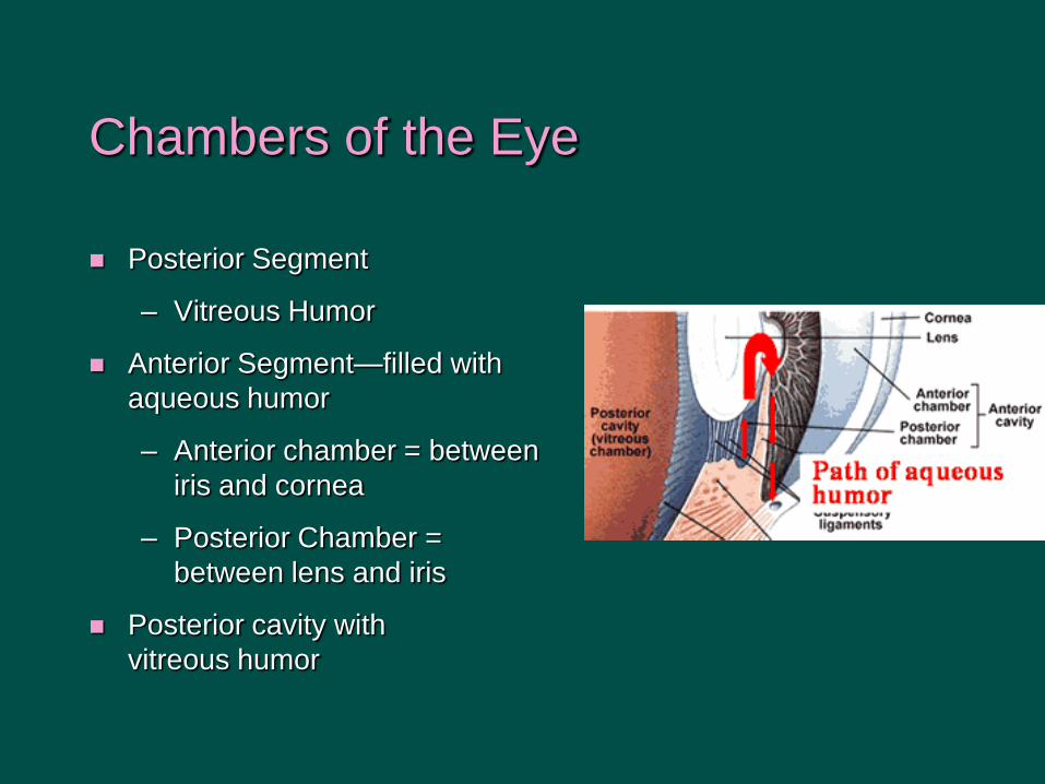
Chambers of the Eye
Posterior Segment
– Vitreous Humor
Anterior Segment—filled with
aqueous humor
– Anterior chamber = between
iris and cornea
– Posterior Chamber =
between lens and iris
Posterior cavity with
vitreous humor

Vision Terminology p 494
Emmetropia = Normal vision
Hyperopia = Farsightedness
Myopia = Nearsightedness
Presbyopia = Poor close-up vision with
aging
Astigmatism = Abnormal shape of the
surface of the lens and/or cornea
Cataract = abnormal crystallization of the
lens, common in diabetes, injury, heredity
Amblyopia = Poor vision in a normal eye
(CNS defect)

Visual Pathway
Optic chiasma - optic nerves partially cross (right side of the field of each eye combining and going to the lateral geniculate on the right, those from the left to the left)
To superior colliculus and thalamus and visual cortex in occipital lobe
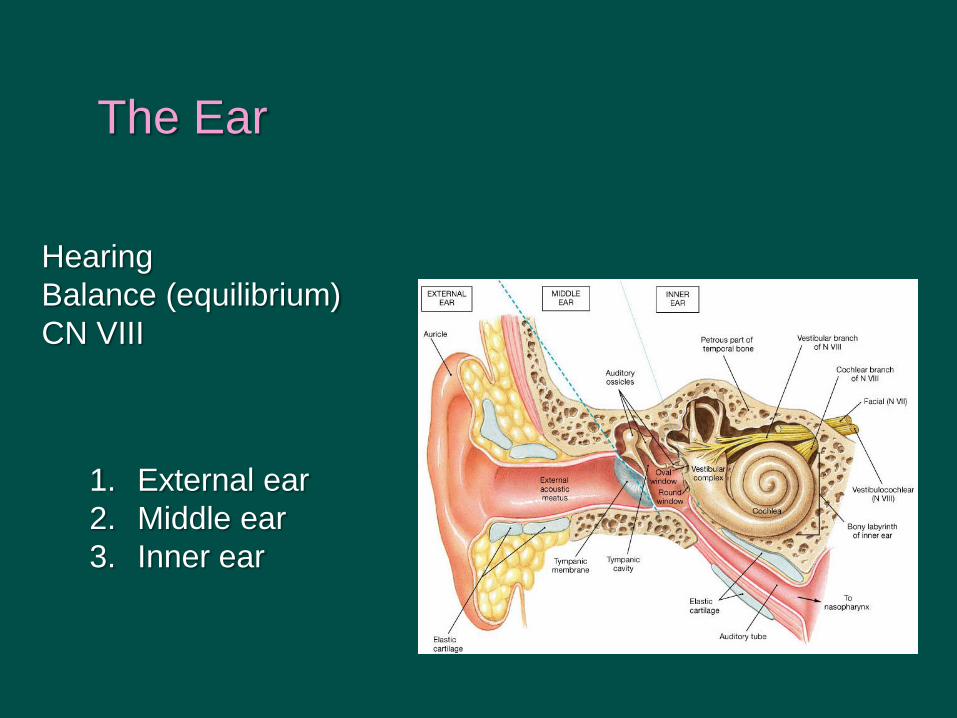
The Ear
Hearing
Balance (equilibrium)
CN VIII
1. External ear
2. Middle ear
3. Inner ear
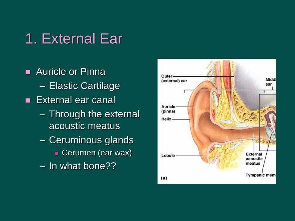
1. External Ear
Auricle or Pinna
– Elastic Cartilage
External ear canal
– Through the external
acoustic meatus
– Ceruminous glands
Cerumen (ear wax)
– In what bone??

2. Middle Ear
•Tympanic membrane
•Three Auditory Ossicles
•Incus, Malleus Stapes
•Transmit Vibrations to Inner
Ear
•Eustachian Tube = Auditory
Tube = Pharyngotympanic Tube
Otitis media

3. Inner Ear
•Cochlea
•Vestibular
complex

Structure of cochlea: 2.5 turns of ducts

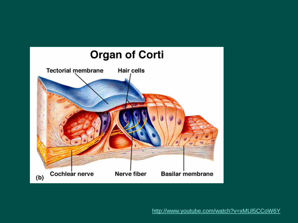
Organ of Corti
Basilar membrane on which sit hair cells with stereocilia
Tectorial membrane above the hair cells
Sound causes hair cells to bounce and touch tectorial membrane causing
transduction

Vestibular Complex
•Vestibule
•Saccule
•Utricle
•Static equilibrium
•Three semicircular canals
with ampullae (mutually
perpendicular)
•Linear acceleration
Each has a
macula with
receptors

Two Receptor Organs of vestibule
•Two Maculae
•or: macula of saccule plus
macula of utricle
•Vertical and horizontal
orientation
•Contain otoliths that move
according to gravity
•Hair cells conduct impulse
to CN VIII

Semicircular Canals
•Oriented perpendicular
•Anterior
•Posterior
•Lateral
•Each has an ampulla
•Crista ampullaris
bends
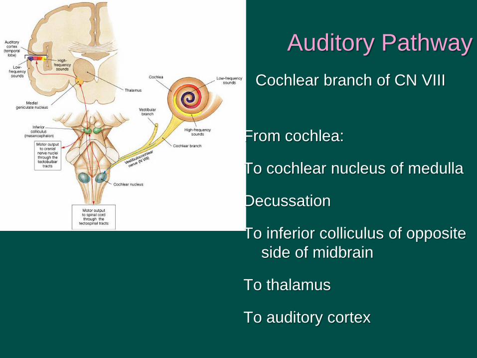
Cochlear branch of CN VIII
From cochlea:
To cochlear nucleus of medulla
Decussation
To inferior colliculus of opposite
side of midbrain
To thalamus
To auditory cortex
Auditory Pathway