chapter 17 magnetic particles for biomedical applications book chapter.pdf · 17 magnetic particles...
TRANSCRIPT

Chapter 17Magnetic Particles for Biomedical Applications
Raju V. Ramanujan
17.1 Introduction
This chapter discusses applications of magnetic materials in bioengineering andmedicine [1–8]. Magnetism and magnetic materials have been used for manydecades in many modern medical applications, and several new applications arebeing developed in part because of the availability of superior electromagnets, super-conducting magnets and permanent magnets [9–12]. Advances in the synthesis andcharacterization of magnetic particles, especially nanomagnetic particles, have alsoaided in the use of magnetic biomaterials [6–12]. We begin with an introductionto magnetism and magnetic materials, followed by a discussion of the characteri-zation, synthesis techniques and applications of magnetic biomaterials [8, 9]. Mag-netic materials can be applied to cell separation, immunoassay, magnetic resonanceimaging (MRI), drug and gene delivery, minimally invasive surgery, radionuclidetherapy, hyperthermia and artificial muscle applications [1–5, 7]. Physical proper-ties which make magnetic materials attractive for biomedical applications are, first,that they can be manipulated by an external magnetic field – this feature is useful forseparation, immunoassay and drug targeting, and second, hysteresis and other lossesoccur in alternating magnetic fields – this is useful in hyperthermia applications.
In biology, there has been much interest in the possible use by bees and pigeonsof magnetic materials as biological compasses for navigation. Some magnetotacticbacteria are known to respond to a magnetic field, they contain chains of smallmagnetite particles and they can navigate to the surface or bottom of the pools thatthey live in using these particles. These particles can be obtained by disruption ofthe cell wall followed by magnetic separation; the presence of the lipid layer makesthese particles biocompatible and they can be readily functionalized for a variety ofbiomedical applications.
The earliest known biomedical use of naturally occurring magnetic materialsinvolves magnetite (Fe3O4) or lodestone which was used by the Indian surgeonSucruta around 2,600 years ago. He wrote in the book Ayurveda that magnetitecan be used to extract an iron arrow tip. Current areas in medicine to which mag-netic biomaterials can be applied include molecular and cell biology, cardiology,neurosurgery, oncology and radiology.
R. Narayan (ed.), Biomedical Materials,DOI 10.1007/978-0-387-84872-3 17, C© Springer Science+Business Media, LLC 2009
477

478 R.V. Ramanujan
In the human body, there is a constant movement of ions within and outside thecells as well as across cellular membranes. This electrical activity is responsible formagnetic fields, called biomagnetic fields, which we can measure using sensitiveinstruments placed outside the body. The study of such fields, called biomagnetism,is a fascinating area related to magnetism which is not covered in this chapter dueto lack of space. Of course, the effect of magnetic fields on humans and animalsis also the focus of many studies, examples being the effect of the electromagneticfield produced by power lines and cell phones on humans. Of course, we are allimmersed in the earth’s magnetic field of about 0.5 × 10⁻⁵ T while the magneticfield of a neutron star is of the order of 108 T!
Here we focus on the origin of magnetism in materials, the types of magneticmaterials used in medicine, and contemporary and future applications of magneticbiomaterials.
17.2 Magnetism and Magnetic Materials
Magnetism is known to all of us from childhood as the phenomenon by whichsome materials attract or repel other materials from a distance; examples ofsuch materials include iron, lodestone and some steels. Broadly, magnetic forcesare generated by moving charged particles, leading to magnetic fields. Thereare a number of excellent references to magnetism and magnetic materials, andan introduction is provided in Callister, from which the following discussion isderived [10].
Consider a material placed in an external magnetic field. The atoms in this mate-rial possess an atomic moment which responds to this external field. It is usefulto think of magnetic dipoles existing in magnetic materials; these dipoles can beconsidered to be small bar magnets with north and south poles. The dipoles pos-sess a magnetic dipole moment which can respond to the external magnetic field.Some field vectors are needed to understand this response: the external magneticfield strength is denoted by H (units A/m), the magnetic induction in the material isdenoted by B (units tesla) and the magnetization by M (units A/m). B, H and M arerelated by
B = μ0 (H + M) (17.1)
where μ0 is the permeability of free space (its magnitude is 1.257 × 10⁻⁶ H/m)and M is the magnetic moment m per unit volume of the material. Thevalue of M depends on the type of material and the temperature and can berelated to the field H through the volumetric magnetic susceptibility χ by therelation
M = χ H (17.2)

17 Magnetic Particles for Biomedical Applications 479
17.2.1 Categories of Magnetic Materials
We now discuss the magnetic response of bulk material. In simple cases we canunderstand in a straightforward fashion this response in terms of the behavior ofindividual atoms. In other cases, interactions between individual atoms makes thepicture more complicated. The magnetic response results in materials being classi-fied as either diamagnetic, paramagnetic or ferromagnetic. Antiferromagnetism andferrimagnetism fall within the broad category of ferromagnetism.
For most bulk materials, the response to an external magnetic field is weak, e.g.,in diamagnetic and paramagnetic materials. Diamagnetism is very weak and notpermanent; it persists only as long as the external field is present. It occurs due toa change in the orbital motion of electrons due to the external field, the directionof the induced magnetic moment is opposite to the field. In an inhomogenous field,such materials are attracted towards regions where the field is weak (Fig. 17.1). Inparamagnetism, each atom has a permanent dipole moment because of incompletecancellation of its electron magnetic moments. When a field is applied these atomicdipoles individually tend to align with the field, much as a compass needle alignswith the earth’s magnetic field.
Diamagnetic and paramagnetic materials exhibit magnetization only in the pres-ence of an external field; the low values of susceptibility χ imply that the magneticinduction in such materials is very weak. Typical values of susceptibility, at roomtemperature, for diamagnetic copper is −0.96 × 10⁻⁵, for paramagnetic aluminumis 2.07 × 10⁻⁵ and paramagnetic manganese sulfate is 3.7 × 10⁻³ [10].
Diamagnetic
Vacuum
Paramagnetic
Ferromagnetic
Magnetic field strength, H
Flu
x d
ensi
ty, B
Fig. 17.1 Schematic of the flux density B as a function of H for various materials [10]

480 R.V. Ramanujan
Feromagnetism is the most familiar type of magnetism. It occurs, for example,in body centred cubic (b.c.c.) iron, cobalt, nickel, and in many alloy compositionsbased on Fe, Co and Ni. Ferromagnetic materials, unlike dia- and para- magneticmaterials, show permanent magnetic moments even in the absence of an externalfield. The susceptibility values are very high compared to those of para- and dia-magnetic materials, reaching up to 106. The magnetic moments in such materialsarise mainly from atomic spin magnetic moments. More importantly, interactionsbetween atoms cause spin magnetic moments to align with one another in a coop-erative fashion. Thus, large regions in a crystal can have atoms with their spinsaligned with one another. When all the magnetic dipoles are aligned the magnetiza-tion reaches its saturation value (Ms), e.g., the magnitude of Ms for nickel is 5.1 ×10⁵ A/m.
As mentioned earlier, ferromagnetism results from a cooperative parallel align-ment of spins. In other materials, e.g., MnO. The magnetic moment couplingbetween atoms (or ions) results in the spin moments of neighboring atoms beingaligned in opposite directions. Such materials are antiferromagnetic. In the case ofMnO, the moments of adjacent Mn2+ ions are antiparallel, thus the material has nonet magnetic moment (Fig. 17.2).
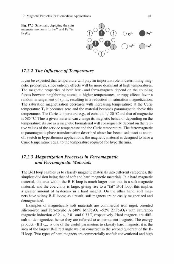
Some materials, including the magnetic biomaterial magnetite (Fe3O4) men-tioned in the introduction, exhibit ferrimagnetic behavior [10]. Hexagonal ferritesand garnets are other ceramic materials that fall in this category. Cubic ferrites, suchas magnetite, can be represented as MFe2O4, where M is a metal. In the case ofFe3O4, Fe ions exist in both the +2 and +3 valence states. The magnetic moments ofthe two types of Fe ions differs; in this case, there is a net magnetic moment becausefor the solid as a whole the spin moments are not completely cancelled; althoughthe spin moments of the Fe3+ ions cancel one another, the magnetization arises fromthe parallel alignment of the moments of the Fe2+ ions (Fig. 17.3). By adding otherions such as Ni2+ and Co2+ to Fe3O4, ferrites having a range of magnetic propertiescan be produced. This flexibility can be used to tune the magnetic properties forhyperthermia applications by creating cubic mixed-ferrite material.
Mn2+
O2–
Fig. 17.2 Schematic ofantiparallel alignment of spinmagnetic moments inantiferromagnetic MnO
17 Magnetic Particles for Biomedical Applications 481
O2– Fe2+
Fe3+Fe3+
Fig. 17.3 Schematic depicting the spinmagnetic moments for Fe3+ and Fe2+inFe3O4
17.2.2 The Influence of Temperature
It can be expected that temperature will play an important role in determining mag-netic properties, since entropy effects will be more dominant at high temperatures.The magnetic properties of both ferri- and ferro-magnets depend on the couplingforces between neighboring atoms; at higher temperatures, entropy effects favor arandom arrangement of spins, resulting in a reduction in saturation magnetization.The saturation magnetization decreases with increasing temperature; at the Curietemperature Tc it becomes zero and the material becomes paramagnetic above thistemperature. The Curie temperature, e.g., of cobalt is 1,120 ◦C and that of magnetiteis 585 ◦C. Thus a given material can change its magnetic behavior depending on thetemperature; its use as a magnetic biomaterial will consequently depend on the rela-tive values of the service temperature and the Curie temperature. The ferromagneticto paramagnetic phase transformation described above has been used to act as an on-off switch in hyperthermia applications; the magnetic material is designed to have aCurie temperature equal to the temperature required for hyperthermia.
17.2.3 Magnetization Processes in Ferromagneticand Ferrimagnetic Materials
The B-H loop enables us to classify magnetic materials into different categories, thesimplest division being that of soft and hard magnetic materials. In a hard magneticmaterial, the area within the B-H loop is much larger than that in a soft magneticmaterial, and the coercivity is large, giving rise to a “fat” B-H loop; this impliesa greater amount of hysteresis in a hard magnet. On the other hand, soft mag-nets have skinny B-H loops; as a result, soft magnets are be easily magnetized anddemagnetized.
Examples of magnetically soft materials are commercial iron ingot, orientedsilicon-iron and Ferroxcube A (48% MnFe2O4 –52% ZnFe2O4) with saturationmagnetic induction of 2.14, 2.01 and 0.33 T, respectively. Hard magnets are diffi-cult to demagnetize, hence they are referred to as permanent magnets. The energyproduct, (BH)max is one of the useful parameters to classify hard magnets; it is thearea of the largest B-H rectangle we can construct in the second quadrant of the B-H loop. Two types of hard magnets are commercially useful: conventional and high
482 R.V. Ramanujan
energy. Examples of conventional hard magnets are Cunife (60% Cu-20% Ni-20%Fe) with a remanence of 0.54 T and an energy product of 12 kJ/m3 and sintered fer-rite 3 (BaO-6 Fe2O3) with a remanence of 0.32 T and an energy product of 20 kJ/m3.The usual range of energy product of conventional hard magnets is between 2 and80 kJ/m3, while that of high energy magnets is greater than 80 kJ/m3. Examplesof the latter are samarium–cobalt magnets (SmCo5) and neodymium–iron–boron(Nd2Fe14B) magnets with typical remanence values of 0.92 and 1.16 T respectivelyand energy product values of 170 and 255 kJ/m3 respectively [10].
17.2.4 Factors Affecting Magnetic Properties
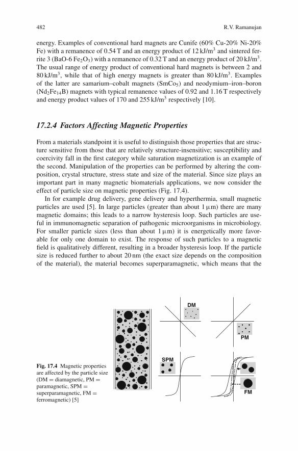
From a materials standpoint it is useful to distinguish those properties that are struc-ture sensitive from those that are relatively structure-insensitive; susceptibility andcoercivity fall in the first category while saturation magnetization is an example ofthe second. Manipulation of the properties can be performed by altering the com-position, crystal structure, stress state and size of the material. Since size plays animportant part in many magnetic biomaterials applications, we now consider theeffect of particle size on magnetic properties (Fig. 17.4).
In for example drug delivery, gene delivery and hyperthermia, small magneticparticles are used [5]. In large particles (greater than about 1 μm) there are manymagnetic domains; this leads to a narrow hysteresis loop. Such particles are use-ful in immunomagnetic separation of pathogenic microorganisms in microbiology.For smaller particle sizes (less than about 1 μm) it is energetically more favor-able for only one domain to exist. The response of such particles to a magneticfield is qualitatively different, resulting in a broader hysteresis loop. If the particlesize is reduced further to about 20 nm (the exact size depends on the compositionof the material), the material becomes superparamagnetic, which means that the
DM
PM
SPM
FM
Fig. 17.4 Magnetic propertiesare affected by the particle size(DM = diamagnetic, PM =paramagnetic, SPM =superparamagnetic, FM =ferromagnetic) [5]

17 Magnetic Particles for Biomedical Applications 483
magnetic moment of the particle fluctuates because of the thermal energy (∼kT); atthe atomic level the individual atomic moments continue to be ordered relative toeach other. Importantly, the remanence is zero, the result is a B-H curve showingno hysteresis; this property is important for reducing the tendency of the particles toagglomerate. The physical basis for the fluctuation of the magnetic moments can beunderstood as a battle between ΔE, the energy barrier to moment reversal and thethermal energy (kT). In the simplest approximation the energy barrier is the prod-uct of the anisotropy energy density K and the volume V. When the particle sizeis small (small V), the KV term is small and comparable to the thermal energy;this leads to flipping of the magnetic moment. The “blocking” temperature TB canbe regarded as the temperature above which the material becomes superparamag-netic. Superparamagnetic particles are useful as magnetic biomaterials, some are,physiologically well tolerated; an example is dextran-magnetite. Iron oxide coatedwith dextran is commercially available for MRI, for cell separation and cell labelingapplications.
17.3 Physical Principles
We now consider the physical principles involved in the applications of magneticparticles in bioengineering applications. In the case of targeted drug delivery weintroduce drug coated magnetic particles into a blood vessel and then apply an exter-nal magnetic field. This field attracts and retains the particles at the site of the disease(Fig. 17.5). The blood vessel will exhibit a paramagnetic response to the field fromentities such as the hemoglobin. It will also exhibit a diamagnetic response because
Magnetic particles
Blo
od
V
essel
Magnet
Tissu
e
Fig. 17.5 Directed motion of magnetic particles by external magnetic field [5]

484 R.V. Ramanujan
of proteins that contain carbon, hydrogen, nitrogen and oxygen atoms. These tworesponses are much smaller than the response by the magnetic particles. What kindof magnetic field should be applied to target the magnetic particles to the site of thedisease? A magnetic field that is uniform gives rise to a torque, but usually we wishto direct the particles in a specific direction, i.e., provide translation motion; thiscan be accomplished by means of a field gradient. If an appropriate magnetic fieldgradient is present, a force acts on the particles driving them in a direction whichcan be chosen to so that the particles are targeted to the site of the disease (Fig.17.6). Consider a magnetic particle in such a magnetic field gradient; for the caseof magnetic nanoparticles suspended in water the magnetic force on the particle hasbeen shown by Pankhurst to be
Fm = Vm(χm − χw)∇(1
2B.H ) (17.3)
where Vm is the volume of the particle, χm and χw are the susceptibility of theparticle and water respectively, and the quantity 1
2 B.H is the magnetostatic fieldenergy density [5]. Assuming that the susceptibility of the particle is greater thanthat of water, this equation shows that the force is proportional to the differentialin the energy density, and recalling the geometrical meaning of the ∇ operator, thismagnetic force on the particle acts in the direction of steepest ascent of the energydensity field.
In the case of magnetic hyperthermia applications, a different principle isinvolved; we wish to raise the temperature to about 43 ◦C in a localized area inorder to destroy cancer cells selectively [3]. This can be done by applying a mag-netic field which varies with time; ferro- and ferri-magnetic material will be repeat-edly cycled through the B-H loop, resulting in hysteresis and other losses whichare then converted to thermal energy and result in an increase in temperature.
Arterial feedCatheter
Magnet
Magnetic carrierTissue/Organ
Fig. 17.6 Magnetic carrier for drug targeting and drug delivery [6]

17 Magnetic Particles for Biomedical Applications 485
Superparamagnetic materials can also be heated using this technique; the loss mech-anisms differ from those observed in ferro- and ferri-magnetic materials.
17.4 Examples and Property Requirementsof Magnetic Biomaterials
A common example of a magnetic biomaterial is magnetite. Interestingly, this is thesame material used by Sucruta more than 2,000 years ago. Magnetite is found inmany biological entities, from bacteria to people. It is an example of cubic ferriteswhich have an inverse spinel structure. It is ferrimagnetic with a Curie tempera-ture of 578 ◦C and a saturation magnetization of 4.76 × 10⁵ A/m. Another exampleof a magnetic biomaterial is maghemite (gamma Fe2O3), which is formed whenmagnetite is oxidized. It has a structure similar to that of magnetite, the differencebeing that all or most of the iron is trivalent, the saturation magnetization is 4.26× 10⁵ A/m. Ferritin is a protein that stores iron in humans. It contains typically4,500 iron atoms in an approximately spherical 12 nm diameter molecule. It hasa 12 subunit protein shell containing a ferrihydrite core and an antiferromagneticcore. Gadolinium(III) chelates are commonly used in MRI applications. Iron coatedwith activated carbon has recently been tried for magnetic drug targeting for thetreatment of hepatocellular carcinomas.
The magnetic biomaterial can, in principle, also be Ni, Co, b.c.c. Fe, magneticalloys of Fe, Co, Ni, Nd–Fe–B or samarium–cobalt materials. In all cases, however,issues of biocompatibility and toxicity limit the choice of materials; however, theuse of coatings may make the use of these materials feasible. The hard magneticmaterials Nd–Fe–B and samarium–cobalt have the disadvantage that large externalfields are required to influence these materials. Materials with high magnetizationand high susceptibility are preferred for applications such as drug targeting andmagnetic separation. Most of the examples of useful magnetic biomaterials are inpowder form, and usually the particles are suitably coated before use. Ideally, themagnetic material should be non-toxic and non-immunogenic. In the case of drugdelivery, the particle sizes should be small enough to be injected into the blood-stream and then to pass through the required capillary systems. For in vitro appli-cations, the requirements are more relaxed, larger particle sizes can be used, andbiocompatibility and toxicity issues may be less important.
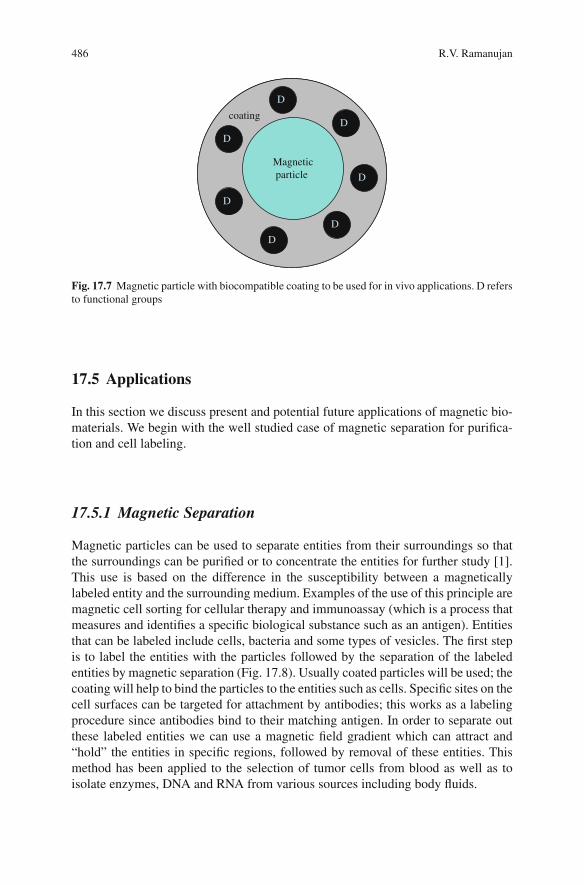
In many cases, it is required to coat the magnetic particles; this is usually doneby coating with a biocompatible polymer or with other coatings such as gold, acti-vated carbon or silica (Fig. 17.7). The coating reduces aggregation and prevents themagnetic particle from being exposed directly to the body. In addition, the polymercan be used as a matrix in which drugs, radionuclides or genetic material can bedissolved or as a site for binding of drugs; thus the magnet-coating system can actas “carrier” to deliver useful material to the targeted region. Some examples of com-mon coatings are derivatives of dextran, polyethylene glycol (PEG) and polyethy-lene oxide (PEO), phospholipids and polyvinyl alcohol [2, 4].
486 R.V. Ramanujan
coating
Magnetic particle
D
D
D
D
D
D
D
coating
Magneticparticle
D
D
D
D
D
D
D
Fig. 17.7 Magnetic particle with biocompatible coating to be used for in vivo applications. D refersto functional groups
17.5 Applications
In this section we discuss present and potential future applications of magnetic bio-materials. We begin with the well studied case of magnetic separation for purifica-tion and cell labeling.
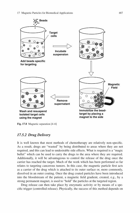
17.5.1 Magnetic Separation
Magnetic particles can be used to separate entities from their surroundings so thatthe surroundings can be purified or to concentrate the entities for further study [1].This use is based on the difference in the susceptibility between a magneticallylabeled entity and the surrounding medium. Examples of the use of this principle aremagnetic cell sorting for cellular therapy and immunoassay (which is a process thatmeasures and identifies a specific biological substance such as an antigen). Entitiesthat can be labeled include cells, bacteria and some types of vesicles. The first stepis to label the entities with the particles followed by the separation of the labeledentities by magnetic separation (Fig. 17.8). Usually coated particles will be used; thecoating will help to bind the particles to the entities such as cells. Specific sites on thecell surfaces can be targeted for attachment by antibodies; this works as a labelingprocedure since antibodies bind to their matching antigen. In order to separate outthese labeled entities we can use a magnetic field gradient which can attract and“hold” the entities in specific regions, followed by removal of these entities. Thismethod has been applied to the selection of tumor cells from blood as well as toisolate enzymes, DNA and RNA from various sources including body fluids.
17 Magnetic Particles for Biomedical Applications 487
Magnetic Field
Beads
Target cells
Add beads specific for targeting
Incubatesuspension
Immobilize the bead-target by placing a magnet to the side
Removesupernatant
Wash and resuspend isolated target cells using the magnet
Fig. 17.8 Magnetic separation [4–6]
17.5.2 Drug Delivery
It is well known that most methods of chemotherapy are relatively non-specific.As a result, drugs are “wasted” by being distributed to areas where they are notrequired, and this can lead to undesirable side effects. What is required is a “magicbullet” which can be used to carry the drugs to the area where they are required.Additionally, it will be advantageous to control the release of the drug once thecarrier has reached the target. Much of the work which has been performed so farrelates to targeting cancerous tumors. In this case, the magnetic particle first actsas a carrier of the drug which is attached to its outer surface or, more commonly,dissolved in an outer coating. Once the drug coated particles have been introducedinto the bloodstream of the patient, a magnetic field gradient, created, e.g., by astrong permanent magnet, is used to “hold” the particles at the targeted region.
Drug release can then take place by enzymatic activity or by means of a spe-cific trigger (controlled release). Physically, the success of this method depends on
488 R.V. Ramanujan
the velocity of the particles in the bloodstream, the circulation time as well as thestrength of the magnetic field in the region of interest. It is easier to immobilizelarger particles in the micron size range; they are less likely to “swept” away bythe blood flow. On the other hand, there are many other advantages in having areduced particle size. It appears that the optimum size may be in the 5–100 nm sizerange. Typically, a coated magnetite particle is used; noble metals such as gold havebeen used as a coating in addition to the usual polymer based coatings. Preliminarystudies using metallic magnetic particles have also been undertaken. Several studieshave reported success in tumor remission (the period during which the symptoms ofa disease decrease) in animal models. The difficult case of targeting cytotoxic (cellkilling) drugs to brain tumors has also been demonstrated in rats. Human trials usingthis technique for treating liver tumors have also been performed.
17.5.3 Radionuclide Delivery
Radionuclides (e.g., β–emitters, β is the symbol used to denote an electron) can alsobe attached to the magnetic particles and this system can then be targeted in the sameway as described in the previous section on drug delivery, since the radionuclidedoes not have to be released in the same way as the drug; one restriction of drugdelivery, i.e., control of drug release, is absent.
17.5.4 Gene Delivery
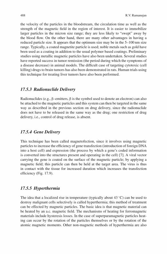
This technique has been called magnetofection, since it involves using magneticparticles to increase the efficiency of gene transfection (introduction of foreign DNAinto a host cell) and expression (the process by which a gene’s coded informationis converted into the structures present and operating in the cell) [7]. A viral vectorcarrying the gene is coated on the surface of the magnetic particle; by applying amagnetic field; this particle can then be held at the target area. The virus is thusin contact with the tissue for increased duration which increases the transfectionefficiency (Fig. 17.9).
17.5.5 Hyperthermia
The idea that a localized rise in temperature (typically about 43 ◦C) can be used todestroy malignant cells selectively is called hyperthermia; this method of treatmentcan be effected by magnetic particles. The basic idea is that magnetic material canbe heated by an a.c. magnetic field. The mechanisms of heating for ferromagneticmaterials include hysteresis losses. In the case of superparamagnetic particles heat-ing can occur by the rotation of the particles themselves or by the rotation of theatomic magnetic moments. Other non-magnetic methods of hyperthermia are also
17 Magnetic Particles for Biomedical Applications 489
Magneticparticle
Nucleicacid Cell Magnetic field
gradient
Magnet
Fig. 17.9 Principle of gene delivery using magnetic particles. The cells to be transfected are posi-tioned between the magnetic field gradient and the nucleic acids which are attached to the particles.The field causes the particles to move towards the target cells, the cell entry is accomplished, e.g.,by endocytosis [7]
available. The advantage of using magnetic particles should by now be familiar, i.e.,we can target the particles to the targeted region and then heat up the particles byusing an external a.c. magnetic field. According to Pankhurst, typically a heat depo-sition rate of 100 mW/cm3 is required, and the frequency of the field should be inthe kHz range with an amplitude of a few kA/m [5]. Generally iron oxides are usedfor hyperthermia applications.
17.5.6 Magnetic Resonance Imaging Contrast Agent
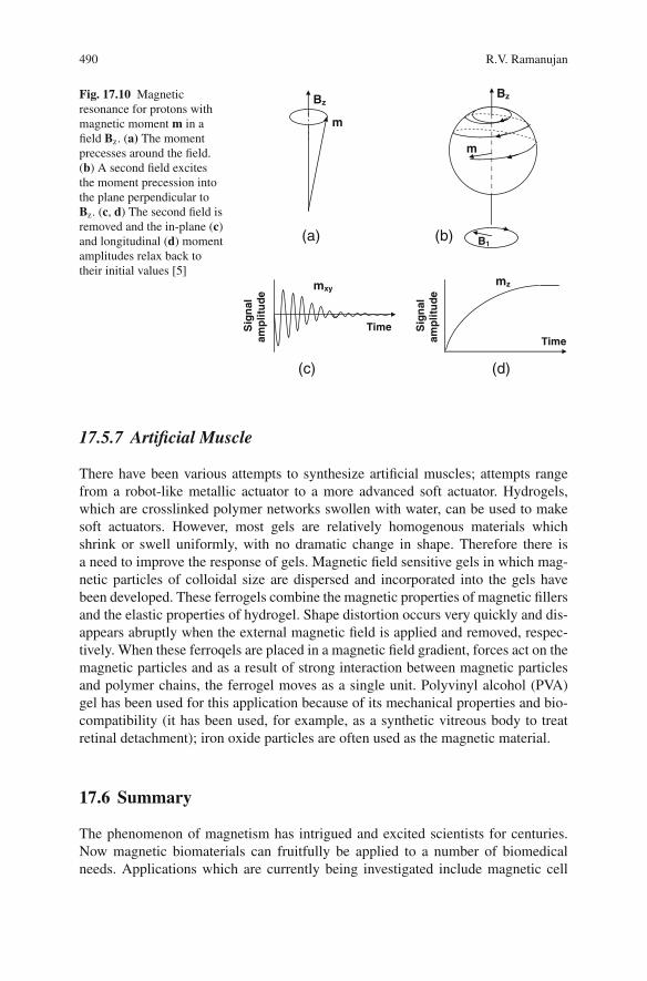
So far, we have only considered magnetic properties associated with the electronsin the material. However, protons also have a magnetic moment, and this can beutilized in the powerful imaging technique of magnetic resonance imaging (MRI)[5]. The principle is as follows. We first apply a steady field of about 1 T to a mate-rial, causing a very small fraction of protons to line up parallel to the field. The netmagnetic moment precesses like a top around the direction of this field (Fig. 17.10).In order to measure the signal produced as a result of this alignment, we now applya transverse radio frequency magnetic field. The frequency is carefully chosen andits effect is to make the magnetic moment precess in the plane perpendicular to thesteady state field. When this second field is switched off, the amplitudes of the mag-netic moments relax back to their initial values. This relaxation of the response ismeasured by pick-up coils. Typically, the relaxation time can be reduced by meansof a magnetic particle. Thus if a region is tagged using the magnetic particles, therelaxation time will be lower compared to untagged regions; thus “contrast” is pro-vided and the particle acts as a contrast agent. Usually paramagnetic Gd based mate-rials are used; superparamagnetic iron oxide particles, usually coated with dextran,have also been used for this purpose.
490 R.V. Ramanujan
Bz
m
m
Bz
B1
mxy
Time
mz
Sig
nal
amp
litu
de
Sig
nal
amp
litu
de
Time
(a) (b)
(c) (d)
Fig. 17.10 Magneticresonance for protons withmagnetic moment m in afield Bz. (a) The momentprecesses around the field.(b) A second field excitesthe moment precession intothe plane perpendicular toBz. (c, d) The second field isremoved and the in-plane (c)and longitudinal (d) momentamplitudes relax back totheir initial values [5]
17.5.7 Artificial Muscle
There have been various attempts to synthesize artificial muscles; attempts rangefrom a robot-like metallic actuator to a more advanced soft actuator. Hydrogels,which are crosslinked polymer networks swollen with water, can be used to makesoft actuators. However, most gels are relatively homogenous materials whichshrink or swell uniformly, with no dramatic change in shape. Therefore there isa need to improve the response of gels. Magnetic field sensitive gels in which mag-netic particles of colloidal size are dispersed and incorporated into the gels havebeen developed. These ferrogels combine the magnetic properties of magnetic fillersand the elastic properties of hydrogel. Shape distortion occurs very quickly and dis-appears abruptly when the external magnetic field is applied and removed, respec-tively. When these ferroqels are placed in a magnetic field gradient, forces act on themagnetic particles and as a result of strong interaction between magnetic particlesand polymer chains, the ferrogel moves as a single unit. Polyvinyl alcohol (PVA)gel has been used for this application because of its mechanical properties and bio-compatibility (it has been used, for example, as a synthetic vitreous body to treatretinal detachment); iron oxide particles are often used as the magnetic material.
17.6 Summary
The phenomenon of magnetism has intrigued and excited scientists for centuries.Now magnetic biomaterials can fruitfully be applied to a number of biomedicalneeds. Applications which are currently being investigated include magnetic cell

17 Magnetic Particles for Biomedical Applications 491
separation, immunoassay, hyperthermia, MRI contrast agents and drug, radionu-clide and gene delivery. The greatest advantage of such materials is their ability torespond to and be manipulated by external magnetic fields. Magnetism of an atomcan be thought to arise from uncompensated spins of its electrons. Magnetism asso-ciated with the proton is much weaker but is used for MRI studies. Magnetic bio-materials are usually used in particle form and can be ferro-, para-, ferri-, antiferro-or superpara-magnetic, they are often encapsulated by coatings that can be diamag-netic; this results in a wide range of magnetic responses to an applied field. Themost common magnetic biomaterials are the iron oxides magnetite and maghamite,and there is considerable interest in using magnetically superior metals and alloys.Several magnetic properties are a sensitive function of size. The size can be alteredby changing process parameters or synthesis techniques, and thus magnetic particleswith a wide range of properties can be obtained for a given composition. It has beenfound that nanosized particles often possess such properties and considerable effortis now underway to create commercially attractive applications using such particles.
Since the field of magnetic biomaterials is a rapidly growing, I have relied onjournal, handbook and encylopedia articles the following references are particularlyuseful.
References
1. Safarik I and Safarikova M. Magnetic nanoparticles and biosciences. Monatshefte fur Chemie,2002, 133: 737–759.
2. Berry CC and Curtis ASG. Functionalisation of magnetic nanoparticles for applications inbiomedicine. J Phys D: Appl Phys, 2003, 36: R198–R206.
3. Bahadur D and Giri J. Biomaterials and magnetism. Sadhana, 2003, 28: 639–656.4. Shinkai M. Functional magnetic materials for medical applications. J Biosci Bioeng, 2002,
94: 606–613.5. Pankhurst QA, Connolly J, Jones SK, and Dobson J. Applications of magnetic nanoparticles
in biomedicine. J Phys D Appl Phys, 2003, 36: R167–R181.6. Tartaj P, et al. The preparation of magnetic nanoparticles for applications in biomedicine. J
Phys D: Appl Phys, 2003, 36: R182–R197.7. Plank C, et al. Enhancing and targeting nucleic acid delivery by magnetic force. Expert Opin
Biol Theor, 2003, 3: 1.8. Hafeli U, Schutt W, Teller J, and Zborowski M. Scientific and Clinical Applications of Mag-
netic Carriers, Plenum Press: New York, 1997.9. Andra W and Nowak H, eds. Magnetism in Medicine, Wiley-VCH: New York, 1998.
10. Callister WD. Materials Science and Engineering, Wiley: New York, 2003.11. O’Handley RC. Modern Magnetic Materials, Wiley: New York, 2000.12. Koch CC, ed. Nanostructured Materials, Noyes Publications: New York, 2002.