chapter 2 structure and function of nucleic acids introduction life depends on the genetic...
TRANSCRIPT

Chapter 2 Structure and Function of Nucleic Acids
• Introduction• Life depends on the genetic instructions. This hereditary
information is passed on from a cell to its daughter cells at division, and from one generation of an organism to the next through the organisms’reproductive cells. Genes, the information-containing elements that determine the characteristics of a species as a whole and of the individuals within it.
1 Chemistry of nucleic acids
• Nucleotides are the subunits of DNA and RNA.
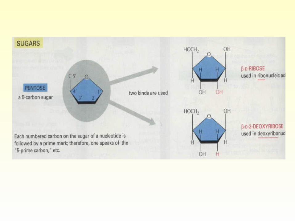
• A nucleotide consists of a base, a five-carbon sugar, and one or more phosphate groups.
•Purines and pyrimidines are nitrogen-containing heterocyclic compounds.
•Their principal derivatives are nucleosides and nucleotides. Cytosine (C), thymine (T), and uracil (U) are called pyrimidine compounds; guaninie (G) and adenine (A) are purine compounds. Each nucleotide is named by reference to the unique base that it contains.
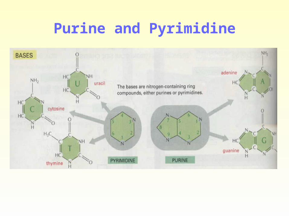
Purine and Pyrimidine

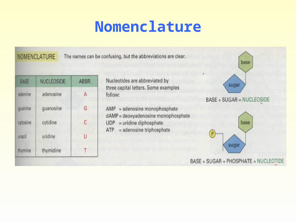
Nomenclature
Diverse physiologic functions of Nucleotides • Specific nucleotides participate in reactions that fulfill
physiologic functions as diverse as protein synthesis, nucleic acid synthesis, regulatory cascades, and intra- and intercellular signal transduction pathways.
• A. Adenosine derivatives: AMP, ADP, ATP, and cAMP. ATP are the major biologic transducer of free energy.
• B. Guanosine derivatives: cGMP serve as the principal second messenger in some cells.
• C. Many coenzyme are nucleotide derives: NAD+, NADP+, FAD, etc.
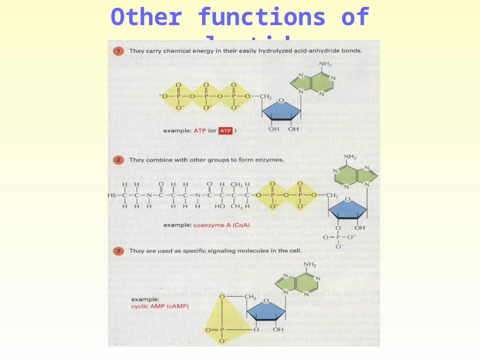
Other functions of nucleotides
2 Deoxyribonucleic acid (DNA)
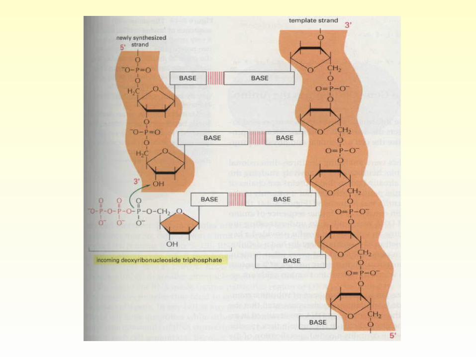
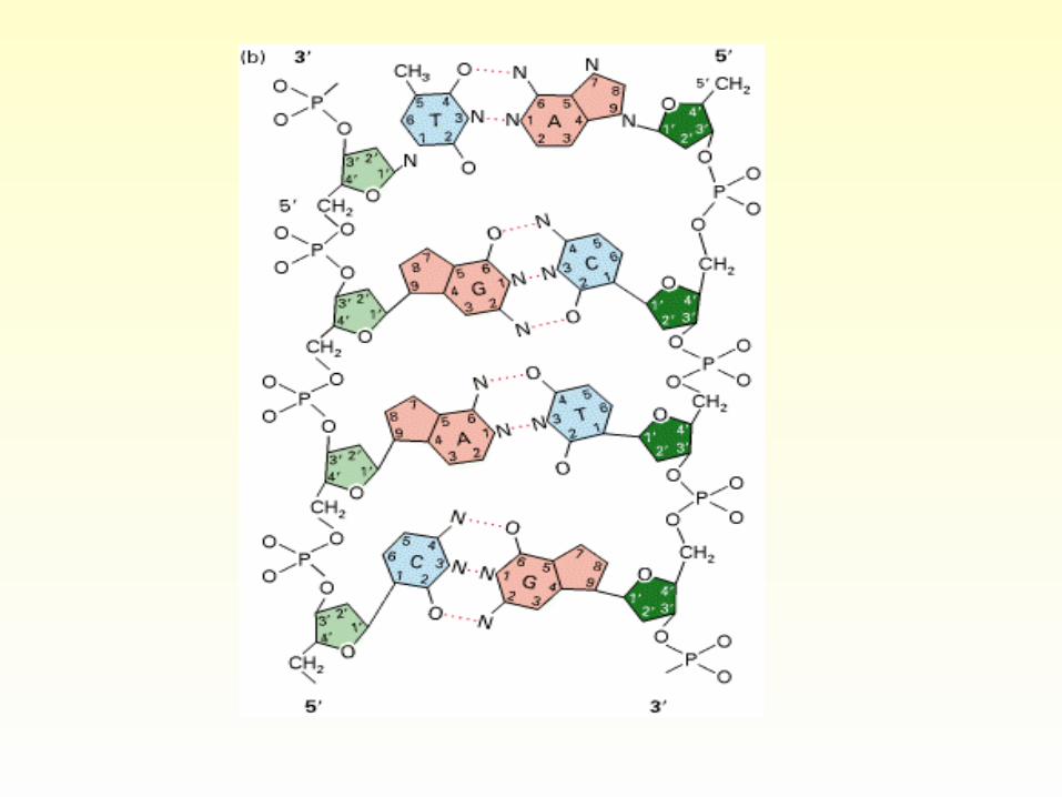
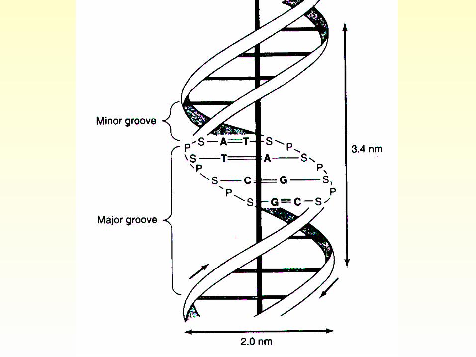
The two strands of the double-helical molecule, each of which possesses a polarity, are antiparallel; ie, one strand runs in the 5’to 3’ direction and the other in the 3’ to 5’direction. The two strands, in which opposing bases are held together by hydrogen bonds, wind around a central axis in the form of a double helix. The genetic information resides in the sequence of nucleotides on one strand, the template strand. This is the strand of DNA that is copied during nucleic acid synthesis.
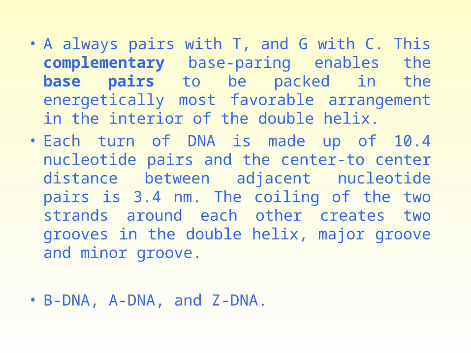
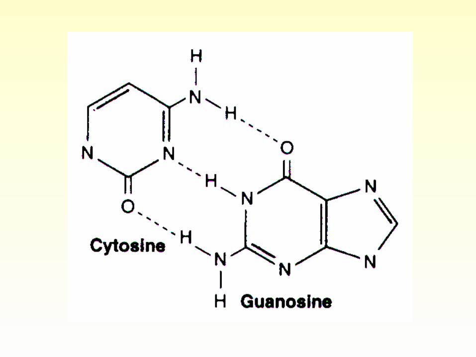
• A always pairs with T, and G with C. This complementary base-paring enables the base pairs to be packed in the energetically most favorable arrangement in the interior of the double helix.
• Each turn of DNA is made up of 10.4 nucleotide pairs and the center-to center distance between adjacent nucleotide pairs is 3.4 nm. The coiling of the two strands around each other creates two grooves in the double helix, major groove and minor groove.
• B-DNA, A-DNA, and Z-DNA.
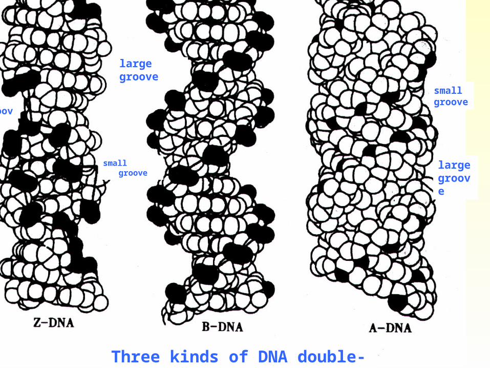
large groove
small groove
groove
large groove
small groove
Three kinds of DNA double-helix
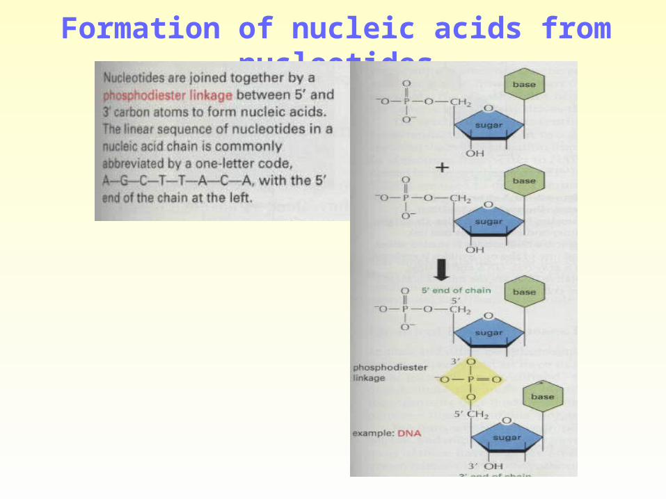
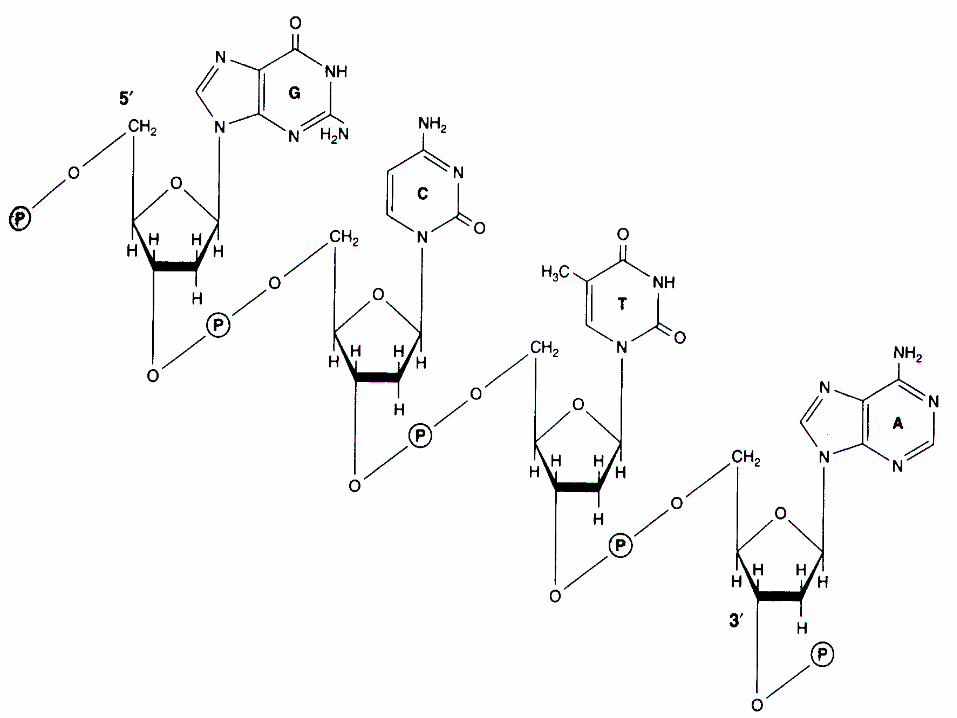
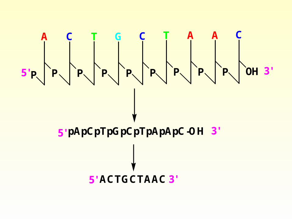
Formation of nucleic acids from nucleotides

P P
A
P
C
P
C
P
T
P
G
OH
C
P
T
P
A
P
A
5' 3'
pApCpTpGpCpTpApApC-OH 3'
5' ACTGCTAAC 3'
5'
Nucleotides absorb ultraviolet light
• The conjugated double bonds of the enterocyclic bases of purines an pyrimidines, and polynucleotides absorbe ultraviolet light. Their spectra are pH-dependent. However, at pH 7.0 all the common nucleotides absorb light at a wavelength close to 260 nm.
DNA supercoiling
• Packaging large DNA molecules to fit into cells requires DNA supercoiling.
• Nucleosome formation
• Chromatin and chromosome.

Circled DNA Supercoiled DNA
Denaturation and Renaturation of DNA
• Denaturation (melting) and Renaturation of DNA
• PCR (Polymerase Chain Reaction)
• cDNA (complementary DNA)
• Southern blotting (DNA/cDNA)
• Northern blotting (DNA/RNA).
• Western Blotting

Figure 4-8. The denaturation and renaturation of double-stranded DNA molecules.
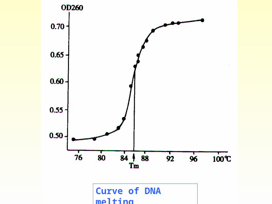
Curve of DNA melting

Transfer of information from DNA to protein
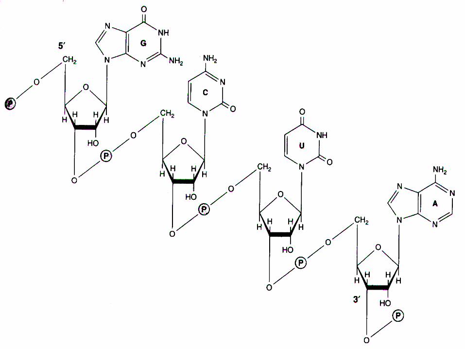
Ribonucleic acid (RNA) RNA is a polymer of purine and pyrimidine ribonucleo
tides linked together by 3’, 5’-phosphodiester bridges analogous to those in DNA. Although sharing many features with DNA, RNA possesses several specific differences.
• (1) Bases are attached to ribose rather than 2’- deoxyribose.
• (2) U, not T.• (3) Single strand. however, capable of folding ba
ck on itself like a hairpin. • (4) Others
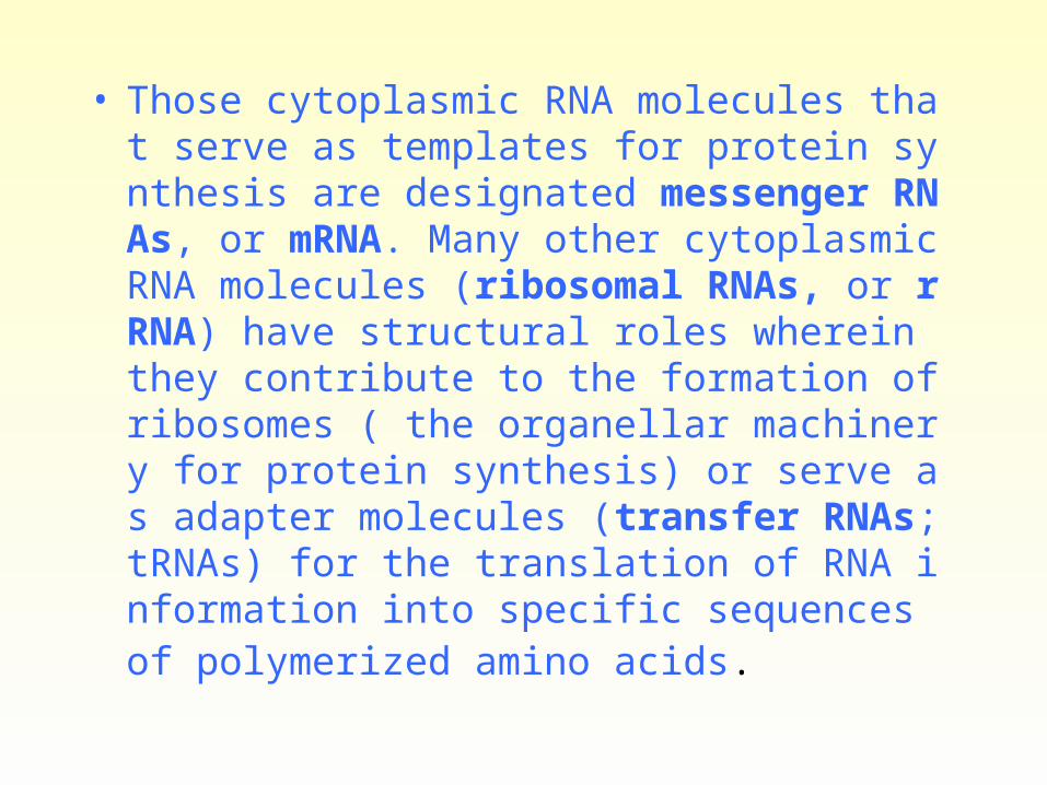
• Those cytoplasmic RNA molecules that serve as templates for protein synthesis are designated messenger RNAs, or mRNA. Many other cytoplasmic RNA molecules (ribosomal RNAs, or rRNA) have structural roles wherein they contribute to the formation of ribosomes ( the organellar machinery for protein synthesis) or serve as adapter molecules (transfer RNAs; tRNAs) for the translation of RNA information into specific sequences of polymerized amino acids.

• Some RNA molecules have intrinsic catalytic activity. The activity of these ribozymes often involves the cleavage of a nucleic acid. An example is the role of RNA in catalyzing the processing of the primary transcript of a gene into mature messenger RNA.
• In human cells, there are small nuclear RNA (snRNA) species that are not directly involved in protein synthesis but that may have roles in RNA processing and the cellular architecture. These relatively small molecules vary in size from 90 to about 300 nucleotides.
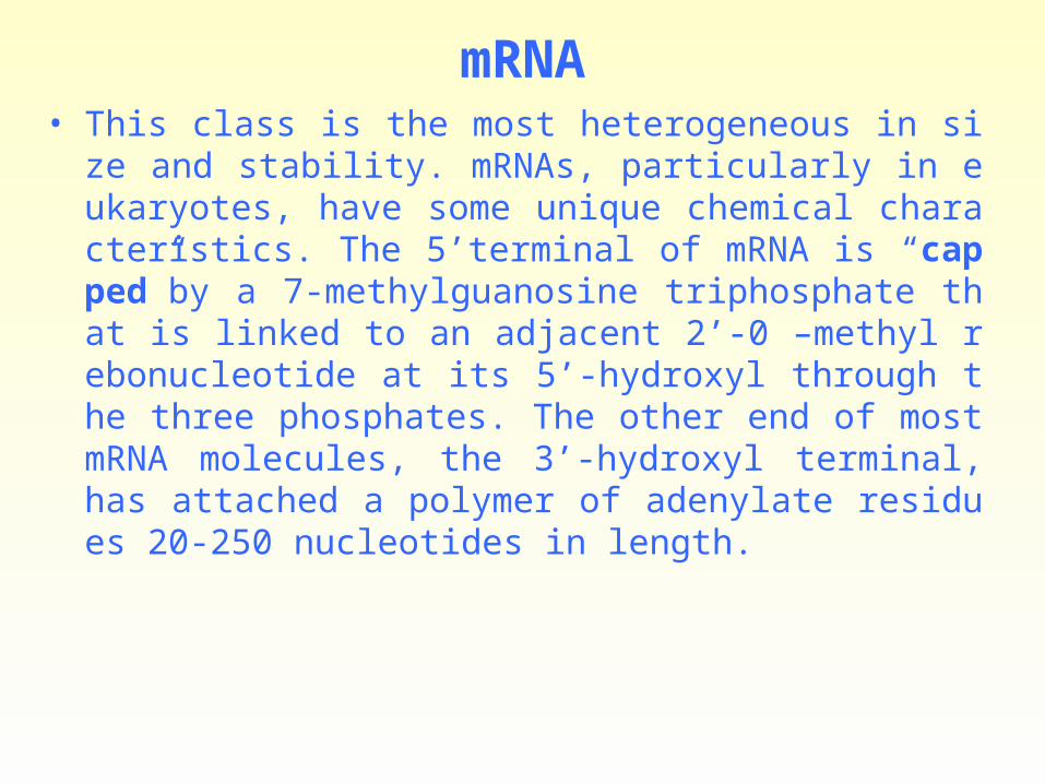
mRNA• This class is the most heterogeneous in size and stability. mRN
As, particularly in eukaryotes, have some unique chemical characteristics. The 5’terminal of mRNA is “capped”by a 7-methylguanosine triphosphate that is linked to an adjacent 2’-0 –methyl rebonucleotide at its 5’-hydroxyl through the three phosphates. The other end of most mRNA molecules, the 3’-hydroxyl terminal, has attached a polymer of adenylate residues 20-250 nucleotides in length.

• The mRNA molecules present in the cytoplasm are not the RNA products immediately synthesized from the DNA template but must be formed by processing from a precursor molecule before entering the cytoplasm. Thus, in mammalian nuclei, the immediate products of gene transcription constitute a fourth class of RNA molecules. These nuclear RNA molecules are very heterogeneous in size and are quite large. The heterogeneous nuclear RNA (hnRNA) molecules may have a molecular weight in excess of 107, whereas the molecule weight of mRNA molecules is generally less than 2×10⁶.
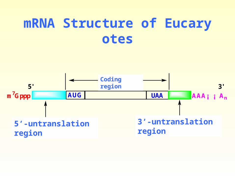
mRNA Structure of Eucaryotes
5' 3'
m7Gppp AAA¡ ¡ An
3'·Ç· ÒëÇø5'·Ç· ÒëÇø
±àÂëÇø
AUG UAA
Coding region
5‘-untranslation region 3‘-untranslation region
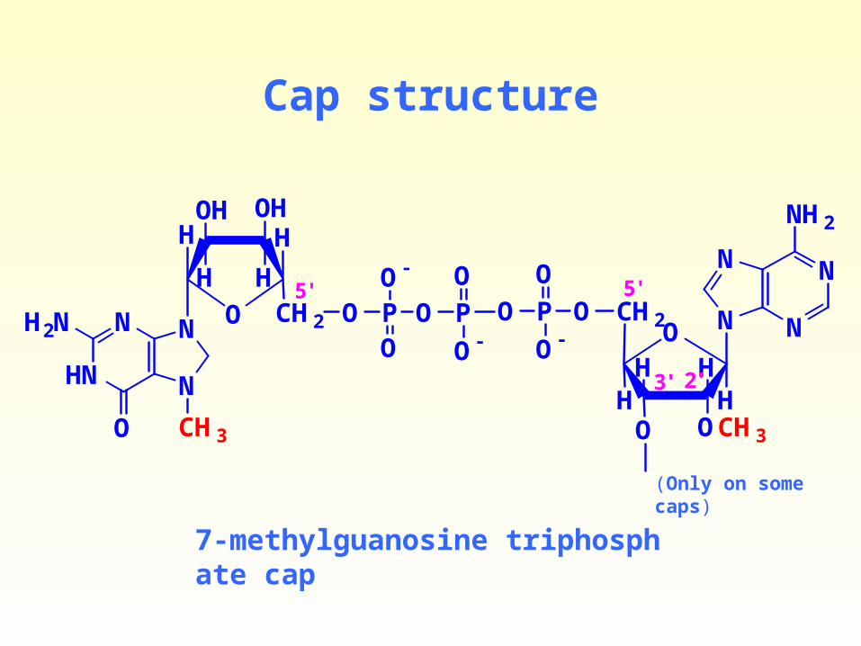
Cap structure
O
N
NN
N
NH2
O
OCH3O
HHH
CH2
H
OP
O-
O
O
HN
N
N
O
H2N N O
OH
H H
H
CH2
HOH
O PO
O-
CH3
P
O-
O5'
2'3'
5'
7-methylguanosine triphosphate cap
(Only on some caps)

Figure 4-18. Structure of the 5′ methylated cap of eukaryotic mRNA.
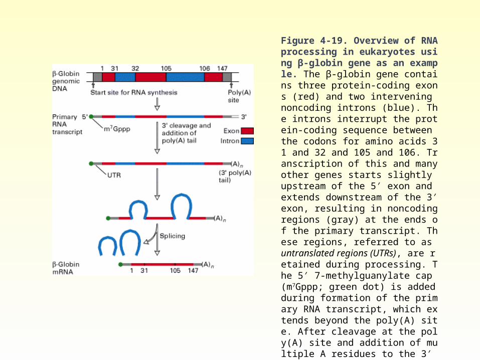
Figure 4-19. Overview of RNA processing in eukaryotes using β-globin gene as an example. The β-globin gene contains three protein-coding exons (red) and two intervening noncoding introns (blue). The introns interrupt the protein-coding sequence between the codons for amino acids 31 and 32 and 105 and 106. Transcription of this and many other genes starts slightly upstream of the 5′ exon and extends downstream of the 3′ exon, resulting in noncoding regions (gray) at the ends of the primary transcript. These regions, referred to as untranslated regions (UTRs), are retained during processing. The 5′ 7-methylguanylate cap (m7Gppp; green dot) is added during formation of the primary RNA transcript, which extends beyond the poly(A) site. After cleavage at the poly(A) site and addition of multiple A residues to the 3′ end, splicing removes the introns and joins the exons. The small numbers refer to positions in the 147-aa sequence of β-globin.
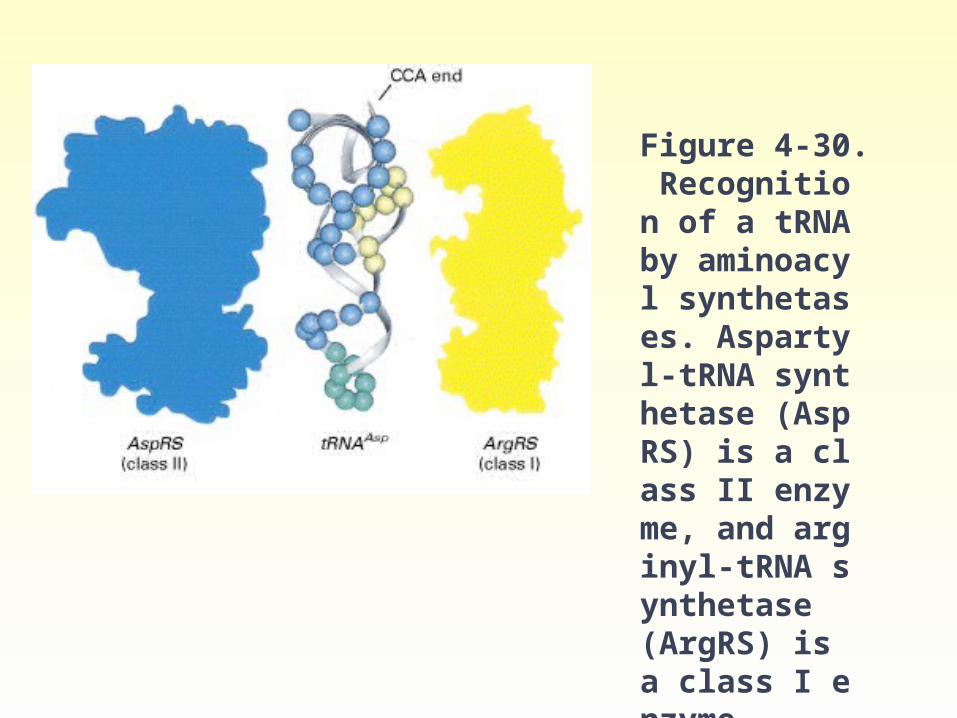
Figure 4-30. Recognition of a tRNA by aminoacyl synthetases. Aspartyl-tRNA synthetase (AspRS) is a class II enzyme, and arginyl-tRNA synthetase (ArgRS) is a class I enzyme.
tRNA
• tRNA molecules vary in length from 74-95 nucleotides. There are at least 20 species of tRNA molecules in every cell, at least one (and often several) corresponding to each of the 20 amino acids required for protein synthesis.
tRN
A structure
D arm
CCA end
Tψ arm
Anticodon armAnticodon

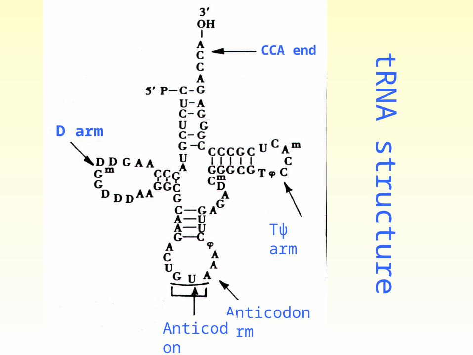
Figure 4-26. Structure of tRNAs.
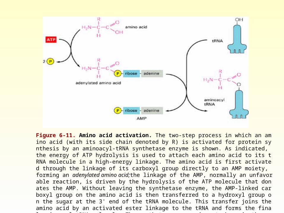
Figure 6-11. Amino acid activation. The two-step process in which an amino acid (with its side chain denoted by R) is activated for protein synthesis by an aminoacyl-tRNA synthetase enzyme is shown. As indicated, the energy of ATP hydrolysis is used to attach each amino acid to its tRNA molecule in a high-energy linkage. The amino acid is first activated through the linkage of its carboxyl group directly to an AMP moiety, forming an adenylated amino acid;the linkage of the AMP, normally an unfavorable reaction, is driven by the hydrolysis of the ATP molecule that donates the AMP. Without leaving the synthetase enzyme, the AMP-linked carboxyl group on the amino acid is then transferred to a hydroxyl group on the sugar at the 3' end of the tRNA molecule. This transfer joins the amino acid by an activated ester linkage to the tRNA and forms the final aminoacyl-tRNA molecule. The synthetase enzyme is not shown in these diagrams.
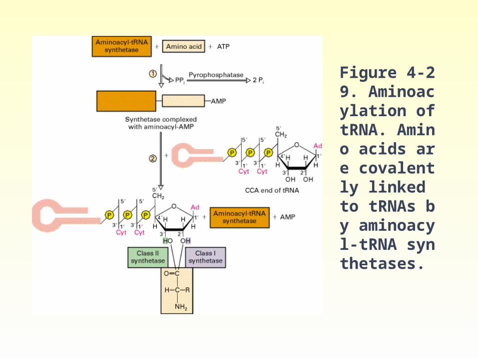
Figure 4-29. Aminoacylation of tRNA. Amino acids are covalently linked to tRNAs by aminoacyl-tRNA synthetases.
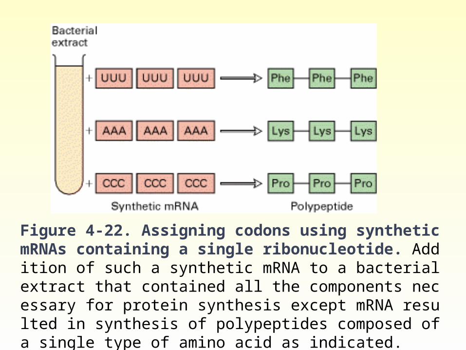
Figure 4-22. Assigning codons using synthetic mRNAs containing a single ribonucleotide. Addition of such a synthetic mRNA to a bacterial extract that contained all the components necessary for protein synthesis except mRNA resulted in synthesis of polypeptides composed of a single type of amino acid as indicated.

rRNA
A ribosome is a cytoplasmic nucleoprotein structure that acts as the machinery for the synthesis of proteins from the mRNA templates. On the ribosomes, the mRNA and tRNA molecules interact to translate into a specific protein molecule information transcribed from the gene.
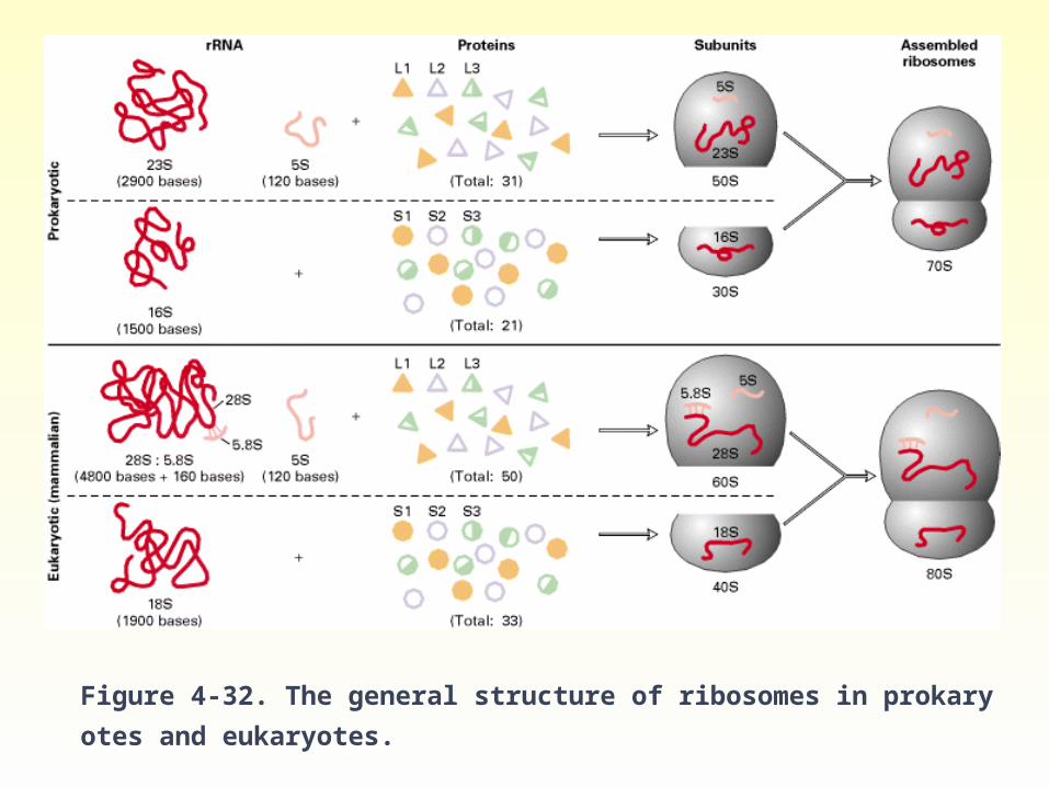
Figure 4-32. The general structure of ribosomes in prokaryotes and eukaryotes.
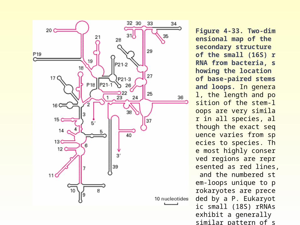
Figure 4-33. Two-dimensional map of the secondary structure of the small (16S) rRNA from bacteria, showing the location of base-paired stems and loops. In general, the length and position of the stem-loops are very similar in all species, although the exact sequence varies from species to species. The most highly conserved regions are represented as red lines, and the numbered stem-loops unique to prokaryotes are preceded by a P. Eukaryotic small (18S) rRNAs exhibit a generally similar pattern of stem-loops, although, as with prokaryotes, a few are unique.
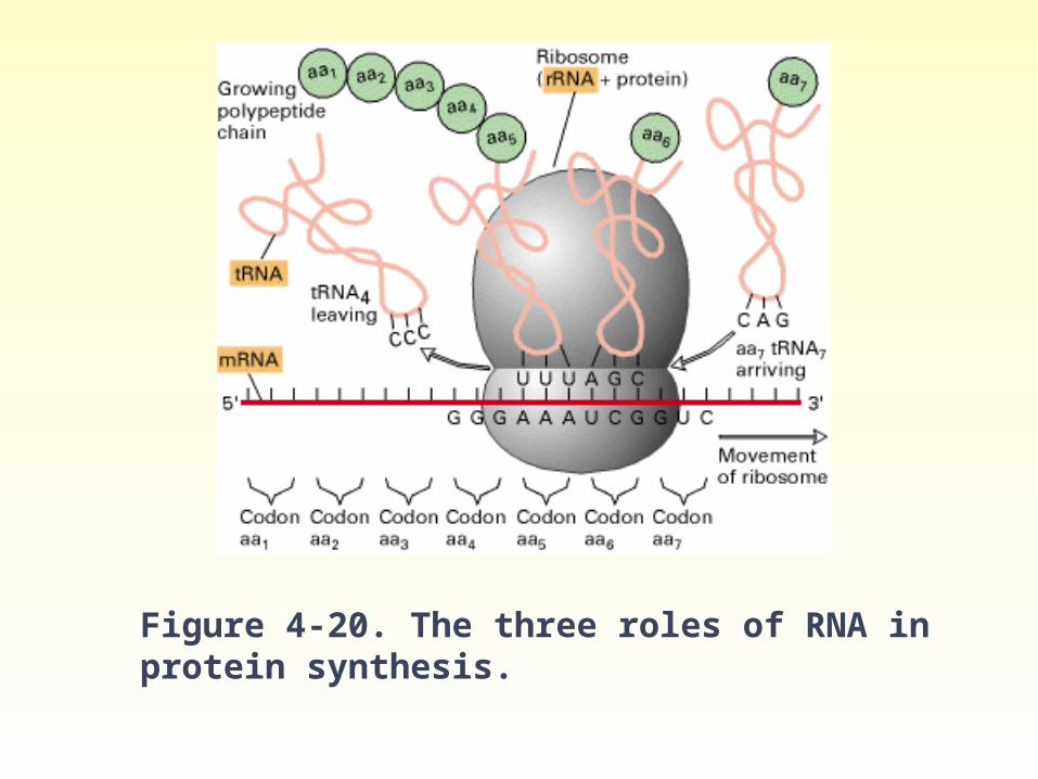
Figure 4-20. The three roles of RNA in protein synthesis.
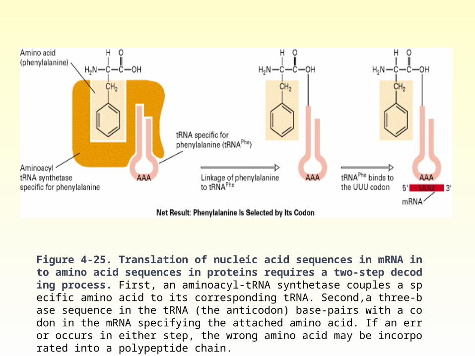
Figure 4-25. Translation of nucleic acid sequences in mRNA into amino acid sequences in proteins requires a two-step decoding process. First, an aminoacyl-tRNA synthetase couples a specific amino acid to its corresponding tRNA. Second,a three-base sequence in the tRNA (the anticodon) base-pairs with a codon in the mRNA specifying the attached amino acid. If an error occurs in either step, the wrong amino acid may be incorporated into a polypeptide chain.
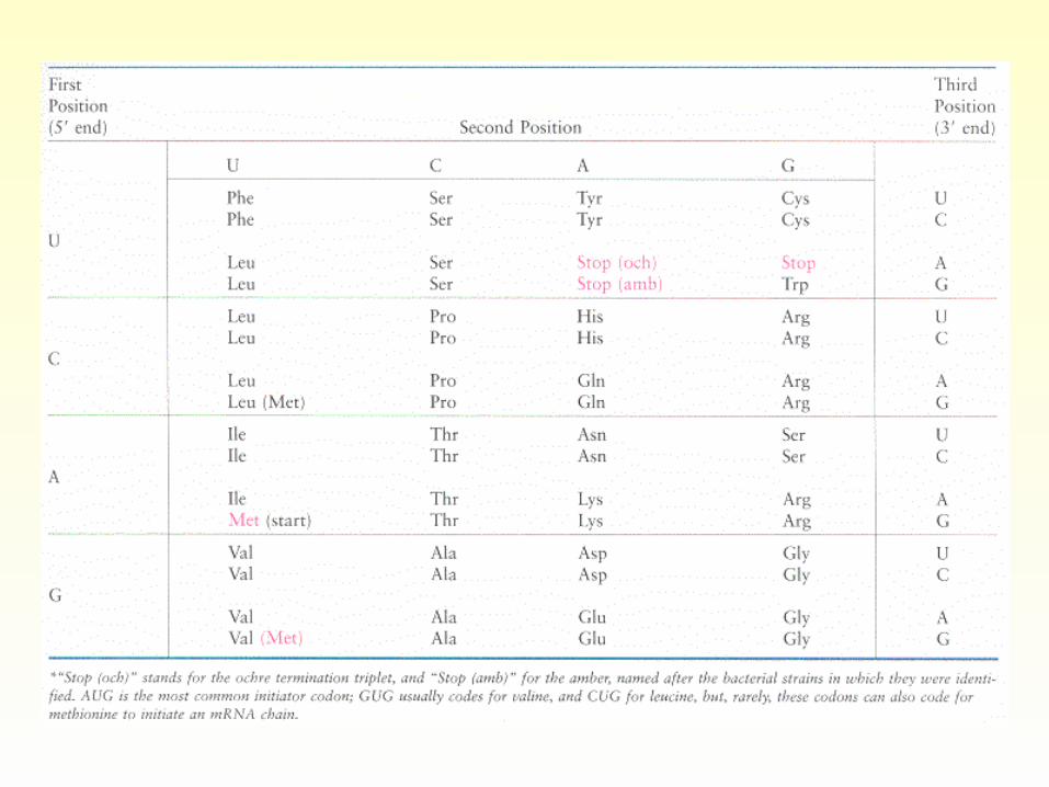
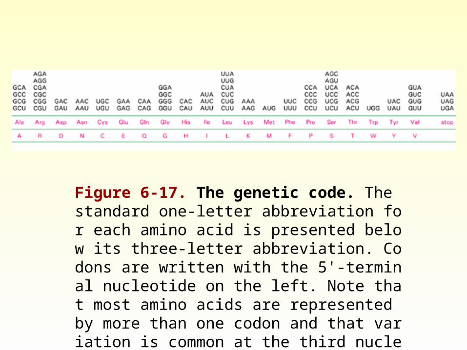
Figure 6-17. The genetic code. The standard one-letter abbreviation for each amino acid is presented below its three-letter abbreviation. Codons are written with the 5'-terminal nucleotide on the left. Note that most amino acids are represented by more than one codon and that variation is common at the third nucleotide (see also Figure 3-16).
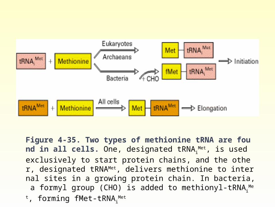
Figure 4-35. Two types of methionine tRNA are found in all cells. One, designated tRNAi
Met, is used exclusively to start protein chains, and the ot
her, designated tRNAMet, delivers methionine to internal sites in a growing protein chain. In bacteria, a formyl group (CHO) is added to methionyl-tRNAi
Met, forming fMet-tRNAiMet
Small stable RNA• A large number of discrete, highly conserved, and
small stable RNA species are found in eukaryotic cells. The majority of these molecules exist as ribonucleoproteins and are distributed in the nucleus, in the cytoplasm, or in both. They range in size from 90 to 300 nucleotides and are present in 100,000-1,000,000 copies per cell. Small nuclear RNA (snRNA) are significantly involved in mRNA processing (intron removal and processing of hnRNA into mRNA) and gene regulation.
Nuclease• Enzymes capable of degrading nucleic acids. • Deoxyribonucleases (DNAase) and Ribonucleases
(RNAase).• Exonuclease and Endonuclease. • Endonuclease: enzymes capable of cleaving internal
phosphodiester bonds to produce either 3’-hydoxyl and 5’-phosphoryl terminals or 5’-hydroxyl and 3’-phosphoryl terminals.
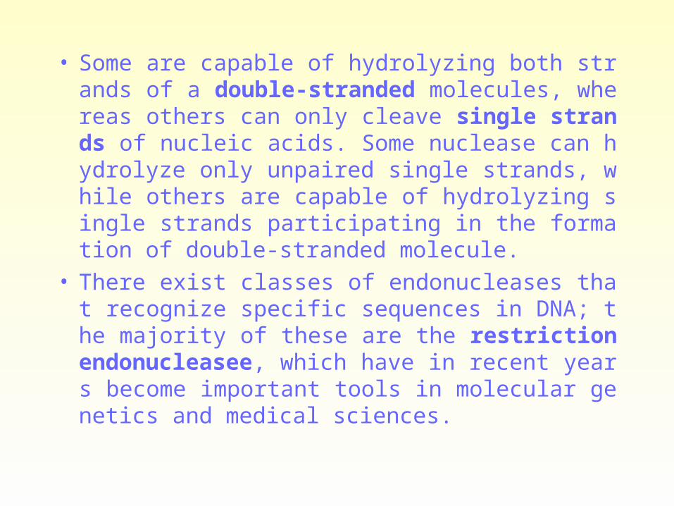
• Some are capable of hydrolyzing both strands of a double-stranded molecules, whereas others can only cleave single strands of nucleic acids. Some nuclease can hydrolyze only unpaired single strands, while others are capable of hydrolyzing single strands participating in the formation of double-stranded molecule.
• There exist classes of endonucleases that recognize specific sequences in DNA; the majority of these are the restriction endonucleasee, which have in recent years become important tools in molecular genetics and medical sciences.
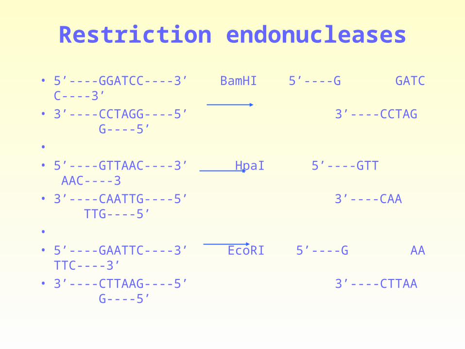
Restriction endonucleases
• 5’----GGATCC----3’ BamHI 5’----G GATCC----3’
• 3’----CCTAGG----5’ 3’----CCTAG G----5’
•
• 5’----GTTAAC----3’ HpaI 5’----GTT AAC----3
• 3’----CAATTG----5’ 3’----CAA TTG----5’
•
• 5’----GAATTC----3’ EcoRI 5’----G AATTC----3’
• 3’----CTTAAG----5’ 3’----CTTAA G----5’
• Palindromic: The nucleotide sequence is the same if the helix is turned 180 degrees around the center of the short region of the helix that is recognized.
• Cohesive ends: Some restriction nucleases produce staggered cuts, which leave short single-stranded tails at the two ends of each fragment. Ends of this type ar known as chohesive ends, as each tail can form complementary base pairs with the tail at any other end by the same enzyme. The cohesive ends generated by restriction enzymes allow any two DNA fragments to be easily joined together, as long as the fragments were generated with the same restriction enzymes (or with another nuclease that produces the same cohesive ends).
选择题练习核酸化学
1. The element that could be used in nucleic acid quantitation is ( )
A. C
B. O
C. N
D. H
E. P
2. The basic unit composition of nucleic acid is ( )
A. Ribose and deoxyribose
B. phosphoric acid and pentaglucose
C. Pentaglucose and basic group
D. mononucleotide
E. phosphoric acid , pentose and basic group
3 .脱氧核糖核苷酸彻底水解,生成的产物的产物是 ( )
A 核糖和磷酸
B 脱氧核糖和碱基
C 脱氧核糖和磷酸
D 磷酸,核糖和碱基
E 脱氧核糖,磷酸和碱基
4 .在核酸分子中核苷酸之间的连接方式是 ( )
A. 3’,3’ -磷酸二酯键
B. 糖苷键
C. 2’,5’ -磷酸二酯键
D. 肽键
E. 3’,5’ -磷酸二酯键
5. The ultraviolet absorption maximum of nucleic acid is about ( )
A. 220nm
B. 240nm
C. 260nm
D. 280nm
E. 300nm
6. 含有稀有碱基比例较多的核酸是 ( )
A. mRNA
B. DNA
C. tRNA
D. rRNA
E. hnRNA
7. 核酸分子中储存、传递遗传信息的关键部分是 ( )
A. 核苷
B. 戊糖
C. 磷酸
D. 碱基序列
E. 戊糖磷酸骨架
8 . DNA 分子碱基含量关系哪种是错误的?
A. A+T=C+G
B. A+G=C+T
C. G=C
D. A=T
E. A/T=G/C
9. ATP 的生理功能不包括 ( )
A. 为生物反应供能
B. 合成 RNA
C. 贮存化学能
D. 合成 DNA
E. 转变为 cAMP
10. 下列哪种核酸的二级结构具有”三叶草”型 ?
A. mRNA
B. 质粒 DNA
C. tRNA
D. 线粒体 DNA
E. rRNA
11. 关于 mRNA 的论述不正确的是 ( )
A. mRNA 分子中含有生物遗传信息
B. mRNA 在生物细胞内种类最多
C. 各种 mRNA3’ 末端和 5’ 末端都有相同的结构
D. mRNA 的碱基序列可以指导多肽链的合成
E. mRNA 的所有碱基都有编码氨基酸的作用
12. The protein not in nucleosome core particle is ( )
A. H1
B. H2A
C. H2B
D. H3
E. H4
13. DNA 变性是指 ( )
A. 多核苷酸链解聚
B. DNA 分子由超螺旋变为双螺旋
C. 分子中磷酸二酯键断裂
D. 碱基间氢键断裂
E. 核酸分子的完全水解

14. DNA Tm 值较高是由于下列哪组核苷酸含量较高所致 ?
A. G+A
B. C+G
C. A+T
D. C+T
E. A+C
15. Where does DNA reside in?
A. Golgi's body
B. rough endoplasmic reticulum
C. mitochondrium
D. chromosome
E. lysosome

16. 含有腺苷酸的辅酶有 ( )
A.NAD
B.NADP
C.FAD
D.FMN
E.CoA-SH
17. 关于 tRNA 的论述不正确的是 ( )
A. 分子中含有稀有碱基
B. 分子中含有密码环
C. 是细胞中含量最多的是 RNA
D. 主要存在于胞液
E. 其二级结构为倒 L 型
18. 维持 DNA 双螺旋结构的稳定因素包括 ( )
A. 分子中的磷酸二酯键
B. 碱基对之间的氢键
C. 碱基平面间的堆积力
D. 磷酸戊糖骨架的支撑力
E. 骨架上磷酸之间的负电排斥力

19. DNA 变性的实质是 ( )
A. 多核苷酸链解聚
B. 碱基的甲基化
C. 磷酸二酯键断裂
D. 加热使碱基对间氢键断裂
E. 使 DNA 双螺旋结构松散 , 变成单链

20. What does the Tm refer to about DNA?
A. optimum temperature
B. hydrolytic temperature
C. Renaturation temperature
D. melting temperature
E. denaturation temperature

Thank you!