chapter 27 bacteriology wang hui. content the purpose of a clinical bacterial laboratory diagnosis...
TRANSCRIPT

Chapter 27
Bacteriology
Wang Hui

Content
• THE PURPOSE OF A CLINICAL BACTERIAL LABORATORY DIAGNOSIS
• APPROACH TO BACTERIAL DIAGNOSIS• BACTERIAL DETECTION OF
– BACTREMIA (SEPSIS)– CENTRAL NERVOUS SYSTEMS INFECTION – GASTROINTESTINAL TRACT INFECTION – RESPIRATORY TRACT INFECTION – GENITAL TRACT INFECTION– URINE TRACT INFECTION

THE PURPOSE OF A CLINICAL BACTERIAL LABORATORY DIAGNOSIS
– to provide accurate, timely, clinically relevant information about the microorganisms
– appropriate selection, collection, and transport of the specimen to the laboratory.
– communication between the laboratory and the clinician

APPROACH TO BACTERIAL DIAGNOSIS
• Specimen•• Stain of smear Culture and isolation Enriched culture
Anaerobic cultivation Cultivation with carbon dioxide Anaerobic cultivation•
Observation of colonial morphology• • Picking colony Stain of smear and microscopic detection
• Serological Biochemical Antimicrobial• identification identification susceptibility testing • • Determination of pathogenic bacteria and results reporting

BACTERIAL DETECTION OF BACTREMIA (SEPSIS)
• Principle– Blood collection and blood culture have been seen
in the chart 27.
• Clinical Significance Of Blood Culture– "True positive" or "False positive"– " Nosocomial " or " Community "– " Primary " or " Secondary "– " Polymicrobial " or " Unimicrobial "

"True Positive" or "False Positive"
• Parameters for evaluating positive blood cultures
• Parameter Considerations– Veracity True-positive vs. false-positive– Duration Transient vs. intermittent vs.
continuous– Pattern of occurrence Single episode vs. persistent vs. recurrent– Clinical severity Inconsequential vs. life threatening– Intensity (CFU/ml) High grade vs. low grade– Lethality Crude mortality vs. attributable mortality– Site of acquisition Community acquired vs nosocomial– Source Primary vs secondary– No. of microorganisms Polymicrobial vs unimicrobial

" Nosocomial " or " Community "
• nosocomial bloodstream infection – onset on or after the third day of hospitalization.
• community-acquired and nosocomial bacteremia.– S. aureus and E. coli
• community-acquired – Streptococcus pneumoniae, H. influenza, and N.
meningitidis • Opportunistic pathogens cause nosocomial
bloodstream infection– Pseudomonas aeruginosa, Serratia marcescens, and
Candida albicans typically.

" Primary " or " Secondary "
• Primary bacteremia or fungemia, – refers to cases without an identifiable source.
• secondary bacteremias or fungemias – the body's normal microbial flora or microbial
adherence phenomenon. ( Streptococcus pneumoniae, E. coli, N. gonorrhoeae )
• Catheter-related septicemias are often both intense and sustained. Hence, metastatic infections can occur in any organ or tissue.

" Polymicrobial " or " Unimicrobial "
• Polymicrobial, – more than one microorganism– hospital acquired, – to arise from the bowel or from multiple locations, – to be associated with tumors or other serious underlying
diseases.– Enterobacteriaceae, Pseudomonadaceae, nongroup A
streptococci, and anaerobes, fungemia• Infections that are usually unimicrobial
– endocarditis, meningitis, pyelonephritis, hematogenous osteomyelitis, septic arthritis, and pneumonia not due to the aspiration of "mouth flora."

BACTERIAL DETECTION OF CENTRAL NERVOUS SYSTEMS INFECTION
• Principle– TB meningitis, PCR testing of CSF– Fungal CNS disease, direct smears,
antigen and antibody testing, and culture. – Viral meningitis and encephalitis, Most
difficult to diagnose them.

The processing of CSF for common bacterial isolates
• CSF
• Make smears by cytospin or centrifuge at 3000 ×g for 20 min
• Acid-fast stain ink stain Gram stain •
+ -• Report positive result •
Perform bacterial antigen test•
+ -• • Report positive result Growth on culture•
+ -• • Perform identification and AST Reincubate for 5-7 days,• or longer for fungi or mycobacteria
+ -
• Perform identification and AST Report no growth

Clinical Significance
• positive CSF results should be reported to the physician immediately.
• Clinical manifestation is very important to determine if it is a contaminant.
• The clinical relevance of a specific isolate is based on clinical, microbiological, and epidemiologic criteria.

Pathogens causing acute meningitis differ from age groups.
population
Neonates
infants (less than 2 month)
young adults patients aged (10 y to 18 y )
adults ( 18 y to 70y)
elderly patients
immunocompromised patients ( AIDS patients, SLE patients )
Common pathogens
Escherichia coli, Streptococcus agalactiae (group B streptococci),
Listeria monocytogenes, herps simplex 2 virus).
S. agalactiae, L. monocytogenes and E.coli
virus, H.influenzae,Streptococcuspneumoniae, and N. meningitidis .
Virus and N. meningitides ,S. pneumoniae and N. meningitidis
S. pneumoniae, grm-negative bacilli, and L. monocytogenes
C. neoformans

BACTERIAL DETACTION OF GASTROINTESTINAL TRACT INFECTION
– Specimen Collection and Transport– Direct Detection Gastroenteritis in Feces– Culture of Fecal Material for Isolation of
Etiologic Agents– Laboratory diagnosis of Clostridium difficile-
associated Diarrhea – Specimen Collection and Transport

Specimen Collection and Transport
• appropriate guidelines for specimen collection and transport is imperative.
• Certain infectious agents, such as Giardia, may be difficult to detect, requiring the processing of multiple specimens

Direct Detection of Agents of Gastroenteritis in Feces
• Wet Mounts
• Stains
• Antigen Detection
• Molecular Biological Techniques

Wet Mounts
• the fastest method – detection of motile trophozoites of parasites – but may alert the microbiologist finding other
parasites, such as Entamoeba coil, Endolimax nana, Chilomastix mesnili, and Trichomonas hominis.

Stains
• Gram stain, – detection of certain etiologic agents. – polymorphonuclear cell
• acid-fast stain – detect Cryptosporidium spp., mycbacteria, and
Isospora spp.

Antigen Detection
• detect numerous microorganisms that cause GI tract infection.
• indirect fluorescent antibody stain – giardiasis and cryptosporidiosis
• Enzyme immunoassays (EIAs)
• latex agglutination

Molecular Biological Techniques
• amplification techniques
• direct detection of many enteric pathogens
• probe technology – the organism itself is not available for
susceptibility testing, – important for certain bacterial pathogens (e.g.,
Shigella ) for which susceptibility patterns vary.

Culture of Fecal Material for Isolation of Etiologic Agents
• Organisms for Routine Culture.
• Helicobacter pylori screen test

Organisms for Routine Culture
• Stools routinely cultured: – Campylobacter, Salmonella, and Shigella spp.. Aeromonas spp.
• Other bacteria in the area served by the Lab– Yersinia enterocolitia and Viibrio spp.
• Protocols for culture of enterohemorrhagic Eschericbia coil (e.g., E.coilO157:H7) – Selective or screening media:
• 1% sorbitol-containing medium (most O157:H7 E.coil are sorbitol-negative)
• trypticase blood agar (Unipath GmbH, Wesel, Germany) • Rainbow Agar O157 (Biolog, Inc., Hayward, Calif.).

Helicobacter pylori screen test
• antral or duodenal biopsy specimen: – be placed in 5mL of sterile saline and submitted to the laboratory
within 30 minutes of collection. Store at 4 up to 4 hours.℃
• Upon its arrival, split the specimen (tissue) into two portions: – one portion in sterile saline or Brucella broth – the second portion in 0.5 ml of 2% Christensen’s urea broth as a direct
screen.
• Colonies selected for testing : be positive for oxidase, catalase, and appear typical on the Gram stain.
• Emulsify colonies into urea broth and incubate at 35 , ℃• The direct screen is read at 1-hour intervals for color chance to red or pink.

Laboratory Diagnosis of Clostridium difficile-associated Diarrhea
• The definitive diagnosis of C.difficile-associated diarrhea – clinical criteria – laboratory testing.
• pseudomembranous colitis – Visualization of a characteristic pseudo-membrane or plaque
on endoscopy – history of prior antibiotic use
• currently available for routine use;– culture,– detection of cytoxin by tissue culture– antigen detection assays (e.g., enzyme immuno-assay, latex
agglutination) for C.difficile toxin(s).

Bactrial Detection of Respiratory Tract Infection - Streptococcal Pharyngitis(1)
• Streptococcus pyogenes(group A streptococci[GAS])- A common cause of bacterial pharyngitis
• Routine laboratory diagnosis of bacterial pharyngitis should consist of procedures which are able to detect low numbers of GAS.

Bactrial Detection of Respiratory Tract Infection - Streptococcal Pharyngitis(2)
• Specimen• throat swab of the tonsillar area and/or posterior pharynx
• Materials• blood agar plate (BAP) • Selective BAP containing sulfamethoxazole and
trimethoprim (SXT).

Bactrial Detection of Respiratory Tract Infection - Streptococcal Pharyngitis(3)
• Procedure• Most procedures are based on extraction of group A
antigen from the swab by enzymatic or chemical means followed by an antigen detection step using coagglutination, enzyme immunoassay, or latex particle agglutination.
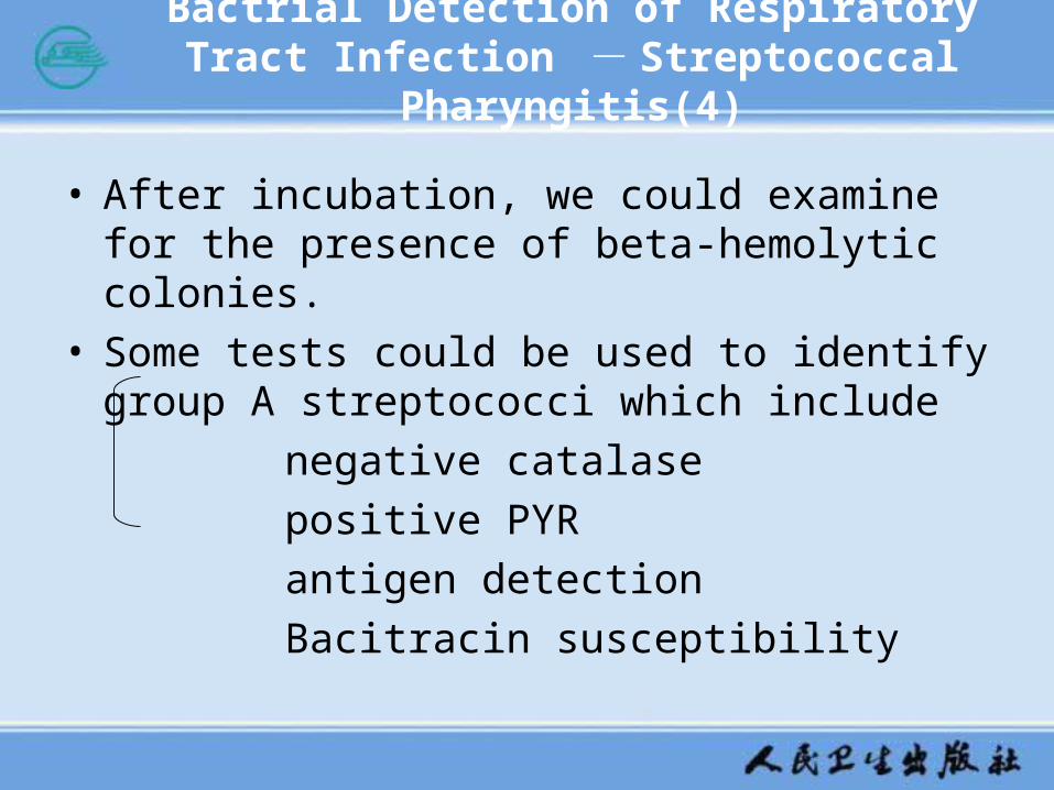
Bactrial Detection of Respiratory Tract Infection - Streptococcal Pharyngitis(4)
• After incubation, we could examine for the presence of beta-hemolytic colonies.
• Some tests could be used to identify group A streptococci which include
negative catalase
positive PYR
antigen detection
Bacitracin susceptibility

Bactrial Detection of Respiratory Tract Infection - Diphtheria(1)
• Specimen and Materials• The specimens could be respiratory specimens that
include both throat and nasopharyngeal specimens and cutaneous specimens that include skin, throat and nasopharyngeal specimens.

Bactrial Detection of Respiratory Tract Infection - Diphtheria(2)
specimen
smear CTBA BAP LAS
Procedure
If CTBA was inoculated, examine it at 24 and 48 h. Gram stain suspicious black colonies and perform catalase test. C. diphtheriae produces a garlic-like odor on CTBA. Also other Corynebacterium spp., staphylococci, and some streptococci can also reduce tellurite to tellurium, therefore producing black colonies on CTBA. Suspicious colonies should be subcultured to BAP for identification.
LMB stain

Bactrial Detection of Respiratory Tract Infection - Diphtheria(3)
• C. diphtheriae Identification• pleomorphic gram-positive bacilli and present metachromatic
granules on LAS slant.
• The colonial morphology on CTBA is grayish black(gunmetal gray) colonies approximately 1 to 3 mm in diameter or black colonies with or without gray rims.
• Biochemical tests at minimum include glucose, sucrose, starch, glycogen, nitrate, and urea.
• If reagents are available in the laboratory, a modified Elek test may be performed.
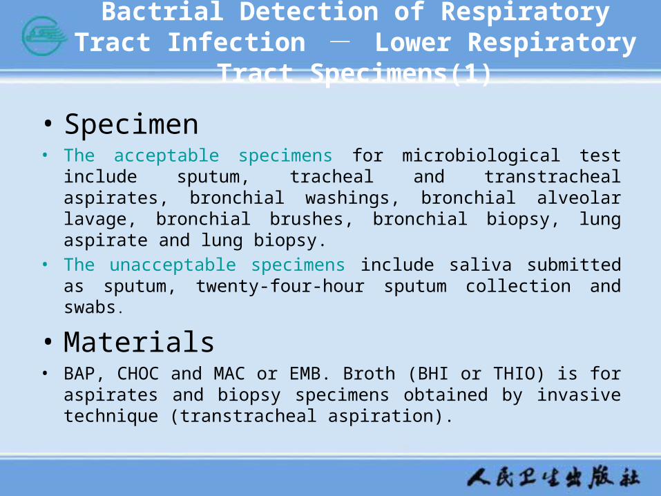
Bactrial Detection of Respiratory Tract Infection - Lower Respiratory Tract Specimens(1)
• Specimen• The acceptable specimens for microbiological test include sputum, tracheal
and transtracheal aspirates, bronchial washings, bronchial alveolar lavage, bronchial brushes, bronchial biopsy, lung aspirate and lung biopsy.
• The unacceptable specimens include saliva submitted as sputum, twenty-four-hour sputum collection and swabs.
• Materials • BAP, CHOC and MAC or EMB. Broth (BHI or THIO) is for aspirates and
biopsy specimens obtained by invasive technique (transtracheal aspiration).

Bactrial Detection of Respiratory Tract Infection - Lower Respiratory Tract Specimens(2)
• General Consideration• Although lower respiratory tract infections are a major cause of
mortality and morbidity, diagnosis of these infections is often complicated by the contamination of specimens with upper respiratory tract secretions during collection. Because the upper respiratory tract may be colonized with potential pathogens not involved in infection of the lower respiratory tract, the laboratory must ensure that an appropriate specimen is processed.

Bactrial Detection of Respiratory Tract Infection - Lower Respiratory Tract Specimens(3)
under low power
(10×objective): • fungal structures or any other
unusual findings
• assess sputum quality: squamous epithelial cells(SECs) and PMNs. Operator should examine a minimum of 10 representative low-power fields(10×objective), In general, there should be a ratio of greater than 2:1 WBCs to SECs.
Microscopic Examination
under oil immersion (100×objective):
The inspectors should semiquantitate the cells (SECs and WBCs) as few, moderate, or many and semiquantitate the organisms as few, moderate, or many.

Bactrial Detection of Respiratory Tract Infection - Lower Respiratory Tract Specimens(4)
• Culture• Probable pathogens (PP) from acceptable-quality sputum and
other noninvasive specimens should be discussed. PP may be defined as any microorganisms that have been associated with infections of the lower respiratory tract. These include both hospital-acquired pathogens, such as members of the family Enterobacteriaceae, P. aeruginosa, other gram-negative bacilli, and fungi, and non-hospital-acquired S. pneumoniae and H. influenzae.

BACTERIAL DETACTION OF GENITAL TRACT INFECTION(1)
• Specimen• If the culture site allows, aspirated material is preferred.
• Swabs should be submitted in a suitable transport medium such as Stuart or Amies medium.
• Dry swabs are unacceptable.
• Specimens must be transported anaerobically if anaerobes are suspected. If an abscess is present, material should be transported anaerobically. Do not process vaginal specimens for anaerobes.
• An appropriate transport system should be used for the isolation of N. gonorrhoeae, such as a charcoal media. Do not refrigerate.
• All specimens should be labeled appropriately and transported promptly to the laboratory.

BACTERIAL DETACTION OF GENITAL TRACT INFECTION(2)
• Materials • BAP, CHOC with IsoVitaleX or other
enriched CHOC, Modified Thayer-Martin (MTM) or other selective gonoccal agar, MAC or other differential gram-negative agar, Selective gram-positive agar, Enriched and selective anaerobic agar.

BACTERIAL DETACTION OF GENITAL TRACT INFECTION(3)
• Specimen processing• Aerobic media should be incubated in ambient air at
35℃ and CHOC should be placed in 5 to 10% CO2 at
35℃. • Anaerobic media should be incubated under anaerobic
conditions. • The information provided by the direct smear may
suggest the inclusion of special media for primary plating and/or provide useful interpretative information when growth is evaluated.

BACTERIAL DETACTION OF GENITAL TRACT INFECTION(4)
• Culture• The presence of pathogens must be evaluated. Identification
tests for the following should be performed: N. gonorrhoeae, S. agalactiae, S. pyogenes, L. monocytogenes, H. ducreyi.
• The presence of probable pathogens should be evaluated. If there is heavy growth, if isolated as the predominant microorganism, or if isolated in quantities greater than or equal to the normal genital microbiota, lab assistant should perform identification tests for the following PPs: Gram-negative bacilli, S. aureus, Streptococcus pneumoniae, Haemophilus spp., N. meningitides, Anaerobes, including Actinomyces spp. Actinomyces should be selectively identified and reported in women with intrauterine devices (IUDs)
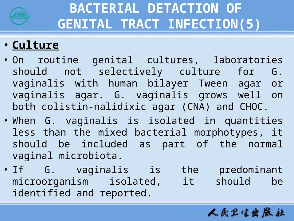
BACTERIAL DETACTION OF GENITAL TRACT INFECTION(5)
• Culture• On routine genital cultures, laboratories should not
selectively culture for G. vaginalis with human bilayer Tween agar or vaginalis agar. G. vaginalis grows well on both colistin-nalidixic agar (CNA) and CHOC.
• When G. vaginalis is isolated in quantities less than the mixed bacterial morphotypes, it should be included as part of the normal vaginal microbiota.
• If G. vaginalis is the predominant microorganism isolated, it should be identified and reported.

BACTERIAL DETACTION OF GENITAL TRACT INFECTION(6)
• Screening culture- N. gonorrhoeae • For materials, use of a self-contained nutritive system
consisting of selective gonococcal medium that is held and transported in an environment containing CO2 is appropriate
and cost-effective
• N. gonorrhoeae is Gram-negative diplococci, small, translucent, grayish, convex colonies, Oxidase is positive, Catalase is positive. Confirmatory tests include Carbohydrate degradation: glucose, maltose, sucrose, and lactose, Commercial identification systems.

BACTERIAL DETACTION OF GENITAL TRACT INFECTION(7)
• Screening culture-S. agalactiae • inoculate both swabs together into the selective broth
medium, incubate at 35℃ for 18 to 24h, subculture broth to BAP and incubate at 35℃ for 18 to 24h in 5% CO2.
• S. agalactiae is beta-hemolytic or nonhemolytic, Gram-positive cocci, Catalase is negative and CAMP is positive. Confirmatory identification includes agglutination tests and antigen detection tests.

BACTERIAL DETACTION OF URINE TRACT INFECTION(1)
• Specimen-avoid contamination• There are some methods to collect specimen. The
usual specimen is clean-voided midstream urine. Catheter urine also could be processed.
• A straight catheter (non-indwelling) is used by a physician or trained practitioner to obtain urine directly from the bladder. This procedure is not routinely recommended because there is a risk of introducing microorganisms into the bladder.

BACTERIAL DETACTION OF URINE TRACT INFECTION(2)
• Specimen-avoid contamination• Urine collection by suprapubic needle aspiration of the bladder
avoids contamination associated with the collection of voided urine. This is the preferred method for infants and for patients for whom the interpretation of results of voided urine is difficult and is also preferred when anaerobic bacteria are the suspected cause of infection.
• Foley catheter tips are unacceptable for culture
• Early-morning specimens should be obtained whenever possible because bacterial counts increase after overnight incubation in the bladder
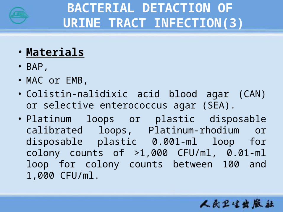
BACTERIAL DETACTION OF URINE TRACT INFECTION(3)
• Materials• BAP,• MAC or EMB, • Colistin-nalidixic acid blood agar (CAN) or selective
enterococcus agar (SEA). • Platinum loops or plastic disposable calibrated loops,
Platinum-rhodium or disposable plastic 0.001-ml loop for colony counts of >1,000 CFU/ml, 0.01-ml loop for colony counts between 100 and 1,000 CFU/ml.

BACTERIAL DETACTION OF URINE TRACT INFECTION(4)
• Gram stain
0.01ml of well-mixed, uncentrifuged urine is placed onto a glass slide
air dry without spreading
Determine the number of organisms per oil immersion field (OIF)
Report the number of microorganisms per OIF
The presence of one or more microorganisms per OIF correlates with a colony count of ≥105 CFU/ml.
The presence of many squamous epithelial cells and different microbial morphotypes indicates contamination. Request a repeat specimen.
BACTERIAL DETACTION OF URINE TRACT INFECTION(5)
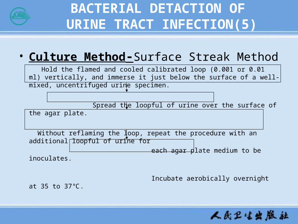
• Culture Method-Surface Streak Method Hold the flamed and cooled calibrated loop (0.001 or 0.01 ml) vertically, and immerse it just
below the surface of a well-mixed, uncentrifuged urine specimen.
Spread the loopful of urine over the surface of the agar plate.
Without reflaming the loop, repeat the procedure with an additional loopful of urine for
each agar plate medium to be inoculates.
Incubate aerobically overnight at 35 to 37℃.

BACTERIAL DETACTION OF URINE TRACT INFECTION(6)
• Examination of Culture Media• Examine cultures that have been incubated overnight. If
there is no visible growth and the specimen was collected by voiding or with a catheter, report as follows: “No growth at less than 1,000 CFU/ml” (0.001-ml inoculum) or “No growth at less than 100 CFU/ml” (0.01-ml inoculum).
•

BACTERIAL DETACTION OF URINE TRACT INFECTION(7)
• Examination of Culture Media
• If the specimen was collected by an invasive technique, e.g., suprapubic bladder aspiration, reincubate the culture media for an additional 24h. Reincubate culture plates with tiny or scant colonies that are not discernible.
• For positive cultures, examine culture media for the quantity and morphological type of organisms present. With 0.001-ml loop, one colony equals 1,000 CFU/ml. With 0.01-ml loop, one colony equals 100 CFU/ml. Perform additional testing based on the colony count method of urine collection.
BACTERIAL DETACTION OF URINE TRACT INFECTION(8)
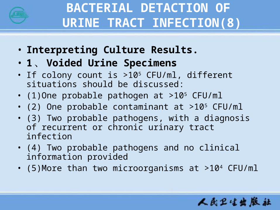
• Interpreting Culture Results.• 1 、 Voided Urine Specimens• If colony count is >105 CFU/ml, different situations should be
discussed:• (1)One probable pathogen at >105 CFU/ml • (2) One probable contaminant at >105 CFU/ml • (3) Two probable pathogens, with a diagnosis of recurrent or
chronic urinary tract infection • (4) Two probable pathogens and no clinical information
provided • (5)More than two microorganisms at >104 CFU/ml

BACTERIAL DETACTION OF URINE TRACT INFECTION(9)
• Interpreting Culture Results.• 1 、 Voided Urine Specimens• If colony count is ≤105 CFU/ml, different situations should
be discussed:• (1)One probable pathogen at ≤105 CFU/ml • (2)One probable contaminant at ≤105 CFU/ml • (3)Two or more microorganisms present at <104 CFU/ml • (4)One or more microorganisms at <104 CFU/ml

BACTERIAL DETACTION OF URINE TRACT INFECTION(10)
• 2 、 Catheterized Urine Specimens • If colony count is >104 CFU/ml, different situations should
be discussed.:• (1)Two or more probable pathogens present at >104
CFU/ml • (2) One or two probable contaminants present at >104
CFU/ml • (3) One probable pathogen and one probable contaminant • (4) Three or more microorganisms
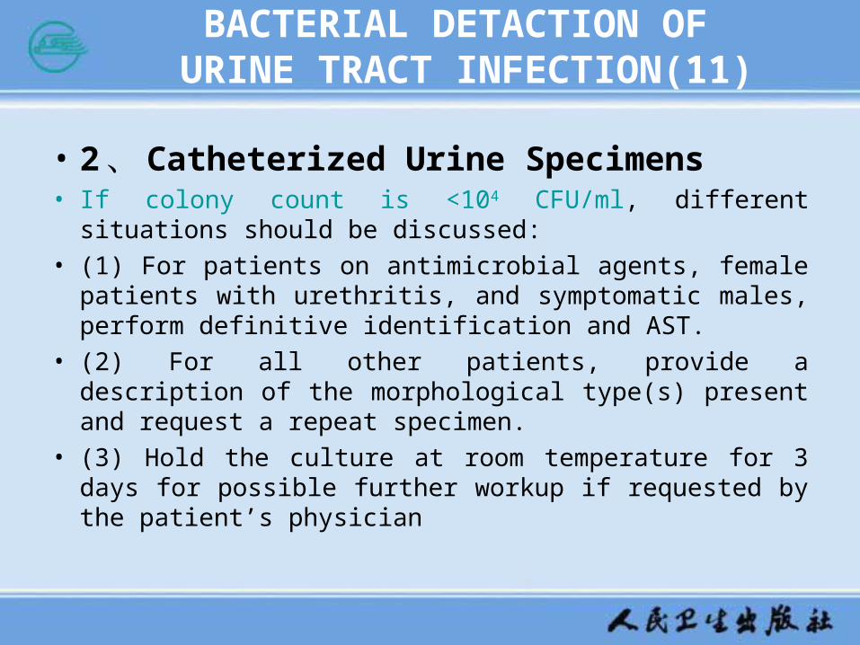
BACTERIAL DETACTION OF URINE TRACT INFECTION(11)
• 2 、 Catheterized Urine Specimens • If colony count is <104 CFU/ml, different situations should be
discussed:
• (1) For patients on antimicrobial agents, female patients with urethritis, and symptomatic males, perform definitive identification and AST.
• (2) For all other patients, provide a description of the morphological type(s) present and request a repeat specimen.
• (3) Hold the culture at room temperature for 3 days for possible further workup if requested by the patient’s physician
BACTERIAL DETACTION OF URINE TRACT INFECTION(12)
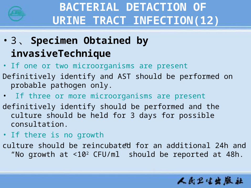
• 3 、 Specimen Obtained by invasiveTechnique
• If one or two microorganisms are present
Definitively identify and AST should be performed on probable pathogen only.
• If three or more microorganisms are present
definitively identify should be performed and the culture should be held for 3 days for possible consultation.
• If there is no growth
culture should be reincubated for an additional 24h and “No growth at <102 CFU/ml” should be reported at 48h.