chapter 27 care of patients with skin problems mrs. kreisel msn, rn nu130 adult health 1 summer 2011
TRANSCRIPT

Chapter 27
Care of Patients with Skin Problems
Mrs. Kreisel MSN, RNNU130 Adult Health 1Summer 2011


Epidermis
• The epidermis is the outer layer of skin. The thickness of the epidermis varies in different types of skin. It is the thinnest on the eyelids at .05 mm and the thickest on the palms and soles at 1.5 mm.
• The epidermis contains 6 layers. From bottom to top the layers are named:
• stratum basale • stratum spinosum • stratum granulosum • stratum licidum • Stratum corneum


The bottom layer, the stratum basale, has cells that are shaped like columns. In this layer the cells divide and push already formed cells into higher layers. As the cells move into the higher layers, they flatten and eventually die.
The top layer of the epidermis, the stratum corneum, is made of dead, flat skin cells that shed about every 2 weeks.
Specialized Epidermal CellsThere are three types of specialized cells in the epidermis. The melanocyte produces pigment (melanin) The Langerhans' cell is the frontline defense of the immune system in the skin The Merkel's cell's function is not clearly known

DermisThe dermis also varies in thickness depending on the location of the skin. It is .3 mm on the eyelid and 3.0 mm on the back. The dermis is composed of three types of tissue that are present throughout - not in layers. The types of tissue are: collagen elastic tissue reticular fibers
Layers of the DermisThe two layers of the dermis are the papillary and reticular layers. The upper, papillary layer, contains a thin arrangement of collagen fibers. The lower, reticular layer, is thicker and made of thick collagen fibers that are arranged parallel to the surface of the skin.

Specialized Dermal CellsThe dermis contains many specialized cells and structures. The hair follicles are situated here with the erector pili muscle that attaches to each follicle. Sebaceous (oil) glands and apocrine (scent) glands are associated with the follicle. This layer also contains eccrine (sweat) glands, but they are not associated with hair follicles. Blood vessels and nerves course through this layer. The nerves transmit sensations of pain, itch, and temperature. There are also specialized nerve cells called Meissner's and Vater-Pacini corpuscles that transmit the sensations of touch and pressure.

Subcutaneous TissueThe subcutaneous tissue is a layer of fat and connective tissue that houses larger blood vessels and nerves. This layer is important is the regulation of temperature of the skin itself and the body. The size of this layer varies throughout the body and from person to person. The skin is a complicated structure with many functions. If any of the structures in the skin are not working properly, a rash or abnormal sensation is the result. The whole specialty of dermatology is devoted to understanding the skin, what can go wrong, and what to do if something does go wrong.

Xerosis (Dryness)
• A common problem among older patients• Fine flaking of the stratum corneum• Generalized pruritus• Scratching may result in secondary skin
lesions, excoriations (abrasiains of the epideris by trauma, checmicals, burns, or other causes lichenification (cutaneous thicking & hardedning from continued irritation), and infection

Collaborative Management
• Nursing interventions aim to rehydrate the skin and relieve itching.
• Bathing with moisturizing soaps, oils, and lotions may reduce dryness.
• Water softens the outer skin layers; creams and lotions seal in the moisture provided by water.
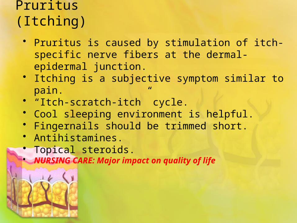
Pruritus (Itching)
• Pruritus is caused by stimulation of itch-specific nerve fibers at the dermal-epidermal junction.
• Itching is a subjective symptom similar to pain.• “Itch-scratch-itch” cycle.• Cool sleeping environment is helpful.• Fingernails should be trimmed short.• Antihistamines.• Topical steroids. • NURSING CARE: Major impact on quality of life

Atopic Dermatitis
• “Itch-scratch-itch” cycle.• STOP THE PRURITUS• Scratch->lesion->infection->pain
Sunburn
• First-degree, superficial burn• Cool baths• Soothing lotions• Antibiotic ointments for blistering and
infected skin• Topical corticosteroids for pain

Urticaria (Hives)
• Urticaria—presence of white or red edematous papules or plaques of varying sizes
• Removal of triggering substances• Antihistamines helpful• Avoidance of overexertion, alcohol
consumption, and warm environments, which can worsen symptoms

Trauma
• Phases of wound healing:• Inflammatory phase• Fibroblastic or connected tissue repair
phase• Maturation or remodeling phase

Process of Wound Healing

Process of Wound Healing (Cont’d)
• First intention resulting in a thin scar• Second intention (granulation) and
contraction—a deeper tissue injury or wound
• Third intention (delayed closure)—high risk for infection with a resultant scar

Partial-Thickness Wounds
• Involve damage to the epidermis and upper layers of the dermis
• Heal by re-epithelialization within 5 to 7 days
• Skin injury immediately followed by local inflammation

Re-epithelialization

Full-Thickness Wounds
• Damage extends into the lower layers of the dermis and underlying subcutaneous tissue.
• Removal of the damaged tissue results in a defect that must be filled with granulation tissue to heal.
• Contraction develops in healing process.

Pressure Ulcer
• Tissue damage caused when the skin and underlying soft tissue are compressed between a bony prominence and an external surface for an extended period.
• Mechanical forces that create ulcers: • Pressure• Friction• Shear

Shearing Force
Identification of High-Risk Patients
• Mental status changes• Independent mobility• Nutritional status• Incontinence

Pressure-Relieving Techniques
• Adequate pressure relief key to prevention of pressure ulcers
• Capillary closing pressure• Pressure-relief products and devices• Positioning Turn & Position q2h• Nutritional Status: Supplements &
frequent snacks

Wound Assessment
• Pressure ulcers and their features are classified and assessed in four stages:• Stage I• Stage II• Stage III• Stage IV

Four Stages of Pressure Ulceration

Wound Assessment
• Location• Size• Color• Extent of tissue involvement• Cell types in the wound base and margins• Exudate• Condition of surrounding tissue• Presence of foreign bodies

Wound Contamination/Wound Infection
• A wound that is exposed is always contaminated but not always infected. Contamination is the presence of organisms without any manifestations of infection.
• Wound infection is contamination with pathogenic organisms to the degree that growth and spread cannot be controlled by the body’s immune defenses.

Nonsurgical Management
• Dressings:• Mechanical débridement• Natural chemical débridement• Hydrophobic material• Hydrophilic material

Nonsurgical Therapy
• Physical therapy• Drug therapy• Nutrition therapy• New technologies:
• Electrical stimulation• Vacuum-assisted wound closure (VAC) • Hyperbaric oxygen (HBO) • Topical growth factors• Skin substitutes
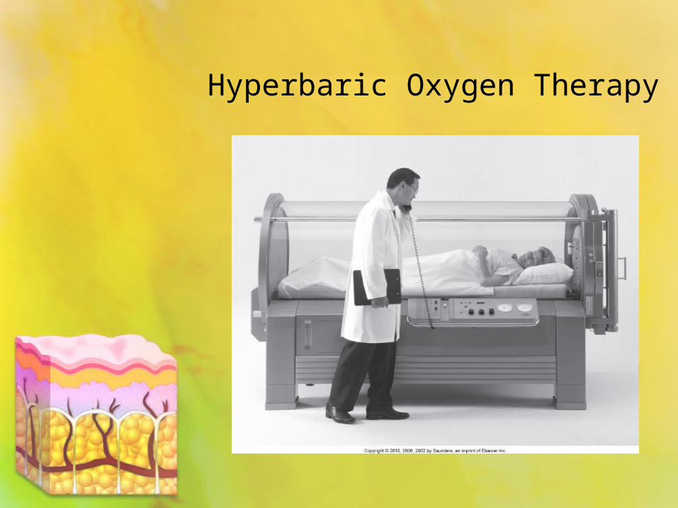
Hyperbaric Oxygen Therapy
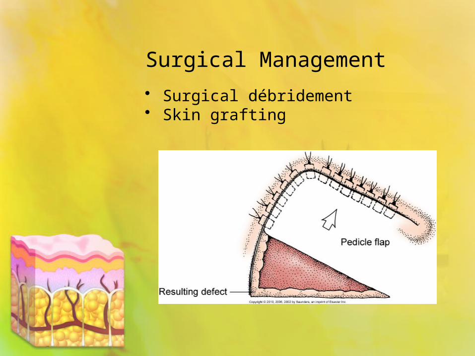
Surgical Management
• Surgical débridement• Skin grafting

Community-Based Care
• Home care management• Health teaching• Health care resources
Bacterial Infections
• Folliculitis—superficial infection involving only the upper portion of the follicle
• Furuncle (boil)—much deeper infection in the follicle
• Cellulitis—generalized infection with either Staphylococcus or Streptococcus involving deeper connective tissue

Furuncle

Cellulitis

Herpes Simplex Virus • Type 1 herpes simplex virus (HSV-1)—
classic recurring cold sore• Type 2 herpes simplex virus (HSV-2)—
genital herpes• Herpes zoster (shingles)• Herpetic whitlow—a form of herpes
simplex infection occurring on the fingertips of medical personnel who have come in contact with viral secretions

Herpes Zoster/Shingles
• Caused by reactivation of the dormant varicella-zoster virus in patients who have previously had chickenpox.
• Multiple lesions occur in a segmental distribution on the skin area innervated by the infected nerve.
• Eruption lasts several weeks.• Postherpetic neuralgia occurs after lesions
have resolved.

Nursing Care of Herpes• Priority: Antiviral Therapy
• pain management • Neuralgia
Itching, spreading, recurrence= nursing education

Fungal Infections (Dermatophyte)
• Tinea pedis: fungul infection of the foot• Tinea manus:fungul infection of the hand• Tinea cruris: fungus disease in the
scrotal, anal or genital areas• Tinea capitis: fungul infection of the scalp• Tinea corporis: red, elevated, scaly
patches over the body• Candida albicans
Tinea = fungus/ring worm

Assessment
• History • Laboratory assessment:
• Tzanck smear: BX of tissue to determine cell type in a vesicular disease
• Swab culture• Potassium hydroxide (KOH) test if positive
eliminates need for a C&S b/c it takes to long

Interventions
• Skin care with proper cleansing• Isolation Precautions• Drug therapy

Skin Care
• Bathe daily with an antibacterial soap.• Remove any pustules or crusts gently.• Apply warm compress twice a day to
furuncles or areas of cellulitis.• Apply Burow's solution (drying agent for
weeping skin lesions) to viral lesions.• Avoid excessive moisture.• Ensure optimal patient positioning.

Drug Therapy for Skin Disorders
• Antibacterial drugs• Antifungal drugs• Anti-inflammatory drugs

Cutaneous Anthrax
• Infection caused by the spores of the bacterium Bacillus anthracis
• Diagnosis based on appearance of the lesions and culture or anthrax antibodies in the blood
• Oral antibiotics for 60 days—ciprofloxacin or doxycycline

Cutaneous Anthrax

Pediculosis
• Pediculosis—infestation by human lice:• Head lice—pediculosis capitis• Body lice—pediculosis corporis• Pubic or crab lice—pediculosis pubis
• Pruritus most common symptom• Drugs• Laundering of clothing and bed linen

Scabies
• Scabies is a contagious skin disease caused by mite infestations.
• Scabies is transmitted by close and prolonged contact or infested bedding.
• Examine skin between fingers and on the palms.
• Infestation is confirmed by an examination of a scraping of a lesion under a microscope.
Common Inflammations
• Contact dermatitis, atopic dermatitis• Interventions include:
• Steroids• Avoidance of oil-based products• Antihistamines• Compresses and baths
Psoriasis
• Lifelong disorder with exacerbations and remissions
• Scaling disorder with underlying dermal inflammation; possibly an autoimmune reaction
• Psoriasis vulgaris most often seen• Exfoliative psoriasis—an explosively
eruptive and inflammatory form of the disease

Psoriasis Vulgaris
Treatment of Psoriasis
• Corticosteroids • Tar preparations• Other topical therapies • Ultraviolet light therapy• Systemic therapy:
• Biologic agents• Cytotoxic agents• Immunosuppressants
• Emotional support: Life long illness, stress can exacerbate breakouts, other autoimmune diseases may develop
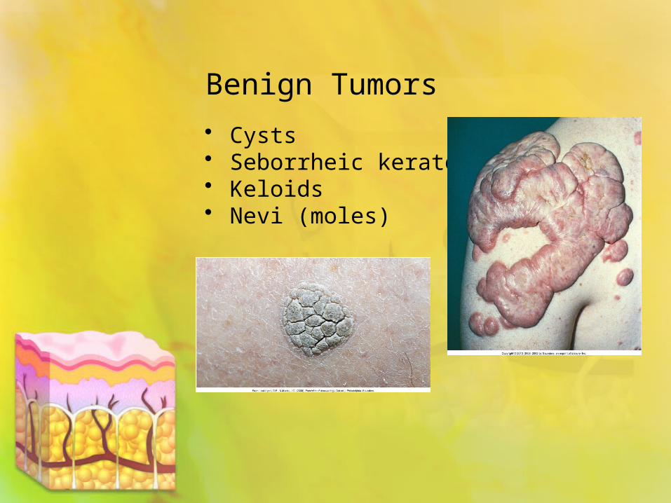
Benign Tumors
• Cysts• Seborrheic keratoses • Keloids• Nevi (moles)
Skin Cancer
• Actinic keratoses (caused by exposure to radiation)
• Squamous cell carcinomas• Basal cell carcinomas• Melanomas—highly metastatic; survival
depends on early diagnosis and treatment
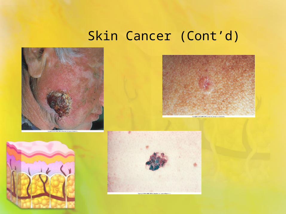
Skin Cancer (Cont’d)

Surgical Management of Skin Cancer
Surgical management:• Cryosurgery • Curettage and electrodesiccation• Excision• Mohs’ surgery: mainly used for basil cell ca.
step by step dissection until clear removal• Wide excision

Nonsurgical Management of Skin Cancer
• Drug therapy• Radiation therapy

Plastic Surgery
• Rhytidectomy (face-lift)• Rhinoplasty (reconstruction of the
nose)• Post op ABC• HOB elevated• Do not administer any
anticoagulants• Do not remove nasal packing• Assess for excessive bleeding
(swallowing)

Acne
• Red pustular eruption affecting the sebaceous glands of the skin
• Progressive disorder that manifests as noninflammatory comedones, inflammatory papules, pustules, and cysts
• Topical agents• Systemic antibiotics and possibly
isotretinoin (Accutane) helpful

Other Skin Disorders
• Lichen planus with itchy papules• Pemphigus vulgaris with chronic blistering• Toxic epidermal necrolysis—a rare, acute
drug reaction• Stevens-Johnson syndrome: multiforms of
erythema• Leprosy; Very contagious. Skin lesions,
muscle weakness, paralysis

NCLEX TIME
Question 1
Melanoma accounts for what percentage of all cancers?
A. 2%B. 4%C. 6%D. 8%

Question 2
What is the ideal environment for healing pressure ulcers?
A. A clean, dry ulcer surface B. A clean, slightly moist ulcer surfaceC. A clean and very moist ulcer surface D. A sterile and dry ulcer surface

Question 3
The nurse discovers an area of intact but reddened skin over a patient’s ankle. When the nurse presses firmly with her fingers at the center of the area of redness, the area blanches, or lightens,
with pressure and then returns to color when pressure is removed.
This assessment finding indicates that the color change is due to:
A. Tissue damageB. Tissue inflammation C. The presence of infection D. Blood vessel dilation

Question 4
While examining the results of a wound culture from a pressure ulcer on a patient’s sacrum, the wound care
nurse tells the nursing student that the wound is “contaminated.” This contamination indicates that:
A. The pressure ulcer is infected. B. The pressure ulcer has organisms
present but is not infected. C. Sepsis may be occurring.D. The culture was done improperly.

Question 5
An older adult patient has been brought to the hospital for generalized weakness after a fall in the nursing home. He is confused, unable to eat, and cannot turn himself in bed. The assessment examination reveals a pressure ulcer over his coccyx that is 3 cm wide and 4 cm long, with eschar (sloughing of tissue) present. Which technique will be used to remove the necrotic tissue?
A. Surgical removal of the tissueB. A biologic dressingC. A continuous dry gauze dressingD. Dressings along with a topical enzyme preparation