chapter 29 epigenetic effects are inherited. 29.1 introduction epigenetic effects can result from...
TRANSCRIPT
Chapter 29Epigenetic Effects Are Inherited

29.1 Introduction• Epigenetic effects can result from modification of a
nucleic acid after it has been synthesized or by the perpetuation of protein structures.
Figure 29.01: Replication of a methylated site produces hemimethylated DNA, in
which only the parental strand is methylated.
Figure 29.02: Heterochromatin is created by proteins that associate with
histones.

29.1 Introduction
• prion – A proteinaceous infectious agent that behaves as an inheritable trait, although it contains no nucleic acid.– Examples are PrPSc, the agent of scrapie in sheep
and bovine spongiform encephalopathy, and PSI, which confers an inherited state in yeast.

29.2 Heterochromatin Propagates from a Nucleation Event
• Heterochromatin is nucleated at a specific sequence and the inactive structure propagates along the chromatin fiber.
• Genes within regions of heterochromatin are inactivated.• The length of the inactive region varies from cell to cell;
as a result, inactivation of genes in this vicinity causes position effect variegation (PEV).

29.2 Heterochromatin Propagates from a Nucleation Event
Figure 29.04: Extension of heterochromatin inactivates genes.

29.2 Heterochromatin Propagates from a Nucleation Event
• Similar spreading effects occur at telomeres (telomeric silencing) and at the silent cassettes in yeast mating type.
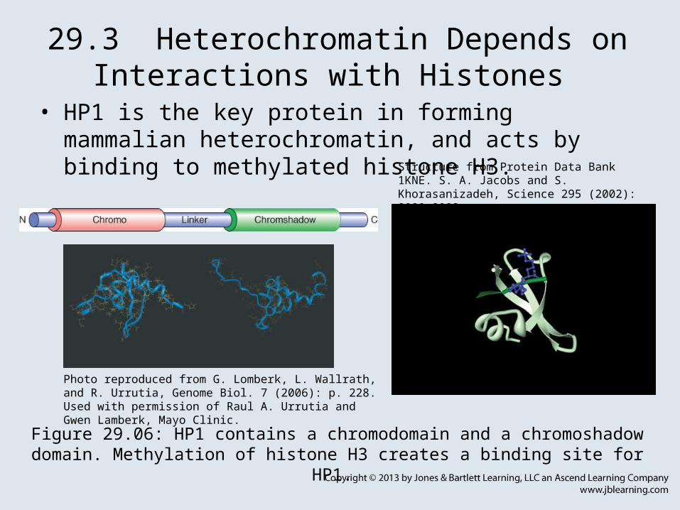
29.3 Heterochromatin Depends on Interactions with Histones
• HP1 is the key protein in forming mammalian heterochromatin, and acts by binding to methylated histone H3.
Figure 29.06: HP1 contains a chromodomain and a chromoshadow domain. Methylation of histone H3 creates a binding site for HP1.
Photo reproduced from G. Lomberk, L. Wallrath, and R. Urrutia, Genome Biol. 7 (2006): p. 228. Used with permission of Raul A. Urrutia and Gwen Lamberk, Mayo Clinic.
Structure from Protein Data Bank 1KNE. S. A. Jacobs and S. Khorasanizadeh, Science 295 (2002): 2080-2083.

29.3 Heterochromatin Depends on Interactions with Histones
Figure 29.07: Binding of HP1 to methylated histone H3 forms a trigger for silencing.
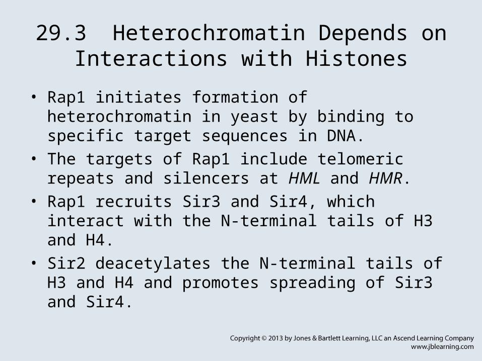
29.3 Heterochromatin Depends on Interactions with Histones
• Rap1 initiates formation of heterochromatin in yeast by binding to specific target sequences in DNA.
• The targets of Rap1 include telomeric repeats and silencers at HML and HMR.
• Rap1 recruits Sir3 and Sir4, which interact with the N-terminal tails of H3 and H4.
• Sir2 deacetylates the N-terminal tails of H3 and H4 and promotes spreading of Sir3 and Sir4.

29.3 Heterochromatin Depends on Interactions with Histones
Figure 29.08: Formation of heterochromatin is initiated when Rap1
binds to DNA.

29.3 Heterochromatin Depends on Interactions with Histones
• RNAi pathways promote heterochromatin formation at centromeres.

29.4 Polycomb and Trithorax Are Antagonistic Repressors and Activators
• Polycomb group proteins (Pc-G) perpetuate a state of repression through cell divisions.
Figure 29.09: Pc-G proteins do not initiate repression, but are responsible for maintaining it.

29.4 Polycomb and Trithorax Are Antagonistic Repressors and Activators
• The PRE is a DNA sequence that is required for the action of Pc-G.
• The PRE provides a nucleation center from which Pc-G proteins propagate an inactive structure.
• Trithorax group proteins (trxG) antagonize the actions of the Pc-G.
• Pc-G and trxG can bind to the same PRE with opposing effects.

29.5 X Chromosomes Undergo Global Changes
• dosage compensation – Mechanisms employed to compensate for the discrepancy between the presence of two X chromosomes in one sex but only one X chromosome in the other sex.
Figure 29.10: Different means of dosage compensation are used to
equalize X chromosome expression in male and female.
Figure 29.11: X-linked variegation is caused by the random inactivation of one X chromosome in each precursor cell.

29.5 X Chromosomes Undergo Global Changes
• constitutive heterochromatin – The inert state of permanently nonexpressed sequences, such as satellite DNA.
• facultative heterochromatin – The inert state of sequences that also exist in active copies; for example, one mammalian X chromosome in females.

29.5 X Chromosomes Undergo Global Changes
• One of the two X chromosomes is inactivated at random in each cell during embryogenesis of eutherian mammals.
• single X hypothesis – The theory that describes the inactivation of one X chromosome in female mammals.
• In exceptional cases where there are >2 X chromosomes, all but one are inactivated (the n–1 rule).

29.5 X Chromosomes Undergo Global Changes
• The Xic (X inactivation center) is a cis-acting region on the X chromosome that is necessary and sufficient to ensure that only one X chromosome remains alive.
• Xic includes the Xist gene, which codes for an RNA that is found only on inactive X chromosomes.
Figure 29.12: X-inactivation involves stabilization of Xist RNA, which coats the inactive chromosome.
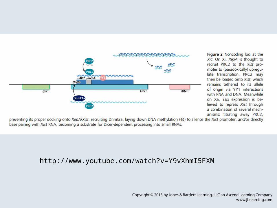
http://www.youtube.com/watch?v=Y9vXhmI5FXM

RNAi
• http://www.nature.com/nrg/multimedia/rnai/animation/index.html

Crispr
http://www.youtube.com/watch?v=9IgLrOEsauk
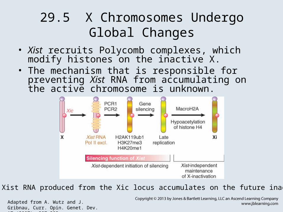
29.5 X Chromosomes Undergo Global Changes
• Xist recruits Polycomb complexes, which modify histones on the inactive X.
• The mechanism that is responsible for preventing Xist RNA from accumulating on the active chromosome is unknown.
Figure 29.13: Xist RNA produced from the Xic locus accumulates on the future inactive X (Xi).
Adapted from A. Wutz and J. Gribnau, Curr. Opin. Genet. Dev. 17 (2007): 387-393.

29.6 Chromosome Condensation Is Caused by Condensins
• SMC (structural maintenance of chromosome) proteins are ATPases that include condensins and cohesins.
• A heterodimer of SMC proteins associates with other subunits.
Figure 29.15: (A) The basic architecture of condensin and cohesin complexes. (B) Condensin and cohesin consist of V-
shaped dimers of two SMC proteins interacting through their hinge domains.
Adapted from T. Hirano, Nat. Rev. Mol. Cell Biol. 7 (2006): 311-322.
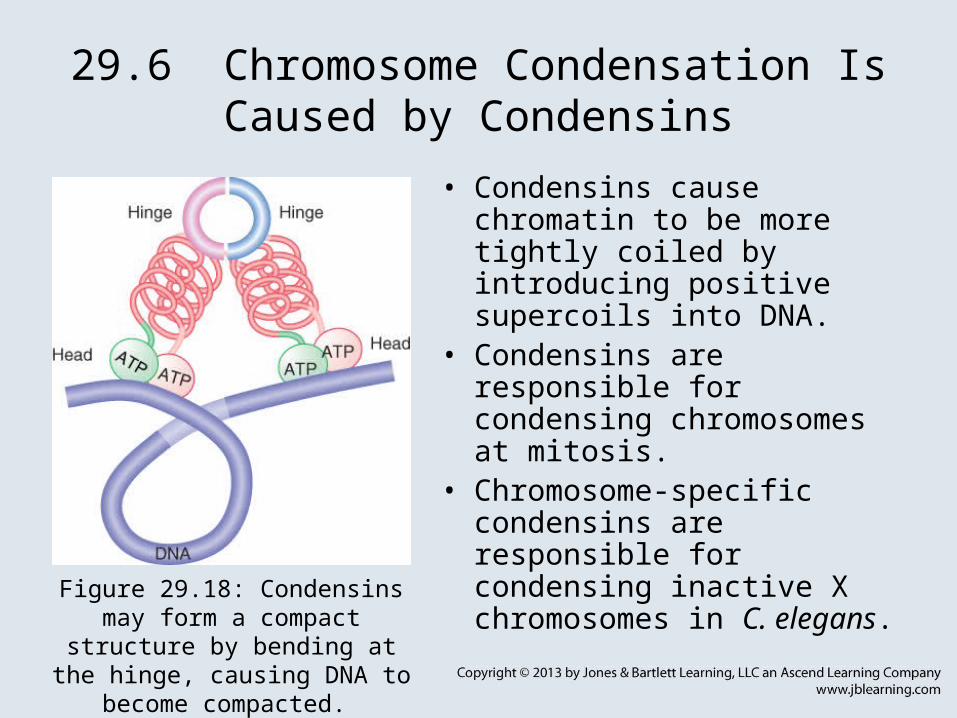
29.6 Chromosome Condensation Is Caused by Condensins
• Condensins cause chromatin to be more tightly coiled by introducing positive supercoils into DNA.
• Condensins are responsible for condensing chromosomes at mitosis.
• Chromosome-specific condensins are responsible for condensing inactive X chromosomes in C. elegans.
Figure 29.18: Condensins may form a compact structure by bending at the
hinge, causing DNA to become compacted.

29.7 CpG Islands Are Subject to Methylation
• Most methyl groups in DNA are found on cytosine on both strands of the CpG doublet.
• Replication converts a fully methylated site to a hemimethylated site.
• DNA methyltransferase – An enzyme that adds a methyl group to a specific target sequence in DNA.
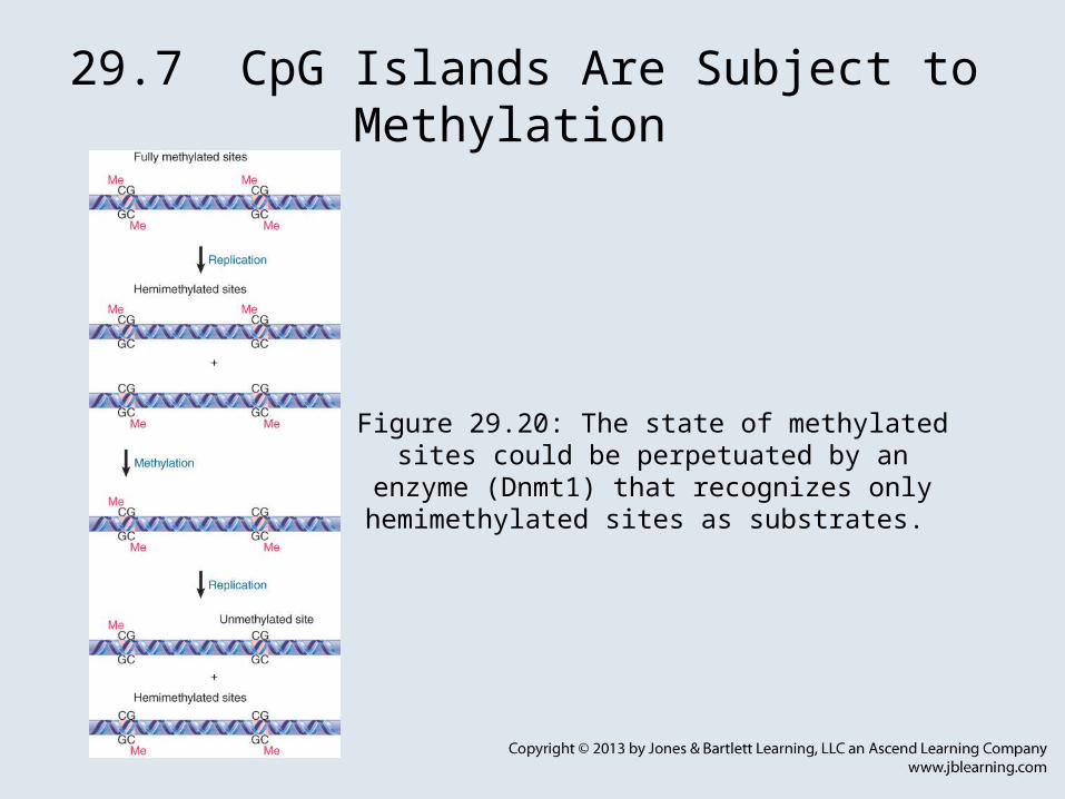
29.7 CpG Islands Are Subject to Methylation
Figure 29.20: The state of methylated sites could be perpetuated by an enzyme (Dnmt1) that recognizes
only hemimethylated sites as substrates.

29.7 CpG Islands Are Subject to Methylation
• demethylase – An enzyme that removes a methyl group, typically from DNA, RNA, or protein.
• de novo methyltransferase – An enzyme that adds a methyl group to an unmethylated target sequence on DNA.
• Hemimethylated sites are converted to fully methylated sites by a maintenance methyltransferase.
• TET proteins convert 5-methylcytosine to 5-hydroxymethylcytosine to lead to DNA demethylation.

29.7 CpG Islands Are Subject to Methylation
Figure 29.21: The state of methylation is controlled by three types of enzyme.

29.8 DNA Methylation Is Responsible for Imprinting
• Paternal and maternal alleles may have different patterns of methylation at fertilization.
• Methylation is usually associated with inactivation of the gene.
• When genes are differentially imprinted, survival of the embryo may require that the functional allele is provided by the parent with the unmethylated allele.
Figure 29.23: The typical pattern for imprinting is that a methylated
locus is inactive.

29.8 DNA Methylation Is Responsible for Imprinting
• Survival of heterozygotes for imprinted genes is different, depending on the direction of the cross.
• Imprinted genes occur in clusters and may depend on a local control site where de novo methylation occurs unless specifically prevented.

29.9 Oppositely Imprinted Genes Can Be Controlled by a Single Center
• Imprinted genes are controlled by methylation of cis-acting sites.
• Methylation may be responsible for either inactivating or activating a gene.
Figure 29.24: The ICR is methylated on the paternal allele, where Igf2 is
active and H19 is inactive.
29.10 Epigenetic Effects Can Be Inherited
• Epigenetic effects can result from modification of a nucleic acid after it has been synthesized or by the perpetuation of protein structures.
• Epigenetic effects may be inherited through generations (transgenerational epigenetics).
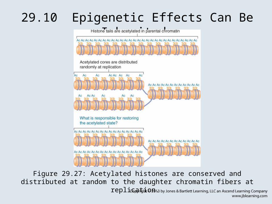
29.10 Epigenetic Effects Can Be Inherited
Figure 29.27: Acetylated histones are conserved and distributed at random to the daughter chromatin fibers at replication.

29.11 Yeast Prions Show Unusual Inheritance
• The Sup35 protein in its wild-type soluble form is a termination factor for translation.
• Sup35 can also exist in an alternative form of oligomeric aggregates, in which it is not active in protein synthesis.
Figure 29.28: The state of the Sup35 protein determines whether
termination of translation occurs.

29.11 Yeast Prions Show Unusual Inheritance
• The presence of the oligomeric form causes newly synthesized protein to acquire the inactive structure.
Figure 29.29: Newly synthesized Sup35 protein is converted into the [PSI+] state by the presence of
preexisting [PSI+] protein.

29.11 Yeast Prions Show Unusual Inheritance
• amyloid fibers – Insoluble fibrous protein polymers with a cross β-sheet structure, generated by prions or other dysfunctional protein aggregations (such as in Alzheimer’s).
• Conversion between the two forms is influenced by chaperones.
• The wild-type form has the recessive genetic state psi– and the mutant form has the dominant genetic state PSI+.

29.12 Prions Cause Diseases in Mammals
• kuru – A human neurological disease caused by prions.• The protein responsible for scrapie exists in two forms:
the wild-type noninfectious form PrPC, which is susceptible to proteases, and the disease-causing PrPSc, which is resistant to proteases.

29.12 Prions Cause Diseases in Mammals
• The neurological disease can be transmitted to mice by injecting the purified PrPSc protein into mice.
• The recipient mouse must have a copy of the PrP gene coding for the mouse protein.
Figure 29.31: A PrpSc protein can only infect an animal that has the same type of endogenous PrPC protein.

29.12 Prions Cause Diseases in Mammals
• The PrPSc protein can perpetuate itself by causing the newly synthesized PrP protein to take up the PrPSc form instead of the PrPC form.
• Multiple strains of PrPSc may have different conformations of the protein.