chapter 3: cells and tissues, part 2 file10/1/2013 1 chapter 6: the muscular system 3 types of...
TRANSCRIPT

10/1/2013
1
CHAPTER 6: THE MUSCULAR SYSTEM
3 types of muscle tissue
Skeletal muscle structure • Each muscle fiber is wrapped
in connective tissue called an endomysium
• Several muscle fibers are then wrapped in a fibrous membrane called a perimysium to form a fascicle
• Another connective tissue called the epimysium wraps around the whole muscle
• The epimysium connects to tendons or aponeuroses, which attach muscles to bones

10/1/2013
2
Smooth muscle
• Not striated like cardiac or skeletal muscle
• Controls involuntary movements
• Cells tend to be arranged in layers running in different directions, as shown here
• Smooth muscle contraction is slow and occurs without fatigue
Cardiac muscle
• Only found in the heart
• Striated like skeletal muscle and involuntary like smooth muscle
• Arranged in bundles that allow the heart to act as a pump
Functions of muscle
• Producing movement
• Maintaining posture
• Stabilizing joints
• Generating heat
• Protect internal organs
• Regulate passage of substances through the body

10/1/2013
3
MICROSCOPIC ANATOMY OF SKELETAL MUSCLE & SKELETAL MUSCLE ACTIVITY
Anatomy of a muscle fiber
A myofibril is made up of repeating patterns of proteins called a sarcomere

10/1/2013
4
Sarcomeres are made from actin and myosin
Motor units
• The activity of muscle fibers is controlled by nerve cells called motor neurons
• A single motor neuron & all the muscle fibers it controls are called a motor unit
The neuromuscular junction
• The neuromuscular junction is where a motor neuron comes in close contact with a muscle fiber
• Electrical signals from the spinal cord travel through the motor neuron to the neuromuscular junction

10/1/2013
5
BUILDING MUSCLE
Skeletal muscle is built through training
• Muscle cells will become larger with exercise training
• Need amino acids and calories to build muscle
• Muscle atrophy will occur when training stops
10/1/2013
6
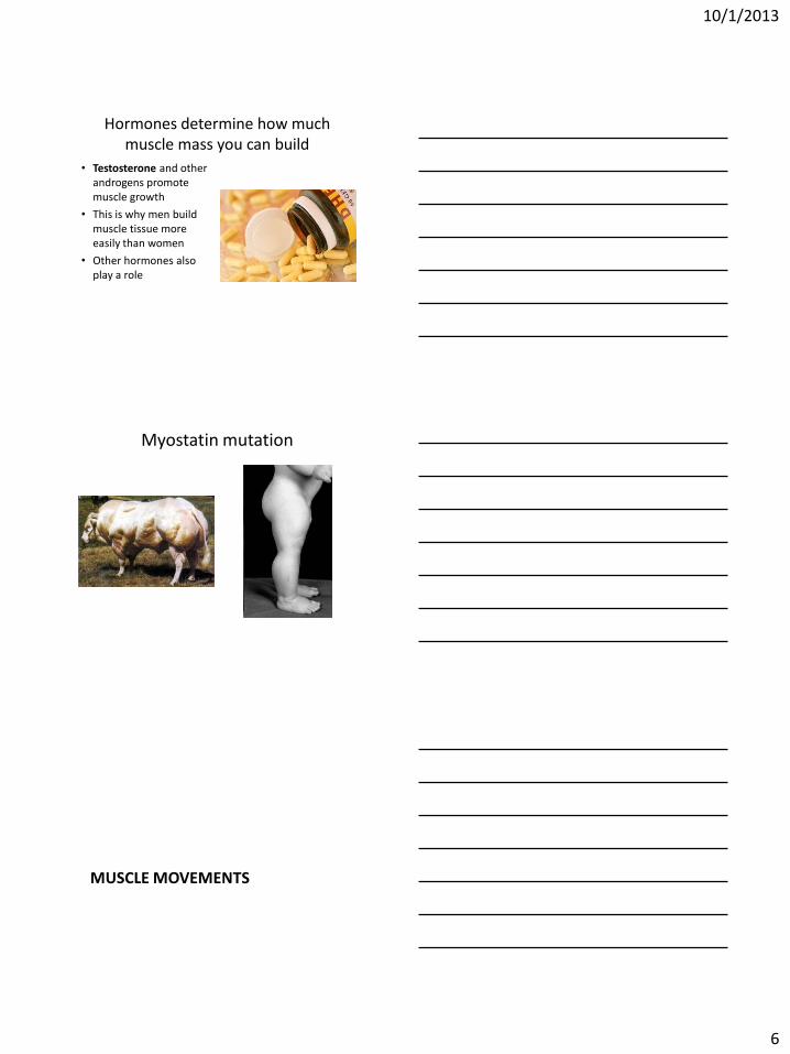
Hormones determine how much muscle mass you can build
• Testosterone and other androgens promote muscle growth
• This is why men build muscle tissue more easily than women
• Other hormones also play a role
Myostatin mutation
MUSCLE MOVEMENTS

10/1/2013
7
Muscle attachments
• All skeletal muscles have two or more connections to bone or other connective tissue
• The point of connection to the less movable bone is called the origin; the point to the more movable bone is called the insertion
Muscle movement
• All muscle movements are the result of muscles contracting
• Opposing muscle groups move limbs in opposite directions
• Flexors are muscles that bring two bones closer together when contracted (example: biceps)
• Extensors are muscles that bring two bones further apart when contracted (example: triceps)
• When the biceps contracts, the triceps relaxes, and vice versa
Flexion & extension

10/1/2013
8
Rotation
• Movement of a bone around its longitudinal axis
• Common for ball-and-socket joints
Abduction and Adduction
• Abduction moves a limb away from the midline
• Adduction is the opposite
Special movements

10/1/2013
9
Special movements
GROSS ANATOMY OF SKELETAL MUSCLES
Muscles of the head & neck • Facial muscles include:
– Frontalis: allows you to raise eyebrows & wrinkle forehead
– Orbicularis Oculi: allows you to close eyes & squint
– Orbicularis Oris: the “kissing muscle”
– Buccinator: flattens the cheek & helps with chewing
– Zygomaticus: the “smiling muscle”

10/1/2013
10
Muscles of the head & neck • Chewing muscles include:
– Masseter: closes the jaw by elevating the mandible
– Temporalis: helps the masseter close the jaw
• Neck muscles include:
– Platysma: helps pull the corners of the mouth down
– Sternocleidomastoids: move the neck
Muscles of the anterior trunk
• Pectoralis major: adducts and flexes the arm
• Intercostal muscles (not shown): found between the ribs and help expand the ribcage for breathing
Abdominal muscles
• Rectus abdominus: flex the vertebral column & compress abdominal contents during childbirth & defecation
• External obliques & internal obliques: Allow rotation and lateral bending of the trunk
• Transversus abdominus: compresses abdominal contents; deeper than other abdominal muscles
10/1/2013
11
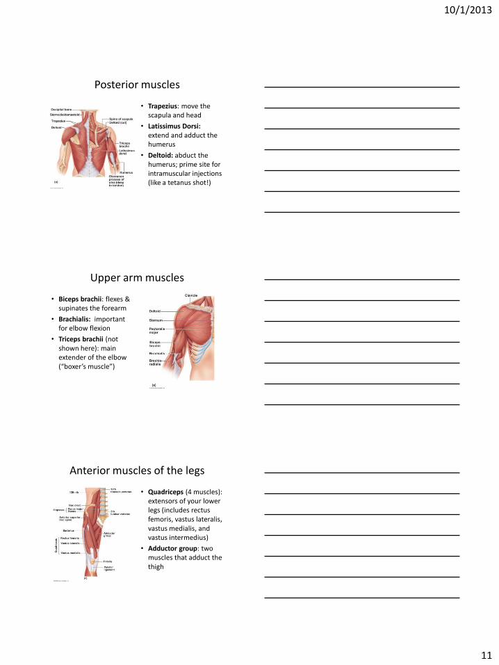
Posterior muscles
• Trapezius: move the scapula and head
• Latissimus Dorsi: extend and adduct the humerus
• Deltoid: abduct the humerus; prime site for intramuscular injections (like a tetanus shot!)
Upper arm muscles
• Biceps brachii: flexes & supinates the forearm
• Brachialis: important for elbow flexion
• Triceps brachii (not shown here): main extender of the elbow (“boxer’s muscle”)
Anterior muscles of the legs
• Quadriceps (4 muscles): extensors of your lower legs (includes rectus femoris, vastus lateralis, vastus medialis, and vastus intermedius)
• Adductor group: two muscles that adduct the thigh

10/1/2013
12
Posterior muscles of the legs • Gluteus maximus & gluteus
minimus help us stand upright and extending the leg, as in climbing
• Hamstring group: 3 muscles that control thigh extension and knee flexion (includes biceps femoris, semitendinosus & semimembranosus)
• Gastrocnemius: responsible for plantar flexion of the foot (“toe dancer’s muscle”)