chapter 33-10-06 x-rays in the healing arts
TRANSCRIPT

CHAPTER 33-10-06X-RAYS IN THE HEALING ARTS
Section33-10-06-01 Scope33-10-06-02 Definitions33-10-06-03 General Requirements33-10-06-04 General Requirements for All Diagnostic X-Ray Systems33-10-06-05 Fluoroscopic X-Ray Systems33-10-06-06 Radiographic Systems Other Than Fluoroscopic, Dental Intraoral, Bone Densitometry
or Computed Tomography X-Ray Systems33-10-06-07 Intraoral Dental Radiographic Systems33-10-06-08 Therapeutic X-Ray Systems of Less Than One Megaelectronvolt (MeV) [Repealed]33-10-06-09 X-Ray and Electron Therapy Systems With Energies of One Megaelectronvolt (MeV)
and Above [Repealed]33-10-06-10 Veterinary Medicine Radiographic Installations [Repealed]33-10-06-11 Computed Tomography X-Ray Systems33-10-06-12 Bone Densitometry
33-10-06-01. Scope.
This chapter establishes requirements, for which a registrant is responsible, for use of x-ray equipment and imaging systems by or under the supervision of an individual authorized by and licensed in accordance with state statutes to engage in the healing arts or veterinary medicine. The requirements of this chapter are in addition to, and not in substitution for, other applicable requirements of this article.
History: Amended effective June 1, 1986; June 1, 1992; March 1, 2003.General Authority: NDCC 23-20.1-04Law Implemented: NDCC 23-20.1-03, 23-20.1-04
33-10-06-02. Definitions.
As used in this chapter, the following definitions apply:
1. "Absorbed dose" means the energy imparted by ionizing radiation per unit mass of irradiated material. The units of absorbed dose are the gray (Gy) and the rad.
2. "Accessible surface" means surface of equipment or of an equipment part that can be touched by persons without the use of a tool.
3. "Added filtration" means any filtration which is in addition to the inherent filtration.
4. "Air kerma" means kerma in air (see "kerma").
5. "Air kerma rate" means the air kerma per unit of time.
6. "AKR" means air kerma rate.
7. "Allied health" means occupations of medical personnel who are not physicians and are qualified by special training to undergo cross-training into x-ray as a limited x-ray machine operator. Refer to appendix G for qualifying professions.
8. "Aluminum equivalent" means the thickness of type 1100 aluminum alloy affording the same attenuation, under specified conditions, as the material in question. (The nominal chemical composition of type 1100 aluminum alloy is ninety-nine percent minimum aluminum, twelve-hundredths percent copper.)
1

9. "As low as reasonably achievable" (ALARA) means making every reasonable effort to maintain the exposures to radiation as far below dose limits as is practicable.
10. "Assembler" means any person engaged in the business of assembling, replacing, or installing one or more components into an x-ray system or subsystem. The term includes the owner of an x-ray system or the employee or agent who assembles components into an x-ray system that is subsequently used to provide professional or commercial services.
11. "Attenuation block" means a block or stack, having dimensions twenty centimeters by twenty centimeters by three and eight-tenths centimeters, of type 1100 aluminum alloy or other materials having equivalent attenuation.
12. "Automatic exposure control" means a device which automatically controls one or more technique factors in order to obtain at a preselected location or locations a required quantity of radiation (includes devices such as phototimers and ion chambers).
13. "Barrier" (see "protective barrier").
14. "Beam axis" means the axis of rotation of the beam-limiting device from the source through the centers of the x-ray fields.
15. "Beam-limiting device" means a device which provides a means to restrict the dimensions of the useful beam.
16. "Biennium" means a two-year cycle.
17. "Board certified" means an individual who has completed an accredited school of medical radiography or chiropractic radiography and has passed a national registry examination.
18. "Board eligible" means an individual who has obtained eligibility to take a national registry examination in radiologic technology or chiropractic radiologic technology.
19. "Bone densitometry system" means a medical device which uses electronically produced ionizing radiation to determine the density of bone structures of human patients.
20. "Calibration" means the determination of:
a. The response or reading of an instrument relative to a series of known radiation values over the range of the instrument; or
b. The strength of a source of radiation relative to a standard.
21. "C-arm x-ray system" means an x-ray system in which the image receptor and x-ray tube housing assembly are connected by a common mechanical support system in order to maintain a desired spatial relationship. This system is designed to allow a change in the projection of the beam through the patient without a change in the position of the patient.
22. "Cephalometric device" means a device intended for the radiographic visualization and measurement of the dimensions of the human head.
23. "Certified components" means components of x-ray systems which are subject to regulations promulgated under the Radiation Control for Health and Safety Act of 1968 [Pub. L. 90-602].
24. "Certified system" means any x-ray system which has one or more certified component or components.
25. "CEU" (see "continuing education unit").
2
26. "CFR" means Code of Federal Regulations.
27. "Changeable filters" means any filter, exclusive of inherent filtration, which can be removed from the useful beam through any electronic, mechanical, or physical process.
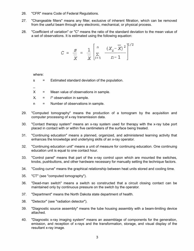
28. "Coefficient of variation" or "C" means the ratio of the standard deviation to the mean value of a set of observations. It is estimated using the following equation:
where:
s = Estimated standard deviation of the population.
_
X = Mean value of observations in sample.
Xi = ith observation in sample.
n = Number of observations in sample.
29. "Computed tomography" means the production of a tomogram by the acquisition and computer processing of x-ray transmission data.
30. "Contact therapy system" means an x-ray system used for therapy with the x-ray tube port placed in contact with or within five centimeters of the surface being treated.
31. "Continuing education" means a planned, organized, and administered learning activity that enhances the knowledge and underlying skills of an x-ray operator.
32. "Continuing education unit" means a unit of measure for continuing education. One continuing education unit is equal to one contact hour.
33. "Control panel" means that part of the x-ray control upon which are mounted the switches, knobs, pushbuttons, and other hardware necessary for manually setting the technique factors.
34. "Cooling curve" means the graphical relationship between heat units stored and cooling time.
35. "CT" (see "computed tomography").
36. "Dead-man switch" means a switch so constructed that a circuit closing contact can be maintained only by continuous pressure on the switch by the operator.
37. "Department" means the North Dakota state department of health.
38. "Detector" (see "radiation detector").
39. "Diagnostic source assembly" means the tube housing assembly with a beam-limiting device attached.
40. "Diagnostic x-ray imaging system" means an assemblage of components for the generation, emission, and reception of x-rays and the transformation, storage, and visual display of the resultant x-ray image.
3
41. "Diagnostic x-ray system" means an x-ray system designed for irradiation of any part of the human or animal body for the purpose of diagnosis or visualization.
42. "Direct scattered radiation" means that scattered radiation which has been deviated in direction only by materials irradiated by the useful beam (see "scattered radiation").
43. "Direct supervision" requires direct observation and observer must be in the room during the time the x-ray image is obtained.
44. "Dose" is a generic term that means absorbed dose, dose equivalent, effective dose equivalent, committed dose equivalent, committed effective dose equivalent, total organ dose equivalent, or total effective dose equivalent. For purposes of these rules, "radiation dose" is an equivalent term.
45. "Dose limits" means the permissible upper bounds of radiation of doses established in accordance with these rules.
46. "Entrance exposure rate" means the radiation exposure free in air per unit time at the point where the center of the useful beam enters the patient.
47. "Equipment" (see "x-ray equipment").
48. "Exposure" means being exposed to ionizing radiation.
49. "Field emission equipment" means equipment which uses an x-ray tube in which electron emission from the cathode is due solely to the action of an electric field.
50. "Filter" means material placed in the useful beam to absorb preferentially selected radiations.
51. "Fluoroscopic imaging assembly" means a subsystem in which x-ray photons produce a visible image. It includes the image receptor or receptors such as the image intensifier and spot-film device, electrical interlocks, if any, and structural material providing linkage between the image receptor and diagnostic source assembly.
52. "Focal spot" means the location projected on the anode of the x-ray tube bombarded by the electrons accelerated from the cathode and from which the useful beam originates.
53. "General diagnostic operator" means an individual who is American registry of radiologic technologists (ARRT) or American chiropractic registry of radiologic technologists (ACRRT) board-certified, is or has been board-eligible, or has the equivalent educational and clinical training and received specific authorization from the department.
54. "General purpose radiographic x-ray system" means any radiographic x-ray system which, by design, is not limited to radiographic examination of specific anatomical regions.
55. "Gonad shield" means a protective barrier for the testes or ovaries.
56. "Gray" (Gy) means the SI unit of absorbed dose. One gray is equal to an absorbed does of one joule per kilogram or 100 rad.
57. "Half-value layer" means the thickness of specified material which attenuates x-radiation or gamma radiation to an extent such that the air kerma rate, exposure rate or absorbed dose rate is reduced to one-half of the value measured without the material at the same point.
58. "Hand-held dental x-ray equipment" (see "x-ray equipment").
59. "Healing arts" means diagnostic or healing treatment of human and animal maladies, including the following which are duly licensed by the state of North Dakota for the lawful practice of
4
medicine and its associated specialties, dentistry, veterinary medicine, osteopathy, chiropractic, and podiatry.
60. "Healing arts screening" means the testing of human beings using x-ray machines for the detection or evaluation of health indications when such tests are not specifically and individually ordered by a licensed practitioner of the healing arts legally authorized to prescribe such x-ray tests for the purpose of diagnosis or treatment.
61. "Heat unit" means a unit of energy equal to the product of the peak kilovoltage, milliamperes, and seconds, i.e., kVp x mA x seconds.
62. "HVL" (see "half-value layer").
63. "Image intensifier" means a device, installed in its housing, which instantaneously converts an x-ray pattern into a corresponding light image of higher energy density.
64. "Image receptor" means any device, such as a fluorescent screen or radiographic film, which transforms incident x-ray photons either into a visible image or into another form which can be made into a visible image by further transformations.
65. "Image receptor support" means, for mammographic systems, that part of the system designed to support the image receptor during a mammographic examination.
66. "Inherent filtration" means the filtration of the useful beam provided by the permanently installed components of the tube housing assembly.
67. "Inspection" means an official examination or observation, including tests, surveys, and monitoring to determine compliance with rules, regulations, requirements, and conditions of the department.
68. "Irradiation" means the exposure of matter to ionizing radiation.
69. "Kerma" means the sum of the initial kinetic energies of all the charged ionizing particles liberated by uncharged ionizing particles per unit mass of a specified material. The SI unit of measure is joule per kilogram, or gray (Gy).
70. "Kilovolts peak" (see "peak tube potential").
71. "kV" means kilovolts.
72. "kVp" (see "peak tube potential").
73. "kWs" means kilowatt second. It is equivalent to 103 kV . mA . s, i.e.,
kWs = (X)kV x (Y)mA x (Z)s x 10-3 = XYZ
103
74. "Lead equivalent" means the thickness of lead affording the same attenuation, under specified conditions, as the material in question.
75. "Leakage radiation" means radiation emanating from the diagnostic or therapeutic source assembly except for:
a. The useful beam; and
b. Radiation produced when the exposure switch or timer is not activated.
5

76. "Leakage technique factors" means the technique factors associated with the diagnostic or therapeutic assembly which are used in measuring leakage radiation. They are defined as follows:
a. For diagnostic source assemblies intended for capacitor energy storage equipment, the maximum-rated peak tube potential and the maximum-rated number of exposures in an hour for operation at the maximum-rated peak tube potential with the quantity of charge per exposure being ten millicoulombs, i.e., ten milliampere seconds, or the minimum obtainable from the unit, whichever is larger.
b. For diagnostic source assemblies intended for field emission equipment rated for pulsed operation, the maximum-rated peak tube potential and the maximum-rated number of x-ray pulses in an hour for operation at the maximum-rated peak tube potential.
c. For all other diagnostic or therapeutic source assemblies, the maximum-rated peak tube potential and the maximum-rated continuous tube current for the maximum-rated peak tube potential.
77. "Light field" means the area illuminated by light, simulating the radiation field.
78. "Limited x-ray machine operator" means any individual who has completed the necessary didactic and clinical training required to perform limited scope x-ray procedures.
79. "Linear attenuation coefficient" or "u" means the quotient of dN/N divided by dl when dN/N is the fraction of uncharged ionizing radiation that experience interactions in traversing a distance dl in a specified material.
80. "mA" means milliampere.
81. "mAs" means milliampere second.
82. "Milliampere" as used in this chapter applies to x-ray tube current.
83. "Milliampere second" as used in this chapter is the product of the tube current and x-ray exposure time measured in seconds.
84. "Mobile x-ray equipment" (see "x-ray equipment").
85. "Monitoring" means the measurement of radiation and the use of the measured results to evaluate potential exposures and doses.
86. "Patient" means an individual or animal subjected to radiation for the purposes of diagnosis or treatment.
87. "PBL" has the same meaning as "positive beam limitation".
88. "Peak tube potential" means the maximum value of the potential difference across the x-ray tube during an exposure.
89. "Phantom" means a volume of material behaving in a manner similar to tissue with respect to the absorption and scattering of the ionizing radiation in question.
90. "Phototimer" means a method for controlling radiation exposures to image receptors by the amount of radiation which reaches a radiation monitoring device. The radiation monitoring device is part of an electronic circuit which controls the duration of time the tube is activated (see "automatic exposure control").
6
91. "Physician" means a medical doctor, doctor of osteopathy, doctor of podiatry, or chiropractor licensed by a state or territory of the United States, the District of Columbia, or the Commonwealth of Puerto Rico.
92. "Portable x-ray equipment" (see "x-ray equipment").
93. "Position indicating device" means a device on dental x-ray equipment used to indicate the beam position and to establish a definite source-surface (skin) distance. It may or may not incorporate or serve as a beam-limiting device.
94. "Positive beam limitation" means the automatic or semiautomatic adjustment of an x-ray beam to the size of the selected image receptor, whereby exposures cannot be made without such adjustment.
95. "Primary dose monitoring system" means a system which will monitor the useful beam during irradiation and which will terminate irradiation when a preselected number of dose monitor units have been delivered.
96. "Primary protective barrier" (see "protective barrier").
97. "Protective apron" means an apron made of radiation attenuating materials used to reduce radiation exposure.
98. "Protective barrier" means a barrier of radiation absorbing material or materials used to reduce radiation exposure. The types of protective barriers are as follows:
a. "Primary protective barrier" means the material, excluding filters, placed in the useful beam; and
b. "Secondary protective barrier" means the material which attenuates stray radiation.
99. "Protective glove" means a glove made of radiation absorbing materials used to reduce radiation exposure.
100. "Qualified expert" means an individual having the knowledge, training, and experience to measure ionizing radiation, to evaluate safety techniques, and to advise regarding radiation protection needs, for example, individuals certified in the appropriate field by the American board of radiology, or the American board of health physics, or the American board of medical physics, or those having equivalent qualifications. With reference to the calibration of radiation therapy equipment, "qualified expert" means an individual having, in addition to the above qualifications, training and experience in the clinical applications of radiation physics to radiation therapy, for example, individuals certified in therapeutic radiological physics or x-ray and radium physics by the American board of radiology, or those having equivalent qualifications.
101. "Radiation" means x-rays and gamma rays, which are capable of producing ions. For purposes of this chapter, ionizing radiation is an equivalent term.
102. "Radiation detector" means a device which in the presence of radiation provides a signal or other indication suitable for use in measuring one or more quantities of incident radiation.
103. "Radiation exposure" means the quotient of dQ by dm where "dQ" is the absolute value of the total charge of the ions of one sign produced in air when all the electrons (negatrons and positrons) liberated by photons in a volume element of air having mass "dm" are completely stopped in air. The SI unit of radiation exposure is the coulomb per kilogram (C/kg). (See section 33-10-01-14 units of exposure, dose, and activity for the special unit equivalent "roentgen" (R).)
7
104. "Radiation exposure rate" means the radiation exposure per unit of time, such as R/min, mR/h, etc.
105. "Radiation machine" means any device capable of producing radiation except those devices with radioactive material as the only source of radiation.
106. "Radiation therapy simulation system" means a radiographic, fluoroscopic, or computed tomography x-ray system intended for localizing the volume to be exposed during radiation therapy and confirming the position and size of the therapeutic irradiation field.
107. "Radiograph" means an image receptor on which the image is created directly or indirectly by an x-ray pattern and results in a permanent record.
108. "Radiographic imaging system" means any system whereby a permanent or temporary image is recorded on an image receptor by the action of ionizing radiation.
109. "Radiological physicist" means an individual who:
a. Is certified by the American board of radiology in therapeutic radiological physics, radiological physics, or x-ray and gamma-ray physics;
b. Has a bachelor's degree in one of the physical sciences or engineering and three year's full-time experience working in therapeutic or diagnostic radiological physics under the direction of a physicist certified by the American board of radiology. The work duties must include duties involving the calibration and spot checks of a medical accelerator or a sealed source teletherapy unit; or
c. Has a master's or a doctor's degree in physics, biophysics, radiological physics, health physics, or engineering; has had one year's full-time training in therapeutic or diagnostic radiological physics; and has had one year's full-time work experience in a radiotherapy facility where the individual's duties involve calibration and spot checks of a medical accelerator or a sealed source teletherapy unit.
110. "Rating" means the operating limits as specified by the component manufacturer.
111. "Recording" means producing a permanent form of an image resulting from x-ray photons.
112. "Registrant" means any person, group, or facility who is registered with the department and is legally obligated to register with the department pursuant to North Dakota Century Code chapter 23-20.
113. "Roentgen" or "(R)" means the special unit of exposure. One roentgen equals two hundred fifty-eight millionth of a coulomb per kilograph of air.
114. "Scattered radiation" means radiation that, during passage through matter, has been deviated in direction (see "direct scattered radiation").
115. "Secondary dose monitoring system" means a system which will terminate irradiation in the event of failure of the primary dose monitoring system.
116. "Secondary protective barrier" (see "protective barrier").
117. "Shutter" means a device attached to the tube housing assembly which can intercept the entire cross-sectional area of the useful beam and which has a lead equivalency not less than that of the tube housing assembly.
118. "SI" means the abbreviation for the international system of units.
8

119. "SID" has the same meaning as "source-image receptor distance".
120. "Source" means the location or material, or both, from which the radiation emanates.
121. "Source-image receptor distance" means the distance from the source to the center of the input surface of the image receptor.
122. "Spot check" means a procedure which is performed to assure that a previous calibration continues to be valid.
123. "Spot film" means a radiograph which is made during a fluoroscopic examination to permanently record conditions which exist during that fluoroscopic procedure.
124. "Spot-film device" means a device intended to transport or position a radiographic image receptor between the x-ray source and fluoroscopic image receptor. It includes a device intended to hold a cassette over the input end of an image intensifier for the purpose of making a radiograph.
125. "Stationary x-ray equipment" (see "x-ray equipment").
126. "Stray radiation" means the sum of leakage and scattered radiation.
127. "Survey" means an evaluation of the radiological conditions which may include tests, physical examinations, and measurements of levels of radiation.
128. "Technique factors" means the conditions of operation. They are specified as follows:
a. For capacitor energy storage equipment, peak tube potential in kilovolts and quantity of charge in milliampere second.
b. For field emission equipment rated for pulsed operation, peak tube potential in kilovolts and number of x-ray pulses.
c. For CT x-ray systems designed for pulsed operation, peak tube potential in kilovolts, scan time in seconds, and either tube current in milliampere, x-ray pulse width in seconds, and the number of x-ray pulses per scan, or the product of tube current, x-ray pulse width, and the number of x-ray pulses in milliampere second.
d. For CT x-ray systems not designed for pulsed operation, peak tube potential in kilovolts, and either tube current in milliampere and scan time in seconds, or the product of tube current and exposure time in milliampere second and the scan time when the scan time and exposure time are equivalent.
e. For all other equipment, peak tube potential in kilovolt and either tube current in milliampere and exposure time in seconds, or the product of tube current and exposure time in milliampere second.
129. "Termination of irradiation" means the stopping of irradiation in a fashion which will not permit continuance of irradiation without the resetting of operating conditions at the control panel.
130. "Tomogram" means the depiction of x-ray attenuation properties of a section through the body.
131. "Traceable to a national standard" means that a quantity or a measurement has been compared to a national standard directly or indirectly through one or more intermediate steps and that all comparisons have been documented.
132. "Tube" means an x-ray tube, unless otherwise specified.
9
133. "Tube housing assembly" means the tube housing with tube installed. It includes high-voltage and/or filament transformers and other appropriate elements when such are contained within the tube housing.
134. "Tube rating chart" means the set of curves which specify the rated limits of operation of the tube in terms of the technique factors.
135. "Useful beam" means the radiation emanating from the tube housing port or the radiation head and passing through the aperture of the beam-limiting device when the exposure controls are in a mode to cause the system to produce radiation.
136. "Variable-aperture beam-limiting device" means a beam-limiting device which has capacity for stepless adjustment of the x-ray field size at a given source-image receptor distance.
137. "Visible area" means that portion of the input surface of the image receptor over which incident x-ray photons are producing a visible image.
138. "Wedge filter" means an added filter effecting continuous progressive attenuation on all or part of the useful beam.
139. "Whole body" means for purposes of external exposure, head, trunk including male gonads, arms above the elbows, or legs below the knees.
140. "X-ray exposure control" means a device, switch, button, or other similar means by which the operator initiates or terminates, or both, the radiation exposure. It may include equipment such as timers, phototimers, automatic brightness stabilizers, and similar devices.
141. "X-ray equipment" means an x-ray system, subsystem, or component thereof. Types of x-ray equipment are as follows:
a. "Mobile x-ray equipment" means x-ray equipment mounted on a permanent base with wheels or casters for moving while completely assembled.
b. "Portable x-ray equipment" means x-ray equipment designed to be hand-carried.
c. "Stationary x-ray equipment" means x-ray equipment which is installed in a fixed location.
d. "Hand-held dental x-ray equipment" means any dental x-ray equipment which is designated to be physically held during x-ray exposure.
142. "X-ray field" means that area of the intersection of the useful beam and any one of the set of planes parallel to and including the plane of the image receptor, whose perimeter is the locus of points at which the radiation exposure rate is one-fourth of the maximum in the intersection.
143. "X-ray high-voltage generator" means a device which transforms electrical energy from the potential supplied by the x-ray control to the tube operating potential. The device may also include means for transforming alternating current to direct current, filament transformers for the x-ray tube, high-voltage switches, electrical protective devices, and other appropriate elements.
144. "X-ray system" means an assemblage of components for the controlled production of x-rays. It includes minimally an x-ray high-voltage generator, an x-ray control, a tube housing assembly, a beam-limiting device, and the necessary supporting structures. Additional components which function with the system are considered integral parts of the system.
145. "X-ray tube" means any electron tube which is designed for the conversion of electrical energy into x-ray energy.
10

History: Amended effective October 1, 1982; June 1, 1986; June 1, 1992; July 1, 1995; May 1, 1998; March 1, 2003; January 1, 2011. General Authority: NDCC 23-20.1-04Law Implemented: NDCC 23-20.1-03, 23-20.1-04
33-10-06-03. General requirements.
1. Administrative controls.
a. Registrant. The registrant shall be responsible for directing the operation of the x-ray systems which have been registered with the department. The registrant or the registrant's agent shall assure that the requirements are met in the operation of the x-ray system.
(1) An x-ray system which does not meet the requirements of this article shall not be operated for diagnostic or therapeutic purposes.
(2) All individuals, except those listed in part 1 of appendix G, prior to operating the x-ray systems shall be adequately instructed in the safe operating procedures and be competent in the safe use of the equipment commensurate with the size, scope, and nature of the service as outlined in appendix F. In addition, all individuals shall meet the specific requirements as outlined in subparagraph a or b. The department may use interview, observation, or testing to determine compliance. Records must be maintained by the registrant to demonstrate compliance with this paragraph.
(a) General diagnostic operators are not limited in scope of practice. Obtaining general diagnostic operator status will consist of one of the following:
[1] Obtain board eligibility or board certification with the American registry of radiologic technologists (ARRT);
[2] Obtain board eligibility or board certification with the American chiropractic registry of radiologic technologists (ACRRT); and only perform x-ray examinations for chiropractic services;
[3] Receive department approval, through individual consideration, by demonstration of an acceptable level of education and clinical training; or
[4] Demonstrate current enrollment in an educational program accredited by a process acceptable to the department, and provide documentation of competency in all routine radiographic procedures and specialty views.
(b) Limited x-ray machine operators are limited in scope of practice to only those procedures listed in appendix I, except as allowed in subparagraph c. Limited x-ray machine operators must meet the prerequisite qualifications, receive training, and demonstrate competence as follows:
[1] Limited x-ray machine operators shall have successfully completed the course of training required by one of the allied health professions listed in part 2 of appendix G;
[2] Complete at least eighty hours of didactic instruction at a single training program providing didactic instruction in accordance with part 1 of appendix H;
[3] Complete the three-hour self-study course designed by the North Dakota state department of health; and
11

[4] Complete the clinical experience requirements in part 2 of appendix H.
(c) Limited x-ray machine operators may only conduct diagnostic x-ray examinations outside the scope of practice of appendix I in accordance with the following:
[1] When it is determined to be an emergency and ordered by individuals listed in part 3 of appendix G. The individual requesting the procedures must comply with subitems a, b, and c.
[a] The requesting individual must provide a written order specifying what types of diagnostic x-ray examinations outside the scope of procedures listed in appendix I are requested. The order shall contain an explanation of the emergency nature or medical reason for the order.
[b] The requesting individual must provide direct supervision during the time the x-ray image is obtained.
[c] The facility must keep records of all emergency x-ray procedures ordered under this subparagraph.
[2] When a practice requires a specific view or examination outside the scope of practice listed in appendix I to be conducted on a routine basis, and the facility has only limited x-ray machine operators, application may be made to the department requesting approval for a limited x-ray machine operator to perform the procedure. This allowance shall be limited to the facility, the specific individual, and the procedure requested. After an allowance has been granted, reapplication and reauthorization are not necessary for the same procedure. The application for allowance should include the following:
[a] Documentation which demonstrates the need for the specific view;
[b] Documentation on forms supplied by the department indicating that each individual for which the request is made has demonstrated competence in the procedure; and
[c] Proof of additional didactic instruction or completion of examination as deemed necessary by the department for each individual.
[3] When it is not a computed tomography examination.
(d) Limited x-ray machine operator implementation period. Individuals who begin taking x-rays after one year from March 1, 2003, will have to meet all of the requirements of this paragraph before operating the x-ray system.
(3) General diagnostic and limited diagnostic x-ray operators shall maintain continuing education units as outlined in appendix K.
(4) A chart shall be provided in the vicinity of the diagnostic x-ray system's control panel, which specifies for all examinations performed with that system the following information:
(a) Patient's body part and anatomical size or body thickness, or age (for pediatrics), versus technique factors to be utilized.
12
(b) Type and size of the film or film-screen combination to be used.
(c) Type and focal distance of the grid to be used, if any.
(d) Source-image receptor distance to be used (except for dental intraoral radiography).
(e) Type and location of placement of gonad shielding to be used.
(f) For mammography, indication of kVp/target/filter combination.
(5) The registrant of a facility shall create and make available to x-ray operators written safety procedures, including patient holding restrictions and any restrictions of the operating technique required for the safe operation of the particular x-ray system. The operator shall be able to demonstrate familiarity with these procedures.
(6) Except for human patients who cannot be moved out of the room, only the staff and ancillary personnel or other persons required for the medical procedure or training shall be in the room during the radiographic exposure. Other than the patient being examined:
(a) All individuals shall be positioned such that no part of the body will be struck by the useful beam unless protected by not less than five-tenths millimeter lead equivalent material.
(b) The x-ray operator, other staff, ancillary personnel, and other persons required for the x-ray procedure shall be protected from the direct scatter radiation by protective aprons or whole body protective barriers of not less than twenty-five one-hundredths millimeter lead equivalent material.
(c) Human patients who cannot be removed from the room shall be protected from the direct scatter radiation by whole body protective barriers of not less than twenty-five one-hundredths millimeter lead equivalent material or shall be so positioned that the nearest portion of the body is at least two meters from both the tube head and the nearest edge of the image receptor.
(7) Gonad shielding of not less than five-tenths millimeter lead equivalent material must be used for human patients who have not passed the reproductive age during radiographic procedures in which the gonads are in the useful beam, except for cases in which this would interfere with the diagnostic procedure.
(8) Individuals may not be exposed to the useful beam except for healing arts purposes and when such exposure has been authorized by a licensed practitioner of the healing arts. This provision specifically prohibits deliberate exposure for the following purposes:
(a) Exposure of an individual for training, demonstration, or other non-healing-arts purposes.
(b) Exposure of an individual for the purpose of healing arts screening except as authorized by paragraph 12.
(9) When a patient or film must be provided with auxiliary support during a radiation exposure:
13

(a) Mechanical holding devices shall be used when the technique permits. The safety rules, required by this section shall list individual projections where holding devices cannot be utilized.
(b) Written safety procedures, as required by paragraph 4, shall indicate the requirements for selecting a holder and the procedure the holder shall follow.
(c) The human holder shall be instructed in personal radiation safety and protected as required by paragraph 6.
(d) No individual shall be used routinely to hold film or patients.
(e) In those cases where the patient must hold the film, except during intraoral examinations, any portion of the body other than the area of clinical interest struck by the useful beam shall be protected by not less than five-tenths millimeter lead equivalent material.
(f) A record shall be made of the examination and shall include the name of the human holder, date of the examination, number of exposures, and technique factors utilized for the exposure.
(g) Each facility shall have leaded aprons and gloves available in sufficient numbers to provide protection to all personnel who are involved with x-ray operations and who are otherwise not shielded.
(10) Procedures and auxiliary equipment designed to minimize patient and personnel exposure commensurate with the needed diagnostic information shall be utilized. This is interpreted to include but not limited to:
(a) The speed of film and screen combinations shall be the fastest speed consistent with the diagnostic objective of the examinations. Film cassettes without intensifying screens shall not be used for any routine diagnostic radiological imaging, with the exception of veterinary radiography, therapeutic portal imaging, and standard film packets for intraoral use in dental radiography.
(b) The radiation exposure to the patient shall be the minimum exposure required to produce images of good diagnostic quality.
(c) Proper film handling and processing procedures. Each installation using a radiographic x-ray system and using analog image receptors (e.g., radiographic film) shall have available suitable equipment for handling and processing radiographic film in accordance with appendix D.
(d) Portable or mobile equipment shall be used only for examinations where it is impractical to transfer the patients to a stationary x-ray installation.
(e) X-ray systems subject to section 33-10-06-06 shall not be utilized in procedures where the source to patient distance is less than thirty centimeters, except for veterinary systems.
(f) If grids are used between the patient and the image receptor to decrease scatter to the film and improve contrast, the grid shall:
[1] Be positioned properly, for example, tube side facing the right direction and grid centered to the central ray; and
14
[2] If the grid is of the focused type, be of the proper focal distance for the source image distances being used.
(11) All individuals who are associated with the operation of an x-ray system are subject to the requirements of section 33-10-04.2-01 [10 CFR 20.1201, 20.1207, and 20.1208]. In addition:
(a) When protective clothing or devices are worn on portions of the body and a monitoring device is required, at least one such monitoring device shall be utilized as follows:
[1] When an apron is worn, the monitoring device shall be worn at the collar outside of the apron.
[2] The dose to the whole body based on the maximum dose attributed to the most critical organ shall be recorded in the reports required by section 33-10-04.2-01 [10 CFR 20.2206]. If more than one device is used and a record is made of the data, each dose shall be identified with the area where the device was worn on the body.
(b) Exposure of a personnel monitoring device to deceptively indicate a dose delivered to an individual is prohibited.
(12) Healing arts screening. Any person proposing to conduct a healing arts screening program shall not initiate such a program without prior approval of the department. When requesting such approval, that person shall submit the information outlined in appendix E. If any information submitted to the department becomes invalid or outdated, the department shall be immediately notified.
b. Information and maintenance record and associated information. The registrant shall maintain the following information for each x-ray system for inspection by the department:
(1) Maximum rating of technique factors.
(2) Model and serial numbers of all major components and user's manuals for those components.
(3) Aluminum equivalent filtration of the useful beam, including any routine variation.
(4) Tube rating charts and cooling curves.
(5) Records of surveys, calibrations, maintenance, and modifications performed on the x-ray system with the names of persons who performed such services.
(6) A scale drawing of the room in which a stationary x-ray system is located with such drawing indicating the use of areas adjacent to the room and an estimation of the extent of occupancy by an individual in such areas. In addition, the drawing shall include:
(a) The results of a survey for radiation levels present at the operator's position and at pertinent points outside the room at specified test conditions; or
(b) The type and thickness of materials, or lead equivalency, of each protective barrier.
(7) A copy of all correspondence with this department regarding that x-ray system.
15
c. X-ray log.
(1) Except for veterinary facilities, each facility shall maintain an x-ray log containing the patient's name, the type of examinations, the dates those examinations were performed, and the name of the x-ray operator. When the patient or film must be provided with human auxiliary support, the name of the human holder shall be recorded.
(2) Veterinary facilities shall maintain an x-ray utilization log indicating the type of examinations, the date of the examinations and if the patient or film was provided with human auxiliary support, the name of the human holder.
2. Plan review.
a. Prior to construction, the floor plans, shielding specifications, and equipment arrangement of all new installations, or modifications of existing installations, utilizing ionizing radiation machines shall be submitted to the department for review and approval. The required information is denoted in appendices A, B, and C.
b. The department may require the applicant to utilize the services of a qualified expert to determine the shielding requirements prior to the plan review and approval.
c. The approval of such plans shall not preclude the requirement of additional modifications should a subsequent analysis of operating conditions indicate the possibility of an individual receiving a dose in excess of the limits prescribed in section 33-10-04.2-01 [10 CFR 20.1201, 20.1207, 20.1208, and 20.1301].
History: Amended effective October 1, 1982; June 1, 1986; June 1, 1992; March 1, 1994; July 1, 1995; May 1, 1998; March 1, 2003; January 1, 2011. General Authority: NDCC 23-20.1-04Law Implemented: NDCC 23-20.1-03, 23-20.1-04
33-10-06-04. General requirements for all diagnostic x-ray systems.
In addition to other requirements of this chapter, all diagnostic x-ray systems shall meet the following requirements:
1. Warning label. The control panel containing the main power switch shall bear the warning statement, legible and accessible to view: "WARNING: This x-ray unit may be dangerous to patient and operator unless safe exposure factors and operating instructions are observed."
2. Battery charge indicator. On battery-powered x-ray generators, visual means shall be provided on the control panel to indicate whether the battery is in a state of charge adequate for proper operation.
3. Leakage radiation from the diagnostic source assembly. The leakage radiation from the diagnostic source assembly measured at a distance of one meter in any direction from the source shall not exceed one hundred milliroentgens in one hour when the x-ray tube is operated at its leakage technique factors. Compliance shall be determined by measurements averaged over an area of one hundred square centimeters with no linear dimension greater than twenty centimeters.
4. Radiation from components other than the diagnostic source assembly. The radiation emitted by a component other than the diagnostic source assembly shall not exceed two milliroentgens in one hour at five centimeters from any accessible surface of the component when it is operated in an assembled x-ray system under any conditions for which it
16

was designed. Compliance shall be determined by measurements averaged over an area of one hundred square centimeters with no linear dimension greater than twenty centimeters.
5. Beam quality.
a. Half-value layer.
(1) The half-value layer (HVL) of the useful beam for a given x-ray tube potential shall not be less than the values shown in table I. If it is necessary to determine such half-value layer at an x-ray tube potential which is not listed in table I, linear interpolation or extrapolation may be made.
TABLE I
Half-Value Layer in Millimeters Aluminum
Design Operating
Range (Kilovolts Peak)
Measured Potential
(Kilovolts Peak)
Dental Intraoral Manufactured Before Aug. 1, 1974, and on
or After Dec. 1, 1980
Diagnostic X-Ray Systems Manufactured
Prior to June 10, 2006
Diagnostic X-Ray Systems Manufactured
on or After June 10, 2006
Below 51 30 N/A 0.3 0.3
40 N/A 0.4 0.4
50 1.5 0.5 0.5
51 to 70 51 1.5 1.2 1.3
60 1.5 1.3 1.5
70 1.5 1.5 1.8
Above 70 71 2.1 2.1 2.5
80 2.3 2.3 2.9
90 2.5 2.5 3.2
100 2.7 2.7 3.6
110 3.0 3.0 3.9
120 3.2 3.2 4.3
130 3.5 3.5 4.7
140 3.8 3.8 5.0
150 4.1 4.1 5.4
(2) For capacitor energy storage equipment, compliance with the requirements of this subsection shall be determined with the system fully charged and a setting of ten mAs for each exposure.
(3) The required minimal aluminum equivalent filtration shall include the filtration contributed by all materials which are permanently present between the source and the patient.
(4) For mammography systems with molybdenum filter and molybdenum target, measured half-value layer (HVL) with compression device in the x-ray beam shall
17

be greater than or equal to the kilovolts peak (kVp) divided by one hundred, millimeters aluminum and less than or equal to the kilovolts peak (kVp) divided by one hundred plus one-tenth millimeter aluminum.
HVL > (kVp/100) mmAl and < (kVp/100) + 0.1 mmAl
b. Filtration controls. For x-ray systems which have variable kilovolts peak and variable filtration for the useful beam, a device shall link the kilovolts peak selector with the filters and shall prevent an exposure unless the minimum amount of filtration required by paragraph 1 of subdivision a is in the useful beam for the given kilovolts peak which has been selected.
6. Multiple tubes. Where two or more radiographic tubes are controlled by one exposure switch, the tube or tubes which have been selected shall be clearly indicated prior to initiation of the exposure. This indication shall be both on the x-ray control panel and at or near the tube housing assembly which has been selected.
7. Mechanical support of tube head. The tube housing assembly supports shall be adjusted such that the tube housing assembly will remain stable during an exposure unless tube housing movement is a designed function of the x-ray system.
8. Technique indicators.
a. The technique factors to be used during an exposure shall be indicated before the exposure begins, except when automatic exposure controls are used, in which case the technique factors which are set prior to the exposure shall be indicated.
b. The requirements of subdivision a may be met by permanent markings on equipment having fixed technique factors. Indication of technique factors shall be visible from the operator's position except in the case of spot films made by the fluoroscopist.
9. Maintaining compliance. Diagnostic x-ray systems and their associated components used on humans and certified pursuant to the federal x-ray equipment performance standard (21 CFR part 1020) shall be maintained in compliance with applicable requirements of that standard.
10. Locks. All position locking, holding, and centering devices on x-ray system components and systems shall function as intended.
11. Structural shielding requirements (see appendix C).
History: Amended effective October 1, 1982; June 1, 1986; June 1, 1992; March 1, 1994; July 1, 1995; May 1, 1998; March 1, 2003; January 1, 2011. General Authority: NDCC 23-20.1-04Law Implemented: NDCC 23-20.1-03, 23-20.1-04
33-10-06-05. Fluoroscopic x-ray systems.
All fluoroscopic x-ray systems shall be image-intensified and meet the following requirements:
1. Limitation of useful beam.
a. Primary barrier.
(1) The fluoroscopic imaging assembly shall be provided with a primary protective barrier which intercepts the entire cross-section of the useful beam at any source-image receptor distance (SID).
18

(2) The x-ray tube used for fluoroscopy shall not produce x-rays unless the barrier is in position to intercept the entire useful beam.
b. X-ray field.
(1) For certified fluoroscopic systems with or without a spot-film device, neither the length nor the width of the x-ray field in the plane of the image receptor shall exceed that of the visible area of the image receptor by more than three percent of the source-image receptor distance. The sum of the excess length and the excess width shall be no greater than four percent of the source-image receptor distance.
(2) For uncertified fluoroscopic systems with a spot-film device, the x-ray beam with the shutters fully opened (during fluoroscopy or spot filming) shall be no larger than the largest spot-film size for which the device is designed. Measurements shall be made at the maximum source image distance available but at no less than twenty centimeters tabletop to the film plane distance.
(3) For uncertified fluoroscopic systems without a spot-film device, the requirements of paragraph 1 apply.
(4) Other requirements for fluoroscopic beam limitation:
(a) Means shall be provided to permit further limitation of the field. Beam-limiting devices manufactured after May 22, 1979, and incorporated in equipment with a variable source-image receptor distance and/or a visible area of greater than three hundred square centimeters shall be provided with means for stepless adjustment of the x-ray field.
(b) All equipment with a fixed source-image receptor distance and a visible area of three hundred square centimeters or less shall be provided with either stepless adjustment of the x-ray field or with means to further limit the x-ray field size at the plane of the image receptor to one hundred twenty-five square centimeters or less. Stepless adjustment shall, at the greatest source-image receptor distance, provide continuous field sizes from the maximum obtainable to a field size of five by five centimeters or less.
(c) For equipment manufactured after February 25, 1978, when the angle between the image receptor and beam axis is variable, means shall be provided to indicate when the axis of the x-ray beam is perpendicular to the plane of the image receptor.
(d) Compliance shall be determined with the beam axis indicated to be perpendicular to the plane of the image receptor. For noncircular x-ray fields used with circular image receptors, the error in alignment shall be determined along the length and width dimensions of the x-ray field which pass through the center of the visible area of the image receptor.
(5) Spot-film devices shall meet the following additional requirements:
(a) Means shall be provided between the source and the patient for adjustment of the x-ray field size in the plane of the film to the size of that portion of the film which has been selected on the spot-film selector. Such adjustment shall be automatically accomplished except when the x-ray field size in the plane of the film is smaller than that of the selected portion of the film. For spot-film devices manufactured after June 21, 1979, if the x-ray field size is less than the size of the selected portion of the film, the means for adjustment of the field size shall be only at the operator's option.
19
(b) Neither the length nor the width of the x-ray field in the plane of the image receptor shall differ from the corresponding dimensions of the selected portion of the image receptor by more than three percent of the source-image receptor distance when adjusted for full coverage of the selected portion of the image receptor. The sum, without regard to sign, of the length and width differences shall not exceed four percent of the source-image receptor distance.
(c) It shall be possible to adjust the x-ray field size in the plane of the film to a size smaller than the selected portion of the film. The minimum field size at the greatest source-image receptor distance shall be equal to, or less than, five centimeters by five centimeters.
(d) The center of the x-ray field in the plane of the film shall be aligned with the center of the selected portion of the film to within two percent of the source-image receptor distance.
(e) On spot-film devices manufactured after February 25, 1978, if the angle between the plane of the image receptor and beam axis is variable, means shall be provided to indicate when the axis of the x-ray beam is perpendicular to the plane of the image receptor, and compliance shall be determined with the beam axis indicated to be perpendicular to the plane of the image receptor.
(6) If a means exists to override any of the automatic x-ray field size adjustments required in subdivision b of subsection 1 that means:
(a) Must be designed for use only in the event of system failure.
(b) Must incorporate a signal visible at the fluoroscopist's position which will indicate whenever the automatic field size adjustment is overridden.
(c) Must be clearly and durably labeled as follows:
FOR X-RAY FIELDLIMITATION SYSTEM FAILURE
2. Activation of the fluoroscopic tube. X-ray production in the fluoroscopic mode shall be controlled by a device which requires continuous pressure by the fluoroscopist for the entire time of any exposure. When recording serial fluoroscopic images, the fluoroscopist shall be able to terminate the x-ray exposure or exposures at any time, but means may be provided to permit completion of any single exposure of the series in process.
3. Radiation exposure rate limits.
a. Entrance exposure rate allowable limits.
(1) Fluoroscopic equipment which is provided with automatic exposure rate control:
(a) The radiation exposure measured at the point where the center of the useful beam enters the patient shall not exceed two and fifty-eight hundredths millicoulomb per kilogram [10 roentgens] per minute, except during recording of fluoroscopic images or when provided with optional high level control.
(b) When provided with optional high level control, the equipment shall not be operable at any combination of tube potential and current which will result in a radiation exposure rate in excess of one and twenty-nine hundredths millicoulomb per kilogram [5 roentgens] per minute at the point where the
20
center of the useful beam enters the patient unless the high level control is activated.
[1] When the high level control is activated, the equipment shall not be operable at any combination of tube potential and current that will result in an exposure rate in excess of five and sixteen hundredths millicoulomb per kilogram [20 roentgens] per minute at the point where the center of the useful beam enters the patient.
[2] Special means of activation of high level controls shall be required. The high level control shall only be operable when continuous manual activation is provided by the operator.
[3] A continuous signal audible to the fluoroscopist shall indicate that the high level control is being employed.
(2) Fluoroscopic equipment which is not provided with automatic exposure rate control:
(a) The radiation exposure measured at the point where the center of the useful beam enters the patient shall not exceed one and twenty-nine hundredths millicoulomb per kilogram [5 roentgens] per minute, except during recording of fluoroscopic images or when provided with an optional high level control and the high level control is activated.
[1] When the high level control is activated, the equipment shall not be operable at any combination of tube potential and current that will result in an exposure rate in excess of five and sixteen hundredths millicoulomb per kilogram [20 roentgens] per minute at the point where the center of the useful beam enters the patient.
[2] Special means of activation of high level controls shall be required. The high level control shall only be operable when continuous manual activation is provided by the operator.
[3] A continuous signal audible to the fluoroscopist shall indicate that the high level control is being employed.
(3) Compliance with the requirements of subsection 3 of this section shall be determined as follows:
(a) Movable grids and compression devices shall be removed from the useful beam during the measurement.
(b) If the source is below the table, the radiation exposure rate shall be measured one centimeter above the tabletop or cradle.
(c) If the source is above the table, the radiation exposure rate shall be measured at thirty centimeters above the tabletop with the end of the beam-limiting device or spacer positioned as closely as possible to the point of measurement.
(d) In a C-arm type of fluoroscope, both stationary and mobile units shall meet the entrance exposure rate limits specified in paragraphs 1, 2, and 3 of subdivision a of subsection 3, shall be measured thirty centimeters from the input surface of the fluoroscopic imaging assembly with the source positioned at any available source-image receptor distance provided that the end of the
21

spacer assembly or beam-limiting device is not closer than thirty centimeters from the input surface of the fluoroscopic imaging assembly.
(e) In a lateral type of fluoroscope, the exposure rate shall be measured at a point fifteen centimeters from the centerline of the x-ray table and in the direction of the x-ray source with the end of the beam-limiting device or spacer positioned as closely as possible to the point of measurement. If the tabletop is movable, it shall be positioned as closely as possible to the lateral x-ray source, with the end of the beam-limiting device or spacer no closer than fifteen centimeters to the centerline of the x-ray table.
(4) Periodic measurement of entrance exposure rate shall be performed by a qualified expert for both typical and maximum values as follows:
(a) Such measurements shall be made annually or after any maintenance of the system which might affect the radiation exposure rate.
(b) Results of these measurements shall be posted where any fluoroscopist may have ready access to such results while using the fluoroscope and in the record required in paragraph 5 of subdivision b of subsection 1 of section 33-10-06-03. Results of the measurements shall include the roentgen per minute, as well as the technique factors used to determine such results. The name of the person performing the measurements and the date the measurements were performed shall be included in the results.
(c) Conditions of periodic measurements of typical entrance exposure rate are as follows:
[1] The measurement shall be made under the conditions that satisfy the requirements of paragraph 4.
[2] The kilovolts peak, mA, and other selectable parameters shall be the settings typical of clinical use on a twenty-three centimeters thick abdominal patient.
[3] The x-ray systems that incorporate automatic exposure control shall have sufficient material placed in the useful beam to produce a milliamperage or kilovoltage, or both, to satisfy the conditions of item 2.
[4] X-ray systems that do not incorporate an automatic exposure control shall utilize a milliamperage typical of clinical use of the x-ray system. Materials should be placed in the useful beam when conducting these periodic measurements to protect the imaging system.
(d) Conditions of periodic measurements of maximum entrance exposure rate are as follows:
[1] The measurement shall be made under the conditions that satisfy the requirements of paragraph 3.
[2] The kVp, mA, and other selectable parameters shall be the maximum selectable parameters of clinical use of the x-ray system.
[3] The x-ray systems that incorporate automatic exposure control shall have sufficient material placed in the useful beam to produce a kVp, mA, and other selectable parameters to satisfy the conditions of item 2.
22

[4] X-ray systems that do not incorporate an automatic exposure control shall utilize the maximum kVp, mA, and other selectable parameters of clinical use of the x-ray system. Materials should be placed in the useful beam when conducting these periodic measurements to protect the imaging system.
4. Barrier transmitted radiation rate limits.
a. The radiation exposure rate due to transmission through the primary protective barrier with the attenuation block in the useful beam, combined with radiation from the image intensifier, shall not exceed five hundred sixteen thousandths microcoulomb per kilogram [2 milliroentgens] per hour at ten centimeters from any accessible surface of the fluoroscopic imaging assembly beyond the plane of the image receptor for each roentgen (C/kg) per minute of entrance exposure rate.
b. Measuring compliance of barrier transmission.
(1) The radiation exposure rate due to transmission through the primary protective barrier combined with radiation from the image intensifier shall be determined by measurements averaged over an area of one hundred square centimeters with no linear dimension greater than twenty centimeters.
(2) If the source is below the tabletop, the measurement shall be made with the input surface of the fluoroscopic imaging assembly positioned thirty centimeters above the tabletop.
(3) If the source is above the tabletop and the source-image receptor distance is variable, the measurement shall be made with the end of the beam-limiting device or spacer as close to the tabletop as it can be placed, provided that it shall not be closer than thirty centimeters.
(4) Movable grids and compression devices shall be removed from the useful beam during the measurement.
5. Indication of potential and current. During fluoroscopy and cinefluorography, the kilovolt and the milliampere shall be continuously indicated.
6. Indication of air kerma rate and cumulative air kerma. Machines manufactured on or after June 10, 2006, shall provide displays of values of air kerma rate and cumulative air kerma and shall be viewable from the x-ray operator position.
a. When the x-ray tube is activated and the number of images produced per unit time is greater than six images per second, the air kerma rate in mGy/minute shall be continuously displayed and updated at least once every second.
b. The cumulative air kerma in units of mGy shall be displayed either within five seconds of termination of an exposure or displayed continuously and updated at least once every five seconds.
c. The display of the air kerma rate shall be clearly distinguishable from the display of the cumulative air kerma.
d. The air kerma rate and cumulative air kerma shall represent the value of conditions of free-in-air irradiation at one of the following reference locations specified according to the type of fluoroscope.
23
(1) For fluoroscopies with x-ray source below the x-ray table, x-ray source above the table, or of lateral type, the reference locations shall be the respective locations specified in subparagraphs 33-10-05.3(3)(b), (c), and (e) for measuring compliance with air kerma rate limits.
(2) For C-arm fluoroscopies, the reference location shall be fifteen cm from the isocenter toward the x-ray source along the beam axis. Alternatively, the reference location shall be at a point specified by the manufacturer to represent the location of the intersection of the x-ray beam with the patient’s skin.
e. Means shall be provided to reset to zero the display of cumulative air kerma prior to the commencement of a new examination or procedure.
f. The displayed air kerma rate and cumulative air kerma shall not deviate from the actual values by more than plus or minus thirty-five percent over the range of six mGy/minute (0.6 R/min) and one hundred mGy (10 R) to the maximum indication of air kerma rate and cumulative air kerma, respectively. Compliance shall be determined with an irradiation time greater than three seconds.
7. Source-skin distance. The source to skin distance shall not be less than:
a. Thirty-eight centimeters on stationary fluoroscopes installed after August 1, 1974.
b. Thirty-five and one-half centimeters on stationary fluoroscopes which were in operation prior to August 1, 1974.
c. Thirty centimeters on all mobile fluoroscopes.
d. Twenty centimeters for all mobile fluoroscopes used for specific surgical applications.
8. Fluoroscopic timer.
a. Means shall be provided to preset the cumulative on-time of the fluoroscopic tube. The maximum cumulative time of the timing device shall not exceed five minutes without resetting.
b. A signal audible to the fluoroscopist shall indicate the completion of any preset cumulative on-time. Such signal shall continue to sound while x-rays are produced until the timing device is reset.
9. Control of scattered radiation.
a. Fluoroscopic table designs when combined with procedures utilized shall be such that no unprotected part of any staff or ancillary individual's body shall be exposed to unattenuated scattered radiation which originates from under the table. The attenuation required shall be not less than twenty-five one-hundredths millimeter lead equivalent.
b. Equipment configuration when combined with procedures shall be such that no portion of any staff or ancillary individual's body, except the extremities, shall be exposed to the unattenuated scattered radiation emanating from above the tabletop unless that individual:
(1) Is at least one hundred twenty centimeters from the center of the useful beam; or
(2) The radiation has passed through not less than twenty-five one-hundredths millimeter lead equivalent material, including, but not limited to, drapes, bucky-slot cover-sliding or folding panel, or self-supporting curtains, in addition to any lead
24
equivalency provided by the protective apron referred to in paragraph 5 of subdivision a of subsection 1 of section 33-10-06-03.
c. The department may grant exceptions to subdivision b of this subsection in some special procedures where a sterile field will not permit the use of the normal protective barriers. Where the use of prefitted sterilized covers for the barriers is practical, the department shall not permit such exception.
10. Spot-film exposure reproducibility. Fluoroscopic systems equipped with spot-film mode shall meet the exposure reproducibility requirements of subsection 5 of section 33-10-06-06 when operating in the spot-film mode.
11. Radiation therapy simulation system. Radiation therapy simulation systems shall be exempt from all the requirements of subsections 1, 3, 4, and 8 of section 33-10-06-05 provided that:
a. Such systems are designed and used in such a manner that no individual other than the patient is in the x-ray room during periods of time when the system is producing x-rays; and
b. Such systems as do not meet the requirements of subsection 7 of section 33-10-06-05 are provided with a means of indicating the cumulative time that an individual patient has been exposed to x-rays. Procedures shall require in such cases that the timer be reset between examinations.
12. Structural shielding requirements (see appendix E).
History: Amended effective October 1, 1982; June 1, 1986; June 1, 1992; March 1, 1994; July 1, 1995; May 1, 1998; March 1, 2003; January 1, 2011. General Authority: NDCC 23-20.1-04Law Implemented: NDCC 23-20.1-03, 23-20.1-04
33-10-06-06. Radiographic systems other than fluoroscopic, dental intraoral, bone densitometry, or computed tomography x-ray systems.
1. Beam limitation requirements for systems without positive beam limitation including portable x-ray systems. The useful beam shall be limited to the area of clinical interest.
a. General purpose stationary and mobile x-ray systems including veterinary systems (other than portable) installed after January 1, 1998.
(1) There shall be provided a means for independent length and width stepless adjustment to the size of the x-ray field.
(2) Means shall be provided for visually defining the perimeter of the x-ray field. The total misalignment of the edges of the visually defined field with the respective edges of the x-ray field along either the length or width of the visually defined field shall not exceed two percent of the distance from the source to the center of the visually defined field when the surface upon which it appears is perpendicular to the axis of the x-ray beam.
(3) The department may grant an exemption to paragraphs 1 and 2 on noncertified x-ray systems, provided the registrant makes a written application for such exemption and demonstrates in the application:
(a) That it is impractical to comply with paragraphs 1 and 2; and
25
(b) The purpose of paragraphs 1 and 2 will be met by other means.
b. Additional requirements for stationary general purpose x-ray systems. In addition to the requirements of subdivision a, all stationary x-ray systems both certified and noncertified shall meet the following requirements:
(1) Means shall be provided to indicate when the axis of the x-ray beam is perpendicular to the plane of the image receptor, to align the center of the x-ray field with respect to the center of the image receptor to within two percent of the source-image receptor distance, and to indicate the source-image receptor distance to within two percent.
(2) The beam-limiting device shall numerically indicate the field size in the plane of the image receptor to which it is adjusted.
(3) Indication of field size dimensions and source-image receptor distances shall be specified in inches or centimeters, and shall be such that aperture adjustments result in x-ray field dimensions in the plane of the image receptor which correspond to those indicated by the beam-limiting device to within two percent of the source-image receptor distance when the beam axis is indicated to be perpendicular to the plane of the image receptor.
c. X-ray systems designed for one image receptor size. Radiographic equipment designed for only one image receptor size at the fixed source-image receptor distance shall be provided with means to limit the field at the plane of the image receptor to dimensions no greater than those of the image receptor, and to align the center of the x-ray field with the center of the image receptor to within two percent of the source-image receptor distance, or shall be provided with means to both size and align the x-ray field such that the x-ray field at the plane of the image receptor does not extend beyond any edge of the image receptor.
d. Systems designed for or provided with special attachments for mammography. Radiographic systems designed only for mammography shall be provided with means to limit the useful beam such that the x-ray field at the plane of the image receptor does not extend beyond any edge of the image receptor at any designated source-image receptor distance except the edge of the image receptor designed to be adjacent to the chest wall where the x-ray field may not extend beyond this edge by more than two percent of the source-image receptor distance. This requirement can be met with a system which performs as prescribed in paragraph 3 of subdivision e. When the beam-limiting device and image receptor support device are designed to be used to immobilize the breast during a mammographic procedure and the source-image receptor distance may vary, the source-image receptor distance indication specified in subparagraphs a and b of paragraph 3 of subdivision e shall be the maximum source-image receptor distance for which beam-limiting device or aperture is designed. In addition, each image receptor support intended for installation on a system designed only for mammography shall have clear and permanent markings to indicate the maximum image receptor size for which it is designed.
e. X-ray systems other than those described in subdivisions a, b, c, and d and veterinary systems installed prior to January 1, 1998, and all portable veterinary x-ray systems.
(1) Means shall be provided to limit the x-ray field in the plane of the image receptor so that such field does not exceed each dimension of the image receptor by more than two percent of the source-image receptor distance when the axis of the x-ray beam is perpendicular to the plane of the image receptor.
26
(2) Means shall be provided to align the center of the x-ray field with the center of the image receptor to within two percent of the source-image receptor distance, or means shall be provided to both size and align the x-ray field such that the x-ray field at the plane of the image receptor does not extend beyond any edge of the image receptor. Compliance shall be determined with the axis of the x-ray beam perpendicular to the plane of the image receptor.
(3) Paragraphs 1 and 2 may be met with a system that meets the requirements for a general purpose x-ray system as specified in subsection 1, or, when alignment means are also provided, may be met with either:
(a) An assortment of removable, fixed-aperture, beam-limiting devices sufficient to meet the requirement for each combination of image receptor size and source-image receptor distance for which the unit is designed with each such device having clear and permanent markings to indicate the image receptor size and source-image receptor distance for which it is designed; or
(b) A beam-limiting device having multiple fixed apertures sufficient to meet the requirement for each combination of image receptor size and source-image receptor distance for which the unit is designed. Permanent, clearly legible markings shall indicate the image receptor size and source-image receptor distance for which each aperture is designed and shall indicate which aperture is in position for use.
2. Beam limitation requirements applicable to certified systems only. Diagnostic x-ray systems incorporating one or more certified components shall be required to comply with the following additional requirements which relate to those certified components.
a. Beam limitation for stationary and mobile general purpose x-ray systems.
(1) There shall be provided a means of independent length and width stepless adjustment of the size of the x-ray field. The minimum field size at a source-image receptor distance of one hundred centimeters shall be equal to or less than five centimeters by five centimeters.
(2) When a light localizer is used to define the x-ray field, it shall provide an average illumination of not less than one hundred sixty lux or fifteen foot-candles at one hundred centimeters or at the maximum source-image receptor distance, whichever is less. The average illumination shall be based upon measurements made in the approximate center of each quadrant of the light field.
(3) The edge of the light field at one hundred centimeters or at the maximum source-image receptor distance, whichever is less, shall have a contrast ratio, corrected for ambient lighting, of not less than four in the case of beam-limiting devices designed for use on stationary equipment, and a contrast ratio of not less than three in the case of beam-limiting devices designed for use on mobile equipment. The contrast ratio is defined as I1/I2 where I1 is the illumination three millimeters from the edge of the light field toward the center of field; and I2 is the illumination three millimeters from the edge of the light field away from the center of the field. Compliance shall be determined with a measuring instrument aperture of one millimeter in diameter.
b. Beam limitation for portable x-ray systems. Beam limitation for portable x-ray systems shall meet the beam limitation requirements of subdivision a of subsection 1 and subdivision a of this subsection.
27

c. Beam limitation and alignment on stationary general purpose x-ray systems equipped with positive beam limitation (PBL). The useful beam shall be limited to the area of clinical interest. This shall be deemed to have been met if a positive beam-limiting device meeting manufacturer's specifications and the requirements of this subdivision have been properly used.
(1) Positive beam limitation (PBL), when provided, shall function as described in paragraph 2 whenever all of the following conditions are met:
(a) The image receptor is inserted into a permanently mounted cassette holder.
(b) The image receptor length and width are each less than fifty centimeters.
(c) The x-ray beam axis is within plus or minus three degrees of vertical and the source-image receptor distance is ninety centimeters to one hundred thirty centimeters inclusive, or the x-ray beam axis is within plus or minus three degrees of horizontal and the source-image receptor distance is ninety centimeters to two hundred five centimeters inclusive.
(d) The x-ray beam axis is perpendicular to the plane of the image receptor to within plus or minus three degrees.
(e) Neither tomographic nor steroscopic radiography is being performed.
(f) The positive beam limitation system has not been intentionally overridden. The override provision is subject to paragraph 3.
(2) Positive beam limitation (PBL), when provided, shall prevent the production of x-rays when:
(a) Either the length or width of the x-ray field in the plane of the image receptor differs, except as permitted by paragraph 5, from the corresponding image receptor dimensions by more than three percent of the source-image receptor distance.
(b) The sum of the length and width differences as stated in subparagraph a, without regard to sign, exceeds four percent of the source-image receptor distance.
(c) The beam-limiting device is at a source-image receptor distance for which positive beam limitation (PBL) is not designed for sizing.
(3) If a means of overriding the positive beam limitation (PBL) system exists, that method:
(a) If located in a position that the operator would consider it part of the operational controls or if it is referenced in the operator's manual or in other materials intended for the operator.
[1] Must require that a key be utilized to defeat the positive beam limitation;
[2] Must require that the key remain in place during the entire time the positive beam limitation system is overridden; and
[3] Must require that the key or key switch be clearly and durably labeled as follows:
28

FOR X-RAY FIELD LIMITATIONSYSTEM FAILURE
(b) Must include a label visible to the operator that override of the positive beam limitation system is engaged.
(4) Compliance with paragraph 2 must be determined when the requirements of paragraph 1 are met. Compliance must be determined no sooner than five seconds after insertion of the image receptor.
(5) The positive beam limitation system must be capable of operation, at the discretion of the operator, such that the size of the field may be made smaller than the size of the image receptor through stepless adjustment of the field size. The minimum field size at the source-image receptor distance of one hundred centimeters must be equal to or less than five centimeters by five centimeters.
(6) The positive beam limitation system must be designed such that if a change in image receptor does not cause an automatic return to positive beam limitation function as described in paragraph 2, then any change of image receptor size or source-image receptor distance must cause the automatic return.
3. Radiation exposure control.
a. Exposure initiation. Means shall be provided to initiate the radiation exposure by a deliberate action on the part of the operator, such as the depression of a switch. Radiation exposure shall not be initiated without such an action. In addition, it shall not be possible to initiate an exposure when the timer is set to a "zero" or "off" position if either position is provided.
b. Exposure indication. Means shall be provided for visual indication observable at or from the operator's protected position whenever x-rays are produced. In addition, a signal audible to the operator shall indicate that the exposure has terminated.
c. Exposure termination. Means shall be provided to terminate the exposure at a preset time interval, preset product of current and time, a preset number of pulses, or a preset radiation exposure to the image receptor. Except for dental panoramic systems, termination of an exposure shall cause automatic resetting of the timer to its initial setting or to "zero". It shall not be possible to make an exposure when the timer is set to a zero or off position if either position is provided.
(1) Manual exposure control. An x-ray control which shall be the equivalent of a dead-man switch shall be incorporated into each x-ray system such that an exposure can be terminated by the operator at any time except for:
(a) Exposure of one-half second or less; or
(b) During serial radiography when means shall be provided to permit completion of any single exposure of the series in process.
(2) Automatic exposure controls. When an automatic exposure control is provided:
(a) Indication shall be made on the control panel when this mode of operation is selected;
29
(b) If the x-ray tube potential is equal to or greater than fifty kVp, the minimum exposure time for field emission equipment rated for pulsed operation shall be equal to or less than a time interval equivalent to two pulses;
(c) The minimum exposure time for all equipment other than that specified in subparagraph b shall be equal to or less than one-sixtieth second or a time interval required to deliver five mAs, whichever is greater;
(d) Either the product of peak x-ray tube potential, current, and exposure time shall be limited to not more than sixty kWs per exposure, or the product of x-ray tube current and exposure time shall be limited to not more than six hundred mAs per exposure except that, when the x-ray tube potential is less than fifty kVp, the product of x-ray tube current and exposure time shall be limited to not more than two thousand mAs per exposure; and
(e) A visible signal shall indicate when an exposure has been terminated at the limits required by subparagraph d, and manual resetting shall be required before further automatically timed exposures can be made.
d. Exposure duration (timer) linearity. For systems having independent selection of exposure time settings, the average ratios [X1] of exposure to the indicated timer setting, in units of coulombs per kilogram per second [milliroentgen per second], obtained at any two clinically used timer settings shall not differ by more than ten-hundredths times their sum. This is written as:
(X1 - X2) < 0.1 (X1 + X2)
where X1 and X2 are the average C kg-1s-1 (mR/s) values.
e. Exposure control location. The x-ray exposure control shall be so placed that the operator can view the patient while making exposures (see appendix B).
f. Operator protection, except veterinary systems.
(1) Stationary systems. Stationary x-ray systems shall be required to have the x-ray control permanently mounted in a protected area so that the operator is required to remain in that protected area during the entire exposure (see appendix B).
(2) Mobile and portable systems. Mobile and portable x-ray systems which are:
(a) Used continuously for greater than one week in the same location, i.e., a room or suite, shall meet the requirements of paragraph 1; and
(b) Used for less than one week at the same location shall be provided with either a protective barrier at least two meters [6.5 feet] high for operator protection during exposures, or means shall be provided to allow the operator to be at least two and seven-tenths meters [9 feet] from the tube housing assembly during the exposure.
(3) Mammography systems shall be operable from a shielded position.
g. Operator protection for veterinary systems. All stationary, mobile, or portable x-ray systems used for veterinary work shall be provided with either a two-meter [6.5-foot] high protection barrier for operator protection during exposures, or shall be provided with means to allow the operator to be at least two and seven-tenths meters [9 feet] from the tube housing assembly during exposures.
30

4. Source-to-skin distance. All mobile or portable radiographic systems shall be provided with means to limit the source-to-skin distance equal to or greater than thirty centimeters, except for veterinary systems.
5. Radiation exposure reproducibility. When all technique factors are held constant, including control panel selections associated with automatic exposure control systems, the coefficient of variation of exposure for both manual and automatic exposure control systems shall not exceed five hundredths. This requirement applies to clinically used techniques. This requirement shall be deemed to have been met if, when four radiation exposures are made at identical technique factors, the value of the average radiation exposure (E) is greater than or equal to five times the maximum radiation exposure (Emax) minus the minimum radiation exposure (Emin),
_
E> 5(Emax - Emin)
6. Radiation from capacitor energy storage equipment in standby status. Radiation emitted from the x-ray tube when the system is fully charged and the exposure switch or timer is not activated shall not exceed a rate of two milliroentgens per hour at five centimeters from any accessible surface of the diagnostic source assembly, with the beam-limiting device fully open.
7. Accuracy. Deviation of measured technique factors from indicated values of kVp and exposure time shall not exceed the limits specified for that system by its manufacturer. In the absence of manufacturer's specifications, the deviation shall not exceed ten percent of the indicated value for kVp and twenty percent for time.
8. mA/mAs linearity. The following requirements apply when the equipment is operated on a power supply as specified by the manufacturer for any fixed x-ray tube potential within the range of forty percent to one hundred percent of the maximum rated:
a. Equipment having independent selection of x-ray tube current (mA). The average ratios (X1) of exposure to the indicated milliampere-seconds product in units of coulombs per kilogram per milliampere second (or milliroentgen per milliampere seconds) obtained at any two consecutive tube current settings shall not differ by more than ten hundredths times their sum:
X1 - X2 ≤ 0.10 (X1 + X2)
where X1 and X2 are the average values obtained at each of two consecutive tube current settings, or at two settings differing by no more than a factor of two where the tube current selection is continuous.
b. Equipment having a combined x-ray tube current-exposure time product (mAs) selector, but not a separate tube current (mA) selector. The average ratio (X1) of exposure to the indicated milliampere-seconds product, in units of coulombs per kilogram per milliampere second (or milliroentgen per milliampere seconds), obtained at any two consecutive mAs selector settings shall not differ by more than ten hundredths times their sum:
X1 - X2 < 0.10 (X1 + X2)
where X1 and X2 are the average values obtained at any two consecutive mAs selector settings, or at two settings differing by no more than a factor of two where the mAs selector provided continuous selection.
31

c. Measuring compliance. Determination of compliance shall be based on four exposures taken within a time period of one hour, at each of the two settings. These two settings may include any two focal spot sizes except where one is equal to or less than forty-five hundredths millimeters and the other is greater than forty-five hundredths millimeters. For purposes of this requirement, focal spot size is the nominal focal spot size specified by the x-ray tube manufacturer.
9. Other requirements:
a. Transmission limit for image receptor supporting devices used for mammography. For x-ray systems manufactured after September 5, 1978, which are designed only for mammography, the transmission of the primary beams through the image receptor support provided with the system will be limited such that the exposure five centimeters from any accessible surface beyond the plane of the image receptor supporting device does not exceed twenty-five and eight-tenths microcoulomb per kilogram [.01 milliroentgen] for each activation of the tube. Exposure shall be measured with the system operated at the minimum source-image receptor distance for which it is designed. Compliance shall be determined at the maximum rated peak tube potential for the system and at the maximum rated product of tube current and exposure time (milliampere second) for that peak tube potential. Compliance shall be determined by measurements averaged over an area of one hundred square centimeters with no linear dimension greater than twenty centimeters.
b. Tube stands for portable x-ray systems. A tube stand or other mechanical support shall be used for portable x-ray systems, so that the x-ray tube housing assembly need not be handheld during exposures.
History: Amended effective October 1, 1982; June 1, 1986; June 1, 1992; March 1, 1994; July 1, 1995; May 1, 1998; March 1, 2003. General Authority: NDCC 23-20.1-04Law Implemented: NDCC 23-20-04, 23-20.1-03, 23-20.1-04
33-10-06-07. Intraoral dental radiographic systems.
In addition to the requirements of sections 33-10-06-03 and 33-10-06-04, the requirements of this section apply to x-ray equipment and associated facilities used for dental radiography. Criteria for extraoral dental radiographic systems are covered in section 33-10-06-06. Only systems meeting the requirements of this section shall be used.
1. Source-to-skin distance. X-ray systems designed for use with an intraoral image receptor shall be provided with means to limit source-to-skin distance to not less than:
a. Eighteen centimeters if operable above fifty kilovolts peak.
b. Ten centimeters if operable at fifty kilovolts peak only.
2. Beam limitation. Radiographic systems designed for use with an intraoral image receptor shall be provided with means to limit the x-ray beam such that:
a. The x-ray beam, at the minimum source-to-skin distance, shall be containable in a circle having a diameter of no more than seven centimeters.
b. An open-ended shielded position indicating device shall be used. The shielding shall be equivalent to the requirements of subsection 4 of section 33-10-06-04.
3. Radiation exposure control.
32

a. Exposure initiation.
(1) Means shall be provided to initiate the radiation exposure by a deliberate action on the part of the operator, such as the depression of a switch. Radiation exposure shall not be initiated without such an action.
(2) It shall not be possible to make an exposure when the timer is set to a "zero" or "off" position if either position is provided.
b. Exposure indication. Means shall be provided for visual indication observable at or from the operator's protected position whenever x-rays are produced. In addition, a signal audible to the operator shall indicate that the exposure has terminated.
c. Exposure termination.
(1) Means shall be provided to terminate the exposure at a preset time interval, preset product of current and time, a preset number of pulses, or a preset radiation exposure to the image receptor.
(2) An x-ray exposure control shall be incorporated into each x-ray system such that an exposure can be terminated by the operator at any time, except for exposures of one-half second or less.
(3) Termination of an exposure shall cause automatic resetting of the timer to its initial setting or to "zero".
d. Exposure duration (timer) linearity. For systems having independent selection of exposure time settings, the average ratios (X1) of exposure to the indicated timer setting, in units of coulombs per kilogram per second [milliroentgen per second], obtained at any two clinically used timer settings shall not differ by more than ten hundredths times their sum.
(X1 - X2) < 0.1 (X1 + X2)
where X1 and X2 are the average values.
e. Exposure control location and operator protection.
(1) Stationary x-ray systems shall be required to have the x-ray exposure control permanently mounted in a protected area, so that the operator is required to remain in that protected area during the entire exposure and so the operator can view the patient while making the exposure.
(2) Mobile and portable x-ray systems which are:
(a) Used for greater than one week in the same location, i.e., a room or suite, shall meet the requirements of paragraph 1.
(b) Used for less than one week in the same location shall be provided with either a protective barrier at least two meters [6.5 feet] high for operator protection, or means to allow the operator to be at least two and seven-tenths meters [9 feet] from the tube housing assembly while making exposures.
(3) Hand-held dental x-ray equipment:
(a) Operators shall use all safety devices and follow safety procedures according to the manufacturer.
33

(b) Operators shall wear a protective apron during exposure in accordance with subparagraph b of paragraph 6 of subdivision a of subsection 1.
(c) Operators shall have dose monitoring in accordance with section 33-10-04.2-01 [10 CFR 20.1502].
4. Reproducibility. When the equipment is operated on an adequate power supply as specified by the manufacturer, the estimated coefficient of variation of radiation exposures shall be no greater than five hundredths for any specific combination of selected technique factors.
5. mA/mAs linearity. The following requirements apply when the equipment is operated on a power supply as specified by the manufacturer for any fixed x-ray tube potential within the range of forty percent to one hundred percent of the maximum rated.
a. Equipment having independent selection of x-ray tube current (mA). The average ratios (X1) of exposure to the indicated milliampere-seconds product, in units of coulombs per kilogram per milliampere second (or milliroentgen per milliampere seconds), obtained at any two consecutive tube current settings shall not differ by more than ten hundredths times their sum:
X1 - X2 < 0.10 (X1 + X2)
where X1 and X2 are the average values obtained at any two consecutive tube current settings, or at two settings differing by no more than a factor of two where the tube current selection is continuous.
b. Equipment having a combined x-ray tube current-exposure time product (mAs) selector, but not a separate tube current (mA) selector. The average ratios (X1) of exposure to the indicated milliampere-seconds product, in units of coulombs per kilogram per milliampere second (or mR/mAs), obtained at any two consecutive mAs selector settings shall not differ by more than ten hundredths times their sum:
X1 - X2 < 0.10 (X1 + X2)
where X1 and X2 are the average values obtained by any two mAs selector settings, or at two settings differing by no more than a factor of two where the mAs selector provides continuous selection.
c. Measuring compliance. Determination of compliance shall be based on ten exposures taken within a time period of one hour, at each of the two settings. These two settings may include any two focal spot sizes except where one is equal to or less than forty-five hundredths millimeters and the other is greater than forty-five hundredths millimeters. For purposes of this requirement, focal spot size is the nominal focal spot size specified by the x-ray tube manufacturer.
6. Accuracy. Deviation of technique factors from values for kVp and exposure time (if time is independently selectable) shall not exceed the limits specified for that system by its manufacturer. In the absence of manufacturer's specifications, the deviation shall not exceed ten percent of the indicated value for kVp and twenty percent for time.
7. kVp limitations. Dental x-ray machines with a nominal fixed kVp of less than fifty kVp shall not be used to make diagnostic dental radiographs of humans.
8. Beam quality. All dental x-ray systems are subject to the filtration requirements of subdivision a of subsection 5 of section 33-10-06-04.
34
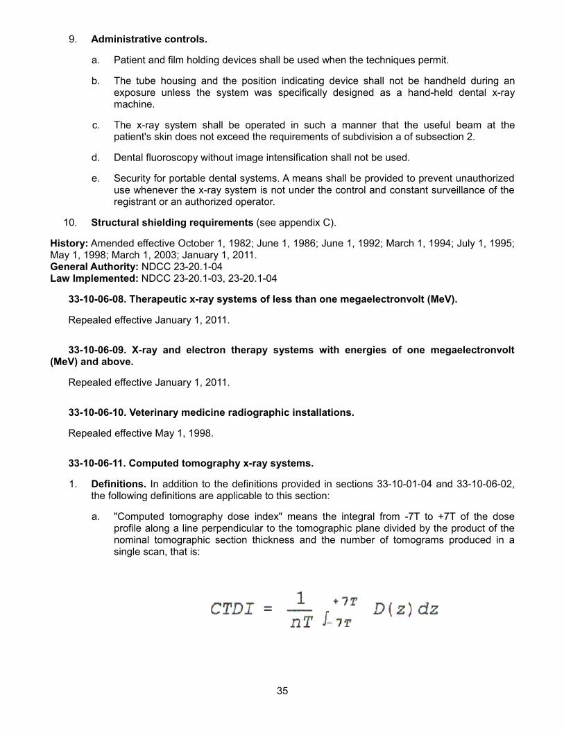
9. Administrative controls.
a. Patient and film holding devices shall be used when the techniques permit.
b. The tube housing and the position indicating device shall not be handheld during an exposure unless the system was specifically designed as a hand-held dental x-ray machine.
c. The x-ray system shall be operated in such a manner that the useful beam at the patient's skin does not exceed the requirements of subdivision a of subsection 2.
d. Dental fluoroscopy without image intensification shall not be used.
e. Security for portable dental systems. A means shall be provided to prevent unauthorized use whenever the x-ray system is not under the control and constant surveillance of the registrant or an authorized operator.
10. Structural shielding requirements (see appendix C).
History: Amended effective October 1, 1982; June 1, 1986; June 1, 1992; March 1, 1994; July 1, 1995; May 1, 1998; March 1, 2003; January 1, 2011. General Authority: NDCC 23-20.1-04Law Implemented: NDCC 23-20.1-03, 23-20.1-04
33-10-06-08. Therapeutic x-ray systems of less than one megaelectronvolt (MeV).
Repealed effective January 1, 2011.
33-10-06-09. X-ray and electron therapy systems with energies of one megaelectronvolt (MeV) and above.
Repealed effective January 1, 2011.
33-10-06-10. Veterinary medicine radiographic installations.
Repealed effective May 1, 1998.
33-10-06-11. Computed tomography x-ray systems.
1. Definitions. In addition to the definitions provided in sections 33-10-01-04 and 33-10-06-02, the following definitions are applicable to this section:
a. "Computed tomography dose index" means the integral from -7T to +7T of the dose profile along a line perpendicular to the tomographic plane divided by the product of the nominal tomographic section thickness and the number of tomograms produced in a single scan, that is:
35
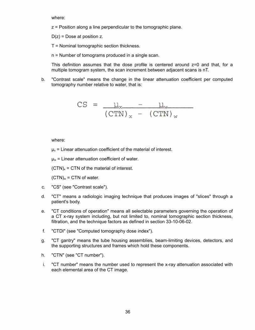
where:
z = Position along a line perpendicular to the tomographic plane.
D(z) = Dose at position z.
T = Nominal tomographic section thickness.
n = Number of tomograms produced in a single scan.
This definition assumes that the dose profile is centered around z=0 and that, for a multiple tomogram system, the scan increment between adjacent scans is nT.
b. "Contrast scale" means the change in the linear attenuation coefficient per computed tomography number relative to water, that is:
where:
μx = Linear attenuation coefficient of the material of interest.
μw = Linear attenuation coefficient of water.
(CTN)x = CTN of the material of interest.
(CTN)w = CTN of water.
c. "CS" (see "Contrast scale").
d. "CT" means a radiologic imaging technique that produces images of "slices" through a patient's body.
e. "CT conditions of operation" means all selectable parameters governing the operation of a CT x-ray system including, but not limited to, nominal tomographic section thickness, filtration, and the technique factors as defined in section 33-10-06-02.
f. "CTDI" (see "Computed tomography dose index").
g. "CT gantry" means the tube housing assemblies, beam-limiting devices, detectors, and the supporting structures and frames which hold these components.
h. "CTN" (see "CT number").
i. "CT number" means the number used to represent the x-ray attenuation associated with each elemental area of the CT image.
36

where:
k = A constant (The constant has a normal value of one thousand when the Houndsfield scale of CTN is used.)
μx = Linear attenuation coefficient of the material of interest.
μw = Linear attenuation coefficient of water.
j. "Dose profile" means the dose as a function of position along a line.
k. "Elemental area" means the smallest area within a tomogram for which the x-ray attenuation properties of a body are depicted. (see also "Picture element").
l. "Multiple tomogram system" means a computed tomography x-ray system which obtains x-ray transmission data simultaneously during a single scan to produce more than one tomogram.
m. "Noise" means the standard deviation of the fluctuations in computed tomography number expressed as a percentage of the attenuation coefficient of water. Its estimate (Sn ) is calculated using the following expression:
where:
CS = Contrast scale.
μw = Linear attenuation coefficient of water.
s = Estimated standard deviation of the CTN of picture elements in a specified area of the CT image.
n. "Nominal tomographic section thickness" means the full width at half-maximum of the sensitivity profile taken at the center of the cross-sectional volume over which x-ray transmission data are collected.
o. "Picture element" means an elemental area of a tomogram.
p. "Reference plane" means a plane which is displaced from and parallel to the tomographic plane.
37
q. "Scan" means the complete process of collecting x-ray transmission data for the production of a tomogram. Data can be collected simultaneously during a single scan for the production of one or more tomograms.
r. "Scan increment" means the amount of relative displacement of the patient with respect to the CT x-ray system between successive scans measured along the direction of such displacement.
s. "Scan sequence" means a preselected set of two or more scans performed consecutively under preselected CT conditions of operation.
t. "Scan time" means the period of time between the beginning and end of x-ray transmission data accumulation for a single scan.
u. "Single tomogram system" means a CT x-ray system which obtains x-ray transmission data during a scan to produce a single tomogram.
v. "Tomographic plane" means that geometric plane which is identified as corresponding to the output tomogram.
w. "Tomographic section" means the volume of an object whose x-ray attenuation properties are imaged in a tomogram.
2. Requirements for equipment.
a. Termination of exposure.
(1) Means must be provided to terminate the x-ray exposure automatically by either deenergizing the x-ray source or shuttering the x-ray beam in the event of equipment failure affecting data collection. Such termination must occur within an interval that limits the total scan time to no more than one hundred ten percent of its preset value through the use of either a backup timer or devices which monitor equipment function.
(2) A visible signal must indicate when the x-ray exposure has been terminated through the means required by paragraph 1.
(3) The operator must be able to terminate the x-ray exposure at any time during a scan, or series of scans under computed tomography x-ray system control, of greater than one-half second duration.
b. Tomographic plane indication and alignment.
(1) For any single tomogram system, means must be provided to permit visual determination of the tomographic plane or a reference plane offset from the tomographic plane.
(2) For any multiple tomogram system, means shall be provided to permit visual determination of the location of a reference plane. This reference plane can be offset from the location of the tomographic planes.
(3) If a device using a light source is used to satisfy paragraph 1 or 2, the light source must provide illumination levels sufficient to permit visual determination of the location of the tomographic plane or reference plane under ambient light conditions of up to five hundred lux.
c. Beam-on and shutter status indicators and control switches.
38
(1) The computed tomography x-ray control and gantry must provide visual indication whenever x-rays are produced and, if applicable, whether the shutter is open or closed.
(2) Each emergency button or switch must be clearly labeled as to its function.
d. Indication of computed tomography conditions of operation. The computed tomography x-ray system must be designed such that the computed tomography conditions of operation to be used during a scan or a scan sequence must be indicated prior to the initiation of a scan or a scan sequence. On equipment having all or some of these conditions of operation at fixed values, this requirement may be met by permanent markings. Indication of computed tomography conditions of operation must be visible from any position from which scan initiation is possible.
e. Extraneous radiation. When data are not being collected for image production, the radiation adjacent to the tube port may not exceed that permitted by subsection 3 of section 33-10-06-04.
f. Maximum surface computed tomography dose index identification. The angular position where the maximum surface computed tomography dose index occurs must be identified to allow for reproducible positioning of a computed tomography dosimetry phantom.
g. Additional requirements applicable to computed tomography x-ray systems containing a gantry manufactured after September 3, 1985.
(1) The total error in the indicated location of the tomographic plane or reference plane may not exceed five millimeters.
(2) If the x-ray production period is less than one-half second, the indication of x-ray production must be actuated for at least one-half second. Indicators at or near the gantry must be discernible from any point external to the patient opening where insertion of any part of the human body into the primary beam is possible.
(3) The deviation of indicated scan increment versus actual increment may not exceed plus or minus one millimeter with any mass from zero to one hundred kilograms resting on the support device. The patient support device must be incremented from a typical starting position to the maximum incremented distance or thirty centimeters, whichever is less, and then returned to the starting position. Measurement of actual versus indicated scan increment may be taken anywhere along this travel.
(4) Premature termination of the x-ray exposure by the operator shall necessitate resetting of the computed tomography conditions of operation prior to the initiation of another scan.
h. Facility design requirements.
(1) All CT capable systems shall be required to have the x-ray control permanently mounted in a protected area during the entire exposure (see appendix B).
(2) Aural communication. Provision must be made for two-way aural communication between the patient and the operator at the control panel.
i. Surveys, calibrations, spot checks, and operating procedures.
(1) Surveys.
39

(a) All computed tomography x-ray systems installed after March 1, 1992, and those systems not previously surveyed shall have a survey made by, or under the direction of, a qualified expert. In addition, such surveys must be done after any change in the facility or equipment which might cause a significant increase in radiation hazard.
(b) The registrant shall obtain a written report of the survey from the qualified expert, and a copy of the report must be made available to the department upon request.
(2) Radiation calibrations.
(a) The calibration of the radiation output of the computed tomography x-ray system must be performed by, or under the direction of, a qualified expert who is physically present at the facility during such calibration.
(b) The calibration of a computed tomography x-ray system must be performed at intervals specified by a qualified expert and after any change or replacement of components which, in the opinion of the qualified expert, could cause a change in the radiation output.
(c) The calibration of the radiation output of a computed tomography x-ray system must be performed with a calibrated dosimetry system. The calibration of such system must be traceable to a national standard. The dosimetry system must have been calibrated within the preceding two years.
(d) Computed tomography dosimetry phantoms must be used in determining the radiation output of a computed tomography x-ray system. Such phantoms must meet the following specifications and conditions of use:
[1] Computed tomography dosimetry phantoms must be right circular cylinders of polymethyl methacrylate of density one point nineteen plus or minus point zero one grams per cubic centimeter. The phantoms must be at least fourteen centimeters in length and must have diameters of thirty-two centimeters for testing computed tomography x-ray systems designed to image any section of the body and sixteen centimeters for systems designed to image the head or for whole body scanners operated in the head scanning mode.
[2] Computed tomography dosimetry phantoms must provide means for the placement of a dosimeter along the axis of rotation and along a line parallel to the axis of rotation one centimeter from the outer surface and within the phantom. Means for the placement of dosimeters or alignment devices at other locations may be provided.
[3] Any effects on the doses measured due to the removal of phantom material to accommodate dosimeters must be accounted for through appropriate corrections to the reported data or included in the statement of maximum deviation for the values obtained using the phantom.
[4] All dose measurements must be performed with the computed tomography dosimetry phantom placed on the patient couch or support device without additional attenuation materials present.
(e) The calibration shall be required for each type of head, body, or whole-body scan performed at the facility.
40

(f) Calibration must meet the following requirements:
[1] The dose profile along the center axis of the computed tomography dosimetry phantom for the minimum, maximum, and midrange values of the nominal tomographic section thickness used by the registrant shall be measurable. Where less than three nominal tomographic thicknesses can be selected, the dose profile determination must be performed for each available nominal tomographic section thickness.
[2] The computed tomography dose index (For the purpose of determining the computed tomography dose index, the manufacturer's statement as to the nominal tomographic section thickness for that particular system may be utilized.) along the two axes specified in item 2 of subparagraph d must be measured. The computed tomography dosimetry phantom must be oriented so that the measurement point one centimeter from the outer surface and within the phantom is in the same angular position within the gantry as the point of maximum surface computed tomography dose index identified. The computed tomography conditions of operation must correspond to typical values used by the registrant.
[3] The spot checks specified in paragraph 3 of subdivision i must be made.
(g) Calibration procedures must be in writing. Records of calibrations performed must be maintained for inspection by the department.
(3) Spot checks.
(a) The spot check procedures must be in writing and must have been developed by a qualified expert.
(b) The spot check procedures must incorporate the use of a computed tomography dosimetry phantom which has a capability of providing an indication of contrast scale, noise, nominal tomographic section thickness, the resolution capability of the system for low and high contrast objects, and measuring the mean computed tomography number for water or other reference material.
(c) All spot checks must be included in the calibration required by paragraph 2 and at time intervals and under system conditions specified by a qualified expert.
(d) Spot checks must include acquisition of images obtained with the computed tomography dosimetry phantoms using the same processing mode and computed tomography conditions of operation as are used to perform calibrations required by paragraph 2 of subdivision i. The images must be retained, until a new calibration is performed, in two forms as follows:
[1] Photographic copies of the images obtained from the image display device; and
[2] Images stored in digital form on a storage medium compatible with the computed tomography x-ray system.
(e) Written records of the spot checks performed shall be maintained for inspection by the department.
(4) Operating procedures.
41

(a) The computed tomography x-ray system may not be operated except by an individual who has been specifically trained in its operation.
(b) Information must be available at the control panel regarding the operation and calibration of the system. Such information must include the following:
[1] Dates of the latest calibration and spot checks and the location within the facility where the results of those tests may be obtained;
[2] Instructions on the use of the computed tomography dosimetry phantoms including a schedule of spot checks appropriate for the system, allowable variations for the indicated parameters, and the results of at least the most recent spot checks conducted on the system;
[3] The distance in millimeters between the tomographic plane and the reference plane if a reference plane is utilized; and
[4] A current technique chart available at the control panel which specifies for each routine examination the computed tomography conditions of operation and the number of scans per examination.
(c) If the calibration or spot check of the computed tomography x-ray system identifies that a system operating parameter has exceeded a tolerance established by the qualified expert, use of the computed tomography x-ray system on patients must be limited to those uses permitted by established written instructions of the qualified expert.
History: Effective June 1, 1992; amended effective May 1, 1998; January 1, 2011.General Authority: NDCC 23-20.1-04Law Implemented: NDCC 23-20.1-03, 23-20.1-04
33-10-06-12. Bone densitometry.
1. Bone densitometry systems shall be:
a. Certified by the manufacturer pursuant to the Medical Device Act and subchapter C - electronic product radiation control (EPRC) of chapter V of the Federal Food, Drug and Cosmetic Act;
b. Registered in accordance with chapter 33-10-02 of these regulations; and
c. Maintained and operated in accordance with the manufacturer's specifications.
2. Equipment requirements. Systems with stepless collimators shall be provided with means to both size and align the x-ray field such that the x-ray field at the plane of the image receptor does not extend beyond two percent of the source-image receptor distance.
3. Operators of bone densitometry systems shall: Complete a training course on the bone densitometry which is acceptable to the department. The training course shall include:
a. Basic radiation protection;
b. Operating procedures for bone densitometry systems, to include use of various system functions, safety, and maintenance; and
c. Patient positioning for the type of examinations performed.
4. During the operation of any bone densitometry system:
42
a. The operator, ancillary personnel, and members of the general public shall be positioned as far away as practical but not less than two meters from the patient and bone densitometry system during the examination.
b. The operator shall advise the patient that the bone densitometry examination is a type of x-ray procedure.
5. The registrant shall keep maintenance records for bone densitometry systems as prescribed by subdivision b of subsection 1 of section 33-10-06-03. These records shall be maintained for inspection by the department.
6. Bone densitometry on human patients shall be conducted only:
a. Under a prescription of a licensed practitioner of the healing arts; or
b. Under a screening program approved by the department.
7. Any person proposing to conduct a bone densitometry screening program shall submit the information outlined in appendix E.
History: Effective March 1, 2003; amended effective January 1, 2011.General Authority: NDCC 23-20.1-04Law Implemented: NDCC 23-20.1-03, 23-20.1-04
43

APPENDIX A
INFORMATION ON RADIATION SHIELDING REQUIRED FOR PLAN REVIEWS
In order for the department to provide an evaluation, technical advice, and official approval on shielding requirements for a radiation installation, the following information shall be submitted:
1. The plans should show, as a minimum, the following:
a. The normal location of the x-ray system's radiation port; the port's travel and traverse limits; general direction or directions of the useful beam; locations of any windows and doors; the location of the operator's booth; and the location of the x-ray control panel.
b. Structural composition and thickness or lead equivalent of all walls, doors, partitions, floor, and ceiling of the room or rooms concerned.
c. The dimensions of the room or rooms concerned.
d. The type of occupancy of all adjacent areas inclusive of space above and below the room or rooms concerned. If there is an exterior wall, show distance to the closest area or areas where it is likely that individuals may be present.
e. The make and model of the x-ray equipment and the maximum technique factors.
f. The type of examinations or treatments which will be performed with the equipment, e.g., dental, orthodontal, chest, gastrointestinal, fluoroscopic, podiatry, fixed therapy, rotational therapy, etc.
2. Information on the anticipated workload of the x-ray systems.
3. If the services of a qualified expert have been utilized to determine the shielding requirements, a report, including all basic assumptions used, must be submitted with the plans.
History: Amended effective June 1, 1992.
44

APPENDIX B
MINIMUM DESIGN REQUIREMENTS FOR AN X-RAY MACHINE OPERATOR'S BOOTH
1. Space requirements.
a. The operator shall be allotted not less than seven and five-tenths square feet [0.697 square meter] of unobstructed floor space in the booth.
b. The operator's booth may be any geometric configuration with no dimension of less than two feet [0.61 meters].
c. The space shall be allotted excluding any encumbrance by the console, such as overhang, cables, or other similar encroachments.
d. The booth must be located or constructed such that unattenuated direct scatter radiation originating on the examination table or at the wall cassette not reach the operator's station in the booth.
2. Structural requirements.
a. The booth walls shall be permanently fixed barriers of at least seven feet [2.13 meters] high.
b. When a door or movable panel is used as an integral part of the booth structure, it must have an interlock which will prevent an exposure when the door or panel is not closed.
c. Shielding must be provided to meet the requirements of chapter 33-10-04.1 of these rules.
3. X-ray control placement. The x-ray control for the system shall be fixed within the booth and:
a. Shall be at least forty inches [1.02 meters] from any open edge of the booth wall which is nearest to the examining table.
b. Shall allow the operator to use the majority of the available viewing windows.
4. Viewing system requirements.
a. Each booth shall have at least one viewing device which will:
(1) Be so placed that the operator can view the patient during any exposure; and
(2) The device shall be so placed that the operator can have full view of any occupant of the room and should be so placed that the operator can view any entry into the room. If any door, which allows access to the room, cannot be seen from the booth, then that door must have an interlock controlling the exposure which will prevent the exposure if the door is not closed.
b. When the viewing system is a window, the following requirements also apply:
(1) The viewing area must be at least one square foot [0.0929 square meter].
(2) The design of the booth must be such that the operator's expected position when viewing the patient and operating the x-ray system is at least eighteen inches [0.457 meter] from the edge of the booth.
(3) The material constituting the window must have the same lead equivalence as that required in the booth's wall in which it is mounted.
45
c. When the viewing system is by mirrors, the mirrors must be so located as to accomplish the general requirements of subdivision a.
d. When the viewing system is by electronic means:
(1) The camera shall be so located as to accomplish the general requirements in subdivision a; and
(2) There shall be an alternate viewing system as a backup for the primary system.
History: Amended effective June 1, 1986; June 1, 1992; January 1, 2011.
46
APPENDIX C
STRUCTURAL SHIELDING REQUIREMENTS
1. General requirements.
a. Each installation must be provided with such primary or secondary barriers as are necessary to assure compliance with section 33-10-04.2-01 [10 CFR 20.1201, 20.1207, 20.1208, and 20.1301]. This requirement must be deemed to be met if the thicknesses of such barriers are equivalent to those as computed in accordance with appendices B, C, and D of the National Council on Radiation Protection and Measurements Report No. 49, "Medical X-Ray and Gamma-Ray Protection for Energies Up to 10 MeV," modified to meet current dose limits.
b. Lead barriers must be mounted in such manner that they will not sag or cold-flow because of their own weight and shall be protected against mechanical damage.
c. Joints between different kinds of protective materials must be designed so that the overall protection of the barrier is not impaired.
d. Joints at the floor and ceiling must be so designed that the overall protection is not impaired.
e. Windows, window frames, doors, and door frames must have the same lead equivalent as that required of the adjacent wall.
f. Holes in protective barriers must be covered so that overall attenuation is not impaired.
2. Fluoroscopic x-ray systems. Ordinarily only secondary barriers are necessary except combined fluoroscopic-radiographic installations.
3. Radiographic systems other than fluoroscopic, dental intraoral, or veterinarian systems:
a. All wall, floor, and ceiling areas exposed to the useful beam must have primary barriers. Primary barriers in walls must extend to a minimum height of eighty-four inches [2.13 meters] above the floor.
b. Secondary barriers must be provided in all wall, floor, and ceiling areas not having primary barriers or where the primary barrier requirements are lower than the secondary requirements.
c. The operator's station at the control shall be behind a protective barrier, either in a separate room, in a protected booth, or behind a shield which will intercept the useful beam and any radiation which has been scattered only once.
d. A window of lead equivalent glass equal to that required by the adjacent barrier or a mirror system shall be provided large enough and so placed that the operator can see the patient without having to leave the protected area during exposure.
e. For mobile and portable x-ray systems which are used for greater than one week in one location (one room or suite), the requirements of this appendix shall apply.
4. Intraoral dental radiographic systems.
a. Dental rooms containing x-ray machines shall be provided with primary barriers at all areas struck by the useful beam. Consideration shall be given to the attenuation provided by the patient.
47

b. When dental x-ray units are installed in adjacent rooms or areas, protective barriers shall be provided between the rooms or areas.
Note: In many cases, structural materials of ordinary walls suffice as a protective barrier without addition of special shielding material.
5. Therapeutic x-ray installations. The structural shielding requirements shall be deemed to be met if the barriers have been designed and constructed in accordance with the National Council on Radiation Protection and Measurements Report No. 49, "Medical X-Ray and Gamma-Ray Protection for Energies Up to 10 MeV", modified to meet current dose limits.
6. Veterinary medicine radiographic installations.
a. All wall, floor, and ceiling areas exposed to the useful beam shall have primary barriers. Primary barriers in walls shall extend to a minimum height of eighty-four inches [2.13 meters] above the floor.
b. Secondary barriers shall be provided in all wall, floor, and ceiling areas not having primary barriers or where the primary barrier requirements are lower than the secondary requirements.
History: Amended effective June 1, 1986; June 1, 1992; January 1, 2011.
48
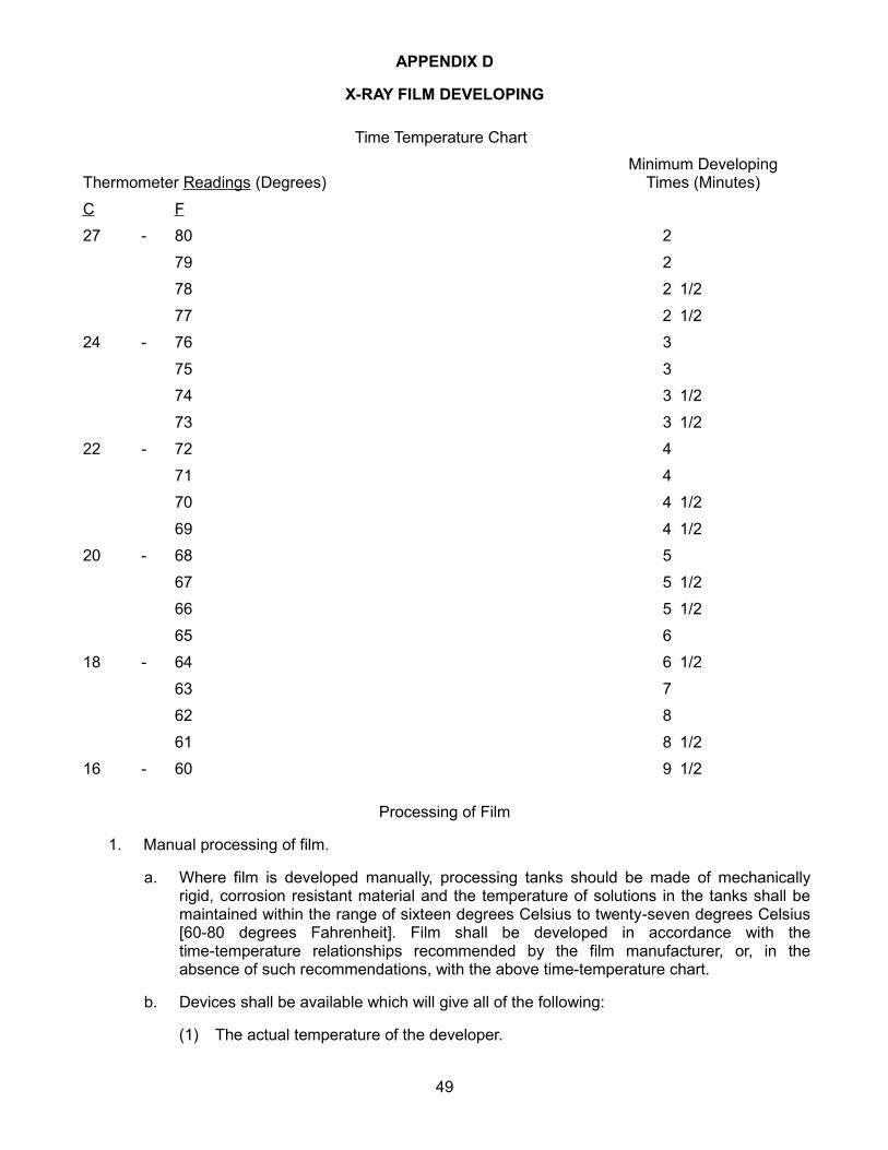
APPENDIX D
X-RAY FILM DEVELOPING
Time Temperature Chart
Thermometer Readings (Degrees)Minimum Developing
Times (Minutes)
C F
27 - 80 2
79 2
78 2 1/2
77 2 1/2
24 - 76 3
75 3
74 3 1/2
73 3 1/2
22 - 72 4
71 4
70 4 1/2
69 4 1/2
20 - 68 5
67 5 1/2
66 5 1/2
65 6
18 - 64 6 1/2
63 7
62 8
61 8 1/2
16 - 60 9 1/2
Processing of Film
1. Manual processing of film.
a. Where film is developed manually, processing tanks should be made of mechanically rigid, corrosion resistant material and the temperature of solutions in the tanks shall be maintained within the range of sixteen degrees Celsius to twenty-seven degrees Celsius [60-80 degrees Fahrenheit]. Film shall be developed in accordance with the time-temperature relationships recommended by the film manufacturer, or, in the absence of such recommendations, with the above time-temperature chart.
b. Devices shall be available which will give all of the following:
(1) The actual temperature of the developer.
49

(2) An audible or visible signal, after a preset time (in minutes of duration).
2. Automatic processors and other closed processing systems.
a. Film shall be developed in accordance with the time-temperature relationships recommended by the film manufacturer.
b. The specified developer temperature and immersion time shall be posted in the darkroom or on the automatic processor.
c. Preventive maintenance shall be performed on the unit, except for extended periods of nonuse, on a frequency basis which is not less than that schedule recommended by the manufacturer. In the event that no schedule is available from the manufacturer a maintenance schedule shall be established which will preserve good film quality.
d. After a full cleansing of the processor a film shall be exposed to a density of approximately one, with one-half of the film protected exposure. It will be developed and then kept near the unit and daily at least one test film (exposed under techniques identical with those used for the original test film) shall be compared with the original test film to evaluate the adequacy of the unit's developing capability and base fog level.
3. Processing deviations from the requirements of appendix D shall be documented by the registrant in such manner that the requirements are shown to be met or exceeded (e.g., extended processing, and special rapid chemistry).
4. Other requirements:
a. Pass boxes, if provided, shall be so constructed as to exclude light from the darkroom when cassettes are placed in or removed from the boxes, and shall incorporate adequate shielding from stray radiation to prevent exposure of undeveloped film.
b. The darkroom shall be light tight and use proper safelighting such that any film type in use exposed in a cassette to x-radiation sufficient to produce an optical density from one to two when processed shall not suffer an increase in density greater than 0.1 (0.05 for mammography) when exposed in the darkroom for two minutes with all safelights on. If used, daylight film handling boxes shall preclude fogging of the film.
c. Darkrooms typically used by more than one individual shall be provided a method to prevent accidental entry while undeveloped films are being handled or processed.
d. Film shall be stored in a cool, dry place and shall be protected from exposure to stray radiation. Film in open packages shall be stored in a light tight container.
e. Film cassettes and intensifying screens shall be inspected periodically and shall be cleaned and replaced as necessary to best assure radiographs of good diagnostic quality.
f. Outdated x-ray film shall not be used for diagnostic radiographs, unless the film has been stored in accordance with the manufacturer's recommendations and a sample of the film passes a sensitometric test for normal ranges of base plus fog and speed.
g. Film developing solutions shall be prepared in accordance with the directions given by the manufacturer, and shall be maintained in strength by replenishment or renewal so that full development is accomplished within the time specified by the manufacturer.
50

APPENDIX E
INFORMATION TO BE SUBMITTED BY PERSONS PROPOSINGTO CONDUCT HEALING ARTS SCREENING
Persons requesting that the department approve a healing arts screening program shall submit the following information and evaluation:
1. Name and address of the applicant and, where applicable, the names and addresses of agents within this state.
2. Diseases or conditions for which the x-ray examinations are to be used in diagnoses.
3. A detailed description of the x-ray examinations proposed in the screening program.
4. Description of the population to be examined in the screening program, i.e., age, sex, physical condition, and other appropriate information.
5. An evaluation of any known alternate methods not involving ionizing radiation which could achieve the goals of the screening program and why these methods are not used instead of the x-ray examinations.
6. An evaluation by a qualified expert on the x-ray systems to be used in the screening program. The evaluation by the qualified expert shall show that such systems do satisfy all requirements of this article. The evaluation shall include a measurement of patient exposures from the x-ray examinations to be performed.
7. A description of the diagnostic x-ray quality control program.
8. A copy of the technique chart for the x-ray examination procedures to be used.
9. The qualifications of each individual who will be operating the x-ray systems.
10. The qualifications of the individual who will be supervising the operators of the x-ray systems. The extent of supervision and the method of work performance evaluation shall be specified.
11. The name and address of the individual who will interpret the radiographs.
12. A description of the procedures to be used in advising the individuals screened and their private practitioners of the healing arts of the results of the screening procedure and any further medical needs indicated.
13. A description of the procedures for the retention or disposition of the radiographs and other records pertaining to the x-ray examinations.
14. An indication of the frequency of screening and the duration of the entire screening program.
History: Effective October 1, 1982; amended effective June 1, 1986; June 1, 1992; May 1, 1998.
51
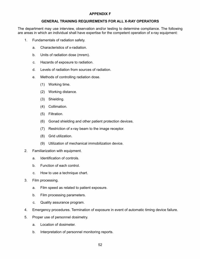
APPENDIX F
GENERAL TRAINING REQUIREMENTS FOR ALL X-RAY OPERATORS
The department may use interview, observation and/or testing to determine compliance. The following are areas in which an individual shall have expertise for the competent operation of x-ray equipment:
1. Fundamentals of radiation safety.
a. Characteristics of x-radiation.
b. Units of radiation dose (mrem).
c. Hazards of exposure to radiation.
d. Levels of radiation from sources of radiation.
e. Methods of controlling radiation dose.
(1) Working time.
(2) Working distance.
(3) Shielding.
(4) Collimation.
(5) Filtration.
(6) Gonad shielding and other patient protection devices.
(7) Restriction of x-ray beam to the image receptor.
(8) Grid utilization.
(9) Utilization of mechanical immobilization device.
2. Familiarization with equipment.
a. Identification of controls.
b. Function of each control.
c. How to use a technique chart.
3. Film processing.
a. Film speed as related to patient exposure.
b. Film processing parameters.
c. Quality assurance program.
4. Emergency procedures. Termination of exposure in event of automatic timing device failure.
5. Proper use of personnel dosimetry.
a. Location of dosimeter.
b. Interpretation of personnel monitoring reports.
52

6. Anatomy and positioning.
a. Relevant human anatomy.
b. Relevant human physiology.
c. Radiographic positioning.
7. The requirements of pertinent federal and state rules.
8. The licensee's or registrant's written operating and emergency procedures.
53

APPENDIX G
The following are individuals that qualify for training exemptions, approved Allied Health professions which qualify for cross-training into diagnostic x-ray as a limited x-ray machine operator and individuals who may order diagnostic x-rays to be taken by a limited x-ray machine operator outside the scope of procedures in appendix I:
1. Individuals exempt from minimum training requirements in subparagraph b of paragraph 2 of subdivision a of subsection 1 of section 33-10-06-03.
a. Medical doctors.
b. Chiropractors.
c. Doctors of osteopathy.
d. Podiatrists.
2. Prerequisite qualification: Individuals who qualify for cross-training as a limited x-ray machine operator.
a. Nurse practitioner, registered nurse, licensed practical nurse.
b. Emergency medical technician paramedic.
c. Physical therapist, physical therapy assistant.
d. Occupational therapist, occupational therapy assistant.
e. Medical technologist, medical lab technician, clinical lab technician.
f. Physician assistant.
g. Orthopedic physician assistant.
3. Individuals who may order emergency x-ray examinations outside the scope of procedures in appendix I to be taken by limited x-ray machine operators:
a. Medical doctor.
b. Doctor of osteopathy.
c. Physician assistant.
d. Nurse practitioner.
e. Chiropractor.
54

APPENDIX H
Limited X-Ray Machine Operator Training Requirements
Students must meet the prerequisite requirements of item 1 of subparagraph b of paragraph 2 of subdivision a of subsection 1 of section 33-10-06-03 and complete the training requirements of this appendix.
Training requirements have been divided into two sections, didactic instruction and clinical experience/supervision. Upon completion of didactic training, the individual must complete the clinical experience requirements of either subdivision a or b of subsection 2 and demonstrate competence for examinations listed in appendix I. Records must be maintained to demonstrate compliance with these requirements.
1. Didactic instruction section: Individuals shall complete a minimum of eighty hours of didactic training at a single course providing the minimum hours of instruction in the subjects below. Correspondence coursework cannot exceed twenty percent of the eighty-hour course (sixteen hours maximum). The course content should approximate the outline below. The eighty-hour course is subject to department approval. Individuals must also complete the three-hour self study course designed by the state health department. An examination is required to demonstrate successful completion of a course.
a. Basic X-Ray Physics 12 hrs.
- general description of production of x-rays
- function of filtration and effects it has on x-ray beam
- collimation
- types and function of beam limiting devices
- design, features and function of x-ray tube
b. Radiobiology 1 hr.
- effects of ionizing radiation to the human body
- factors that cause somatic and genetic damage
c. Radiation Protection 6 hrs.
- ALARA concept
- shielding materials
- radiation quantity and units of measurement
- basic interactions of x-ray with matter
- primary and secondary scatter
- importance of time, distance, shielding
- maximum permissible dose-occupational/public
- latency period
- patient protection
d. Principles of Exposure 15 hrs.
55

- factors that control and influence radiographic quality
- properties of x-rays
- size distortion caused by geometric parameters
- parameters which cause shape distortion
- technique factor selection
- 15% rule, mAs and kVp relationship
- grid-types, ratios, and how they affect image quality
- intensifying screens
- x-ray film
- artifacts
- inverse square law
e. Darkroom Procedure and Processing 4 hrs.
- film storage and handling
- film processing and troubleshooting
- design, features and function of a processor
- silver recovery
- quality assurance/quality control
f. Anatomy and Positioning
1. Chest 4 hrs.
2. Abdomen 4 hrs.
3. Extremity 8 hrs.
4. Spine 8 hrs.
5. Skull 8 hrs.
g. Pediatric 2 hrs.
h. Rules and Regulations 1 hr.
2. Clinical experience/supervision section. Individuals must complete either a or b below. If the individual is unable to demonstrate clinical competence in a procedure due to a lack of opportunity, the student shall complete the three prerequisite examinations required by appendix J using simulation for subdivisions a through k of subsection 1 of appendix J. Final demonstration of competence in subdivisions a through s of subsection 1 of appendix J should be completed as soon as there is a patient requiring the procedure. No individual may perform an unsupervised procedure for which they have not successfully completed the final demonstration of competence.
a. The individual must complete three months of clinical training during which time they may perform x-ray examinations only under direct supervision.
56
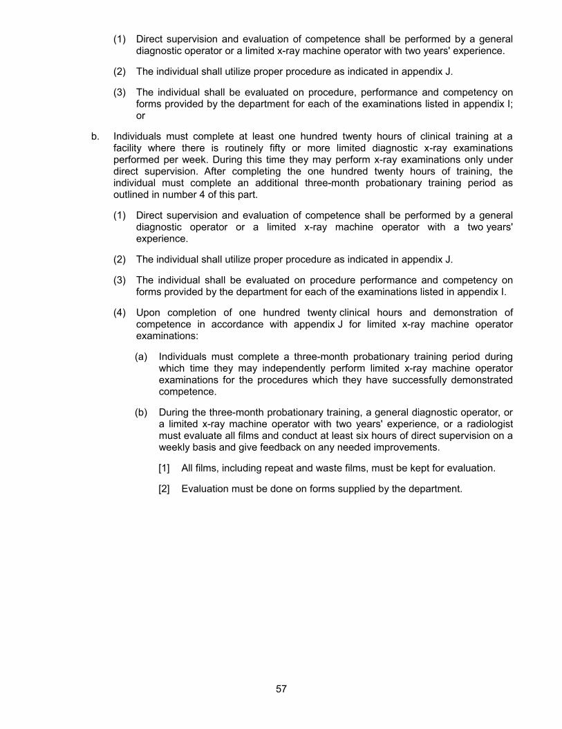
(1) Direct supervision and evaluation of competence shall be performed by a general diagnostic operator or a limited x-ray machine operator with two years' experience.
(2) The individual shall utilize proper procedure as indicated in appendix J.
(3) The individual shall be evaluated on procedure, performance and competency on forms provided by the department for each of the examinations listed in appendix I; or
b. Individuals must complete at least one hundred twenty hours of clinical training at a facility where there is routinely fifty or more limited diagnostic x-ray examinations performed per week. During this time they may perform x-ray examinations only under direct supervision. After completing the one hundred twenty hours of training, the individual must complete an additional three-month probationary training period as outlined in number 4 of this part.
(1) Direct supervision and evaluation of competence shall be performed by a general diagnostic operator or a limited x-ray machine operator with a two years' experience.
(2) The individual shall utilize proper procedure as indicated in appendix J.
(3) The individual shall be evaluated on procedure performance and competency on forms provided by the department for each of the examinations listed in appendix I.
(4) Upon completion of one hundred twenty clinical hours and demonstration of competence in accordance with appendix J for limited x-ray machine operator examinations:
(a) Individuals must complete a three-month probationary training period during which time they may independently perform limited x-ray machine operator examinations for the procedures which they have successfully demonstrated competence.
(b) During the three-month probationary training, a general diagnostic operator, or a limited x-ray machine operator with two years' experience, or a radiologist must evaluate all films and conduct at least six hours of direct supervision on a weekly basis and give feedback on any needed improvements.
[1] All films, including repeat and waste films, must be kept for evaluation.
[2] Evaluation must be done on forms supplied by the department.
57

APPENDIX I
Specific examinations that are allowed in the scope of practice for limited x-ray machine operators.
Chest: PA, lateral, decubitus
Ribs: AP, PA, obliques
Abdomen: KUB, upright abdomen
Hand & fingers: PA, lateral, oblique
Wrist: PA, lateral, oblique
Forearm: AP, lateral
Elbow: AP, lateral
Humerus: AP, lateral
Shoulder: AP, internal & external rotation, y-view
Clavicle: AP, AP axial
Pelvis: AP
Hips: AP, Frog leg lateral, cross-table lateral
Femur: AP, lateral
Knee: AP, lateral, obliques
Patella: AP, lateral, sunrise
Tibia-Fibula: AP, lateral
Ankle: AP, lateral, obliques
Calcaneous: Plantodorsal, lateral
Foot & toes: AP, lateral, obliques
Sinuses: Water's, lateral
Skull: AP/PA, lateral
Facial bones: PA, lateral
Nasal bones: Water's, lateral
C-spine: AP, lateral, odontoid, (not trauma), swimmer's (not trauma)
T-spine: AP, lateral, swimmer's (not trauma)
L-spine: AP, lateral, L5-S1 lateral
Any situation deemed an emergency and requiring a limited x-ray machine operator to conduct procedures not specifically listed above, requires a written order from an individual listed in part 3 of appendix G and direct supervision from the individual ordering the examination in accordance with item 1 of subparagraph c of paragraph 2 of subdivision a of subsection 1 of section 33-10-06-03.
58
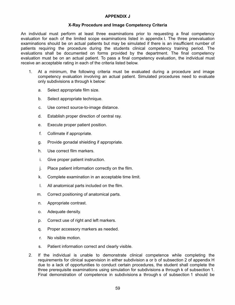
APPENDIX J
X-Ray Procedure and Image Competency Criteria
An individual must perform at least three examinations prior to requesting a final competency evaluation for each of the limited scope examinations listed in appendix I. The three preevaluation examinations should be on actual patients but may be simulated if there is an insufficient number of patients requiring the procedure during the students clinical competency training period. The evaluations shall be documented on forms provided by the department. The final competency evaluation must be on an actual patient. To pass a final competency evaluation, the individual must receive an acceptable rating in each of the criteria listed below.
1. At a minimum, the following criteria must be evaluated during a procedure and image competency evaluation involving an actual patient. Simulated procedures need to evaluate only subdivisions a through k below:
a. Select appropriate film size.
b. Select appropriate technique.
c. Use correct source-to-image distance.
d. Establish proper direction of central ray.
e. Execute proper patient position.
f. Collimate if appropriate.
g. Provide gonadal shielding if appropriate.
h. Use correct film markers.
i. Give proper patient instruction.
j. Place patient information correctly on the film.
k. Complete examination in an acceptable time limit.
l. All anatomical parts included on the film.
m. Correct positioning of anatomical parts.
n. Appropriate contrast.
o. Adequate density.
p. Correct use of right and left markers.
q. Proper accessory markers as needed.
r. No visible motion.
s. Patient information correct and clearly visible.
2. If the individual is unable to demonstrate clinical competence while completing the requirements for clinical supervision in either subdivision a or b of subsection 2 of appendix H due to a lack of opportunities to conduct certain procedures, the student shall complete the three prerequisite examinations using simulation for subdivisions a through k of subsection 1. Final demonstration of competence in subdivisions a through s of subsection 1 should be
59

completed as soon as there is a patient requiring the procedure. No individual may perform an unsupervised procedure for which they have not successfully completed the final demonstration of competence.
60

APPENDIX K
Continuing Education Requirements
Continuing education units (CEUs) are required for all limited x-ray machine operators and general diagnostic operators as defined by subparagraphs a and b of paragraph 2 of subdivision a of subsection 1 of section 33-10-06-03 of the North Dakota radiological health rules. Continuing education unit requirements will be associated with a two-year cycle (biennium).
1. General diagnostic operators shall obtain a minimum of 24 CEUs per biennium.
2. Limited x-ray machine operators shall obtain a minimum of 12 CEUs per biennium.
3. Units from one biennium cannot be carried forward or applied to the following biennium.
4. Determining the beginning of a biennium.
a. For limited x-ray machine operators and general diagnostic operators not certified through an accrediting body, the biennium will begin January 1, 2009.
b. For general diagnostic operators certified through an accrediting body, the biennium will be defined by the registration requirements through their accrediting body and shall begin on the first due date following January 1, 2009.
5. Continuing education unit activities must be approved by an accrediting body.
61