chapter 36 skin integrity and wound healing. 36-2 copyright 2004 by delmar learning, a division of...
TRANSCRIPT

Chapter 36
Skin Integrity and Wound Healing
36-2Copyright 2004 by Delmar Learning, a division of Thomson Learning, Inc.
Normal Structures and Function of Healthy Skin
The skin is the body’s largest organ and the primary defense against pathogenic invasion.
The skin also contributes to temperature regulation, prevents loss of internal fluids, and provides sensory awareness.

36-3Copyright 2004 by Delmar Learning, a division of Thomson Learning, Inc.
Normal Structures and Function of Healthy Skin
Epidermis• Outermost layer of the skin• Primary function is to maintain a barrier
against loss of internal fluids and pathogenic invasion.

36-4Copyright 2004 by Delmar Learning, a division of Thomson Learning, Inc.
Normal Structures and Function of Healthy Skin
Dermal-Epidermal Junction• Anatomic point at which the epidermis
connects with the dermis• Characterized by interdigitating connections
that provide resistance to superficial skin injury.
36-5Copyright 2004 by Delmar Learning, a division of Thomson Learning, Inc.
Normal Structures and Function of Healthy Skin
Dermis• Innermost layer of the skin• Nourishes the basal layer of the epidermis.• Provides sensory awareness.• Contributes to temperature regulation.• Composed primarily of collagen and elastin
fibers.

36-6Copyright 2004 by Delmar Learning, a division of Thomson Learning, Inc.
Normal Structures and Function of Healthy Skin
Hypodermis (Subcutaneous layer)• Consists primarily of adipose tissue and
connective tissue.• Critical role of providing “padding” and even
weight distribution over bony prominences.
36-7Copyright 2004 by Delmar Learning, a division of Thomson Learning, Inc.
Normal Structures and Function of Healthy Skin
Fascia/Muscle Layer• Fascia is a thin layer of connective tissue
covering the muscle.• Muscle layer is composed of contractile
fibers that control position and movement.• Muscle layer is the most metabolically active
layer of the skin and soft tissues.• Muscle layer is most vulnerable to ischemic
damage.

36-8Copyright 2004 by Delmar Learning, a division of Thomson Learning, Inc.
Normal Structures and Function of Healthy Skin
Changes Across the Lifespan• Neonates and Infants• Elderly Adults
36-9Copyright 2004 by Delmar Learning, a division of Thomson Learning, Inc.
Strategies to Maintain Healthy Skin
Nutrition and Hydration Bathing and Lubrication Managing Pruritic Skin
36-10Copyright 2004 by Delmar Learning, a division of Thomson Learning, Inc.
Strategies to Maintain Healthy Skin
Common Skin Lesions• Bacterial Infections• Fungal Infections• Viral Infections

36-11Copyright 2004 by Delmar Learning, a division of Thomson Learning, Inc.
Strategies to Maintain Healthy Skin
Inflammatory Conditions Cutaneous Malignancies

36-12Copyright 2004 by Delmar Learning, a division of Thomson Learning, Inc.
Pressure Ulcer Formation
A pressure ulcer is an area of skin and tissue loss caused by prolonged or excessive soft tissue pressure.
Results in skin breakdown. Increasingly common problem among
clients in all health care settings.
36-13Copyright 2004 by Delmar Learning, a division of Thomson Learning, Inc.
Pressure Ulcer Formation
Pathology of Pressure Ulcers• Tunneling• Friction• Maceration

36-14Copyright 2004 by Delmar Learning, a division of Thomson Learning, Inc.
Pressure Ulcer Formation
Assessment• Use of a research-based risk assessment
tool to screen all non-ambulatory clients- Braden scale- Norton scale
• Nonblanching erythema• Induration with palpation• Extensive tissue damage

36-15Copyright 2004 by Delmar Learning, a division of Thomson Learning, Inc.
Pressure Ulcer Formation
Assessment• Etiologic Risk Factors• Prolonged or High-Intensity Pressure• Shear Force• Compromised Tissue Tolerance

36-16Copyright 2004 by Delmar Learning, a division of Thomson Learning, Inc.
Pressure Ulcer Formation
Nursing Diagnosis• Impaired Skin Integrity Related to
Pressure/Shear Injury

36-17Copyright 2004 by Delmar Learning, a division of Thomson Learning, Inc.
Outcome Identification and Planning
Individualized outcomes are based on the client’s overall physical condition, the stage of the wound, and the client’s risk factors.
Client teaching is an integral part of the planning process.

36-18Copyright 2004 by Delmar Learning, a division of Thomson Learning, Inc.
Implementation
Pressure ulcers can be prevented through a variety of measures.
Early identification of high-risk individuals and contributing factors decrease the possibility of pressure ulcer formation.

36-19Copyright 2004 by Delmar Learning, a division of Thomson Learning, Inc.
Implementation
Appropriate Use and Selection of Support Surfaces• A variety of support surfaces for bed and
chair are designed to reduce interface pressures or to constantly change the pressure points.
36-20Copyright 2004 by Delmar Learning, a division of Thomson Learning, Inc.
Implementation
Measures to Control Moisture and Maceration
Nutritional and Fluid Support Routine Skin Assessment Management for Shear Force Avoidance of Massage of Tissue at Risk
36-21Copyright 2004 by Delmar Learning, a division of Thomson Learning, Inc.
Evaluation
Physical signs of healing and the status of the pressure ulcer
Client’s adaptation to the altered skin integrity
Each intervention should be evaluated for its effectiveness.
Plan of care is revised to reflect most beneficial actions.
36-22Copyright 2004 by Delmar Learning, a division of Thomson Learning, Inc.
Wound Healing
Definitions and Classifications of Wounds• Acute • Chronic

36-23Copyright 2004 by Delmar Learning, a division of Thomson Learning, Inc.
Wound Healing
Definitions and Classifications of Wounds• Partial-thickness wounds involve partial loss
of the skin layers but do not involve the deeper tissues.
• Full-thickness wounds involve total loss of the epidermis and dermis with extension into the subcutaneous tissue and possibly the muscle.

36-24Copyright 2004 by Delmar Learning, a division of Thomson Learning, Inc.
Wound Healing
Partial-Thickness Wound Repair• Brief inflammatory phase• Epithelial cell proliferation and migration• Vertical migration• Collagen synthesis (formation of new
connective tissue)

36-25Copyright 2004 by Delmar Learning, a division of Thomson Learning, Inc.
Wound Healing
Full-Thickness Wound Repair• Inflammatory phase
- Control bleeding- Establish clean wound bed- Release of growth factors- Inflammatory response
36-26Copyright 2004 by Delmar Learning, a division of Thomson Learning, Inc.
Wound Healing
Full-Thickness Wound Repair• Proliferative phase
- Granulation tissue- Epithelialization- Contraction
• Maturation phase (remodeling phase)- 3 months to 2 years- Hypertrophic scarring (keloid formation)

36-27Copyright 2004 by Delmar Learning, a division of Thomson Learning, Inc.
Wound Management
Identify and address etiologic factors. Establish appropriate goals. Provide systemic support and topical
therapy.

36-28Copyright 2004 by Delmar Learning, a division of Thomson Learning, Inc.
Wound Management
Assessment• Location, dimensions and depth• Stage of the wound• Status of wound bed (eschar, slough)• Exudate• Status of wound edges (flat, red, moist,
closed)• Status of surrounding skin• Pain

36-29Copyright 2004 by Delmar Learning, a division of Thomson Learning, Inc.
Assessment
Factors Affecting Wound Healing• Perfusion and Oxygenation• Nutritional Status• Diabetes Mellitus• Corticosteroids• Aging

36-30Copyright 2004 by Delmar Learning, a division of Thomson Learning, Inc.
Assessment
Laboratory Data• Cultures of wound drainage• Elevated WBC count• Decreased leukocyte• Albumin
36-31Copyright 2004 by Delmar Learning, a division of Thomson Learning, Inc.
Nursing Diagnoses
Impaired Tissue Integrity Risk for Infection Pain Disturbed Body Image Deficient Knowledge (wound care)

36-32Copyright 2004 by Delmar Learning, a division of Thomson Learning, Inc.
Outcome Identification and Planning
Targeted outcomes are based on client’s identified needs and individualized on basis of client’s condition.
Focus is on promoting wound healing, preventing infection, and educating the client.

36-33Copyright 2004 by Delmar Learning, a division of Thomson Learning, Inc.
Implementation
Systemic Support Measures• Tissue perfusion and oxygenation• Nutritional support• Glucose levels within normal limits• Compensation for chronic steroid intake

36-34Copyright 2004 by Delmar Learning, a division of Thomson Learning, Inc.
Implementation
Topical Therapy• Wound cleansing• Dressing selection• Debridement of necrotic tissue
36-35Copyright 2004 by Delmar Learning, a division of Thomson Learning, Inc.
Implementation
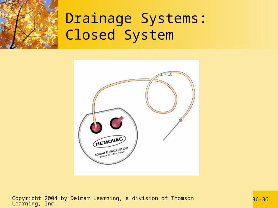
Topical Therapy• Monitor drainage of wounds
- Penrose drains- Jackson-Pratt drains- Hemovac drains
• Maintenance of open proliferative wound edges
36-36Copyright 2004 by Delmar Learning, a division of Thomson Learning, Inc.
Drainage Systems: Closed System

36-37Copyright 2004 by Delmar Learning, a division of Thomson Learning, Inc.
Drainage Systems: Tube and Reservoir System
36-38Copyright 2004 by Delmar Learning, a division of Thomson Learning, Inc.
Evaluation
Achievement or Maintenance of Skin Integrity• Wound healing• Prevention of infection• Client education
36-39Copyright 2004 by Delmar Learning, a division of Thomson Learning, Inc.
Management Guidelines for Specific Wounds
Abrasions and Lacerations Surgical Incisions Skin Tears

36-40Copyright 2004 by Delmar Learning, a division of Thomson Learning, Inc.
Types of Wounds

36-41Copyright 2004 by Delmar Learning, a division of Thomson Learning, Inc.
Management Guidelines for Specific Wounds
Lower Extremity Ulcers• Venous ulcers• Arterial ulcers• Neuropathic ulcers• Atypical ulcers

36-42Copyright 2004 by Delmar Learning, a division of Thomson Learning, Inc.
Management Guidelines for Specific Wounds
Burns• Thermal, chemical, or electrical causes• Epidermal burns• Superficial partial-thickness burns• Deep partial-thickness burns• Full-thickness burns

36-43Copyright 2004 by Delmar Learning, a division of Thomson Learning, Inc.
Contusions, Strains, and Sprains: Management Guidelines
Contusions are bruises of the soft tissues with no break in the skin surface.
Contusions resolve spontaneously and require no active management.
Application of ice for 24 hours following injury can reduce the amount of edema and bruising.

36-44Copyright 2004 by Delmar Learning, a division of Thomson Learning, Inc.
Contusions, Strains, and Sprains: Management Guidelines
Strains represent “stretch” injuries of muscles, tendons, or ligaments.
Application of ice for 24 hours to reduce swelling and bleeding, elevation to reduce swelling, use of an elastic wrap or sling, and aspirin or acetaminophen as needed.

36-45Copyright 2004 by Delmar Learning, a division of Thomson Learning, Inc.
Contusions, Strains, and Sprains: Management Guidelines
First- and second-degree sprains involve trauma to ligaments, tendons, or bones around a joint.
Caused by twisting or pulling forces. Nonsteroidal anti-inflammatory drugs,
ice, elastic wrap or sling, and restricted activity until symptoms resolve

36-46Copyright 2004 by Delmar Learning, a division of Thomson Learning, Inc.
Contusions, Strains, and Sprains: Management Guidelines
Third-degree sprains represent a more serious injury.
Characterized by separation of tendons and ligaments from their bony attachments.
Produce severe bleeding, swelling, pain, and loss of function.

36-47Copyright 2004 by Delmar Learning, a division of Thomson Learning, Inc.
Contusions, Strains, and Sprains: Management Guidelines
Management of Third-Degree Strains• Rest• Crutch to prevent weight bearing during
ambulation• Ice for 24 to 72 hours• Compression with an elastic wrap• Soft cast or sling• Elevation
36-48Copyright 2004 by Delmar Learning, a division of Thomson Learning, Inc.
Contusions, Strains, and Sprains: Management Guidelines
Management of Third-Degree Sprains• Narcotic analgesics for severe pain• Restricted mobility for up to 3 weeks• Surgery may be required for reattachment or
removal of torn tendons and ligaments.• Potential for developing post-traumatic
arthritis

36-49Copyright 2004 by Delmar Learning, a division of Thomson Learning, Inc.
Administer Heat and Cold Therapy
Heat and cold therapies require nursing care that assesses both the vasoconstriction and vasodilation of an individual.

36-50Copyright 2004 by Delmar Learning, a division of Thomson Learning, Inc.
Administer Heat and Cold Therapy
Conditions that necessitate precautions in the use of heat and cold applications:• Neurosensory impairment• Impaired mental status• Impaired circulation• Open wounds, broken skin, scar formation,
edema
36-51Copyright 2004 by Delmar Learning, a division of Thomson Learning, Inc.
Administer Heat and Cold Therapy
Heat Therapy• Promotes vasodilation• Decreases blood viscosity• Increases tissue metabolism• Increases capillary permeability• Reduces muscle tension

36-52Copyright 2004 by Delmar Learning, a division of Thomson Learning, Inc.
Administer Heat and Cold Therapy
Cold Therapy• Promotes vasoconstriction• Increases blood viscosity• Decreases tissue metabolism• Local anesthetic effect• Decreases muscle tension