chapter 4 tissue organization
DESCRIPTION
AnatomyTRANSCRIPT

The Tissue Level of Organization
Chapter 4

Ch 4 Outline
4 major tissue types- functions, features 4 types of membranes & functions Injury Aging

Four Types of Tissues
Tissues- collections of cells that perform specific, limited functions
Histology- study of tissues
4 Types of tissue: Epithelial tissue Connective tissue Muscle tissue Neural tissue

Four Types of Tissues Epithelial tissue
Covers exposed surfaces Lines internal passageways Forms glands
Connective tissue Fills internal spaces Supports other tissues Transports materials Stores energy

Four Types of Tissues
Muscle tissue Specialized for contraction Skeletal muscle, heart muscle, and walls of
hollow organs
Neural tissue Carries electrical signals from one part of the
body to another

Epithelial Tissues
Epithelia- layers of cells covering internal or
external surfaces
Glands- structures that
produce fluid secretions, either
attached or derived from
epithelia

Characteristics of Epithelia
Cellularity- composed almost entirely of cells held together by cell junctions
Polarity- exposed surface faces exterior of body or internal space (apical) and side attached to base- (basal), structural & functional differences

Polarity of Epithelial Cells
Characteristics of Epithelia
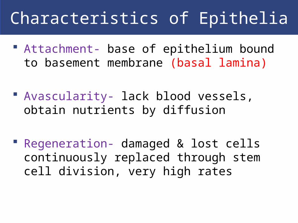
Attachment- base of epithelium bound to basement membrane (basal lamina)
Avascularity- lack blood vessels, obtain nutrients by diffusion
Regeneration- damaged & lost cells continuously replaced through stem cell division, very high rates

Functions of Epithelial Tissues
Provide physical protection- abrasion, dehydration, biological/chemical agent
Control permeability- ions, nutrients
Provide sensation- sensory nerves provide info about external environment
Produce specialized secretions (glandular epithelium)- physical protection, temp regulation, chemical messengers

Specializations of Epithelial Cells
In order to perform functions, highly specialized structure- polarity: cell divided into 2 distinct regions
Apical surfaces: exposed to internal or external environment Microvilli increase absorption or secretion Cilia (ciliated epithelium) move fluid
Basolateral surfaces: Base- cellular attachment Sides- cells contact neighbors

Maintaining the Integrity of Epithelia
To be an effective barrier must form a complete cover:
Intercellular connections
Attachment to basal lamina
Epithelial maintenance and repair

Intercellular Connections Epithelial cells tightly bound together CAMs (cell adhesion molecules)- transmembrane
proteins
Intercellular cement- proteoglycans contain hyaluronan (hyaluronic acid)

Intercellular Connections
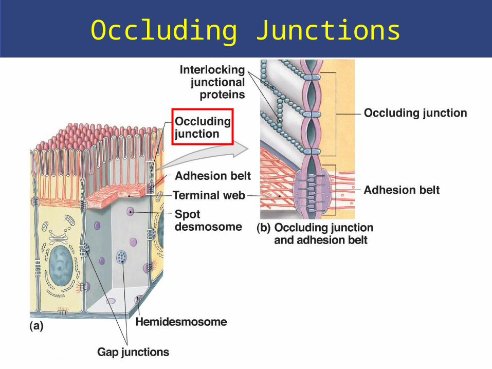
Cell junctions- form bonds with other cells or extracellular material: Occluding (tight) junctions Gap junctions Macula adherens (desmosomes)

Occluding (Tight) junctions Between two plasma membranes
Adhesion belt attaches to terminal web Prevents passage of water and solutes Isolates wastes in the lumen of GI tract
Occluding Junctions

Gap junctions Allow rapid communication
Held together by connexons (channel proteins) Allow ions to pass between cells Coordinate contractions in heart muscle

Gap Junctions

Macula adherens (Desmosomes) CAMs, dense areas, intercellular cement link plasma
membranes Spot desmosomes- tie cells together & allow
bending, twisting Hemidesmosomes- attach cells to the basal lamina
Attached to cytoskeleton

Spot Desmosomes
CAMS
Intercellular cement
Small discs connected to intermediate
filaments, which function to
stabilize cell shape

Hemidesmosome
Attach cell to extracellular filaments in basal lamina
Stabilize cell position and anchor to underlying tissues

Attachment to the Basal Lamina
Lamina lucida- clear layer closest to epithelium Glycoproteins & fine protein filaments Thin layer secreted by epithelia Restricts movement of proteins from underlying
connective tissue into epithelium
Lamina densa- dense layer Thick fibers produced by connective tissue Strength and filtration


Epithelial Maintenance and Repair
Constantly replaced by division of germinative cells (stem cells) near basal lamina
Exposure to enzymes, toxic chemicals, bacteria, mechanical abrasion
Classification of Epithelia
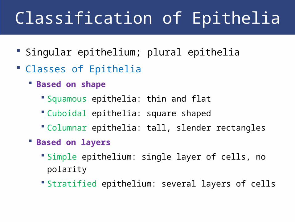
Singular epithelium; plural epithelia
Classes of Epithelia Based on shape
Squamous epithelia: thin and flat
Cuboidal epithelia: square shaped
Columnar epithelia: tall, slender rectangles
Based on layers
Simple epithelium: single layer of cells, no polarity
Stratified epithelium: several layers of cells

Simple Epithelia

Stratified Epithelia

Simple Squamous Epithelia Thin, flat, irregular- smooth surface Simple squamous epithelium
Absorption and diffusion Aveoli, lining body cavities, lining heart &
blood vessels
Mesothelium- lines body cavities
Endothelium- lines heart and blood vessels

Simple Squamous Epithelia

Located where mechanical stress is severe- skin surface, lining of mouth, esophagus, anus
Protects against attacks Keratin- protein adds strength & water resistant

Simple cuboidal epithelium- secretion and absorption (kidney tubules)
Sratified Cuboidal EpitheliaSimple Cuboidal Epithelia

Stratified Cuboidal Epithelia
Stratified cuboidal epithelia- sweat ducts and mammary ducts

-Tolerates repeated cycles of stretching and recoiling and returns to its previous shape without damage
-Appearance changes as stretching occurs
Columnar Epithelia
Simple columnar epithelium Absorption and secretion
Pseudostratified columnar epithelium Cilia movement
Stratified columnar epithelium Protection
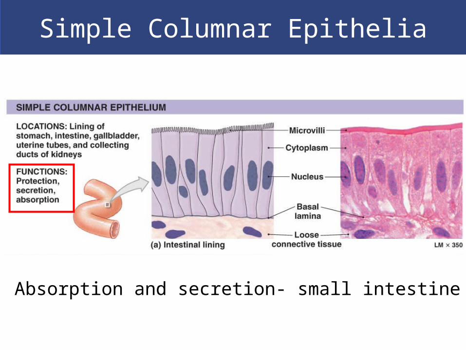
Simple Columnar Epithelia
Absorption and secretion- small intestine

Pseudostratified CiliatedColumnar Epithelia
-Several cell types, varying shapes & functions-Not truly stratified-Cilia movement- nasal cavity, trachea, bronchi
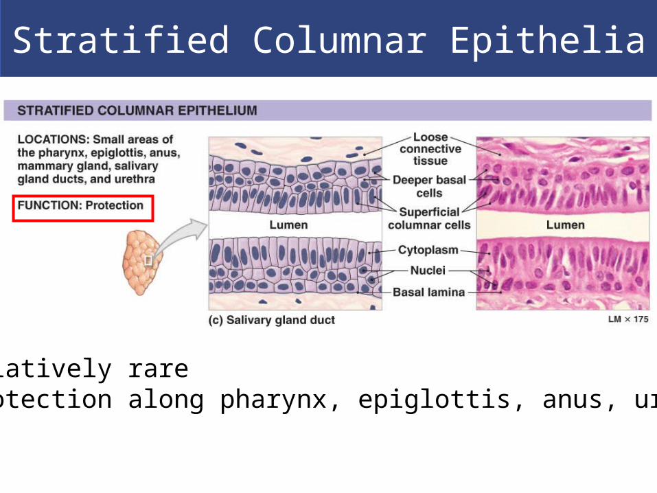
Stratified Columnar Epithelia
-Relatively rare-Protection along pharynx, epiglottis, anus, urethra
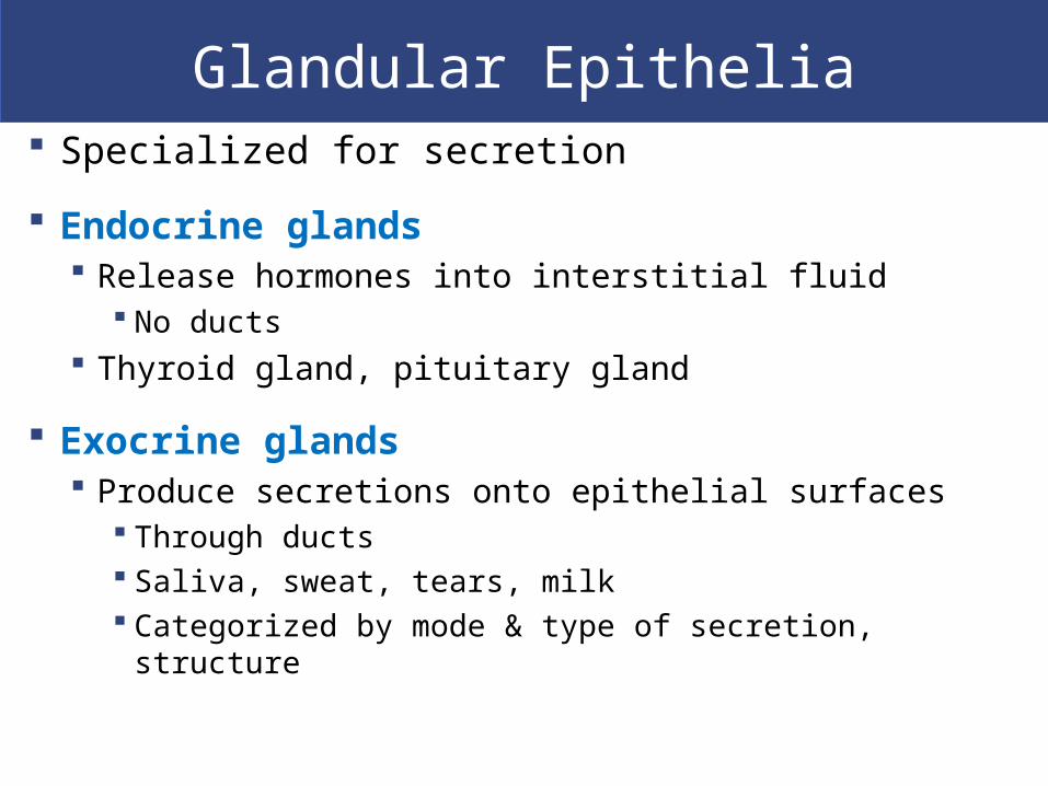
Glandular Epithelia Specialized for secretion
Endocrine glands Release hormones into interstitial fluid
No ducts Thyroid gland, pituitary gland
Exocrine glands Produce secretions onto epithelial surfaces
Through ducts Saliva, sweat, tears, milk Categorized by mode & type of secretion, structure

Modes of Glandular Secretionby Exocrine Glands
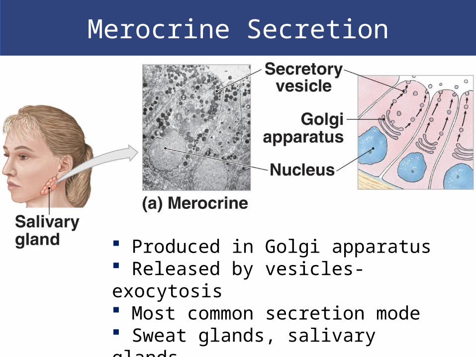
Merocrine Secretion
Produced in Golgi apparatus Released by vesicles- exocytosis Most common secretion mode Sweat glands, salivary glands

Apocrine Secretion
Produced in Golgi apparatus Released by shedding cytoplasm Mammary gland

Holocrine Secretion
Released by cells bursting, killing gland cells Gland cells replaced by stem cells Sebaceous gland- oil coating on hair

Types of Secretion by Exocrine Glands
Serous glands: watery secretions- saliva
Mucous glands: secrete mucins- mucus
Mixed exocrine glands: both serous and mucous

Gland Structure- Exocrine Glands
Unicellular glands- independent, scattered cells Mucous (goblet) cells- intestinal lining
Multicellular glands- glandular epithelia & aggregations of gland cells Structure of the duct- simple or compound
Shape of secretory portion of the gland Tubular (straight/coiled) or blind pockets
Relationship between ducts & glandular areas Branched

Simple Multicellular Exocrine Glands
Simple- single duct does not
divide on way to gland cell

Compound Multicellular Exocrine Glands
Compound- duct divides one or more times on
way to gland cell

Tubular Exocrine Glands
Tubular- form tubes, straight or
coiled

Alveolar/Acinar Exocrine Glands
Alveolar- blind pocketsTubuloalveolar- both tubes and
pockets

Connective Tissues
Connect epithelium to the rest of the body Dense layer of basal lamina produced by connective
tissue
Functions: Provide structure, support- bone Store energy- fat Transport materials- blood Defending body from microbes Protect organs
Have no contact with outside environment

Connective Tissues
Characteristics of Connective Tissues Specialized cells
Solid extracellular protein fibers
Fluid extracellular ground substance
Extracellular components make up the matrix
Majority of connective tissue volume
Determines specialized function
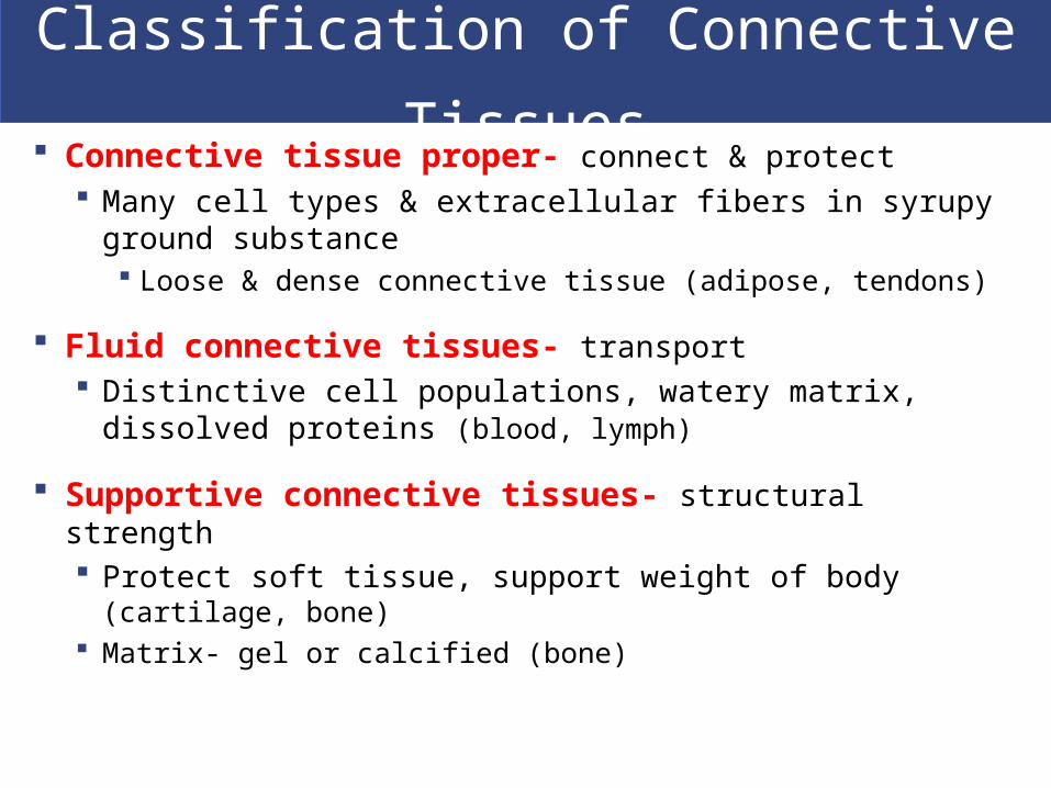
Classification of Connective Tissues Connective tissue proper- connect & protect
Many cell types & extracellular fibers in syrupy ground substance Loose & dense connective tissue (adipose, tendons)
Fluid connective tissues- transport Distinctive cell populations, watery matrix, dissolved
proteins (blood, lymph)
Supportive connective tissues- structural strength Protect soft tissue, support weight of body (cartilage, bone) Matrix- gel or calcified (bone)
Connective Tissues
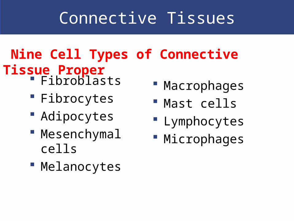
Fibroblasts Fibrocytes Adipocytes Mesenchymal cells Melanocytes
Macrophages Mast cells Lymphocytes Microphages
Nine Cell Types of Connective Tissue Proper

Connective Tissue Proper Cells
Fibroblasts Most abundant cell type:
Always found in all connective tissue proper
Secrete proteins and hyaluronan (cellular cement)
Fibrocytes 2nd most abundant cell type:
Found in all connective tissue proper Maintain the fibers of connective tissue
proper

Connective Tissue Proper Cells
Macrophages “Big eaters” immune system:
Eat pathogens & damaged cells Fixed macrophages- stay in tissue Free macrophages- migrate
Adipocytes Fat cells- each cell stores a single, large fat droplet
Mesenchymal Cells Stem cells that respond to injury or infection:
Differentiate into fibroblasts, macrophages

Connective Tissue Proper Cells
Melanocytes Synthesize and store the brown pigment
melanin
Mast Cells Stimulate inflammation after injury or infection:
Release histamine and heparin
Basophils (white blood cells) also contain histamine and heparin

Connective Tissue Proper Cells Lymphocytes
Specialized immune cells in lymphatic system: B-cells (plasma cells)- produce antibodies T-cells
Microphages (neutrophils, eosinophils) Phagocytic white blood cells:
Respond to signals from macrophages & mast cells

Connective Tissue Fibers
Collagen fibers
Most common fibers in connective tissue proper
Long, straight, unbranched
Strong and flexible
Tendons & ligaments

Connective Tissue Fibers
Reticular fibers Network of interwoven fibers- stroma Strong & flexible Stabilize functional cells (parenchyma) &
blood vessels, nerves Sheaths around organs
Connective Tissue Fibers
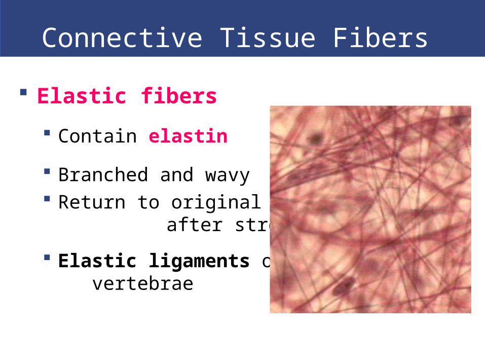
Elastic fibers
Contain elastin
Branched and wavy Return to original length after
stretching
Elastic ligaments of vertebrae

Connective Tissues
Ground Substance
Clear, colorless, viscous (thick)
Fills spaces between cells & slows pathogen
movement
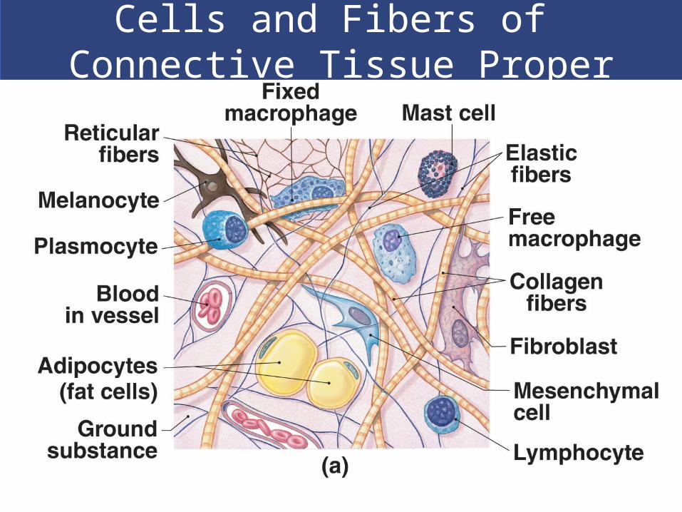
Cells and Fibers of Connective Tissue Proper

Cells and Fibers of Connective Tissue Proper
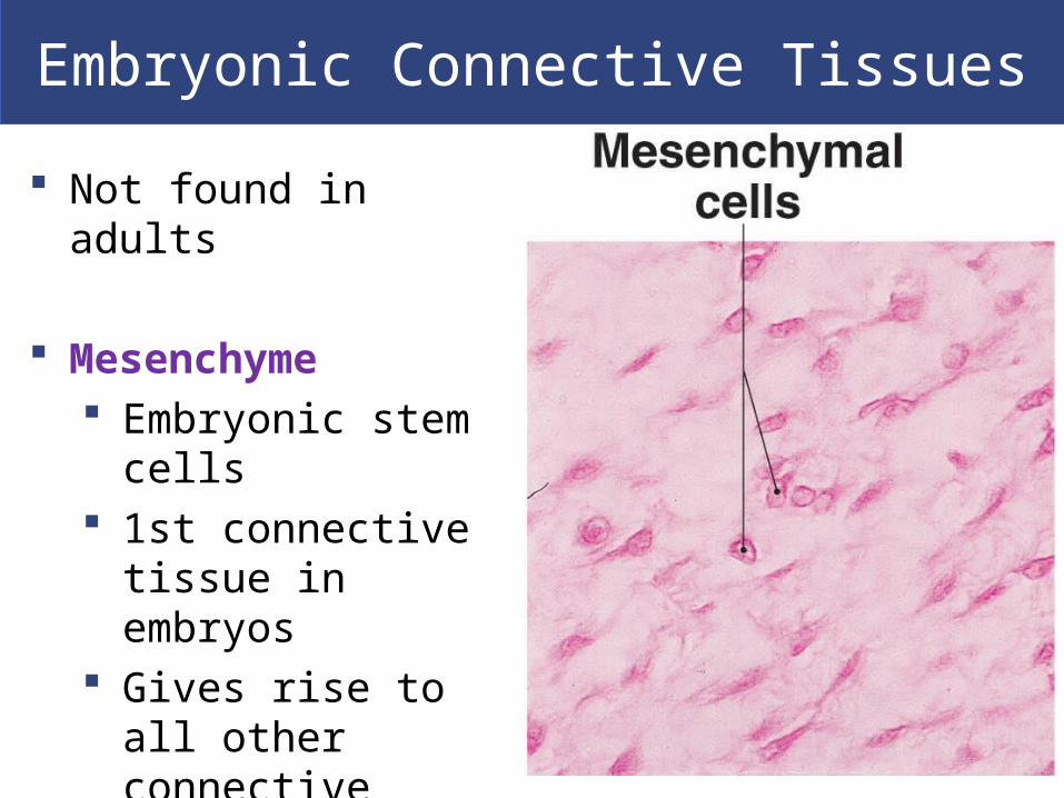
Embryonic Connective Tissues
Not found in adults
Mesenchyme Embryonic stem cells 1st connective tissue
in embryos Gives rise to all other
connective tissue

Embryonic Connective Tissues
Mucous connective tissue Wharton’s jelly Loose embryonic
connective tissue

Loose Connective Tissues
Packing materials of the body
Fill spaces between organs, cushion, stabilize specialized cells in organs, support epithelia, blood vessels, nerves, store lipids
Three types in adults Areolar Adipose Reticular

Areolar TissueLoose Connective Tissues
Least specialized
Open framework Viscous ground substance
most volume
Elastic fibers
Holds blood vessels & capillary beds
Under skin (subcutaneous layer), padding

Adipose TissueLoose Connective Tissues
Contains many adipocytes (fat cells)
Types of adipose tissue White fat:
Most common Stores fat, absorbs shocks, slows heat loss (insulation)
Brown fat: More vascularized Adipocytes have many mitochondria When stimulated by nervous system, fat break down
accelerates, releasing energy Energy absorbed from surrounding tissues- heats body
Adipose Tissue
Adipose cells Adipocytes in adults do not divide:
Expand to store fat
Shrink as fats are released
Mesenchymal cells divide and differentiate: Produce more fat cells when more storage is
needed

Adipose Tissues
Functions- padding, absorbs shock, insulation, packing/filler around structures

Reticular TissueLoose Connective Tissues
Provides support Complex, 3D network- stroma
Support functional cells (parenchyma) Reticular organs
Spleen, liver, lymph nodes, and bone marrow

Dense Connective Tissues
Connective tissues proper, tightly packed with high numbers of collagen or elastic fibers
Dense regular connective tissue
Dense irregular connective tissue
Elastic tissue

Dense Regular Connective Tissues
Tightly packed, parallel collagen fibers Tendons- attach muscles to bones Ligaments- connect bone to bone & stabilize organs Aponeuroses- tendinous sheet that attaches a broad,
flat muscles to another muscle or

Dense Irregular Connective Tissues
Interwoven networks of collagen fibers
Strengthen &support areas subjected to stress from multiple directions, gives skin strength
Sheaths cartilage- perichondrium Sheaths bones- periosteum
Form capsules around some organs (liver, kidneys, spleen)
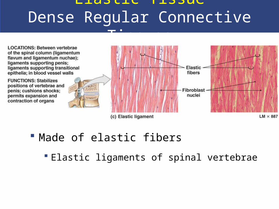
Elastic TissueDense Regular Connective Tissues
Made of elastic fibers
Elastic ligaments of spinal vertebrae

Fluid Connective Tissues
Blood & lymph Watery matrix of dissolved proteins Fluid Elements- extracellular
Plasma Interstitial fluid Lymph
Formed Elements- specific cell types Formed elements of blood
Red blood cells- erythrocytes White blood cells- leukocytes Platelets

Formed Elements of the Blood

Connective Tissues
Lymph- extracellular fluid
Collected from interstitial space
Monitored by immune system
Transported by lymphatic
system
Returned to venous system

Fluid Tissue Transport Systems
Cardiovascular system (blood)
Arteries
Capillaries
Veins
Lymphatic system (lymph)
Lymphatic vessels
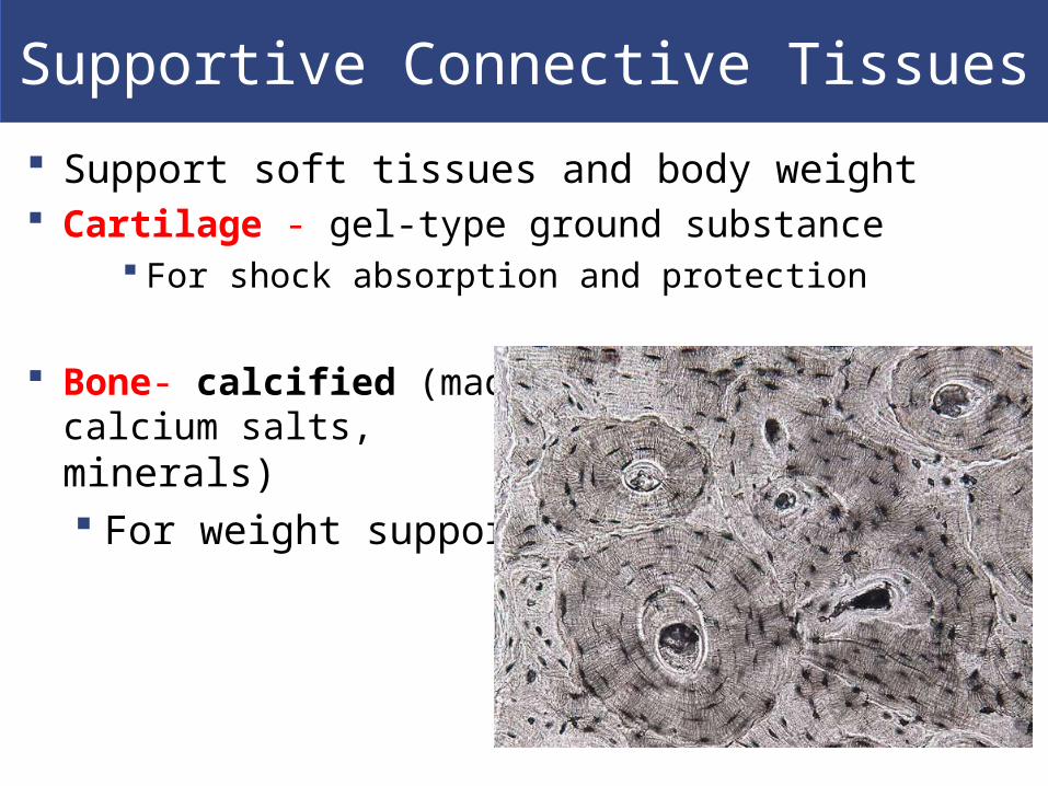
Supportive Connective Tissues
Support soft tissues and body weight Cartilage - gel-type ground substance
For shock absorption and protection
Bone- calcified (made rigid by calcium salts, minerals) For weight support

Cartilage Characteristics
Cartilage Matrix- firm gel contains proteoglycans derived from chondroitin sulfates complexed with ground substance proteins Chondrocytes (cartilage cells)- occupy small
chambers- lacunae
Cartilage Structure- no blood vessels, exchange by diffusion only Chondrocytes produce antiangiogenesis factor
Perichondrium- sets cartilage apart from surrounding tissues
Outer, fibrous layer- strength Inner, cellular layer- growth and maintenance

Interstitial Growth of Cartilage
*Most important during
development
- Enlarges cartilage from
within

Appositional Growth of Cartilage
- Adds new layers of
cartilage to the surface

Hyaline Cartilage
Hyaline cartilage (most common) Stiff, flexible support Reduces friction between bones Found in synovial joints, rib tips, sternum, trachea,
elbows, knees
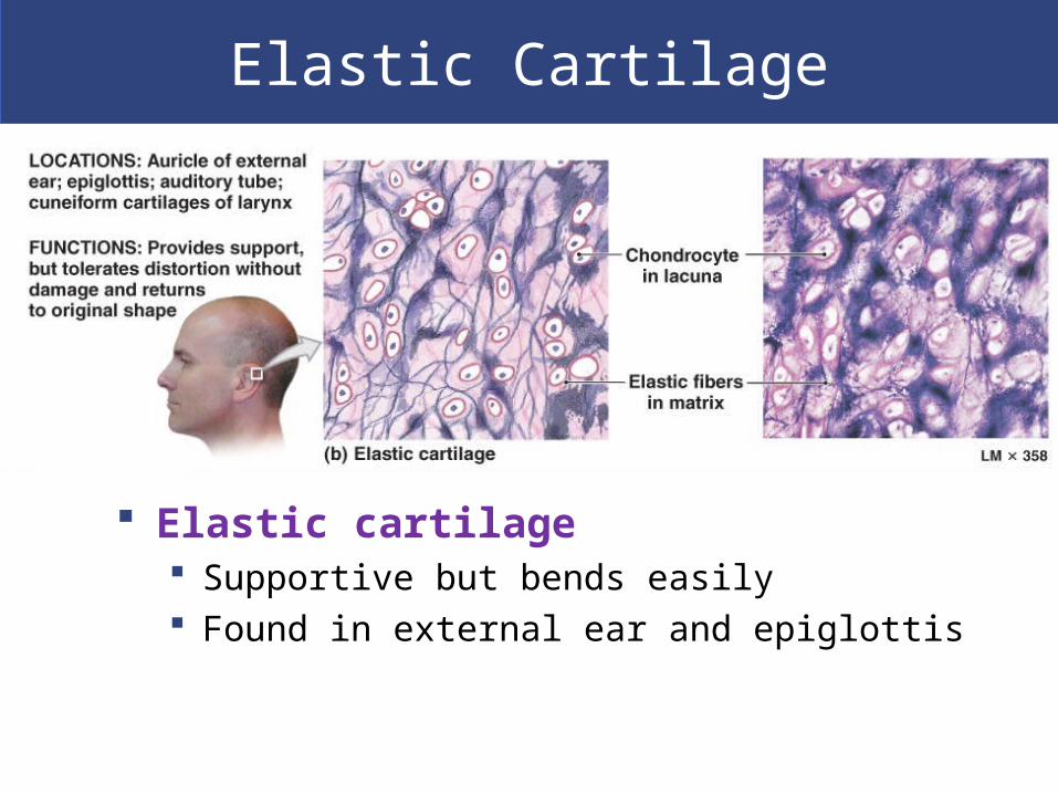
Elastic Cartilage
Elastic cartilage Supportive but bends easily Found in external ear and epiglottis

Fibrous Cartilage
Fibrous cartilage (fibrocartilage) Limits movement Prevents bone-to-bone contact Pads knee joints Found between pubic bones and intervertebral discs
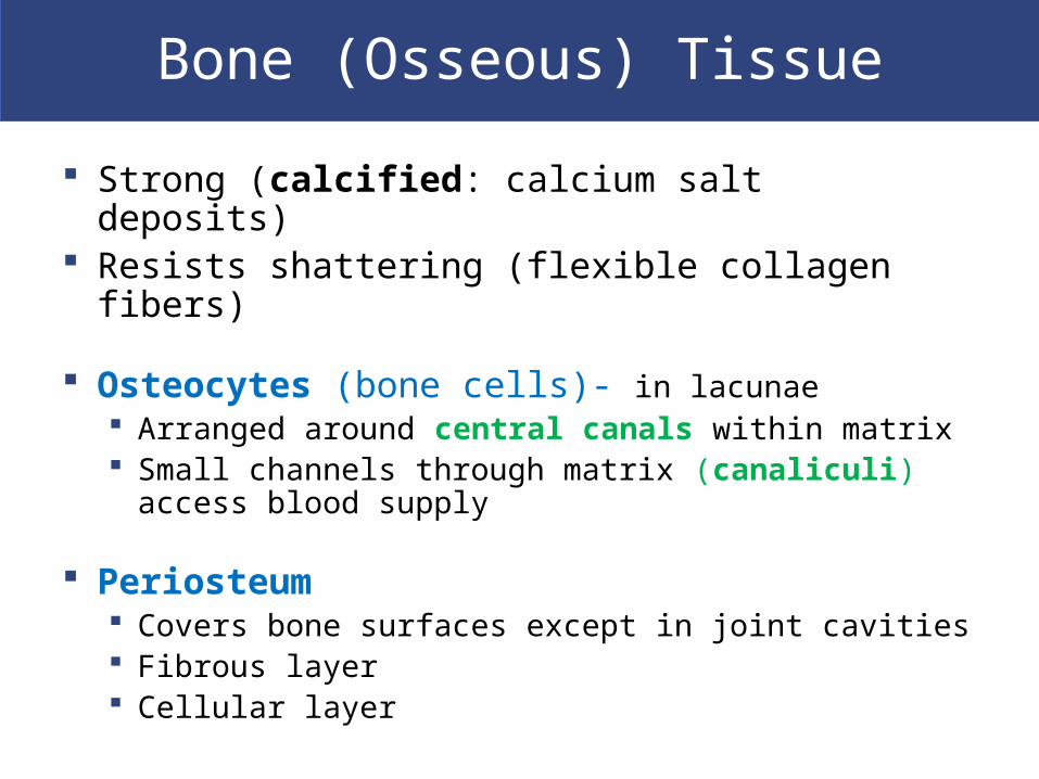
Bone (Osseous) Tissue
Strong (calcified: calcium salt deposits) Resists shattering (flexible collagen fibers)
Osteocytes (bone cells)- in lacunae Arranged around central canals within matrix Small channels through matrix (canaliculi) access
blood supply
Periosteum Covers bone surfaces except in joint cavities Fibrous layer Cellular layer

Supportive Connective Tissue- Bone
Matrix- mixture of calcium salts (calcium phosphate, carbonate) and collagen fibers

Supportive Connective Tissues
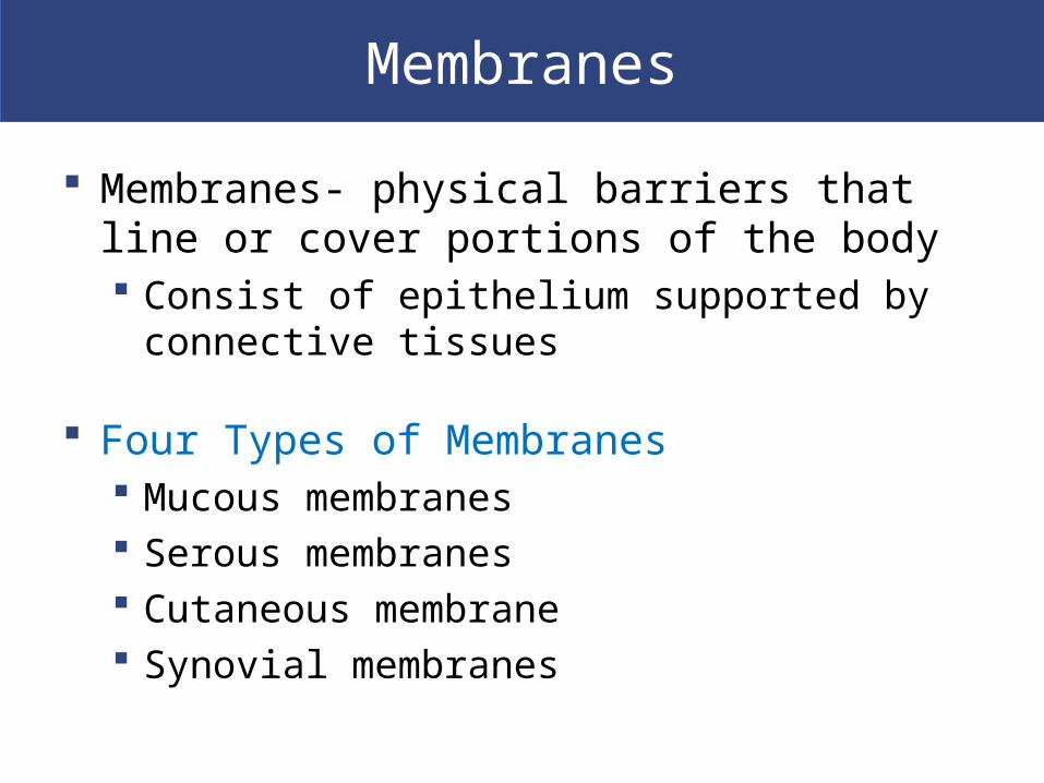
Membranes
Membranes- physical barriers that line or cover portions of the body Consist of epithelium supported by connective
tissues
Four Types of Membranes Mucous membranes Serous membranes Cutaneous membrane Synovial membranes

Mucous Membranes
Line passageways with external connections Digestive, respiratory, urinary, reproductive tracts
Epithelial surfaces must be moist Reduce friction & facilitate absorption and excretion
Lamina propria- areolar tissue

Serous Membranes Line cavities not open to outside Thin but strong Have fluid, transudate, to reduce friction Parietal portion- covers cavity Visceral portion, serosa- covers organs

Three Serous Membranes
Pleura: Lines pleural cavities Covers lungs
Peritoneum: Lines peritoneal cavity Covers abdominal organs
Pericardium: Lines pericardial cavity Covers heart
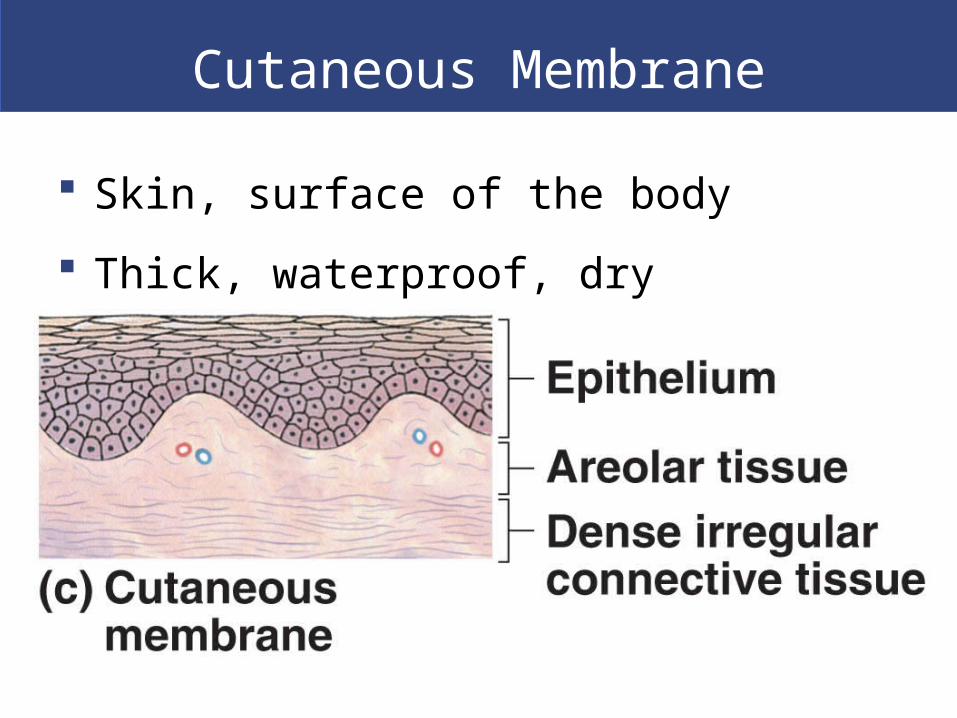
Cutaneous Membrane
Skin, surface of the body
Thick, waterproof, dry

Synovial Membranes
Articulating joint cavities (bones very close or touching) Produce synovial fluid (lubricant)- joint movement is
important in stimulating formation & circulation of fluid
Protect the ends of bones

Internal Framework of the Body
Connective tissues Provide strength and stability Maintain positions of internal organs Provide routes for blood vessels, lymphatic vessels,
and nerves
Fasciae (singular form is fascia)
Connective tissue layers surround, support organs Three types of fasciae
– Superficial fascia– Deep fascia– Subserous fascia


Muscle Tissue
Specialized for contraction
Produces all body movement
Three types of muscle tissue Skeletal muscle
Large body muscles responsible for movement
Cardiac muscle
Found only in the heart
Smooth muscle
Found in walls of hollow, contracting organs (blood vessels; urinary bladder; respiratory, digestive,
reproductive tracts), contractility & support
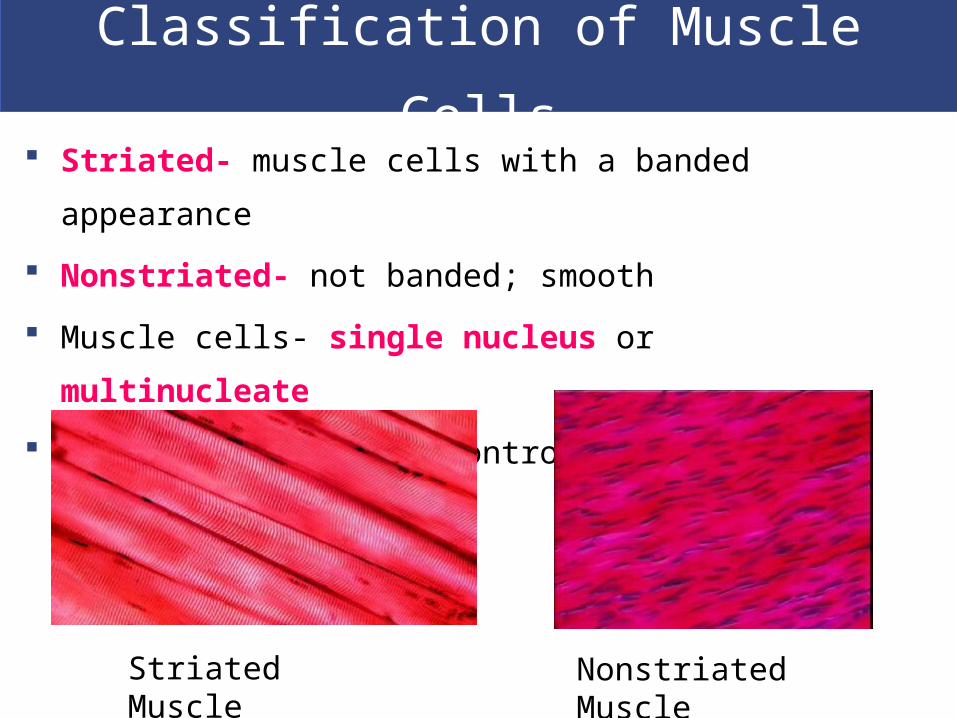
Classification of Muscle Cells
Striated- muscle cells with a banded appearance
Nonstriated- not banded; smooth
Muscle cells- single nucleus or multinucleate
Muscle cells can be controlled voluntarily or involuntarily
Striated Muscle Nonstriated Muscle

Muscle Tissue- Skeletal Long, thin, very large- muscle
fibers Several hundred nuclei! Do NOT divide New fibers- myosatellite cells Striated voluntary muscle-
nervous system

Muscle Tissue- CardiacCardiac cells- cardiocytesForm branching networks connected at intercalated discsRegulated by pacemaker cellsStriated involuntary muscle

Muscle Tissue- Smooth
Smooth muscle cells Small and tapered Divide and regenerate No striations
Nonstriated involuntary muscle

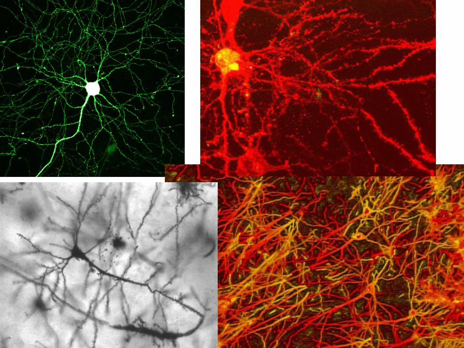
Neural Tissue
Specialized for conducting electrical impulses
Rapidly senses internal or external environment
Processes information and controls responses
Central nervous system Brain Spinal cord

Two Kinds of Neural Cells
Neurons Nerve cells Perform electrical communication
Neuroglia Supporting cells Repair and supply nutrients to neurons
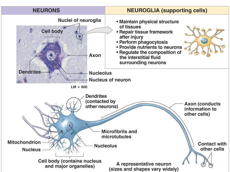

Cell Parts of a Neuron Cell body- nucleus Dendrites- short branches extending from cell body
Receive incoming signals
Axon (nerve fiber)- long, thin extension of cell body Carries outgoing electrical signals

Tissue Injuries and Repair
Tissues respond to injuries to maintain homeostasis
Cells restore homeostasis with two processes
Inflammation- tissues 1st response to injury Regeneration
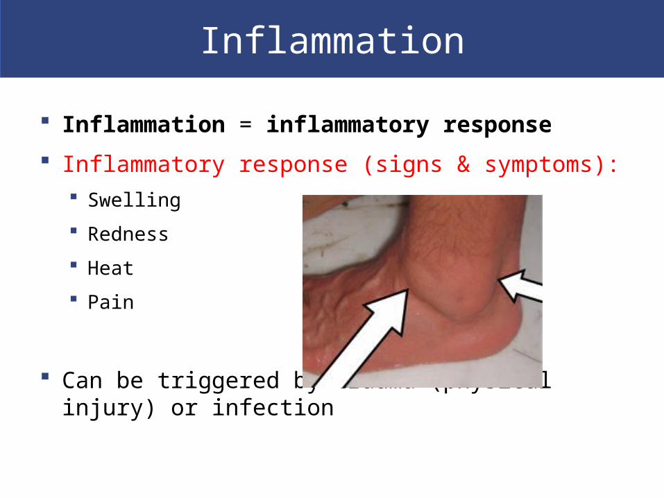
Inflammation
Inflammation = inflammatory response
Inflammatory response (signs & symptoms): Swelling
Redness
Heat
Pain
Can be triggered by trauma (physical injury) or infection

Process of Inflammation Damaged cells release chemical signals into
surrounding interstitial fluid Prostaglandins, proteins, potassium ions

Process of Inflammation
Damaged cells break down, lyse- contents leak out Lysosomes release hydrolytic enzymes Tissue destruction surrounding tissues- necrosis
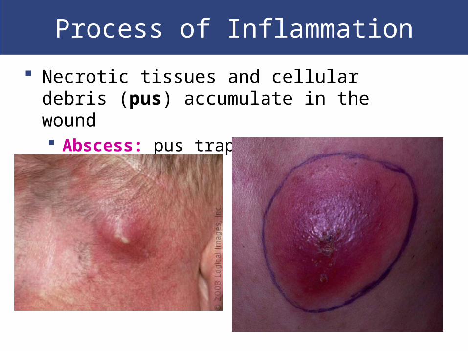
Process of Inflammation
Necrotic tissues and cellular debris (pus) accumulate in the wound Abscess: pus trapped in an enclosed area

Process of Inflammation
Injury stimulates mast cells to release:
Histamine Heparin Prostaglandins

Process of Inflammation
Dilation of blood vessels (redness & heat) Increases blood circulation Brings more nutrients and oxygen to the area Removes wastes
Plasma diffuses into the area Swelling and pain
Phagocytic white blood cells Clean up the area- damaged cells, pathogens

An Introduction to Inflammation

Regeneration Injury or infection is cleaned up healing begins Fibrocytes move into necrotic area
Lay down collagen fibers- bind area together (scar tissue)
New cells migrate into area or mesenchymal stem cells produce them
Not all tissues can regenerate Epithelia & connective tissues regenerate well Cardiac cells & neurons do not regenerate (or regenerate
poorly)
Aging and Tissue Tissue repair speed, efficiency decreases with age:
Slower rate of energy consumption (metabolism) Hormonal alterations Reduced physical activity
Chemical & structural tissue changes Thinning epithelia and connective tissues Increased bruising and bone brittleness Cardiovascular disease- cumulative damage Mental deterioration

Aging and Cancer Incidence
Cancer rates increase with age 1 in 4 people in US develops cancer Cancer is the #2 cause of death in US Environmental chemicals, cigarette smoke
cause cancer Longer exposure, accumulate mutations
– 7 mutations in 1 cell
Plastics (food); pesticides (air, water, soil, food); air pollution (industry, vehicles)