chapter 42 circulation (pretty neat). exchange issues. cells live in aqueous environments. the...
TRANSCRIPT

CHAPTER 42CIRCULATION
(pretty neat)

• Exchange Issues.
• Cells live in aqueous environments.
• The resources needed, move across membrane into cytoplasm.
• Metabolic wastes moved across membrane out of cytoplasm
• Materials must be transported through intracellular space
• Ultimate payoff; SPEED vs. ENERGY!!!!
Introduction

• Gastrovascular cavity either simple (hydra) or branched (Aurelia, flatworms).
• Body Wall MUST BE THIN - diffusion
• The fluid inside the cavity is continuous with the water outside through a single opening, the mouth.
• both the inner and outer tissue layers are bathed in fluid.
Most invertebrates have either a gastrovascular cavity or a circulatory system for internal transport
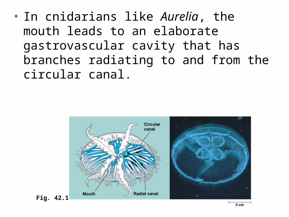
• In cnidarians like Aurelia, the mouth leads to an elaborate gastrovascular cavity that has branches radiating to and from the circular canal.
Fig. 42.1

• For animals with many cell layers, gastrovascular cavities are insufficient WHY???.
• Circulatory systems either open or closed.
• Both have a circulatory fluid (blood), a set of tubes (blood vessels), and a muscular pump (the heart).
• The heart powers circulation by using metabolic power to elevate the hydrostatic pressure of the blood (blood pressure), which then flows down a pressure gradient through its circuit back to the heart.
Circulatory System
• For animals with many cell layers, gastrovascular cavities are insufficient WHY???.
• Circulatory systems either open or closed.
• Both have a circulatory fluid (blood), a set of tubes (blood vessels), and a muscular pump (the heart).
• The heart powers circulation by using metabolic power to elevate the hydrostatic pressure of the blood (blood pressure), which then flows down a pressure gradient through its circuit back to the heart.

• In insects, other arthropods, and most mollusks, blood bathes organs directly in an open circulatory system.
• There is no distinction between blood and interstitial fluid, collectively called hemolymph.
• One or more hearts pump the hemolymph into interconnected sinuses
• Heart an elongated tube
Fig. 42.2a

• In a closed circulatory system, as found in earthworms, squid, octopuses, and vertebrates (us), blood is confined to vessels and is distinct from the interstitial fluid.
• One or more hearts pump blood into large vessels that branch into smaller ones cursing through organs.
• Materials are exchanged by diffusion between the blood and the interstitial fluid bathing the cells.
Fig. 42.2b
• Closed System
• Arteries – pressure
• Capillaries – exchange
• Veins - return
• Heart
• Atria – receive/collection chamber
• Ventricles – pump/pressure chamber
• Valves – ensure one way flow
• NOTE; CV adaptations and metabolic rate intimate!!
Vertebrate Cardiovascular System
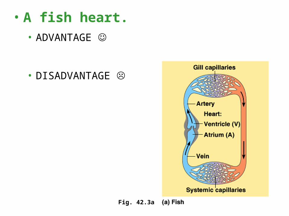
• A fish heart.• ADVANTAGE
• DISADVANTAGE
Fig. 42.3a
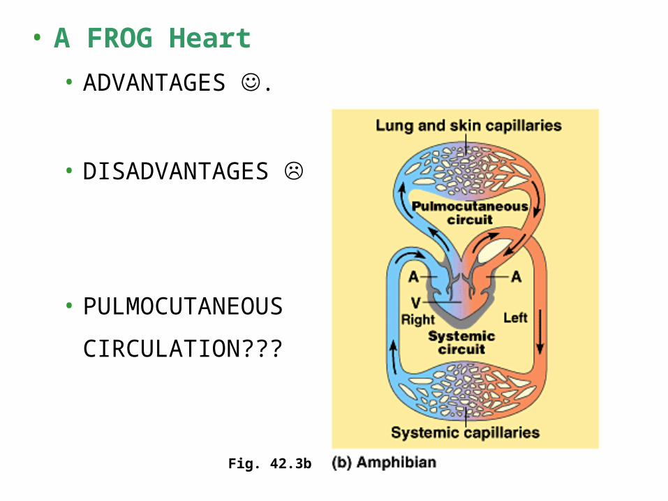
• A FROG Heart
• ADVANTAGES .
• DISADVANTAGES
• PULMOCUTANEOUS
CIRCULATION???
Fig. 42.3b

Crocodilians, birds, and mammals
ADVANTAGES
DISADVANTAGES
ENDOTHERMY VS.
ECTOTHERMY…
Fig. 42.3c

• In the mammalian cardiovascular system, the pulmonary and system circuits operate simultaneously.
• The two ventricles pump almost in unison
• While some blood is traveling in the pulmonary circuit, the rest of the blood is flowing in the systemic circuit.
4. Double circulation in mammals depends on the anatomy and pumping cycle of the heart

MEMORIZE MEMORIZE MEMORIZE MEMORIZE MEMORIZE MEMORIZE MEMORIZE
MEMORIZE MEMORIZE MEMORIZE MEMORIZE MEMORIZE MEMORIZE MEMORIZE
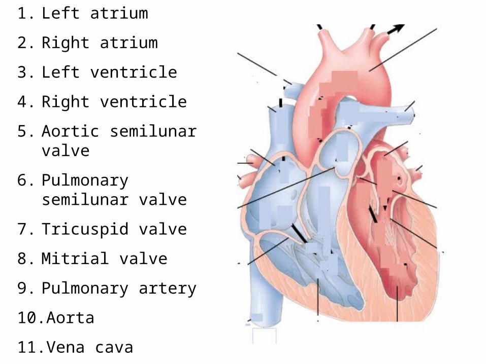
1. Left atrium
2. Right atrium
3. Left ventricle
4. Right ventricle
5. Aortic semilunar valve
6. Pulmonary semilunar valve
7. Tricuspid valve
8. Mitrial valve
9. Pulmonary artery
10. Aorta
11. Vena cava
12. Pulmonary vein

1. Left atrium
2. Right atrium
3. Left ventricle
4. Right ventricle
5. Aortic semilunar valve
6. Pulmonary semilunar valve
7. Tricuspid valve
8. Mitrial valve
9. Pulmonary artery
10. Aorta
11. Vena cava
12. Pulmonary vein

Thursday
• Review
• Quiz
• CV comparisons
• Mammalian circulation
• Systemic/pulmonary circulation
• Measurements
• Weekend assign
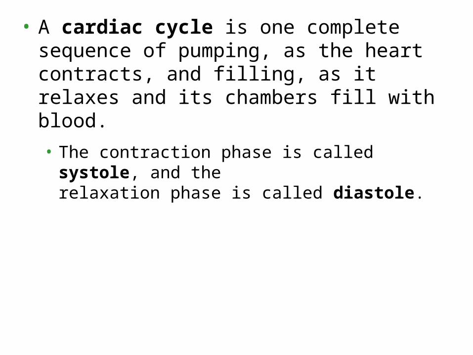
• A cardiac cycle is one complete sequence of pumping, as the heart contracts, and filling, as it relaxes and its chambers fill with blood.
• The contraction phase is called systole, and the relaxation phase is called diastole.
• Cardiac output depends on two factors: the rate of contraction or heart rate (number of beats per second) and stroke volume, the amount of blood pumped by the left ventricle in each contraction.
• The average stroke volume for a human is about 75 mL.
• The typical resting cardiac output, about 5.25 L / min, is about equivalent to the total volume of blood in the human body.
• Cardiac output can increase about fivefold during heavy exercise.
• Heart rate can be measured indirectly by measuring your pulse - the rhythmic stretching of arteries caused by the pressure of blood pumped by the ventricles.

• The heart sounds are caused by the closing of the valves.
• The sound pattern is “lub-dup, lub-dup, lub-dup.”
• The first heart sound (“lub”) is created by the recoil of blood against the closed AV valves.
• The second sound (“dup”) is the recoil of blood against the semilunar valves.

• A sphygmomanometer, an inflatable cuff attached to a pressure gauge, measures blood pressure fluctuations in the brachial artery of the arm over the cardiac cycle.
• The arterial blood pressure of a healthy human oscillates between about 120 mm Hg at systole and 70 mm Hg at diastole.
Fig. 42.11

• A defect in one or more of the valves causes a heart murmur, which may be detectable as a hissing sound when a stream of blood squirts backward through a valve.
• Some people are born with heart murmurs.
• Others are due damage to the valves by infection.
• Most heart murmurs do not reduce the efficiency of blood flow enough to warrant surgery.

Monday –
Goal: Follow Lab Packet Gather Data from/for your partner.
Blood Pressure – in a bunch of different postures
For Tuesday –
Complete Packet
Check out slides 36-37, be able to answer questions!!!!
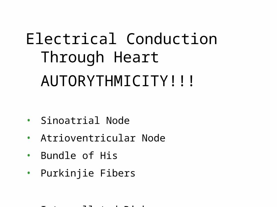
Electrical Conduction Through Heart
AUTORYTHMICITY!!!
• Sinoatrial Node
• Atrioventricular Node
• Bundle of His
• Purkinjie Fibers
• Intercollated Disks

• The cardiac cycle is regulated by electrical impulses that radiate throughout the heart.
• Cardiac muscle cells are electrically coupled by intercalated disks between adjacent cells.
Fig. 42.7


• The impulses generated during the heart cycle produce electrical currents that are conducted through body fluids to the skin.
• Here, the currents can be detected by electrodes and recorded as an electrocardiogram (ECG or EKG).

• While the SA node sets the tempo for the entire heat, it is influenced by a variety of physiological cues.
• Two sets of nerves affect heart rate with one set speeding up the pacemaker and the other set slowing it down.
• Heart rate is a compromise regulated by the opposing actions of these two sets of nerves.
• The pacemaker is also influenced by hormones.
• For example, epinephrine from the adrenal glands increases heart rate.
• The rate of impulse generation by the pacemaker increases in response to increases in body temperature and with exercise.

• The walls of both arteries and veins have three similar layers.• On the outside, a layer of connective tissue with elastic
fibers allows the vessel to stretch and recoil.
• Function ????
• A middle layer has smooth muscle and more elastic fibers.
• Function ????
• Lining the lumen of all blood vessels, including capillaries, is an endothelium, a single layer of SPECIALIZED EPITHELIAL CELLS.
• Function ????
Structure v Function of Vessels
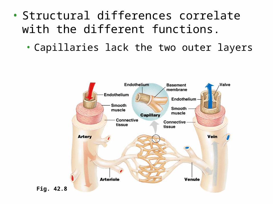
• Structural differences correlate with the different functions.
• Capillaries lack the two outer layers
Fig. 42.8

• The thinner-walled veins convey blood back to the heart at low velocity and pressure.
• Venous blood flow is mostly as a result of skeletal muscle contractions when we move that squeeze blood in veins.
• Within larger veins, flaps of tissues act as one-way valves.
Fig. 42.9
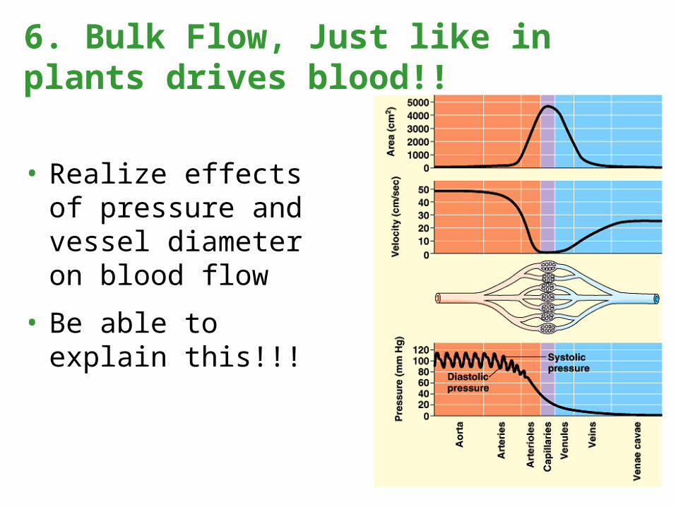
• Realize effects of pressure and vessel diameter on blood flow
• Be able to explain this!!!
6. Bulk Flow, Just like in plants drives blood!!
Today in APB1)BP Lab Discussion – mechanisms of physiological responses
2)Blood pressure vs. blood Flow vs. SA graphs
3)Electrochemical Control of Heart Rate
4)Hormonal Control of CV

• ??
Fig. 42.10
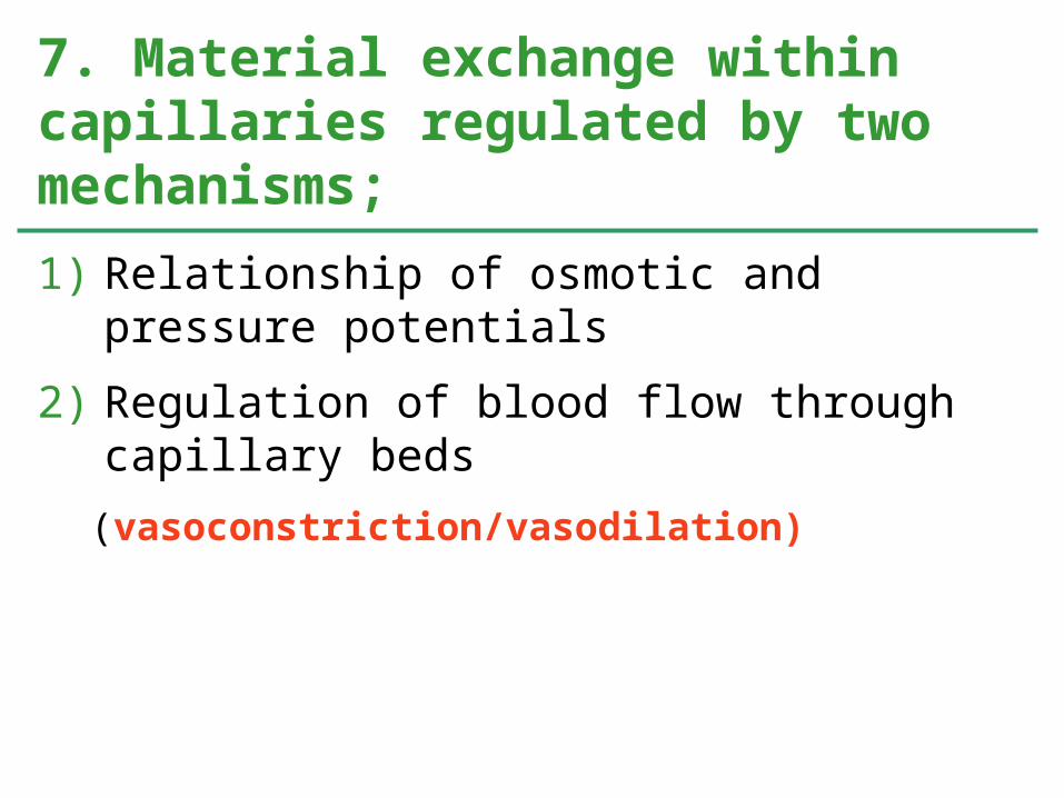
1) Relationship of osmotic and pressure potentials
2) Regulation of blood flow through capillary beds
(vasoconstriction/vasodilation)
7. Material exchange within capillaries regulated by two mechanisms;

Some significant questions for you;1) How do numbers indicated influence fluid flow?2) How would this diagram be different is someone with congestive
heart failure? Which systems would be involved?3) How does liver function affect this diagram?4) Identify at least three hormones involved in this balance.

A significant question for Monday;1) Chronic liver diseases such as liver cirrhosis negatively affect the
liver’s ability to produce albumin – the primary plasma protein. Explain how Chronic liver disease would affect the above diagram. AND THEN, predict how the body might attempt to compensate.

Profuse Sweating… Really severe nose bleed…

• Make a chart for the following blood pressure influencing hormones;
1. Renin
2. Angiotensin
3. Aldosterone
4. Atrial natriuretic hormone
5. Anti diuretic hormone
• Chart should include the following;
• Stimulus for secretion, site of secretion, direct mechanism of action, final effect on blood pressure

wow
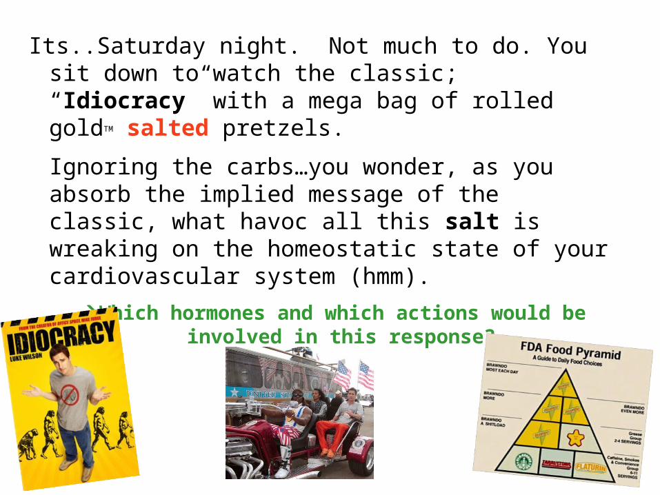
Its..Saturday night. Not much to do. You sit down to watch the classic; “Idiocracy” with a mega bag of rolled goldTM salted pretzels.
Ignoring the carbs…you wonder, as you absorb the implied message of the classic, what havoc all this salt is wreaking on the homeostatic state of your cardiovascular system (hmm).
Which hormones and which actions would be involved in this response?

To know from lab
1)What do systolic and diastolic pressures represent?
2)What is baroreceptor reflex? Why is it important?
3)What is relationship between fitness, heart rate, and blood pressure?
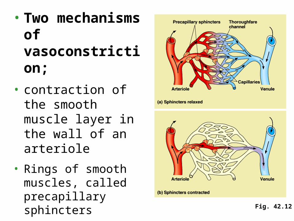
• Two mechanisms of vasoconstriction;
• contraction of the smooth muscle layer in the wall of an arteriole
• Rings of smooth muscles, called precapillary sphincters
• Some blood flows directly from arterioles to venules through thoroughfare channels which are always open. Fig. 42.12

• Fluids and some blood proteins that leak from the capillaries into the interstitial fluid are returned to the blood via the lymphatic system.
• Fluid enters this system by diffusing into tiny lymph capillaries intermingled among capillaries of the cardiovascular system.
• Once inside the lymphatic system, the fluid is called lymph, with a composition similar to the interstitial fluid.
• The lymphatic system drains into the circulatory system near the junction of the venae cavae with the right atrium.
8. The lymphatic system returns fluid to the blood and aids in body defense
• Lymph vessels, like veins, have valves that prevent the backflow of fluid toward the capillaries.
• Rhythmic contraction of the vessel walls help draw fluid into lymphatic capillaries.
• Also like veins, lymph vessels depend mainly on the movement of skeletal muscle to squeeze fluid toward the heart.

• Along a lymph vessels are organs called lymph nodes.
• The lymph nodes filter the lymph and attack viruses and bacteria.
• Inside a lymph node is a honeycomb of connective tissue with spaces filled with white blood cells specialized for defense.
• When the body is fighting an infection, these cells multiply, and the lymph nodes become swollen.
• In addition to defending against infection and maintaining the volume and protein concentration of the blood, the lymphatic system transports fats from the digestive tract to the circulatory system.

• Whole Blood – Components measured as Hematocrit
• Plasma
• Formed Elements
• Erythrocytes
• Leukocytes
• Platelets
• Hematocrit values ~ 30-45+
A bit more about blood

How variable is Hematocrit• Erythropoesis
• Formation of Erythrocytes
• WHERE – Bone Marrow
• HOW – Erythropoetin from Kidneys
• Toying with Erythropoesis
• EPO !!!!!

for Friday• Fully understand the following
Cardiac Cycle
Diastole/systole
Electrical activity
Blood flow
Pathways (vessels)
Regulation
Pressures
Blood volume/Blood osmolality regulation
RAAS, ADH