chapter 5 gases - mr. doyle suis...
TRANSCRIPT
Chapter 4
Cell Structure and
Function Sections 1-6
4.1 Food For Thought
• E. coli O157:H7A, strain of bacteria that causes severe illness
or death, occasionally contaminates foods such as ground
beef and fresh vegetables
• Outbreaks, which occur with disturbing regularity, also have
severe economic impacts
• Food growers and processors are now using procedures that
they hope will reduce E. coli O157:H7 outbreaks
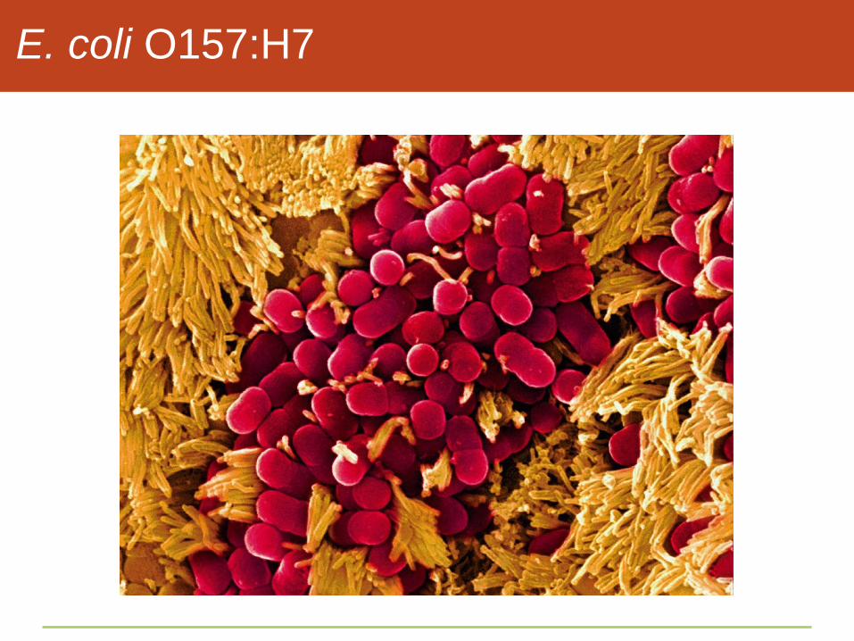
E. coli O157:H7

4.2 Cell Structure
• The cell is the smallest unit that shows the properties of life
• All cells have a plasma membrane and cytoplasm, and all
start out life with DNA

Components of All Cells
• Plasma membrane
• Controls substances passing in and out of the cell
• DNA containing region
• Nucleus in eukaryotic cells
• Nucleoid region in prokaryotic cells
• Cytoplasm
• A semifluid mixture containing cell components
Organelles
• Organelles are structures that carry out special metabolic
functions inside a cell
• Membrane-enclosed organelles compartmentalize tasks such
as building, modifying, and storing substances

Prokaryotic and Eukaryotic Cells
• Eukaryotic cell
• Cell interior is divided into functional compartments,
including a nucleus
• Prokaryotic cell
• Small, simple cells without a nucleus
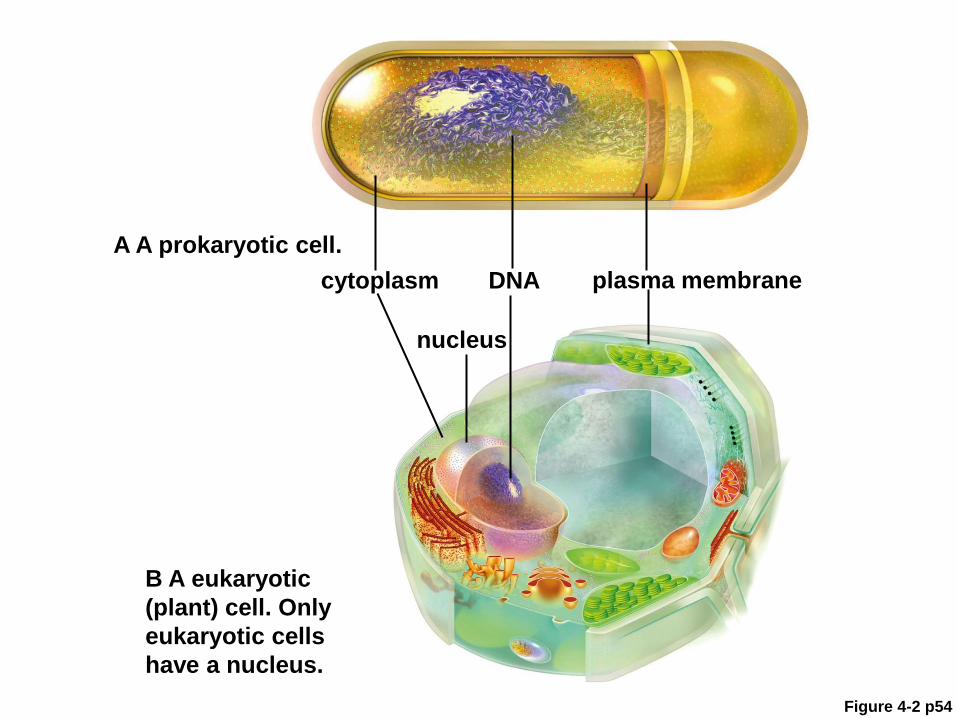
Figure 4-2 p54
A A prokaryotic cell.
cytoplasm DNA plasma membrane
nucleus
B A eukaryotic
(plant) cell. Only
eukaryotic cells
have a nucleus.

ANIMATED FIGURE: Overview of cells
To play movie you must be in Slide Show Mode
PC Users: Please wait for content to load, then click to play
Mac Users: CLICK HERE
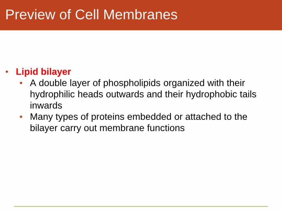
Preview of Cell Membranes
• Lipid bilayer
• A double layer of phospholipids organized with their
hydrophilic heads outwards and their hydrophobic tails
inwards
• Many types of proteins embedded or attached to the
bilayer carry out membrane functions

Figure 4-3 p54
cell’s exterior
plasma membrane
cell’s interior
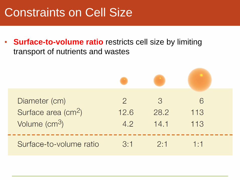
Constraints on Cell Size
• Surface-to-volume ratio restricts cell size by limiting
transport of nutrients and wastes

The Cell Theory Emerges
• Van Leeuwenhoek was the first to describe small organisms
seen through a microscope, which he called animalcules and
beasties
• Hooke was the first to sketch and name cells
• Brown was the first to identify a cell nucleus
Cell Theory
• The cell theory, a foundation of modern biology, states that
cells are the fundamental units of life
• In 1839, Schleiden and Schwann proposed the basic
concepts of the modern cell theory
• All organisms consists of one or more cells
• A cell is the smallest unit with the properties of life
• Each new cell arises from division of a preexisting cell
• Each cell passes its hereditary material to its offspring
Take-Home Message:
How are all cells alike?
• All cells start life with a plasma membrane, cytoplasm, and a
region of DNA, which, in eukaryotic cells only, is enclosed by
a nucleus
• The surface-to-volume ratio limits cell size and influences cell
shape
• Observations of cells led to the cell theory: All organisms
consist of one or more cells; the cell is the smallest unit of life;
each new cell arises from another cell; and a cell passes
hereditary material to its offspring
4.3 How Do We See Cells?
• Most cells are 10–20 micrometers in diameter, about fifty
times smaller than the unaided human eye can perceive
• One micrometer (μm) is one-thousandth of a millimeter, which
is one-thousandth of a meter
• We use different types of microscopes to study different
aspects of organisms, from the smallest to the largest
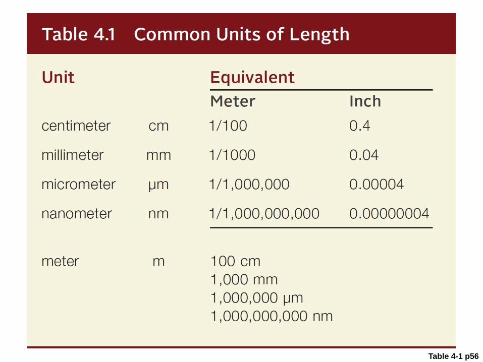
Table 4-1 p56

Modern Microscopes
• Light microscopes
• Phase-contrast microscopes
• Reflected light microscopes
• Fluorescence microscopes
• Electron microscopes
• Transmission electron microscopes
• Scanning electron microscopes

Figure 4-5a p56
path of light rays (bottom to top) to eye
prism that
directs rays to
ocular lens ocular
lens
objective
lenses
specimen
condenser
lens
focusing
knob
light source
(in base)
illuminator
stage
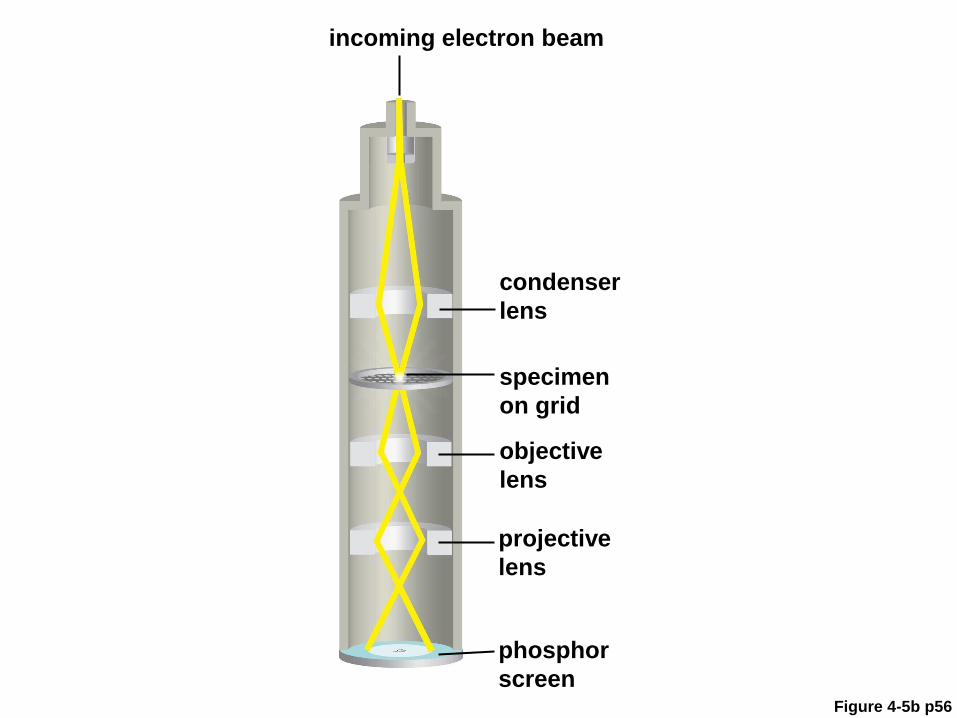
Figure 4-5b p56
incoming electron beam
condenser
lens
specimen
on grid
projective
lens
phosphor
screen
objective
lens

ANIMATED FIGURE: How a light
microscope works
To play movie you must be in Slide Show Mode
PC Users: Please wait for content to load, then click to play
Mac Users: CLICK HERE

ANIMATED FIGURE: How a light
microscope works
To play movie you must be in Slide Show Mode
PC Users: Please wait for content to load, then click to play
Mac Users: CLICK HERE

Different Microscopes,
Different Characteristics
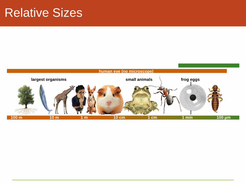
Relative Sizes
human eye (no microscope)
largest organisms small animals frog eggs
100 m 10 m 1 m 10 cm 1 cm 1 mm 100 µm
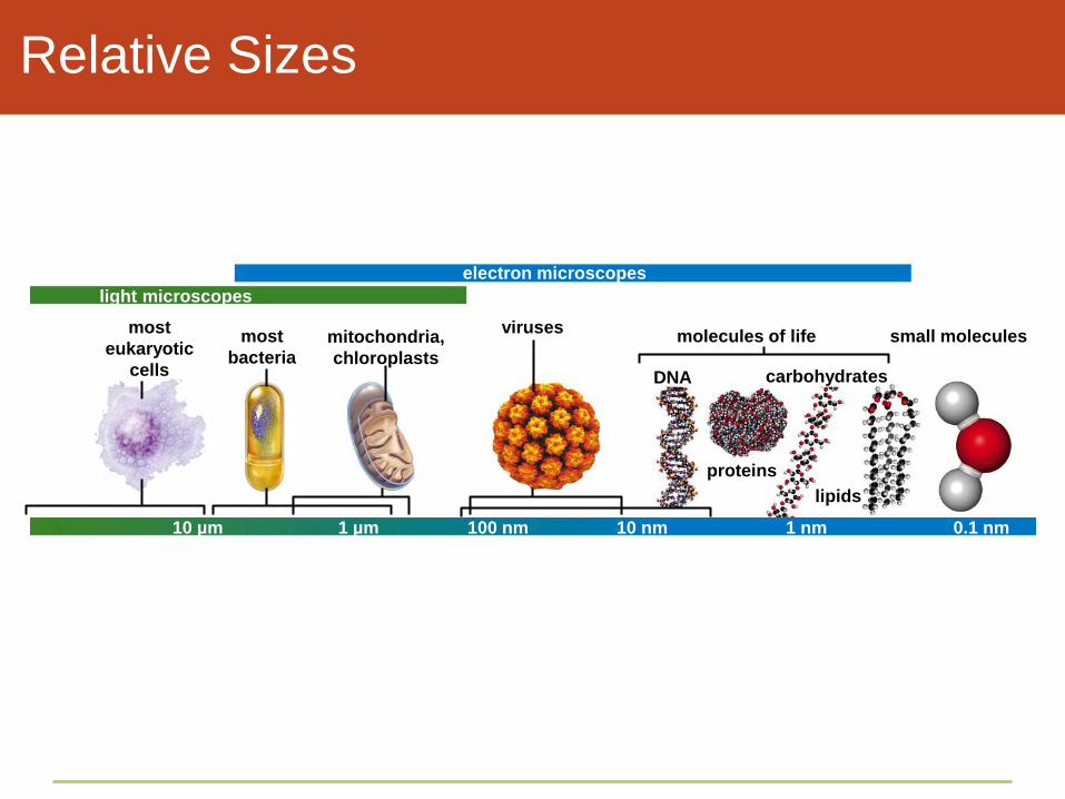
Relative Sizes
electron microscopes
light microscopes
most
eukaryotic
cells
most
bacteria mitochondria,
chloroplasts
viruses molecules of life small molecules
DNA
proteins
carbohydrates
10 µm 1 µm 100 nm 10 nm 1 nm 0.1 nm
lipids
Take-Home Message:
How do we see cells?
• Most cells are visible only with the help of microscopes
• Different types of microscopes reveal different aspects of cell
structure
4.4 Introducing “Prokaryotes”
• Bacteria and archaea are the prokaryotes (“before the
nucleus”), the smallest and most metabolically diverse forms
of life
• Bacteria and archaea are similar in appearance and size, but
differ in structure and metabolism

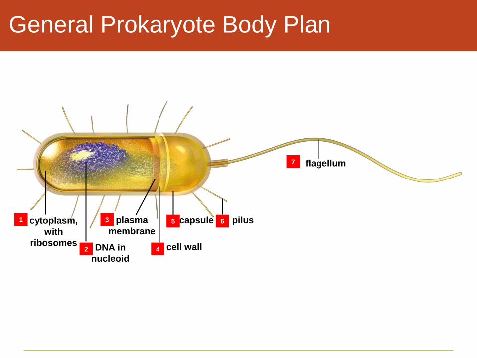
General Prokaryote Body Plan
• Cell wall surrounds the plasma membrane
• Made of peptidoglycan (in bacteria) or proteins (in
archaea) and coated with a sticky capsule
• Flagellum for motion
• Pili help cells move across surfaces
• “Sex” pilus aids in sexual reproduction
General Prokaryote Body Plan
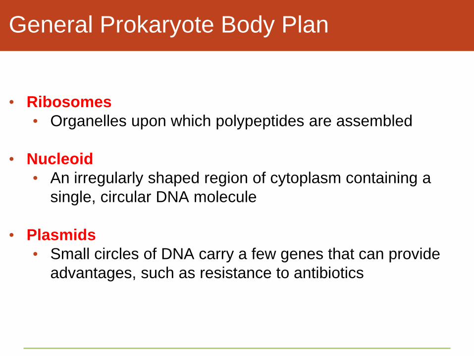
• Ribosomes
• Organelles upon which polypeptides are assembled
• Nucleoid
• An irregularly shaped region of cytoplasm containing a
single, circular DNA molecule
• Plasmids
• Small circles of DNA carry a few genes that can provide
advantages, such as resistance to antibiotics
General Prokaryote Body Plan
flagellum
cytoplasm,
with
ribosomes
plasma
membrane
capsule
DNA in
nucleoid
cell wall
pilus 1
2
3
4
5 6
7

ANIMATED FIGURE: Typical prokaryotic
cell
To play movie you must be in Slide Show Mode
PC Users: Please wait for content to load, then click to play
Mac Users: CLICK HERE
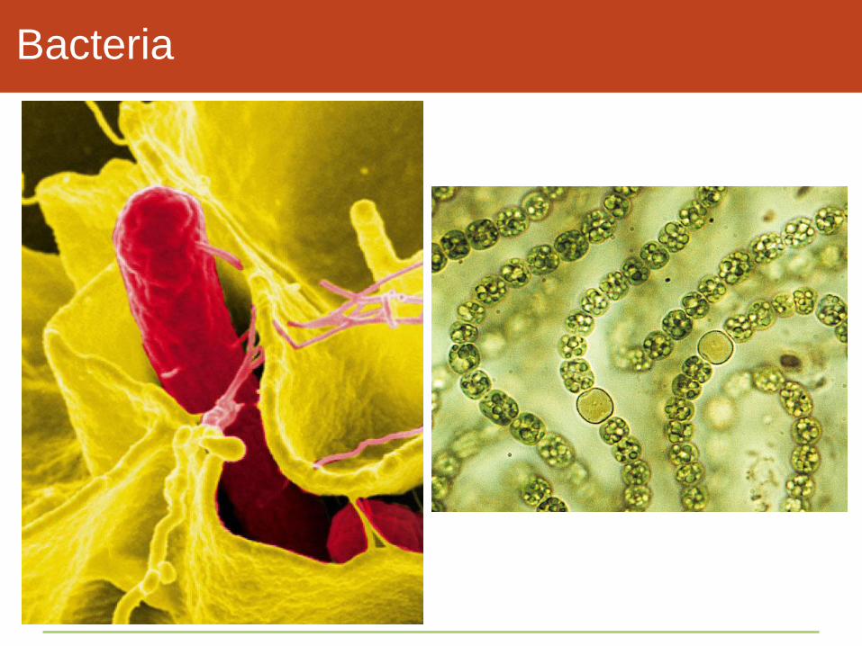
Bacteria

Archaeans

Biofilms
• Although prokaryotes are all single-celled, few live alone
• Biofilm
• Single-celled organisms sharing a secreted layer of
polysaccharides and glycoproteins
• May include bacteria, algae, fungi, protists, and archaeans

Dental Plaque: A Biofilm

Take-Home Message:
How are bacteria and archaea alike?
• Bacteria and archaea do not have a nucleus. Most kinds have
a cell wall around their plasma membrane; the permeable wall
reinforces and imparts shape to the cell body
• The structure of bacteria and archaea is relatively simple, but
as a group these organisms are the most diverse forms of life;
they inhabit nearly all regions of the biosphere
• Some metabolic processes occur at the plasma membrane of
bacteria and archaea; they are similar to complex processes
that occur at certain internal membranes of eukaryotic cells
4.5 Introducing Eukaryotic Cells
• All protists, fungi, plants, and animals are eukaryotes
• Eukaryotic (“true nucleus”) cells carry out much of their
metabolism inside membrane-enclosed organelles
• Organelle
• A structure that carries out a specialized function within a
cell
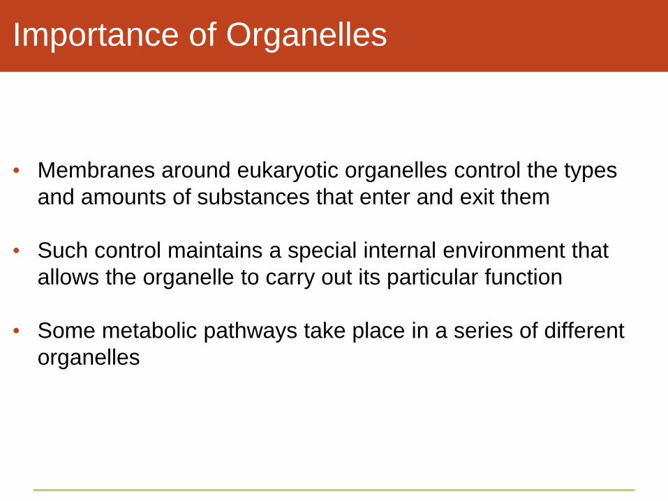
Importance of Organelles
• Membranes around eukaryotic organelles control the types
and amounts of substances that enter and exit them
• Such control maintains a special internal environment that
allows the organelle to carry out its particular function
• Some metabolic pathways take place in a series of different
organelles
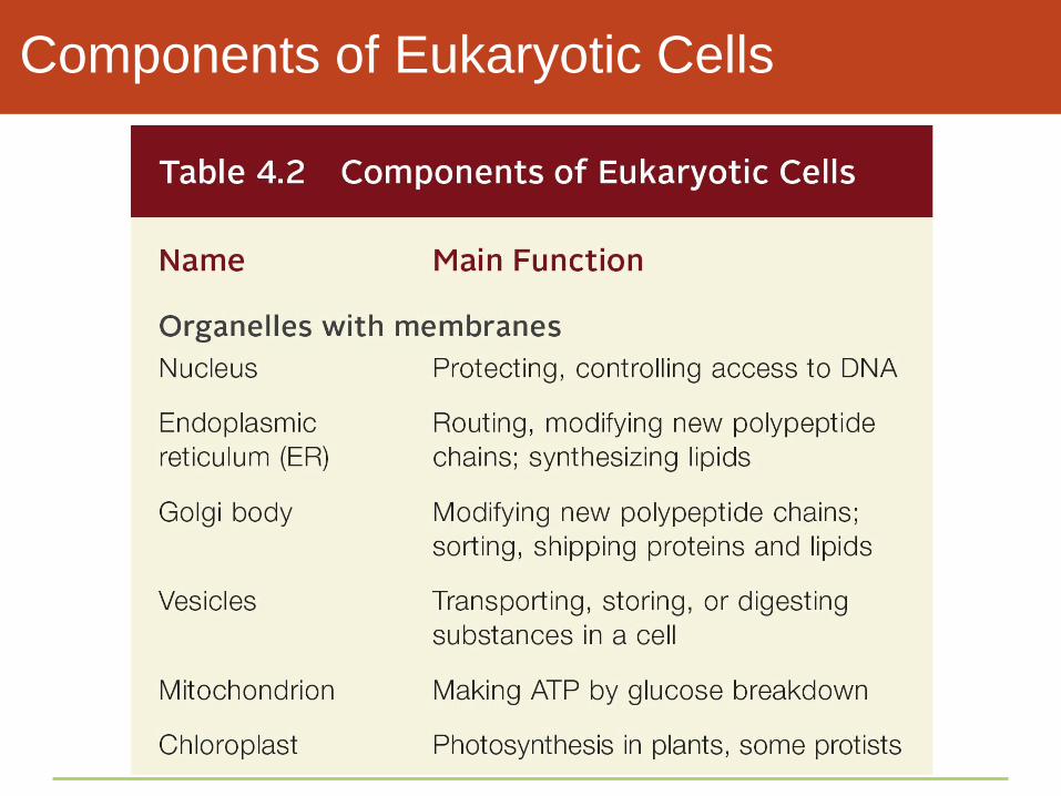
Components of Eukaryotic Cells
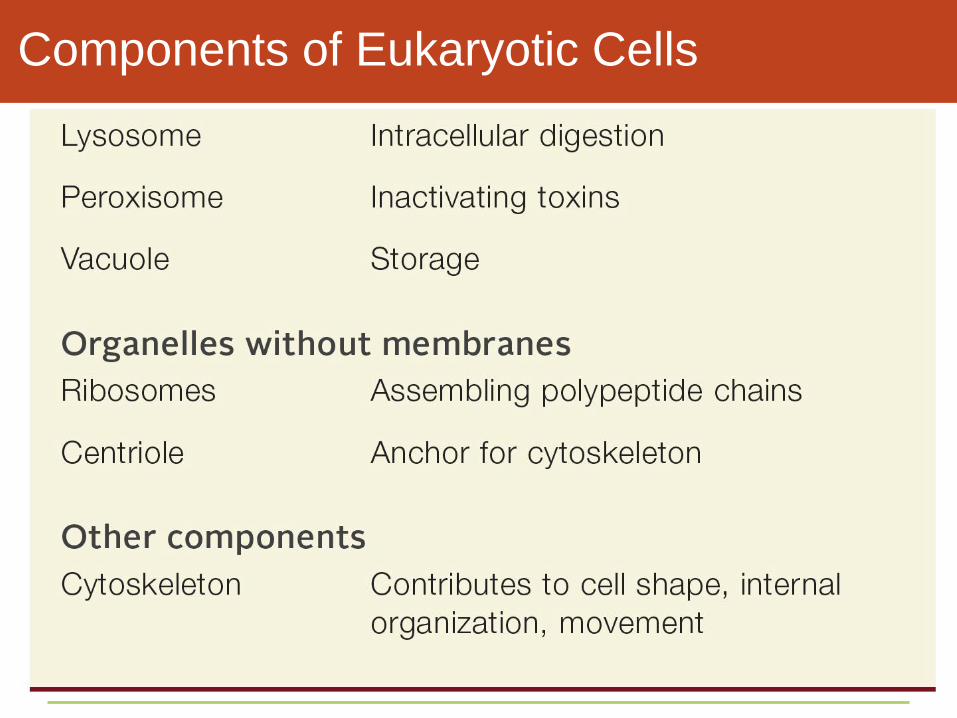
Components of Eukaryotic Cells
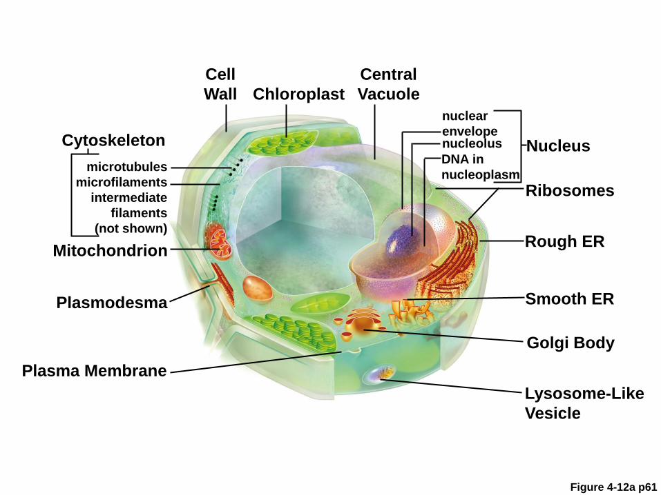
Figure 4-12a p61
Cell
Wall Chloroplast
Central
Vacuole
nuclear
envelope
Nucleus Cytoskeleton
microtubules
microfilaments
intermediate
filaments
(not shown)
Ribosomes
Mitochondrion
Smooth ER Plasmodesma
Golgi Body
Plasma Membrane
Lysosome-Like
Vesicle
Rough ER
DNA in
nucleoplasm
nucleolus
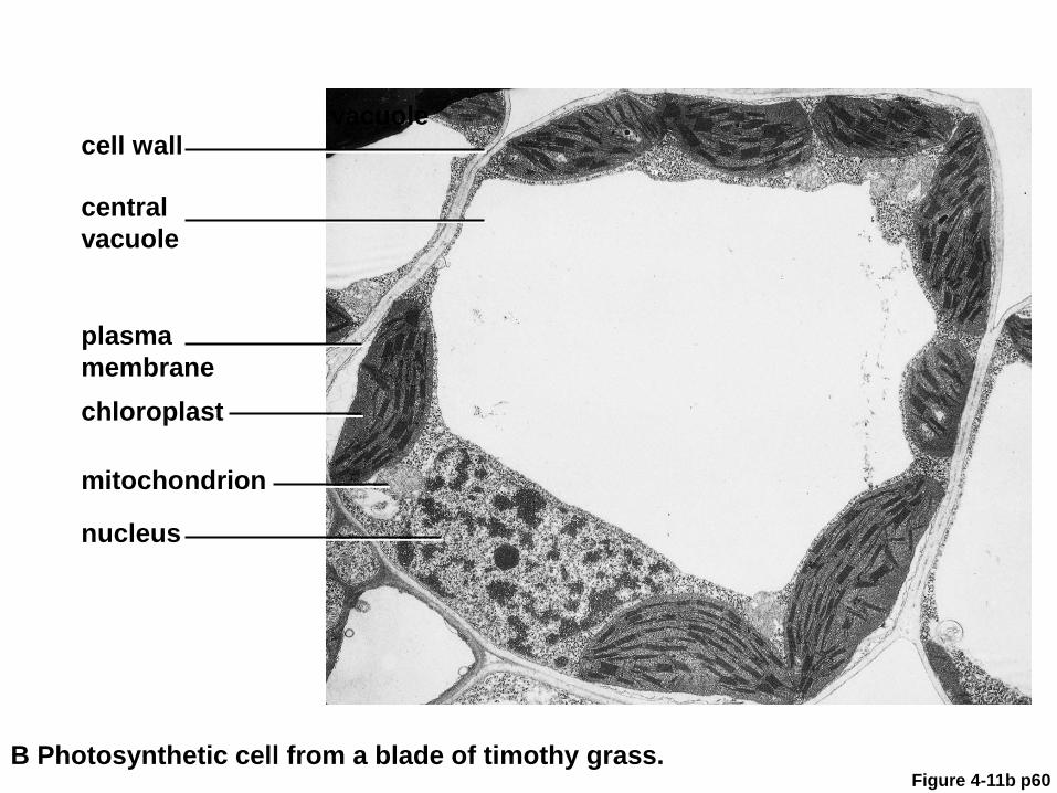
Figure 4-11b p60
cell wall
central
vacuole
plasma
membrane
mitochondrion
nucleus
B Photosynthetic cell from a blade of timothy grass.
vacuole
chloroplast

Makes lipids, breaks down
carbohydrates and fats,
inactivates toxins
Smooth ER
Finishes, sorts, ships lipids,
enzymes, and proteins
Golgi Body
Modifies proteins made by
ribosomes attached to it
Rough ER
Digests, recycles materials
Lysosome
Stepped Art
p64

Figure 4-11a p60
mitochondrion
nucleus
A Human white blood cell.
vacuole
plasma
membrane

Take-Home Message:
What do eukaryotic cells have in common?
• All eukaryotic cells start life with a nucleus and other
membrane-enclosed organelles

ANIMATION: Common eukaryotic
organelles
To play movie you must be in Slide Show Mode
PC Users: Please wait for content to load, then click to play
Mac Users: CLICK HERE
4.6 The Nucleus
• All of a eukaryotic cell’s DNA is in its nucleus
• The nucleus keeps eukaryotic DNA away from potentially
damaging reactions in the cytoplasm
• The nuclear envelope controls when DNA is accessed
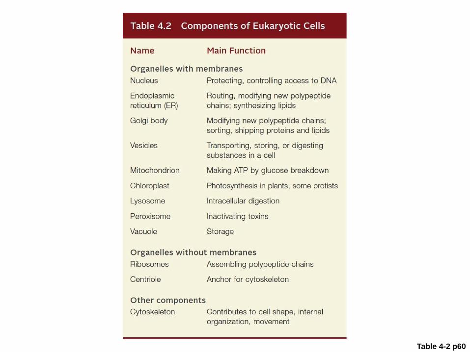
Table 4-2 p60
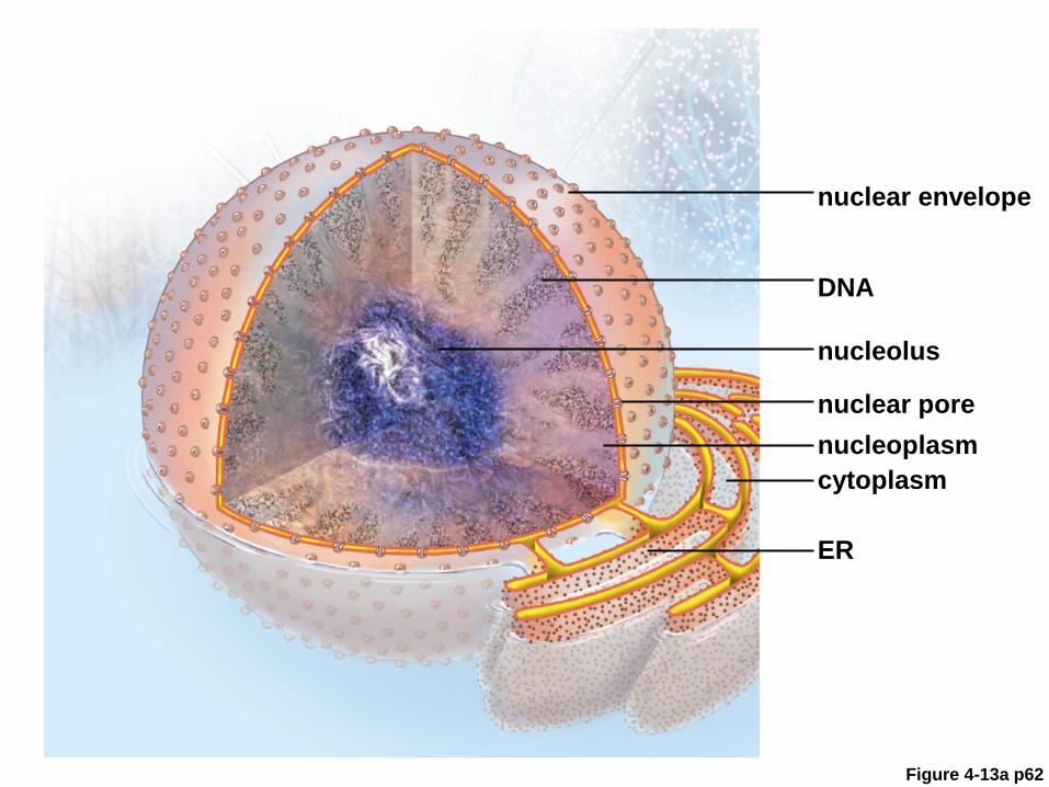
Figure 4-13a p62
nuclear envelope
DNA
nucleolus
nuclear pore
ER
cytoplasm
nucleoplasm

Figure 4-13b p62
nuclear envelope
DNA
nucleolus
nuclear pore
ER
cytoplasm
nucleoplasm

The Nuclear Envelope
• Nuclear envelope
• Two lipid bilayers pressed together as a single membrane
surrounding the nucleus
• Outer bilayer is continuous with the ER
• Nuclear pores allow certain substances to pass through
the membrane

Structure of the Nuclear Envelope
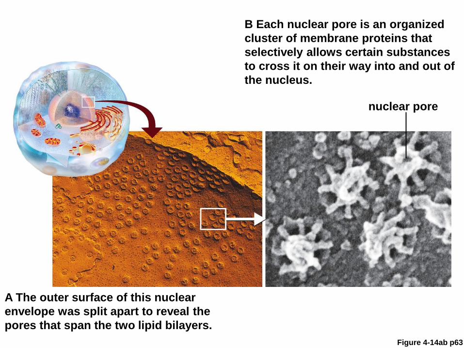
Figure 4-14ab p63
A The outer surface of this nuclear
envelope was split apart to reveal the
pores that span the two lipid bilayers.
B Each nuclear pore is an organized
cluster of membrane proteins that
selectively allows certain substances
to cross it on their way into and out of
the nucleus.
nuclear pore

Figure 4-14b p63
nuclear envelope
(two lipid bilayers)
cytoplasm
nuclear pore

ANIMATED FIGURE: Nuclear envelope
To play movie you must be in Slide Show Mode
PC Users: Please wait for content to load, then click to play
Mac Users: CLICK HERE

The Nucleoplasm and Nucleolus
• Nucleoplasm
• Viscous fluid inside the nuclear envelope, similar to
cytoplasm
• Nucleolus
• A dense region in the nucleus where subunits of
ribosomes are assembled from proteins and RNA

Chromosomes
• Chromatin
• All DNA and its associated proteins in the nucleus
• Chromosome
• A single DNA molecule with its attached proteins
• During cell division, chromosomes condense and become
visible in micrographs
• Human body cells have 46 chromosomes

A Condensed Chromosome

Take-Home Message:
What is the function of the cell nucleus?
• A nucleus protects and controls access to a eukaryotic cell’s
DNA
• The nuclear envelope is a double lipid bilayer. Proteins
embedded in it control the passage of molecules between the
nucleus and the cytoplasm