chapter 5: the integumentary system - …classpages.warnerpacific.edu/bdupriest/bio 221/chapter...
TRANSCRIPT

Copyright © 2010 Pearson Education, Inc.
CHAPTER 5:
THE INTEGUMENTARY SYSTEM

Copyright © 2010 Pearson Education, Inc.
OVERALL SKIN STRUCTURE
3 LAYERS
Copyright © 2010 Pearson Education, Inc.
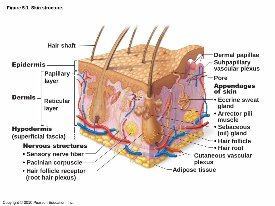
Figure 5.1 Skin structure.
Epidermis
Hair shaft
Dermis Reticular
layer
Papillary
layer
Hypodermis
(superficial fascia)
Dermal papillae
Pore
Subpapillary vascular plexus
Appendages
of skin
• Eccrine sweat gland
• Arrector pili muscle
• Sebaceous (oil) gland
• Hair follicle • Hair root Nervous structures
• Sensory nerve fiber
• Pacinian corpuscle
• Hair follicle receptor (root hair plexus)
Cutaneous vascular plexus
Adipose tissue

Copyright © 2010 Pearson Education, Inc.
EPIDERMIS
4 (or 5) LAYERS

Copyright © 2010 Pearson Education, Inc.
Figure 5.2 The main structural features of the skin epidermis.
Melanocyte
Melanin granule
Tactile
(Merkel)
cell
Sensory
nerve
ending
Epidermal
dendritic
cell
Dermis
Dermis
Keratinocytes
Desmosomes
(b)
(a)
Stratum corneum
Stratum granulosum
Stratum spinosum
Stratum basale
Copyright © 2010 Pearson Education, Inc.
DERMIS
2 LAYERS
Copyright © 2010 Pearson Education, Inc.
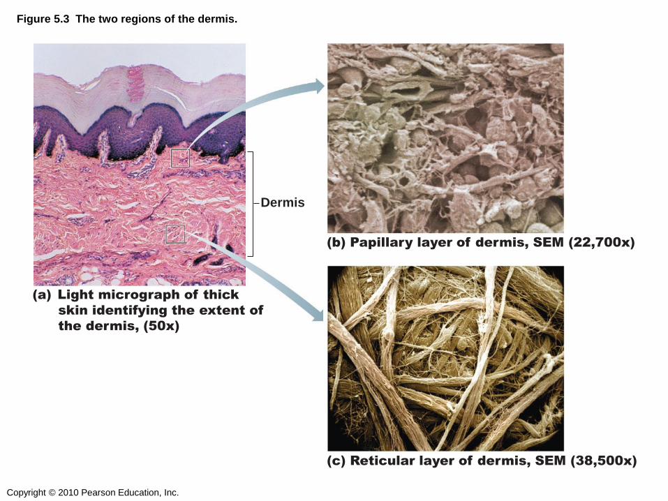
Figure 5.3 The two regions of the dermis.
Dermis
(a) Light micrograph of thick
skin identifying the extent of
the dermis, (50x)
(b) Papillary layer of dermis, SEM (22,700x)
(c) Reticular layer of dermis, SEM (38,500x)

Copyright © 2010 Pearson Education, Inc.
Figure 5.3a The two regions of the dermis.
Dermis
(a) Light micrograph of thick skin identifying
the extent of the dermis, (50x)

Copyright © 2010 Pearson Education, Inc.
Q1: The type of gland which secretes its
products onto a surface is an _______ gland.
1) Endocrine
2) Exocrine
3) Merocrine
4) Holocrine
Copyright © 2010 Pearson Education, Inc.
Q2: The embryonic tissue which gives rise to
muscle and most connective tissue is…
1) Ectoderm
2) Endoderm
3) Mesoderm
Copyright © 2010 Pearson Education, Inc.
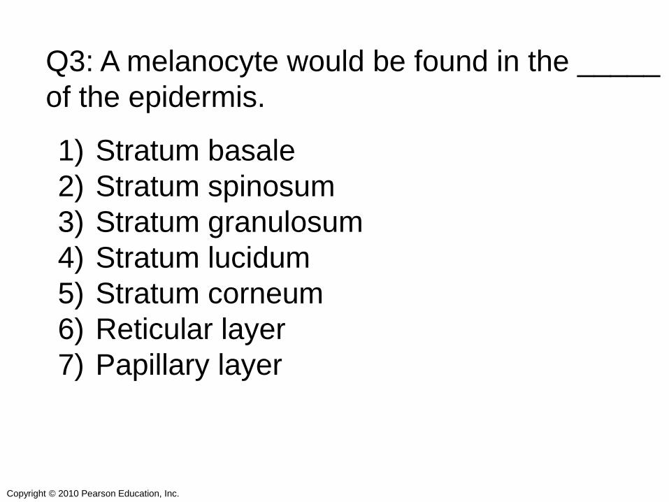
Q3: A melanocyte would be found in the _____
of the epidermis.
1) Stratum basale
2) Stratum spinosum
3) Stratum granulosum
4) Stratum lucidum
5) Stratum corneum
6) Reticular layer
7) Papillary layer

Copyright © 2010 Pearson Education, Inc.
Q4: The main cell type in the epidermis is the...
1) Melanocyte
2) Langerhans Cell
3) Merkel Cell
4) Dendritic Cell
5) Keratinocyte

Copyright © 2010 Pearson Education, Inc.
Q5: The bulk of the dermis is the ______ layer.
1) Epidermal
2) Hypodermal
3) Reticular
4) Papillary
Copyright © 2010 Pearson Education, Inc.
SKIN COLOR
3 PIGMENTS
Copyright © 2010 Pearson Education, Inc.
SKIN APPENDAGES

Copyright © 2010 Pearson Education, Inc.
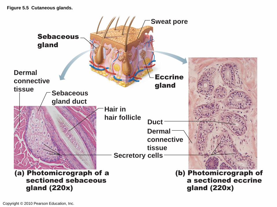
CUTANEOUS GLANDS
Copyright © 2010 Pearson Education, Inc.
Figure 5.5 Cutaneous glands.
(a) Photomicrograph of a
sectioned sebaceous
gland (220x)
(b) Photomicrograph of
a sectioned eccrine
gland (220x)
Sebaceous
gland duct
Hair in
hair follicle
Secretory cells
Dermal
connective
tissue
Dermal
connective
tissue
Duct
Sebaceous
gland
Sweat pore
Eccrine
gland
Copyright © 2010 Pearson Education, Inc.
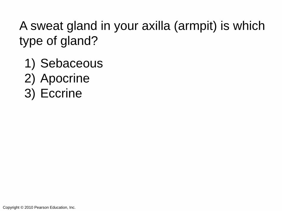
A sweat gland in your axilla (armpit) is which
type of gland?
1) Sebaceous
2) Apocrine
3) Eccrine

Copyright © 2010 Pearson Education, Inc.
HAIR FOLLICLES
Copyright © 2010 Pearson Education, Inc.
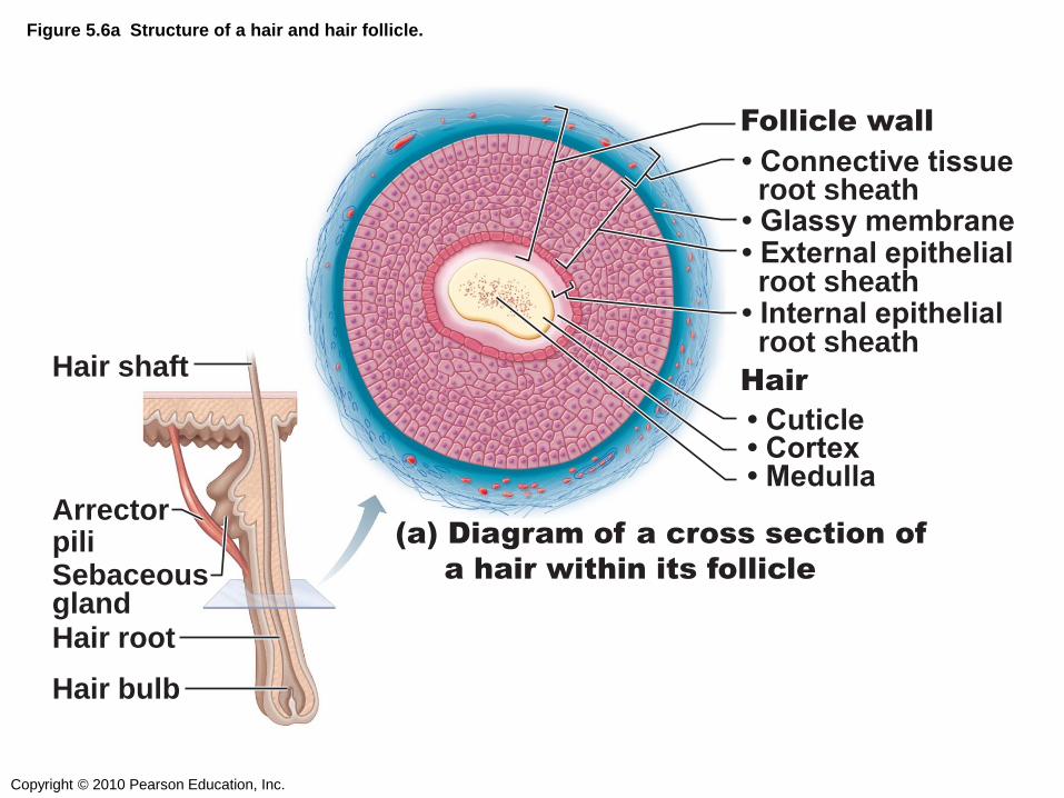
Figure 5.6a Structure of a hair and hair follicle.
Hair shaft
Arrector pili Sebaceous gland Hair root
Hair bulb
(a) Diagram of a cross section of
a hair within its follicle
• Connective tissue root sheath • Glassy membrane
• External epithelial root sheath
• Internal epithelial root sheath
Follicle wall
• Cuticle • Cortex
• Medulla
Hair
Copyright © 2010 Pearson Education, Inc.
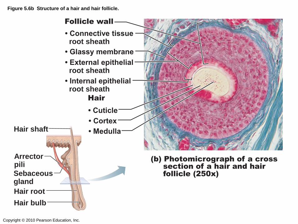
Figure 5.6b Structure of a hair and hair follicle.
(b) Photomicrograph of a cross
section of a hair and hair
follicle (250x)
• Connective tissue root sheath
Follicle wall
• Cuticle
• Glassy membrane
• Cortex
• Medulla
• Internal epithelial root sheath
• External epithelial root sheath
Hair
Hair shaft
Arrector pili
Sebaceous gland
Hair root
Hair bulb
Copyright © 2010 Pearson Education, Inc.
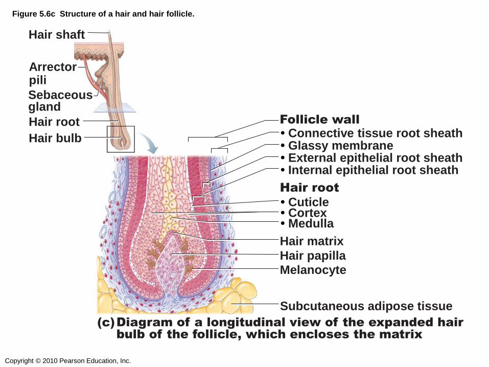
Hair shaft
Arrector pili
Sebaceous gland
Hair root
Hair bulb
(c) Diagram of a longitudinal view of the expanded hair
bulb of the follicle, which encloses the matrix
• Internal epithelial root sheath • External epithelial root sheath
• Connective tissue root sheath Follicle wall
Hair matrix
Melanocyte Hair papilla
Subcutaneous adipose tissue
• Medulla • Cortex • Cuticle
• Glassy membrane
Hair root
Figure 5.6c Structure of a hair and hair follicle.
Copyright © 2010 Pearson Education, Inc.
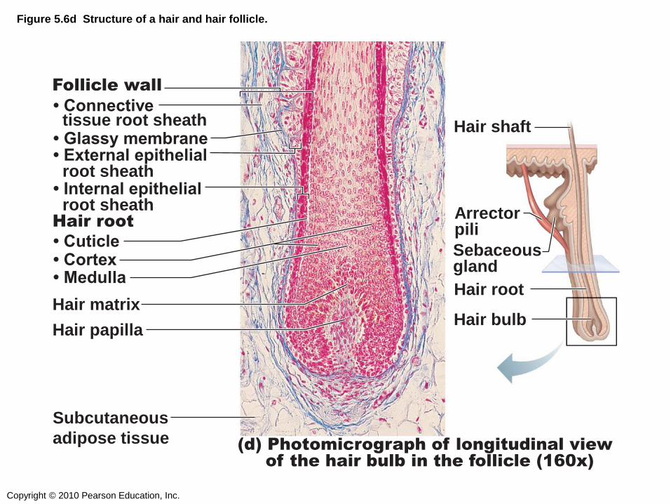
Figure 5.6d Structure of a hair and hair follicle.
(d) Photomicrograph of longitudinal view
of the hair bulb in the follicle (160x)
Follicle wall
Hair matrix
Hair papilla
Subcutaneous
adipose tissue
Hair root
• Connective tissue root sheath
• Glassy membrane • External epithelial root sheath • Internal epithelial root sheath
• Cuticle
• Cortex
• Medulla
Hair shaft
Arrector pili
Sebaceous gland
Hair root
Hair bulb
Copyright © 2010 Pearson Education, Inc.
Which layer of a hair follicle wall is thickest?
1) Connective tissue root sheath
2) External epithelial root sheath
3) Internal epithelial root sheath
Copyright © 2010 Pearson Education, Inc.
NAILS

Copyright © 2010 Pearson Education, Inc.
Figure 5.7 Structure of a nail.
Lateral
nail fold
Lunule
Nail
matrix
Root of nail
Proximal
nail fold
Hyponychium
Nail bed
Phalanx (bone of fingertip)
Eponychium
(cuticle)
Body
of nail
Free edge
of nail
(a)
(b)

Copyright © 2010 Pearson Education, Inc.
What is the scientific name for the cuticle?
1) Eponychium
2) Hyponychium
3) Lunule
4) Nail matrix
Copyright © 2010 Pearson Education, Inc.
FUNCTIONS

Copyright © 2010 Pearson Education, Inc.
DISEASES
(“HOMEOSTATIC IMBALANCES”)
Copyright © 2010 Pearson Education, Inc.
SKIN CANCER
Copyright © 2010 Pearson Education, Inc.
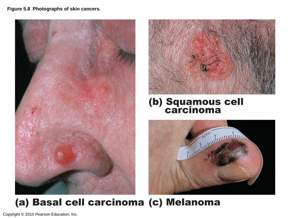
Figure 5.8 Photographs of skin cancers.

Copyright © 2010 Pearson Education, Inc.
True or false: all skin cancers start out
looking like moles.
1) True
2) False
Copyright © 2010 Pearson Education, Inc.
SKIN BURNS

Copyright © 2010 Pearson Education, Inc.
Figure 5.10 Partial thickness and full thickness burns.
(a) Skin bearing partial
thickness burn (1st and
2nd degree burns)
(b) Skin bearing full
thickness burn
(3rd degree burn)
1st degree
burn
2nd degree
burn
3rd degree
burn
Copyright © 2010 Pearson Education, Inc.
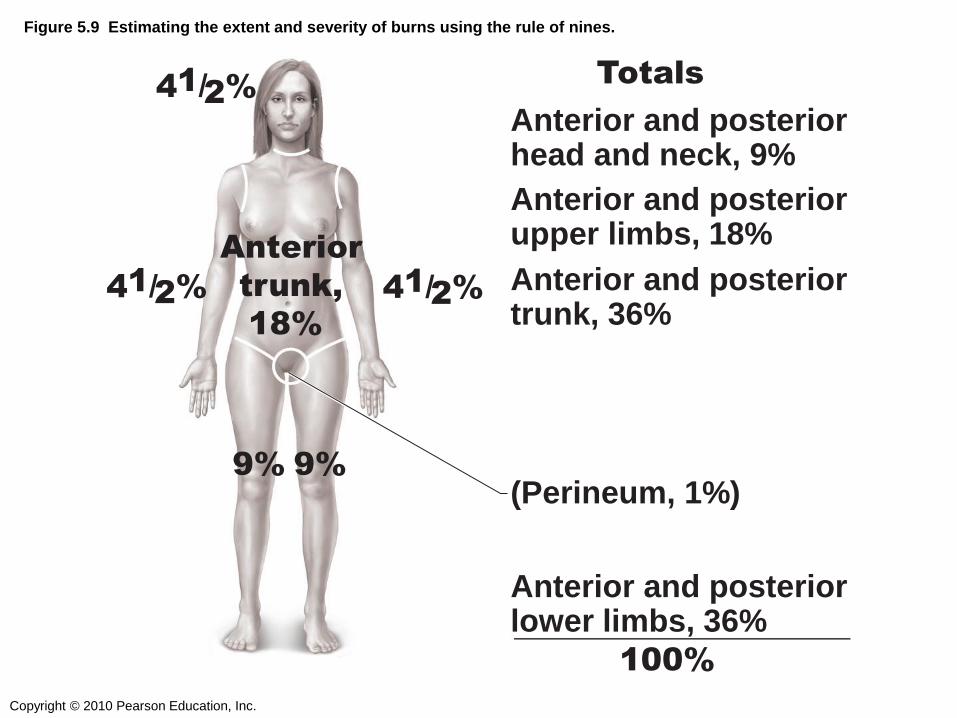
Figure 5.9 Estimating the extent and severity of burns using the rule of nines.
Anterior and posterior head and neck, 9%
4 1 / 2 % 4 1 / 2 %
Anterior and posterior upper limbs, 18%
Anterior and posterior lower limbs, 36%
100%
Totals
Anterior and posterior trunk, 36%
Anterior
trunk,
18%
9% 9% (Perineum, 1%)
4 1 / 2 %

Copyright © 2010 Pearson Education, Inc.
A patient playing with a lighter and a can of
hairspray experienced second- and third-
degree burns covering his entire left arm.
What percent of the body was burned?
1) 4.5 %
2) 9%
3) 18%
4) 8%