chapter 6 pathology of breast carcinoma and ... - amfe …amfe-eg.com/books/book breast cancer...
TRANSCRIPT

Pathology and methods of analysi
Chapter 6Chapter 6Chapter 6Chapter 6
Pathology of Breast CarcinomaPathology of Breast CarcinomaPathology of Breast CarcinomaPathology of Breast Carcinoma
and methods of analysisand methods of analysisand methods of analysisand methods of analysis
Pathology and methods of analysis 71
Microscopic examination is the definitive means of evaluation of
breast disease. The ultimate method of treatment of the patients with breast
carcinoma may be determined on the bases of the pathologic findings in the initial
breast biopsy. The following sampling techniques are used singly or in
combination:
1)1)1)1) Cytopuncture Cytopuncture Cytopuncture Cytopuncture and and and and Fine Needle Aspiration (FNA) Fine Needle Aspiration (FNA) Fine Needle Aspiration (FNA) Fine Needle Aspiration (FNA)
Cytopuncture or FNA may constitute the initial diagnostic procedure for palpable
breast masses. This technique has been also used to evaluate non-palpable
mammographically detected lesions under stereotactic or ultrasound guidance.
This method has two limitations: first its accuracy which depends upon the skill
and experience of the personnel who perform the puncture, the radiologic guidance
for the non palpable lesion, and also the microscopical analysis. Second, the
inability to permit a reliable distinction between in situ and invasive ductal
carcinoma except when the puncture concerns metastatic lymph nodes.
This technique is the simplest and the cheapest diagnostic method, only little
material is required. It is immediately done and gives the possibility to have a
nearly instant diagnosis. By being that easy to perform, this has allowed its
integration to the consultations associating radiologist and clinicians to ensure a
complete diagnosis in one visit. It equally allows a rapid evaluation of the efficacy
of neo-adjuvant chemotherapy. This material guarantees the diagnosis of
carcinoma, the evaluation of its type but also the measurement of its grade. The
immunohistochemistry staining is possible thanks to the cytobloc techniques. The
material, formed essentially of tumor elements, is especially adapted to the
techniques of cytometry, molecular biology and even the microarray.
2222) Core needle biopsies) Core needle biopsies) Core needle biopsies) Core needle biopsies
The biopsied material has considerably progressed thanks to multiple technological
improvements. First, the development of aspiration needles having variable
calibers, starting from 18G, then the aspiration systems which guarantees the
possibility to achieve multiple aspiration materials through only one entry site to
the lesion and finally the instrument and technique of stereotactic detection or ultra
sound allowing the precise millimetric targeting of non palpable lesions.
Pathology and methods of analysis 72
Today, it represents for most of the countries, where an organized mammographic
screening was developed, the first diagnostic method thus allowing the surgical
resections to be only limited to patients having malignant lesions (therapeutic
surgery) or doubtful lesions (diagnostic surgery).
The handling of the specimens by the pathologist requires learning both for: their
management that requires a severely strict protocol having multiple levels of cut
sections and also for their microscopic analysis. The pathologist has a double role,
performs the most precise microscopic diagnosis on the small size and fragmented
material, then confirming the correct representation of the biopsy in relation to the
doubtful image.
3333) Incisional or excisional open biopsy) Incisional or excisional open biopsy) Incisional or excisional open biopsy) Incisional or excisional open biopsy
The pathologic evaluation of the primary excision specimen is a crucial component
in the selection and implementation of breast conservative surgery. The following
recommendations should be adopted for proper evaluation of the breast specimens:
• The specimen should be presented to the pathologist intact and
carefully oriented by means of the suture tags or fixed to a support
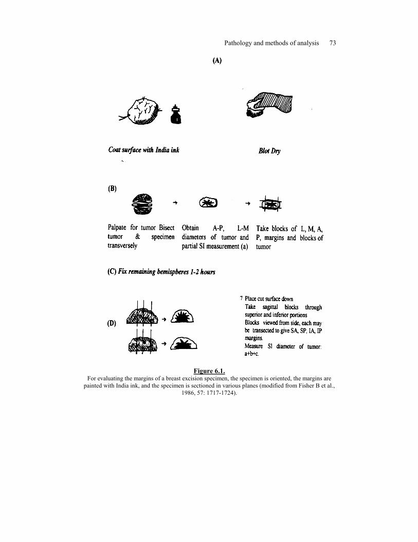
where the anatomical marks are indicated (Fig. 6.1).
• After measurement, it is inspected for gross margin involvement. If
there is evidence that the specimen contains a grossly suspicious
lesion that extends to the surface of the specimen, the surgeon, if still
operating, is immediately notified about the precise location of the
margin involved so that additional tissue can be excised. Prior to
cutting the breast excisional biopsy, the surface should be blotted dry
and then painted with marker (India ink), which will be visible on
the permanent section.
The subsequent steps in processing will depend upon the nature of the specimen,
whether it is obtained because of the presence of a palpable mass, and if carcinoma
is suspected clinically or radiographically. For these specimens, the specimen
should be cut in such a way as to permit examination of the resected margins on
histologic sections. The steps of handling of the specimen are outlined in figure 7.1.
Briefly, the tumor and the specimen are bisected transversely. The anteroposterior
and mediolateral diameters of the tumor are measured. Aliquots of the tumor are
systematically conserved in cryopreserved tumor bank when its size is sufficient.
The standard method of sectioning varies, depending upon the size of excised
specimen.
Pathology and methods of analysis 73
Figure 6.1.
For evaluating the margins of a breast excision specimen, the specimen is oriented, the margins are
painted with India ink, and the specimen is sectioned in various planes (modified from Fisher B et al.,
1986, 57: 1717-1724).
Pathology and methods of analysis 74
For small excisions specimen, the whole tissue is totally included. For larger
specimens, a sampling is necessary. After inking the specimen, single incision is
made through the center of the palpable mass where it most closely approaches the
margin in this way, the size of the tumor and the relationship to the nearest margin
can be quickly determined. The remaining margins can then be entirely removed
from the specimen and submitted for permanent sections so that representative
sections from each of the six surfaces of the specimen are submitted. Multiple
sections can then be made through the remainder of the specimen at 3 to 5 mm
intervals and several sections to the tumor and the adjacent parenchyma or fat
should be submitted for microscopic evaluation. Segments of skin and muscle
should be systematically sampled in order to demonstrate their relationship with the
tumor.
For the specimen excised because of the presence of mammographic abnormality,
in the absence of a palpable mass, the most frequent mammographic abnormalities
promoting biopsy are microcalcifications, mass with or without associated
microcalcifications, and focal asymmetry. These specimens are usually excised
using the hook wire or needle localization technique. Specimen radiography is an
essential component in the evaluation of these specimens in order to both,
document the presence of the lesion detected by mammography or the clip and
localize the suspicious area for histologic examination. The use of frozen sections
is limited because many lesions have been previously evaluated by core needle
biopsy and for the others; their small size renders difficult or even impossible the
realization of a frozen section. After fixation, the entire specimen is submitted for
permanent sections. The specimen can be cut perpendicular or parallel to its main
axis in serial cut sections.
4444) Mastectomy) Mastectomy) Mastectomy) Mastectomy
The skin and the nipple are assessed, with sampling of any isolated cutaneous
lesions. The tumor size is measured and any evidence of multicentricity is
recorded. Adequate histologic sections should be obtained from the tumor mass,
any other identified lesion, the nipple parallel and perpendicular to the axis of the
main lactiferous ducts and systematically each quadrant.
5) 5) 5) 5) AAAAxillary lymph nodes xillary lymph nodes xillary lymph nodes xillary lymph nodes
In mastectomy specimens, the axillary nodes are removed in contiguity with the
breast tissue. In patients treated with breast conserving therapy, an axillary
dissection usually comprises a specimen containing the lymph nodes which is
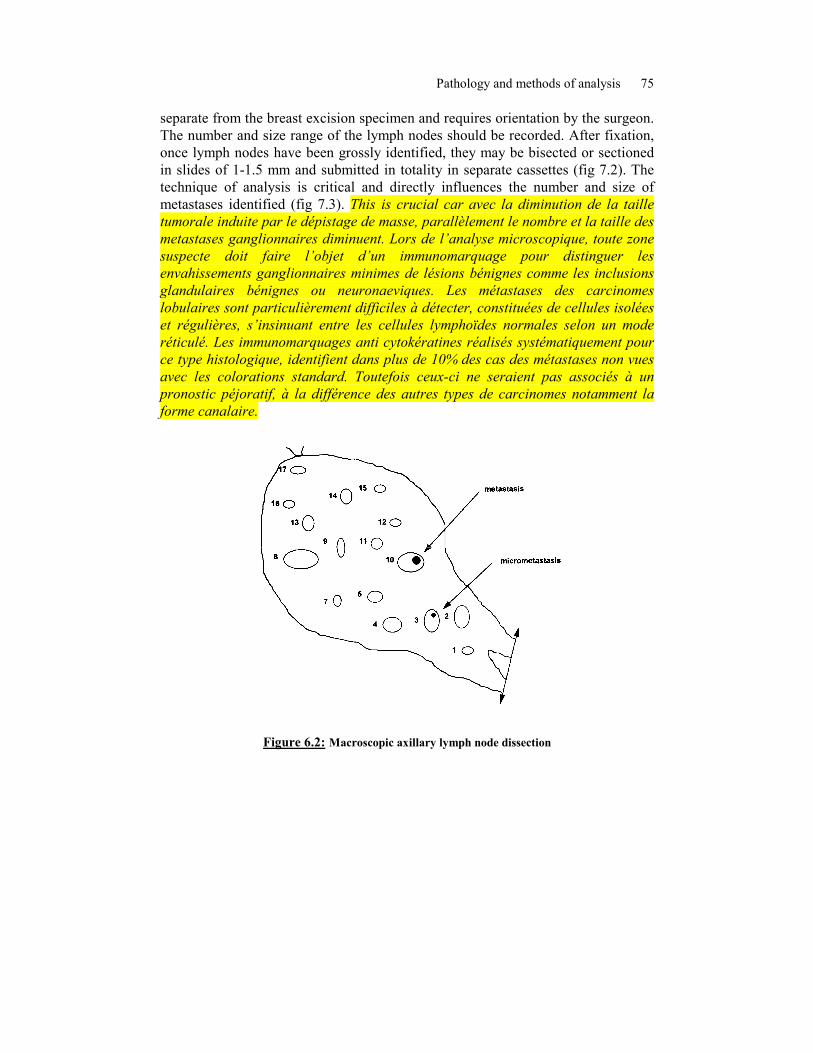
Pathology and methods of analysis 75
separate from the breast excision specimen and requires orientation by the surgeon.
The number and size range of the lymph nodes should be recorded. After fixation,
once lymph nodes have been grossly identified, they may be bisected or sectioned
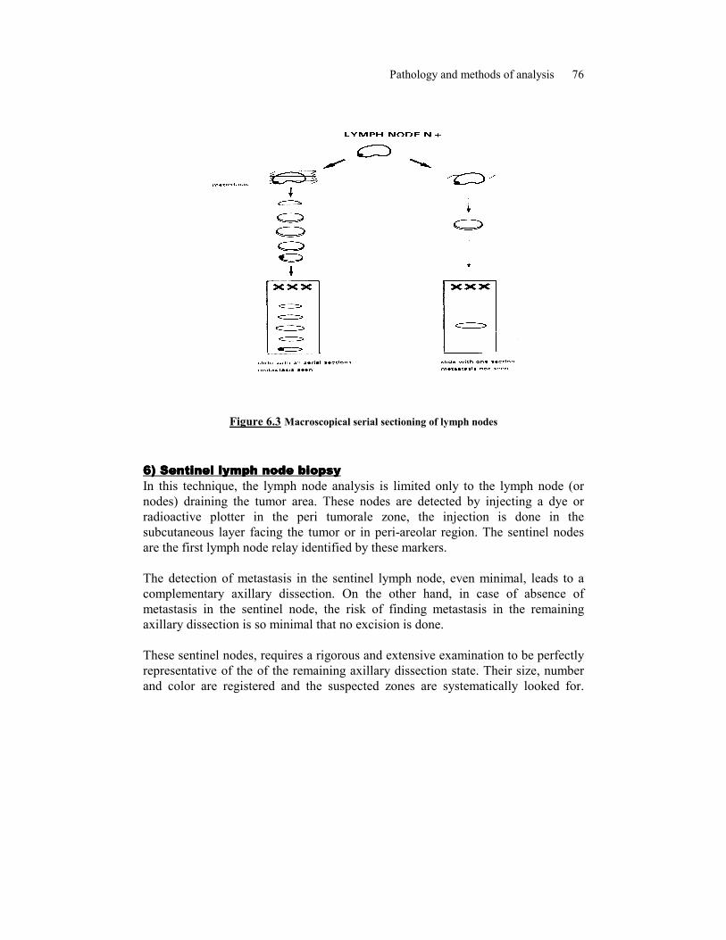
in slides of 1-1.5 mm and submitted in totality in separate cassettes (fig 7.2). The
technique of analysis is critical and directly influences the number and size of
metastases identified (fig 7.3). This is crucial car avec la diminution de la taille
tumorale induite par le dépistage de masse, parallèlement le nombre et la taille des
metastases ganglionnaires diminuent. Lors de l’analyse microscopique, toute zone
suspecte doit faire l’objet d’un immunomarquage pour distinguer les
envahissements ganglionnaires minimes de lésions bénignes comme les inclusions
glandulaires bénignes ou neuronaeviques. Les métastases des carcinomes
lobulaires sont particulièrement difficiles à détecter, constituées de cellules isolées
et régulières, s’insinuant entre les cellules lymphoïdes normales selon un mode
réticulé. Les immunomarquages anti cytokératines réalisés systématiquement pour
ce type histologique, identifient dans plus de 10% des cas des métastases non vues
avec les colorations standard. Toutefois ceux-ci ne seraient pas associés à un
pronostic péjoratif, à la différence des autres types de carcinomes notamment la
forme canalaire.
Figure 6.2: Macroscopic axillary lymph node dissection
Pathology and methods of analysis 76
Figure 6.3 Macroscopical serial sectioning of lymph nodes
6666) S) S) S) Sentinel lymph node biopsyentinel lymph node biopsyentinel lymph node biopsyentinel lymph node biopsy
In this technique, the lymph node analysis is limited only to the lymph node (or
nodes) draining the tumor area. These nodes are detected by injecting a dye or
radioactive plotter in the peri tumorale zone, the injection is done in the
subcutaneous layer facing the tumor or in peri-areolar region. The sentinel nodes
are the first lymph node relay identified by these markers.
The detection of metastasis in the sentinel lymph node, even minimal, leads to a
complementary axillary dissection. On the other hand, in case of absence of
metastasis in the sentinel node, the risk of finding metastasis in the remaining
axillary dissection is so minimal that no excision is done.
These sentinel nodes, requires a rigorous and extensive examination to be perfectly
representative of the of the remaining axillary dissection state. Their size, number
and color are registered and the suspected zones are systematically looked for.

Pathology and methods of analysis 77
They are immediately examined, by opposition, and / or by cryostat cut sections
with the possibility of immunohistochemical examination thanks to the presence of
rapid analysis kit.
Then, they are totally included in separated cassettes and examined on staged cut
section levels stained by immunohistochemistry. As required for their
identification, their management by the pathologist also requires learning.
7777) ) ) ) Frozen sectionFrozen sectionFrozen sectionFrozen section
Despite the obvious limitations, frozen section diagnosis remains to be the most
useful tool in the evaluation of breast lesions and sentinel lymph nodes. The
clinical data, gross morphology, specimen consistency and mammographic
findings, if available, should be taken into consideration. False negative results may
be encountered with sampling error or when dealing with a well differentiated
tubular carcinoma. False positive results may be obtained in lesions exhibiting
sclerosing adenosis and like lesions. In frozen sections of lymph nodes, the largest
and firmest nodes should be selected for sampling. Diagnostic difficulties arise in
the setting of small intrasinusoidal-subcapsular micrometastases, or nodal deposits
of infiltrating lobular carcinoma with pseudoreticular forms.
8888) ) ) ) Investigational ToolsInvestigational ToolsInvestigational ToolsInvestigational Tools
As an adjuvant to routine histopathologic examination of breast tumors, additional
investigational tools can help improve the evaluation. These tools include immuno-
histochemistry, cytogenetics and molecular biopsy tests.
Many protein products can be detected by immunohistochemistry, some including
estrogen and progesterone receptors, HER-2/Neu oncogene done on a routine basis;
other like p53 tumor suppressor gene, proliferation markers, angiogenesis,
apoptosis, basal and luminal cytokeratins, EGFR etc. as a complement or for
research purpose. Their application should be perfectly controlled and in the frame
of using automation, calibrated internal and external control, as part of the quality
assurance in order to ensure constant precision and reproducibility. Different
European organisations suggest a quality control program.
Molecular biology methods have been developed and adapted in order to be able on
fixed and embedded tissues. Fluorescent in situ hybridization (FISH) technique is
today the gold standard method for the determination of amplification of her2 gene.
Research of deletion and mutation of different genes such as EGFR, K-ras are
Pathology and methods of analysis 78
possible by PCR and more recently prognostic and predictive molecular signature
determined by microarray has been developed.
Proliferative disease and in situ carcinomaProliferative disease and in situ carcinomaProliferative disease and in situ carcinomaProliferative disease and in situ carcinoma
Table 6.1.
Simplified WHO Histologic Classification of epithelial proliferation
1111---- Lobular NeoplasiaLobular NeoplasiaLobular NeoplasiaLobular Neoplasia
This type of lesion occurs during a woman’s period of sexual activity, that is to say
while the lobules are fully active, and disappears after the menopause. Its incidence
represents about 1 to 4% of breast carcinoma. Lobular neoplasia is nearly always
diagnosed by incidental microscopic discovery on surgical specimen of patients
with microcalcifications or other abnormal radiographic images connected with
benign pathology. Microscopically, there is a proliferation of globular cells, which
are of the same shape and a little bigger than the neighboring cells, which have
slightly bigger nuclei and slightly more irregular chromatin, with vacuolised
cytoplasm in places. They fill in the center of terminal ectasic ducts and are able to
propagate between the layers of neighboring extralobular ducts in a “Paget”
spreading way. The absence of expression of E-Cadherin facilitates their
Lobular neoplasiaLobular neoplasiaLobular neoplasiaLobular neoplasia
- lobular carcinoma in situ
Intraductal proliferative lesionIntraductal proliferative lesionIntraductal proliferative lesionIntraductal proliferative lesion
- usual ductal hyperplasia
- flat epithelial atypia
- atypical ductal hyperplasia
- ductal carcinoma in situ
Intraductal papillary neoplasmsIntraductal papillary neoplasmsIntraductal papillary neoplasmsIntraductal papillary neoplasms
- central papilloma
- peripheral papilloma
- atypical papilloma
- intraductal papillary carcinoma
- intracystic papillary carcinoma
Pathology and methods of analysis 79
recognition. About 80% of lesions are multifocal and multicentric and 30%
bilateral. Despite their name, they finally represent risk lesions as about 20% of
patients treated by lumpectomy alone develop an infiltrating carcinoma after 10 to
25 years.
According to the intensity of the proliferation and the cytology 3 different
groups are distinguished:
Atypical lobular hyperplasia
This lesion (Page) is characterized with small and irregular cells which
incompletely filled in the acini, or involved less than half the acini of a lobule. The
relative risk is estimated about 4 times the risk of the general population. This
lesion could be more associated with an homolateral rather than a bilateral risk of
cancer.
Lobular in situ carcinoma
The cells are more homogeneous and completely filled the lumen of the majority of
acini of lobules in at least a minimum of 2 lobules.
Despite the term of carcinoma, this lesion is only associated with a high risk of
cancer. After conservative surgery, about 20% of the patients developed homo or
contralateral carcinoma. LCIS’s multifocality is brought to light by all authors, and
is estimated at 50 to 70% depending on the series
Pleomorphic lobular in situ carcinoma or LIN3
This recently variant of LCIS associates LCIS with specific features
large distension of the lumen
presence of comedonecrosis sometimes calcified
neoplastic cells with large and irregular nuclei
These features might be isolated or more often associated. This variant of LCIS is
considered more aggressive, with 20 to 60% of microinvasive areas and unlike the
classic LCIS is generally treated by complete surgical excision.
2222---- Ductal Carcinomas Ductal Carcinomas Ductal Carcinomas Ductal Carcinomas InInInIn----Situ Situ Situ Situ (DCIS)(DCIS)(DCIS)(DCIS)
Until the 1980’s, this type of cancer was detected either by bleeding of the nipple
or a mastosic lump in the breast in which DCIS was discovered, or by Paget’s
nipple disease. In even rarer cases, a focus of microcalcifications was found
accidentally. Today, it is the opposite, for the majority of DCIS’s are discovered
due to an isolated cluster of microcalcifications. In microscopic terms, DCIS
corresponds to a proliferation of cells, which vary in shape and size. These cells
Pathology and methods of analysis 80
proliferate within the lumina of the ducts and do not go beyond the myoepithelial
border, which can be proven by appropriate immuno-staining. At the center of
certain formations, necrotic areas form with some calcareous degeneration as in
comedo-carcinoma. They are characterized by “stick-like” radiological
images.Many microscopic forms of DCIS have been reported, both architectural
(massive, comedomatous, papillary, cribriform, clinging ...), and cytologic (with
big, small, apocrine or clear cells ...).
Until 1970, mostly comedomatous forms were discovered due to bleeding and/or
tumoral masses and the treatment chosen for breast cancers in general, and in situ
forms in particular, was mainly surgical, by mastectomy according to Patey or
Halsted due to widespread diffusion to the mammary gland. But the development of
screening campaigns has gradually brought about patients being operated on due to
radiological signs alone, without any clinical symptoms. These infraclinical forms
are the subject of this chapter, and the proportion of in situ carcinomas is
increasing more and more, parallel to a decrease in size.
At the same time, came the idea of women, with early cancers, keeping their breast.
This brought about a revision of therapeutic protocol. Moreover, after proving that
small infiltrating carcinomas could benefit from “conservative” treatment, it
seemed unethical that women with carcinomas with even better prognoses
“intraductal forms” should continue to undergo mammary amputation. The first
studies carried out confirmed the validity of conservative treatment but also
observed a high number of relapses, linked to different factors like the size of the
carcinoma and its histologic shape. Consequently, from 1988 onwards when
EORTC held an initial consensus meeting, the separation of DCIS into 2 different
types was recommended: the large cell type, or “comedomatous”, and the small cell
type or “non-comedomatous”. This distinction was made from biological and
evolutive characteristics: in fact the comedomatous type is pejorative, as shown by
the over-expression of C-erb B-2, which is much higher (77%) than in the non-
comedomatous type (15%).
Thereafter, another classification was suggested which put forward 3 groups
divided up according to nuclear features and the architectural pattern (polarization
of cells): the well differentiated type, clinging; the intermediate type, and the
poorly differentiated comedo type. Finally in 1995, another classification divided
into 3 groups came about, “Van Nuys classification”, based on nuclear grade and
necrosis (fig). It was separated into: group 1, with neither high nuclear grade nor
necrosis; group 2, without high nuclear grade but with comedomatous necrosis; and
Pathology and methods of analysis 81
group 3 with high nuclear grade, whatever its architectural shape. This
classification is based on a prognostic report study, which confirms the wisdom of
certain radical treatment of comedomatous types.
Differential diagnosis of DCIS is difficult, from atypical hyperplasia to one hand to
invasive carcinoma. L’identification d’une assise de cellules myoépithéliales par
immunohistochimie (smooth muscle actine, p63…) facilite le diagnostic de micro
infiltration. Par contre malgré les espoirs placés dans l’immunohistochimie,
comme l’identification de cytokératines de différents poids moléculaire, pour aider
la distinction entre lésions proliférante, atypique et in situ, il n’existe pas à ce jour
de marqueurs utiles en cas de difficulté diagnostique.
Clinically, and in relation to infiltrating carcinomas, average age is variable,
sometimes significantly younger (50 vs. 54), which gives value to the idea of a
“continuum” between in-situ and infiltrating carcinoma, and sometimes non-
different if there is a high proportion of comedomatous forms in the group studied.
Moreover, asymptomatic forms “T0” are ten times more numerous than in general
breast cancers (55 vs. 5%).
This high multifocality is often underlined in old series which mainly reported
comedomatous DCIS: it reached nearly 80% in an IGR study. More recently
following the work of R. Holland in correlation with radiologists, there appeared a
distinction between multicentricity which, is rare and characterized by the presence
of multiple neoplastic areas, at a distance from the initial tumor and independent of
it; and multifocality, which is more frequent, and corresponds to areas in direct
liaison with the tumor, less than 2 cm from it in 40% of cases, and more than 2%
from it in 10% of cases, suggesting a segmentary distribution or a gradual invasion
(Table 15.3), which reinforces the development of conservative surgery. These
observations contrast with the usual absence of axillary node invasion at this stage.
As far as evolution is concerned, relapse rate is, on the one hand, connected to
histologic classification, which is based on nuclear size and the presence of
necrosis, as shown in the majority of recent studies, and on the other hand, to the
quality of resection. Les rechutes se font pour moitie sous forme infiltrante et pour
moitié reste in situ. Elles comportent les mêmes caractéristiques moléculaires. Ceci
souligne l’importance du choix initial de traitement local et la difficulté à trouver
un consensus pour sélectionner les patientes pouvant bénéficier d’une résection
chirurgicale sans radiothérapie associée, qui représentent de 5% à 30% des
patientes selon les pays.
Pathology and methods of analysis 82
In conclusion, the pathologist in direct liaison with the radiologist and the surgeon,
play a capital role both in the diagnosis of DCIS, distinguishing it from atypical
hyperplasia or micro-invasive carcinomas, as well as in treatment, by giving details
of its boundary of the specimen. However, in reality it is very difficult, even
impossible, in spite of the claims of certain authors, to evaluate the size of these
DCIS, even if the tumor foci are placed as precisely as possible on the glass slides.
The pathologist’s role is to broaden the field, using new techniques offered by
modern biology, and to attempt to forecast the forms, which stay local and may
benefit from “conservative” treatment, and those which will become multifocal
and/or multicentric and which justify mastectomy. Thus, individual treatment of
each patient will be better programmed through communal decision.
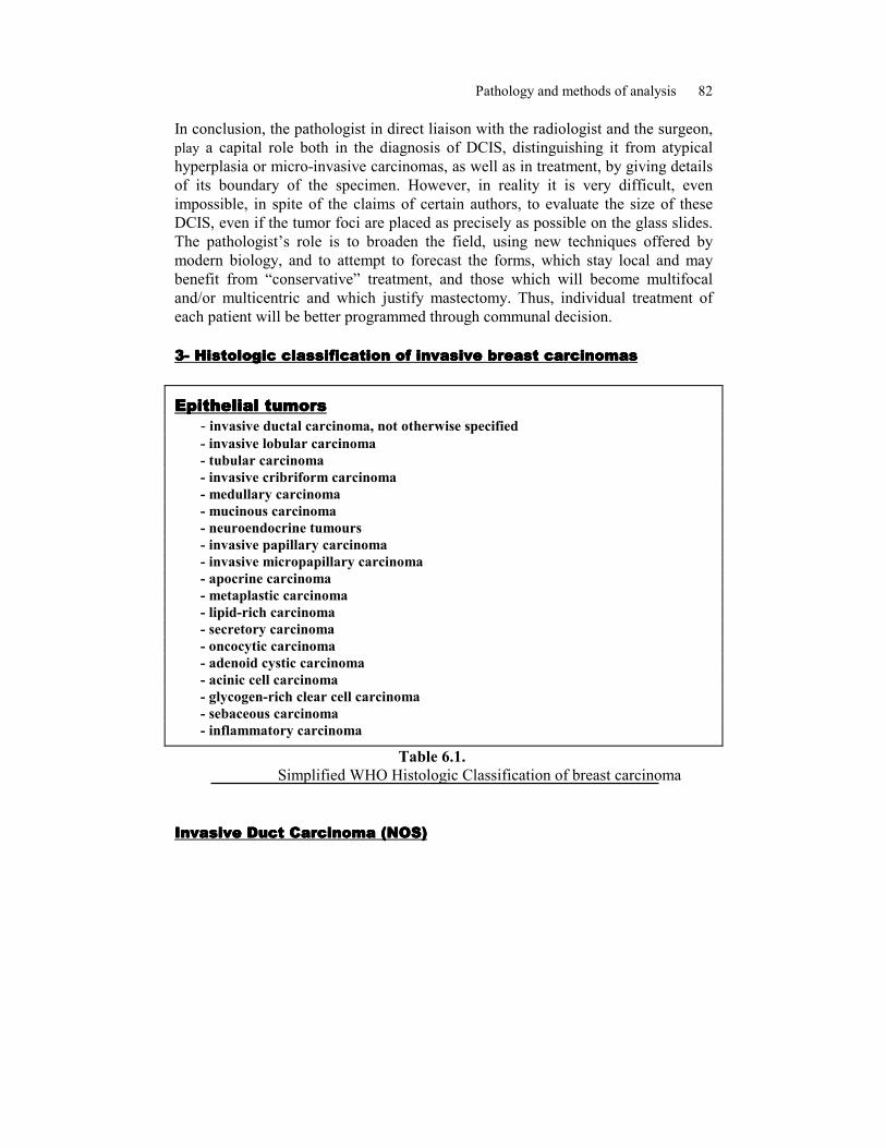
3333---- Histologic classification of invasive breast carcinomaHistologic classification of invasive breast carcinomaHistologic classification of invasive breast carcinomaHistologic classification of invasive breast carcinomassss
Epithelial tumorsEpithelial tumorsEpithelial tumorsEpithelial tumors
- invasive ductal carcinoma, not otherwise specified - invasive lobular carcinoma
- tubular carcinoma
- invasive cribriform carcinoma
- medullary carcinoma
- mucinous carcinoma
- neuroendocrine tumours
- invasive papillary carcinoma
- invasive micropapillary carcinoma
- apocrine carcinoma
- metaplastic carcinoma
- lipid-rich carcinoma
- secretory carcinoma
- oncocytic carcinoma
- adenoid cystic carcinoma
- acinic cell carcinoma
- glycogen-rich clear cell carcinoma
- sebaceous carcinoma
- inflammatory carcinoma
Table 6.1.
Simplified WHO Histologic Classification of breast carcinoma
Invasive Duct Carcinoma (NOS)Invasive Duct Carcinoma (NOS)Invasive Duct Carcinoma (NOS)Invasive Duct Carcinoma (NOS)
Pathology and methods of analysis 83
This represents the most frequently encountered histologic type of breast
carcinoma. Grossly, the tumor appears either well circumscribed, stellate, or shows
a combination of both. The stellate variant is associated with extensive fibrosis
(scirrhous carcinoma). The size of the tumor is variable and its assessment
constitutes an important prognostic parameter. Microscopically, the tumor is
characterized by the presence of irregular or rounded solid clusters of tumor
admixed with single cells and cords of tumor cells. Based on the degree of tubule
formation, nuclear appearance and mitotic activity, the tumor should be graded.
In some tumors, an in-situ ductal carcinoma may be found; alternatively both ductal
and lobular types of in-situ carcinoma may be present.
Infiltrative Duct Carcinoma with Extensive InInfiltrative Duct Carcinoma with Extensive InInfiltrative Duct Carcinoma with Extensive InInfiltrative Duct Carcinoma with Extensive In----situ Componentsitu Componentsitu Componentsitu Component
This variant has been defined as invasive tumor in which 25% of the overall area
involved by the invasive carcinoma is composed of intraductal carcinoma. The
presence of abundant intraductal carcinoma within the tumor was associated with a
tendency to have in-situ component beyond the tumor margin and multicentric
carcinoma. However the risk of local relapse as other tumor type is directly
dependant of the status of the resection margins.
Microinvasive CarcinomaMicroinvasive CarcinomaMicroinvasive CarcinomaMicroinvasive Carcinoma
These are tumors that are predominantly intraductal but focally the tumor cells
have barely broken through the basement membrane, invading the stroma
immediately adjacent to the defective portion of the duct wall but measuring less
than 1mm.
Infiltrating Carcinoma Presenting as an Axillary MassInfiltrating Carcinoma Presenting as an Axillary MassInfiltrating Carcinoma Presenting as an Axillary MassInfiltrating Carcinoma Presenting as an Axillary Mass
Breast carcinoma may present as axillary mass in the absence of a clinically,
detectable breast tumor. It is possible that some of these tumors actually arise from
either the axillary tail of the breast, accessory breast tissue in the axilla, or
heterotopic breast tissue in an axillary lymph node.
InInInInvasive Lobular Carcinomavasive Lobular Carcinomavasive Lobular Carcinomavasive Lobular Carcinoma
The tumor tends to present either as an irregular infiltrating or well circumscribed,
indurated mass. Originally the invasive lobular carcinoma is characterized by a
linear growth pattern of small cells in “Indian file” of one cell width. Subsequently,
additional patterns of invasive lobular carcinoma were defined including: solid
pattern, alveolar pattern, and a mixed group composed of an admixture of one or
more of these patterns. Another pattern designated tubulo-lobular has been
described. In this variant, tubules formations are arranged along with cords of cells
Pathology and methods of analysis 84
arranged in “Indian files”. Cytologic variants such as apocrine, histiocytoid and
pleomorphic have been also described. This latter form differs from the classic type
by its aggressiveness. Infiltrating lobular carcinoma can closely simulate a
lymphoma. Immunostains for cytokeratin and leucocyte common antigen (LCA) are
helpful in establishing the nature of the neoplastic cells. E cadherin, a membranous
antigen which expression is typically absent in lobular carcinoma is helpful for the
recognition of these rare variants.
Tubular CarcinomaTubular CarcinomaTubular CarcinomaTubular Carcinoma
A distinctive variant of mammary carcinoma characterized by proliferation of
angulated tubules separated from each other by a reactive fibroblastic stroma. The
tubules display open lumens and are lined by a single layer of epithelial cells. A
minimum of 75% of the lesion should display this morphology in order to call this
tumor a tubular carcinoma. When such arrangement is present as a minor
component the tumor is referred to as a mixed tubular carcinoma. The recognition
of tubular carcinoma is important because of its good prognosis and the rarity of
axillary node metastases. Tubular carcinoma can be mistaken for sclerosing
adenosis, microglandular adenosis and radial scar.
Invasive cribriforme carcinomaInvasive cribriforme carcinomaInvasive cribriforme carcinomaInvasive cribriforme carcinoma
This rare type grows mainly in a cribriform pattern similar to that seen in
intraductal carcinoma but without any myo-epithelial cells. A minor tubular
component is frequently seen. This form is associated to an excellent prognosis.
Immunohistochemistry is important to distinguish it from an adenoid cystic
carcinoma and intraductal carcinoma.
Medullary CarcinomaMedullary CarcinomaMedullary CarcinomaMedullary Carcinoma
Medullary carcinomas are distinctive tumors with pushing expansile margins.
Occasionally, they may appear encapsulated. The tumors have fleshy soft
consistency and composed of a syncytium of anastomosing cords and sheets of
tumor cells separated by loose connective tissue. The tumor cells are round with
abundant cytoplasm, round vesicular nuclei, containing one or more prominent
nucleoli. Mitosis is common. Squamous metaplasia and atypical tumor giant cells
have been noted in some of these tumors. Typically, the tumor cells in medullary
carcinoma are accompanied by moderate to pronounced lympho-plasmocytic
infiltrate in the supporting and surrounding stroma. Microglandular or tubular
formations are absent. The constant lymphocytic reaction seen in medullary
carcinoma may be linked to the favorable prognosis of medullary carcinoma and
low frequency of nodal metastasis. This form is rare since strict histologic criteria
Pathology and methods of analysis 85
have been used to its identification. It is frequently observed in predisposed
patients for breast carcinoma with BRCA1 germ line mutation.
Mucinous CarcinomaMucinous CarcinomaMucinous CarcinomaMucinous Carcinoma
In this form, the uniform tumor cells are accompanied by large amounts of
extracellular mucin lakes. Pure and mixed variants of mucinous carcinoma have
been recognized. An infiltrating duct carcinoma is the most common associated
tumor in the mixed tumors. It is important to differentiate the pure form from the
mixed type because of the favorable prognosis of the former.
Neuroendocrine Neuroendocrine Neuroendocrine Neuroendocrine TumoursTumoursTumoursTumours
These are mammary carcinomas that display any growth patterns similar to those of
carcinoid tumors that occur in other organs. They express neuroendocrine markers
in more than 50% of the cell population. Hormonal receptors are frequently
identified. If neuroendocrine markers are frequently found in other breast
carcinomas, they are limited to scattered cells. This group comprises different
subtypes, solid neuroendocrine carcinoma, atypical carcinoid, oat cell carcinomas
and large cell neuroendocrine.
Invasive papillary carcinomaInvasive papillary carcinomaInvasive papillary carcinomaInvasive papillary carcinoma
This tumor is generally well delineated, frequently cystic and associated with
hemorrhage. Myoepithelial cells are completely absent. It is now considered that
many of the intra cystic carcinoma published may represent invasive papillary
carcinoma. As they share an excellent prognosis, they have been called also
papillary encapsulated carcinoma.
Invasive micropapillary carcinomaInvasive micropapillary carcinomaInvasive micropapillary carcinomaInvasive micropapillary carcinoma
This newly identified form of carcinoma is characterized by the presence of small
clusters of tumours cells lying in clear stromal spaces mimicking vascular channels.
This is an aggressive form, with frequent and numerous axillary lymph node
metastases and poor prognosis. It may be pure or associated with a ductal
component.
Metaplastic CarcinomaMetaplastic CarcinomaMetaplastic CarcinomaMetaplastic Carcinoma
Several metaplastic forms may be encountered in this category. Pure squamous or
epidermoid carcinoma is rare, a more common presentation being foci of squamous
differentiation in vicinity of necrotic areas in other histologic patterns of tumor.
Spindle cell differentiation and metaplasia such as cartilagenous or osseous
metaplasia are rare in pure form.
Pathology and methods of analysis 86
Adenoid Cystic CarcinomaAdenoid Cystic CarcinomaAdenoid Cystic CarcinomaAdenoid Cystic Carcinoma
In this histologic subtype, a cribriform pattern is seen with rounded hyaline spaces
enclosed in between cellular masses. It associated two cell components one
expressing cytokeratin the other actin. Perineural invasion is a peculiar feature to
this pattern of growth. Generally, this type of neoplasm exhibits a slow rate of
growth and a favorable prognostic outcome.
Lipid Rich CarcinomaLipid Rich CarcinomaLipid Rich CarcinomaLipid Rich Carcinoma
This histologic variant has a particularly unfavorable prognosis. Large tumor cells
exhibit lipid material, best demonstrated in frozen tissue material
Juvenile (Secretory) Carcinoma
It occurs primarily in children and adolescents. This neoplastic variant is highly
differentiated exhibiting excessive PAS positive secretory products.
Inflammatory CarcinomaInflammatory CarcinomaInflammatory CarcinomaInflammatory Carcinoma
This type of breast carcinoma has been given a variety of terms such as mastitis
carcinoma, carcinoma mastitoides, or acute mammary carcinoma. It is considered
as the most malignant type of breast carcinomas, and of high proliferative activity
(PEV 3). It represents 1% to 2% of breast carcinoma in Western literature,
however, in Egypt, North Africa and Tunisia such mammary carcinoma is not
uncommon. Characteristically, the skin of the breast is reddened, warm, edematous
and thickened. During early stage, an underlying breast mass may not be palpable,
and this may cause an erroneous diagnosis of inflammatory non-neoplastic process.
Histologically, the skin dermis is filled with many lymphatic tumor cell emboli.
Blocking of lymphatics causes skin edema.
ClinicalClinicalClinicalClinical VersusVersusVersusVersus PathologPathologPathologPathologic Definitionic Definitionic Definitionic Definition
The criteria for clinical diagnosis of inflammatory carcinoma include diffuse
erythemaedema extending to greater than two thirds of the breast, “peau d’orange”,
tenderness, engorgement, and diffuse breast involvement. The pathologic diagnosis
requires the presence of tumor emboli in the dermal lymphatics, in addition to the
presence of invasive carcinoma with spontaneous necrotic foci. In majority of
cases, the clinical picture and the pathologic picture of dermal lymphatic
involvement coincide. However, all patients with the clinical disease have
demonstrable dermal lymphatic involvement, and there are also cases with dermal
lymphatic involvement but without the clinical inflammatory features. It was found
that clinically occult inflammatory carcinoma i.e. patients without clinical signs of
inflammatory carcinoma but with tumor emboli in the dermal lymphatics followed

Pathology and methods of analysis 87
rapidly deteriorating clinical course, and wide spread metastases similar to
clinically diagnosed inflammatory carcinoma. Furthermore, clinically diagnosed
inflammatory carcinoma without demonstrable dermal lymphatic invasion, behave
aggressively. Therefore, the use of the term “inflammatory carcinoma” is justified
with either the clinical or the pathologic features.
PPPProrororonostic clanostic clanostic clanostic classification ssification ssification ssification
Histologic SBR GradeHistologic SBR GradeHistologic SBR GradeHistologic SBR Grade
Tumor grading regardless of the system used has prognostic importance in breast
cancer. Grading systems have been developed based on nuclear features,
architectural pattern, mitotic rate or combinations of these features. Its use, initially
restricted to the sole invasive ductal carcinoma, now has been extended to every
subtype of invasive carcinoma except the medullary carcinoma. The chief difficulty
with any of these grading systems is their subjective nature and the resultant poor
reproducibility among pathologists, one approach is to use a uniform grading
system with distinct criteria that are easy to reproduce among observers (Table
7.2.). For instance, the mitotic count is evaluated on ten consecutive high power
fields and adjust according to the microscopic field area (table 7.3.). Studies have
indicated that proliferation is the most important prognostic compound of this
system. Other investigators have combined morphologic grade with other features,
including tumor size and nodal status to calculate a prognostic index that appears to
be highly predictive of clinical course.
Axillary Lymph Node examinationAxillary Lymph Node examinationAxillary Lymph Node examinationAxillary Lymph Node examination
Involvement of axillary lymph nodes by metastases in patients with cancer breast is
one of the important markers of prognosis. Pathologic examination of the axillary
nodes in patients with breast cancer is required in order to assess prognosis and
determine the need for adjuvant therapy. For sentinel lymph node, serial sectioning
and the use of immunohistochemical staining is mandatory to ensure the predictive
value for the involvement of the other axillary lymph nodes. One consequence of
this method is to increase the detection metastases, in particular those of limited
size (micrometastases) whose prognostic value is still under discussion. UICC has
modified its classification of these small metastases, based on their size, in order to
be able in the future to determine their prognostic value.
Features Score
Tubule formation
Majority of tumor (>75%) 1
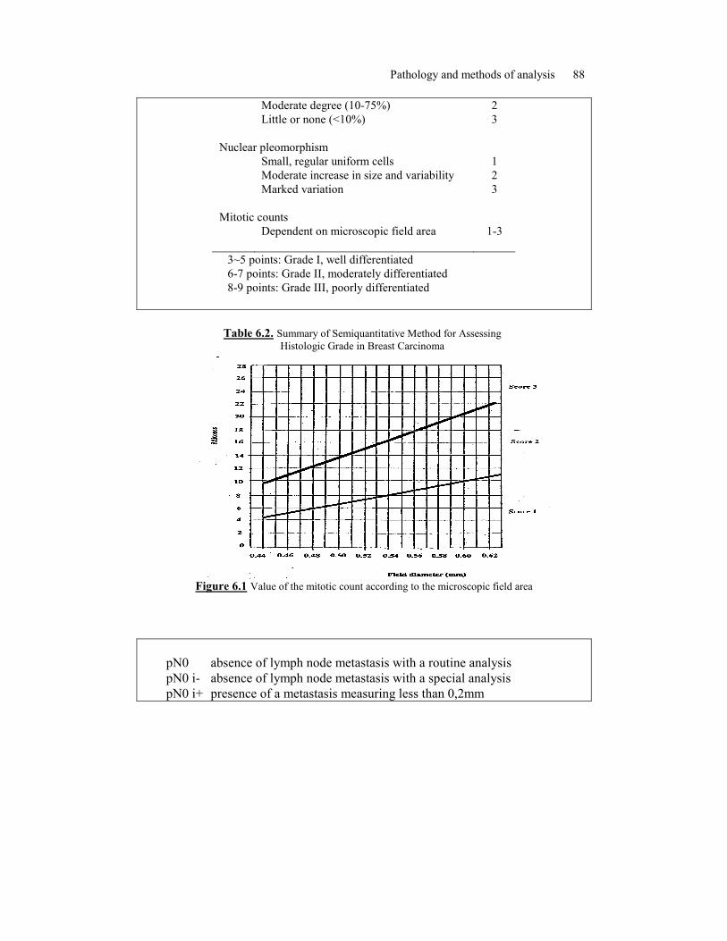
Pathology and methods of analysis 88
Moderate degree (10-75%) 2
Little or none (<10%) 3
Nuclear pleomorphism
Small, regular uniform cells 1
Moderate increase in size and variability 2
Marked variation 3
Mitotic counts
Dependent on microscopic field area 1-3
3~5 points: Grade I, well differentiated
6-7 points: Grade II, moderately differentiated
8-9 points: Grade III, poorly differentiated
Table 6.2. Summary of Semiquantitative Method for Assessing
Histologic Grade in Breast Carcinoma
Figure 6.1 Value of the mitotic count according to the microscopic field area
pN0 absence of lymph node metastasis with a routine analysis
pN0 i- absence of lymph node metastasis with a special analysis
pN0 i+ presence of a metastasis measuring less than 0,2mm
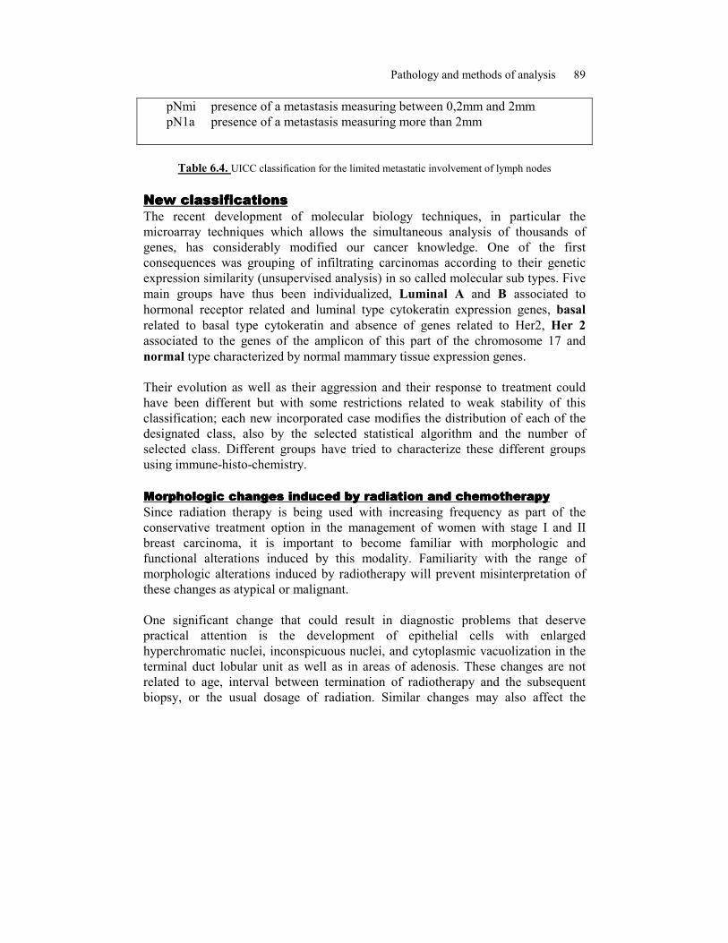
Pathology and methods of analysis 89
pNmi presence of a metastasis measuring between 0,2mm and 2mm
pN1a presence of a metastasis measuring more than 2mm
Table 6.4. UICC classification for the limited metastatic involvement of lymph nodes
New classifiNew classifiNew classifiNew classificationscationscationscations
The recent development of molecular biology techniques, in particular the
microarray techniques which allows the simultaneous analysis of thousands of
genes, has considerably modified our cancer knowledge. One of the first
consequences was grouping of infiltrating carcinomas according to their genetic
expression similarity (unsupervised analysis) in so called molecular sub types. Five
main groups have thus been individualized, Luminal A and B associated to
hormonal receptor related and luminal type cytokeratin expression genes, basal
related to basal type cytokeratin and absence of genes related to Her2, Her 2
associated to the genes of the amplicon of this part of the chromosome 17 and
normal type characterized by normal mammary tissue expression genes.
Their evolution as well as their aggression and their response to treatment could
have been different but with some restrictions related to weak stability of this
classification; each new incorporated case modifies the distribution of each of the
designated class, also by the selected statistical algorithm and the number of
selected class. Different groups have tried to characterize these different groups
using immune-histo-chemistry.
Morphologic changes induced by radiation and chemotherapyMorphologic changes induced by radiation and chemotherapyMorphologic changes induced by radiation and chemotherapyMorphologic changes induced by radiation and chemotherapy
Since radiation therapy is being used with increasing frequency as part of the
conservative treatment option in the management of women with stage I and II
breast carcinoma, it is important to become familiar with morphologic and
functional alterations induced by this modality. Familiarity with the range of
morphologic alterations induced by radiotherapy will prevent misinterpretation of
these changes as atypical or malignant.
One significant change that could result in diagnostic problems that deserve
practical attention is the development of epithelial cells with enlarged
hyperchromatic nuclei, inconspicuous nuclei, and cytoplasmic vacuolization in the
terminal duct lobular unit as well as in areas of adenosis. These changes are not
related to age, interval between termination of radiotherapy and the subsequent
biopsy, or the usual dosage of radiation. Similar changes may also affect the
Pathology and methods of analysis 90
epithelial cells in larger ducts, but less frequently. Variable degrees of lobular
sclerosis may also occur. The radiation induced atypia differs from carcinoma
involving the lobules by the absence of epithelial hyperplasia, mitotic activity, and
luminal necrosis. Furthermore, the cytoplasmic vacuolization and a history of prior
radiotherapy should serve as additional clues.
Similar epithelial changes have also been observed secondary to chemotherapy. An
increase in nuclear size, pleomorphism, vacuolization, and chromatin clumping in
residual tumor cells has been described after chemotherapy. Chemotherapy may
induce nodular fibrosis and areas of fibrohistiocytic proliferation in both the breast
as well as in axillary lymph nodes. Chemotherapy does not induce the vascular and
stromal changes. Preoperative chemotherapy can abolish the tumor leaving no
evidence of residual malignancy in the specimen.
Name: No: Breast: 1) Left 2) Right Specimen: 1) Excisional (for palpable
mass) 2) Mammographic Loc.
3) Incisional (includes core needle and FNA) 4) Re-excisional 5) Mastectomy 6) Chest wall Specimen Size:
Tumor
Pathology and methods of analysis 91
Size(s): Tumor Type
1) DCIS 5) Mixed NOS/ILC
9) Papillary
2)LCIS 6) Tubular 10) Cribriform 3) Infiltrating ductal
(NOS) 7) Mucinous 11) Other.
(specify) 4) Infiltrating lobular 8) Medullary Grade of invasion: 1) I 2) II 3) III Gross margin:
1) Free (specify
distance) 2) Focal 3) Inevaluable
Margins invasive (specify type of margin evaluation) 1) Free (specify
distance) 2) Focal 3) Inevaluable
Margins DCIS (specify type of margin evaluation) 1) Free (specify
distance) 2) Focal 3) Inevaluable
DCIS nuclear morphology 1) High grade 2) Intermediate
grade 3) Low grade
DCIS patterns (specify all that apply)
1) Large areas of central necrosis (comedo) 4) Solid 2) Small areas of central necrosis 5) Micropapillary 3) Cribriform 6) Papillary
Calcification in situ: 1) Absent 2) Prominent in
DCIS 3) Local in DCIS
4) In LCIS 5) Prominent in benign breast tissue 6) Focal in benign breast tissue Peritumoral lymphatic invasion: 1) Absent 2) Present 3) Dermal Peritumoral vascular invasion: 1) Absent 2) Present
Pathology and methods of analysis 92
Extent DCIS within invasive tumor: 1) Absent 2) Slight 3) Moderate-
Marked 4) Tumor primarily DCIS with focal
invasion
Extent DCIS adjacent to invasive tumor:
1) Absent 2) Slight 3) Moderate-
Marked EIC status:
1) EIC negative 2) EIC positive
3) EIC intermediate
Note: If a tumor is primarily DCIS with focal invasion or has a moderate or marked amount of DCIS within the infiltrating tumor and in the -adjacent tissue it is EIC positive Skin:
1) Not sampled 2) Free
3) Invasive
4) Dermal lymphatic Nipple:
1) Not sampled 2) Free
3) Invasive
4) Dermal lymphatic 5) DCIS 6) Paget’s Muscle:
1) Not sampled 2) Free
3) Involved
Mastectomy tumor location: 1) Central 2)UOQ 3) UIQ 4) LOQ 5)LIQ 6) Axillary tail Multiple areas involved: 1) Central 2) UOQ 3) UIQ 4) LOQ 5) LIQ 6) Axillary tail 7) Only one area
involved
Lymph nodes (number of involved nodes in relation to total number examined): Total Level I Level II Level III Other (specify) Extranodal extension 1) Absent 2) Present

Pathology and methods of analysis 93