chapter 6: visual attention - university of...
TRANSCRIPT

Chapter 6: Visual Attention
"Everyone knows what attention is. It is the taking possession by the mind in clear and vivid form, of one out of what seem several simultaneously possible objects or trains of thought...It implies withdrawal from some things in order to deal effectively with others." (William James, Principles of Psychology, 1890)

http://viscog.beckman.uiuc.edu/grafs/demos/15.html
We are aware of only a small portion of information that is impinging upon us.
What determines what we attend to?
What happens in the brain when we attend?
Why is Selective Attention Necessary?
Conscious experience seems to have a limited capacity:We can only attend to one thing at a time.
Attention helps us decide where to move our eyes next.
Our perception of a scene is developed by a combination of attention, eye movements, and memory.

What’s in this upcoming scene?


Saccades: quick eye movements from one fixation location to another.
We make around 3 saccades per second!

Not all parts of a scene are sampled equally
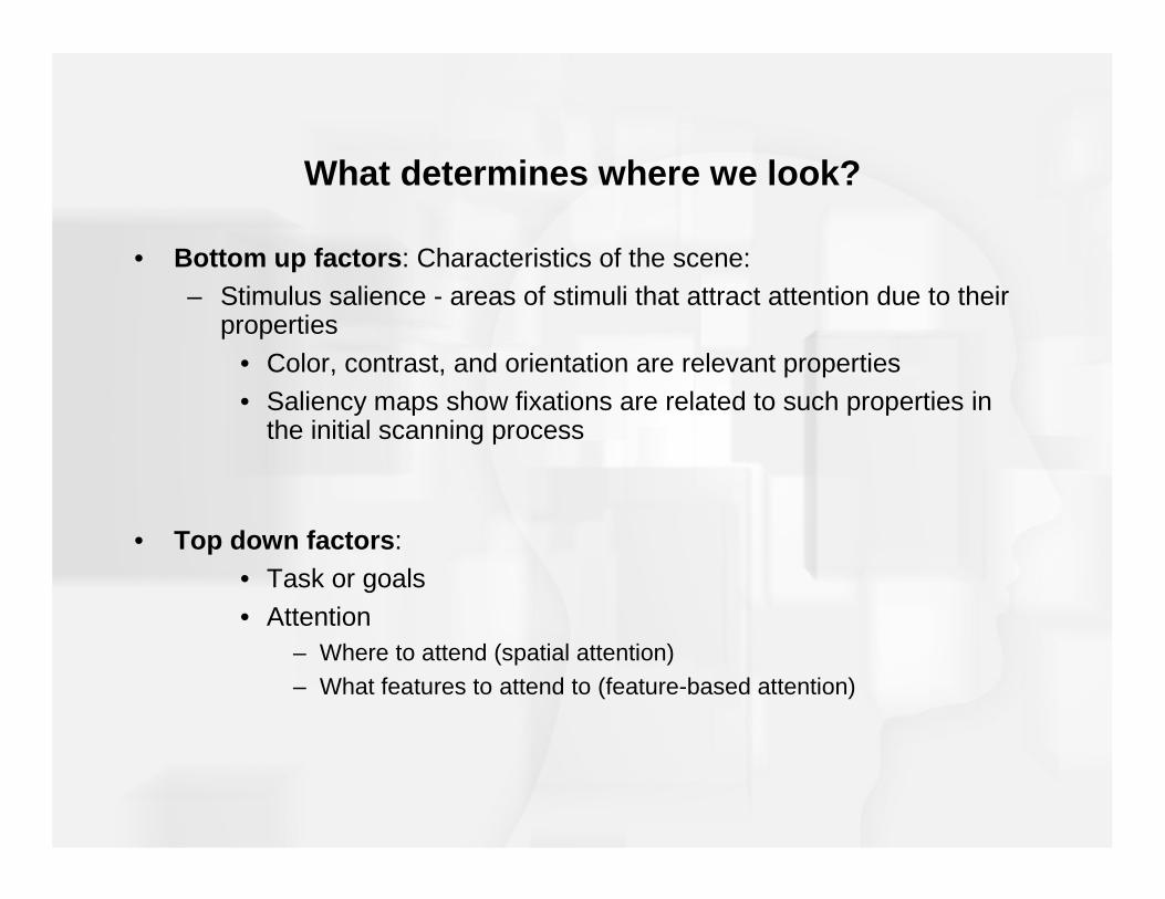
What determines where we look?
• Bottom up factors : Characteristics of the scene:– Stimulus salience - areas of stimuli that attract attention due to their
properties
• Color, contrast, and orientation are relevant properties• Saliency maps show fixations are related to such properties in
the initial scanning process
• Top down factors : • Task or goals
• Attention – Where to attend (spatial attention)– What features to attend to (feature-based attention)
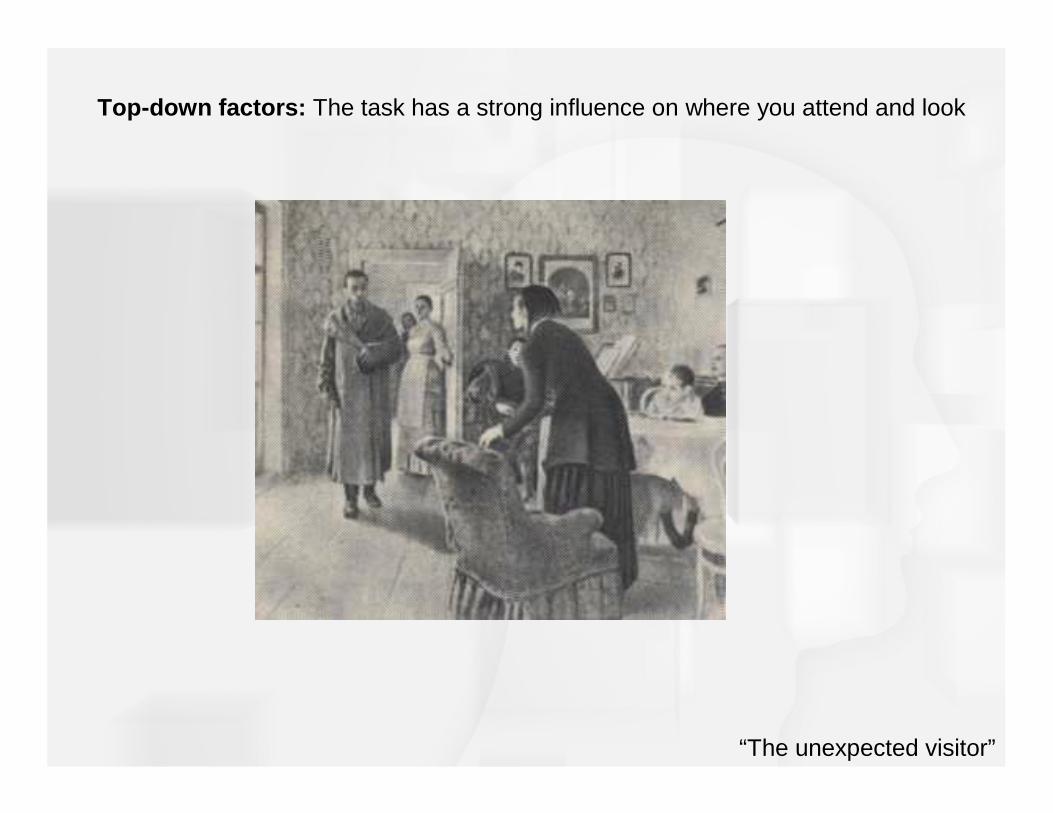
“The unexpected visitor”
Top-down factors: The task has a strong influence on where you attend and look
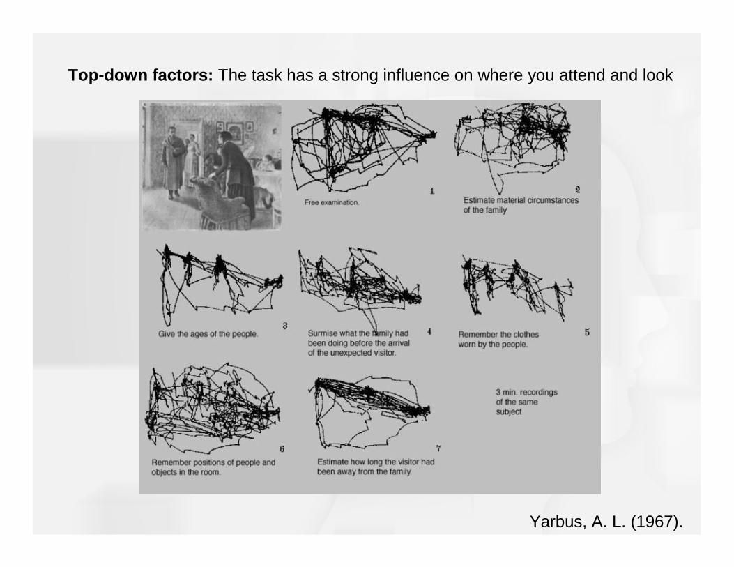
Top-down factors: The task has a strong influence on where you attend and look
Yarbus, A. L. (1967).
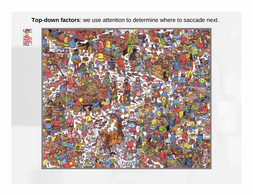
Top-down factors : we use attention to determine where to saccade next.

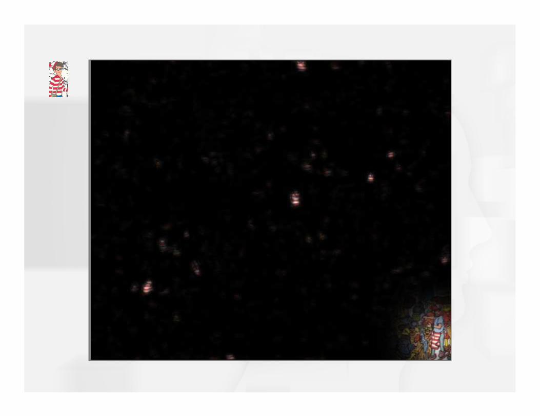
Where are the horizontal red stripey things?
We use feature-based attention to highlight specific features throughout a scene.
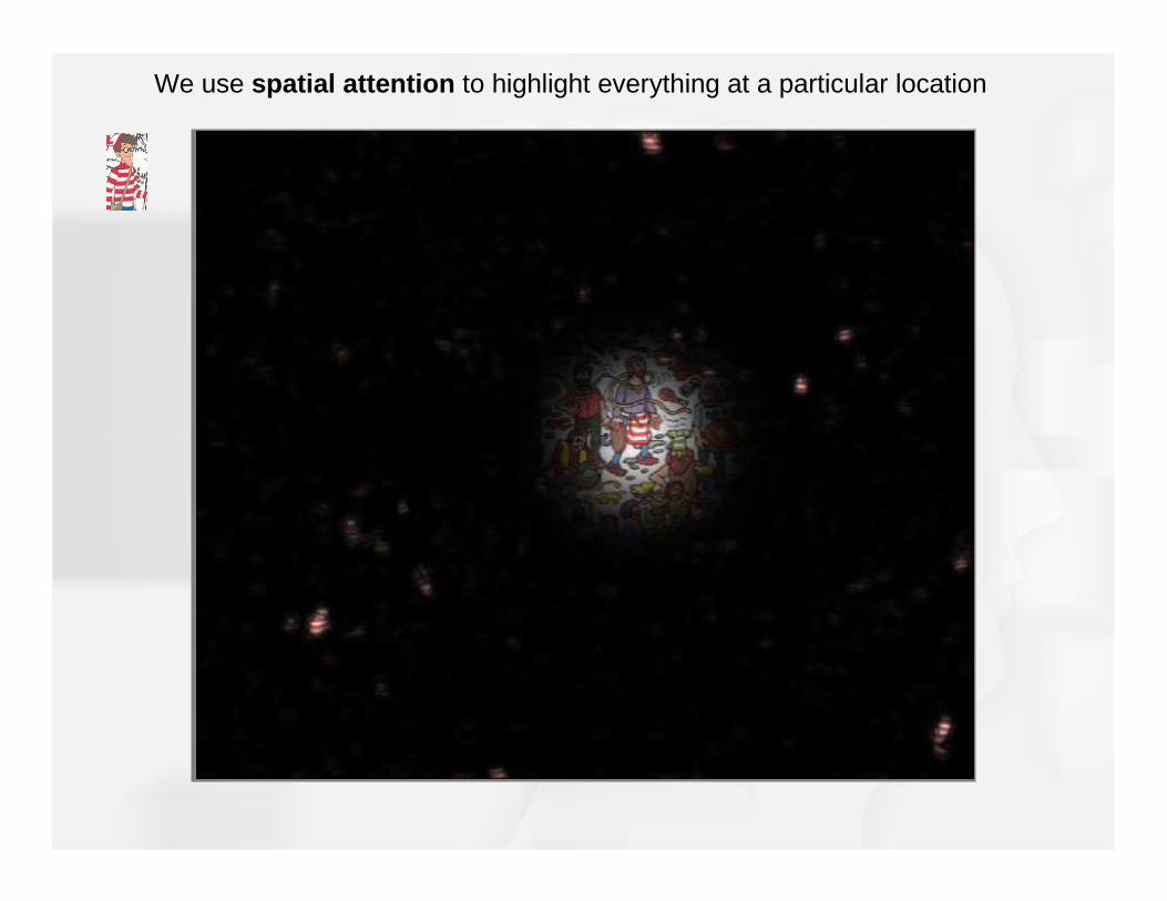
We use spatial attention to highlight everything at a particular location


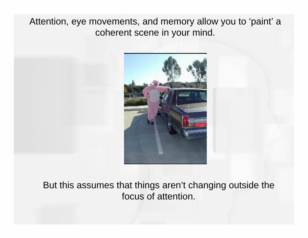
Attention, eye movements, and memory allow you to ‘paint’ a coherent scene in your mind.
But this assumes that things aren’t changing outside the focus of attention.
• Change blindness
– Observers are shown a picture with and without a missing elementin an alternating fashion with a blank screen
– Results show that the pictures had to alternate a number of times before the change was detected
Change blindness demos

Two ways that spatial attention can be directed:
Endogenous: voluntary, or by instruction in laboratory experiments: “attend left”
Exogenous: involuntary, often by a flash, sound or any sudden change.
Spatial attention: Direction of attention to a particular region of space
Feature-based attention:Direction of attention to a particular feature, anywhere in space
Features include:
- Direction of motion- Color- Orientation
How does spatial and feature-based attention affect neuronal responses in the visual cortex?
How does attention affect behavioral performance?
Where in the brain are these top-down influences coming from?
Can attention affect the appearance of things?
Key questions about attention

How does attention help performance?
• Experiment by Posner et al.
– Observers saw a square with two lights on each side
– Precueing was used to indicate on which side the light would turn on (endogenous cue to attention)
– Lights turned on consistent or inconsistent with the cue
– Task was to push button when light was seen

Experiment by Posner et al.
• Results showed that observers responded fastest when cue was consistent with light
• Information processing is most efficient where attention is directed
Invalid Cue Valid Cue0
50
100
150
200
250
300
Res
pons
e T
ime
(ms)
Effects of Attention on Perception
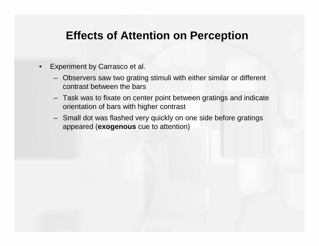
• Experiment by Carrasco et al.
– Observers saw two grating stimuli with either similar or different contrast between the bars
– Task was to fixate on center point between gratings and indicateorientation of bars with higher contrast
– Small dot was flashed very quickly on one side before gratings appeared (exogenous cue to attention)
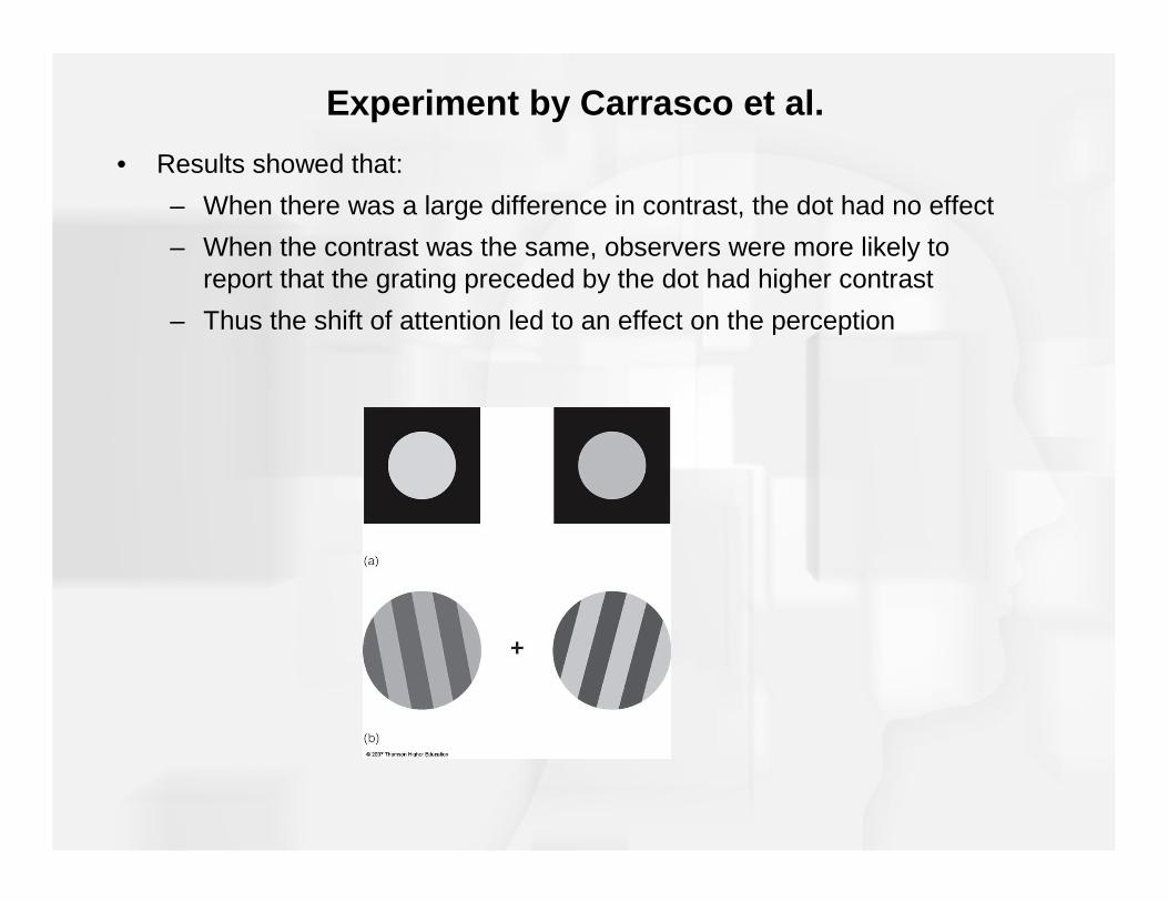
Experiment by Carrasco et al.
• Results showed that:
– When there was a large difference in contrast, the dot had no effect
– When the contrast was the same, observers were more likely to report that the grating preceded by the dot had higher contrast
– Thus the shift of attention led to an effect on the perception


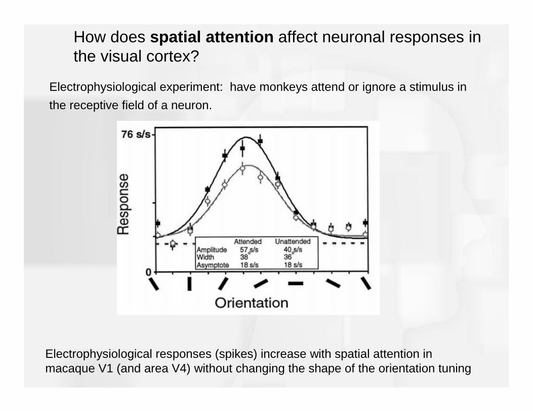
How does spatial attention affect neuronal responses in the visual cortex?
fMRI experiment: present stimuli to left and right side of visual field at the same time.
Have subjects attend to one stimulus at a time (endogenous cue to spatial attention)

How does spatial attention affect neuronal responses in the visual cortex?
fMRI responses in V1 are increased by spatial attention
How does spatial attention affect neuronal responses in the visual cortex?
Electrophysiological responses (spikes) increase with spatial attention in macaque V1 (and area V4) without changing the shape of the orientation tuning
Electrophysiological experiment: have monkeys attend or ignore a stimulus in
the receptive field of a neuron.
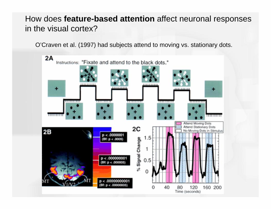
O’Craven et al. (1997) had subjects attend to moving vs. stationary dots.
How does feature-based attention affect neuronal responses in the visual cortex?

How does feature-based attention affect neuronal responses in the visual cortex?
fMRI experiment: present stimuli to left and right side of visual field at the same time.
Have subjects attend to one color at a time on the left (endogenous cue to spatial attention)
On one side, have two different overlapping colors on the left side

Attend green Attend red0
0.5
1
1.5
fMR
I res
pons
e to
igno
red
gree
n st
imul
us
How does feature-based attention affect neuronal responses in the visual cortex?
Attending to a color enhances the fMRI response in V1 and other visual areas to all stimuli having the attended color throughout the visual scene.

Attending to a color enhances the fMRI response to all stimuli having the attended color throughout the visual scene.
More recent studies have shown that the same is true for direction of motionand orientation.
How could this help us search for Waldo?

How does feature-based attention affect neuronal responses in the visual cortex?
Electrophysiological experiment: present two stimuli in the receptive field of a neuron in area MT. (MT has mostly direction selective neurons).
Present dots moving in opposite directions.Have monkey attend to only one of the two dots.
Treue and Maunsell, 1996
Neuron fires only when attended dot moves in the
preferred direction!

How does feature-based attention affect neuronal responses in the visual cortex?
Electrophysiological experiment: present two stimuli in the receptive field of a V4 neuron
Receptive field
This stimulus alone producesa small response (5 Hz).
This stimulus alone produces a big response
(50 Hz).
Presented together, the stimuli produce an intermediate response (20 Hz).
Moran and Desimone, 1995

How does feature-based attention affect neuronal responses in the visual cortex?
Electrophysiological experiment: now have monkeys attend to one of the two stimuli.
Receptive field
Applying feature-based attention (to orientation) is like removing the unattended stimulus from the receptive field.
Attention to this stimulus produces a big response(50 Hz)
Attention to this stimulus produces a small response (10 Hz)
Moran and Desimone, 1995
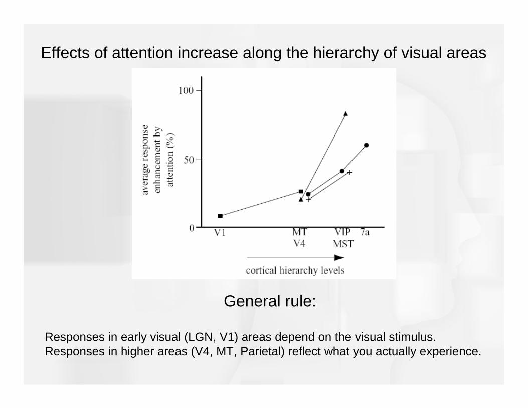
Effects of attention increase along the hierarchy of visual areas
General rule:
Responses in early visual (LGN, V1) areas depend on the visual stimulus.Responses in higher areas (V4, MT, Parietal) reflect what you actually experience.

Where in the brain are these top-down influences coming from?
Balint’s syndrome: caused by bilateral damage to parietal lobes
Symptoms include the inability to voluntarily direct or shift spatial attention.
Often includes simultanagnosia , where scenes containing multiple objects cannot be interpreted as a whole. Instead, patients with simultanagnosiarecognize only portions of the scene at one time, and fail to describe the overall nature of the scene and comprehend its meaning.
The parietal lobe seems to be important:

Visual Awareness
To what extent can it be said that our conscious visual experience fully captures what we perceive?
Dissociations between explicit visual awareness and the control of action suggest that our conscious visual awareness is not all that we perceive.
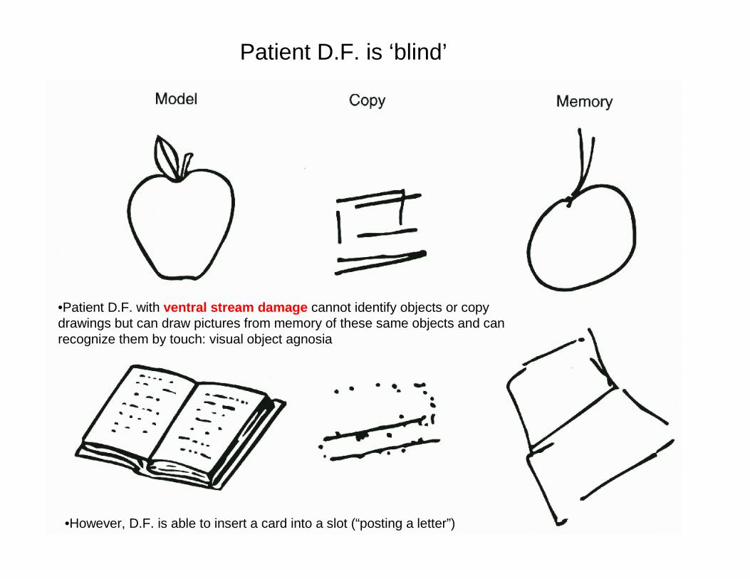
•Patient D.F. with ventral stream damage cannot identify objects or copy drawings but can draw pictures from memory of these same objects and can recognize them by touch: visual object agnosia
•However, D.F. is able to insert a card into a slot (“posting a letter”)
Patient D.F. is ‘blind’

• Individuals who have experienced a stroke that has selectively damaged area V1, or who have had V1 removed surgically, report being blind in the affected part of visual space (e.g., damage to left V1 affects the right visual field).
• However, if these individuals are asked to point to an object to reach out and grasp an object in the “blind” field, they can do so accurately, even though they claim they are just guessing.
Blindsight: What happens when V1 is damaged?
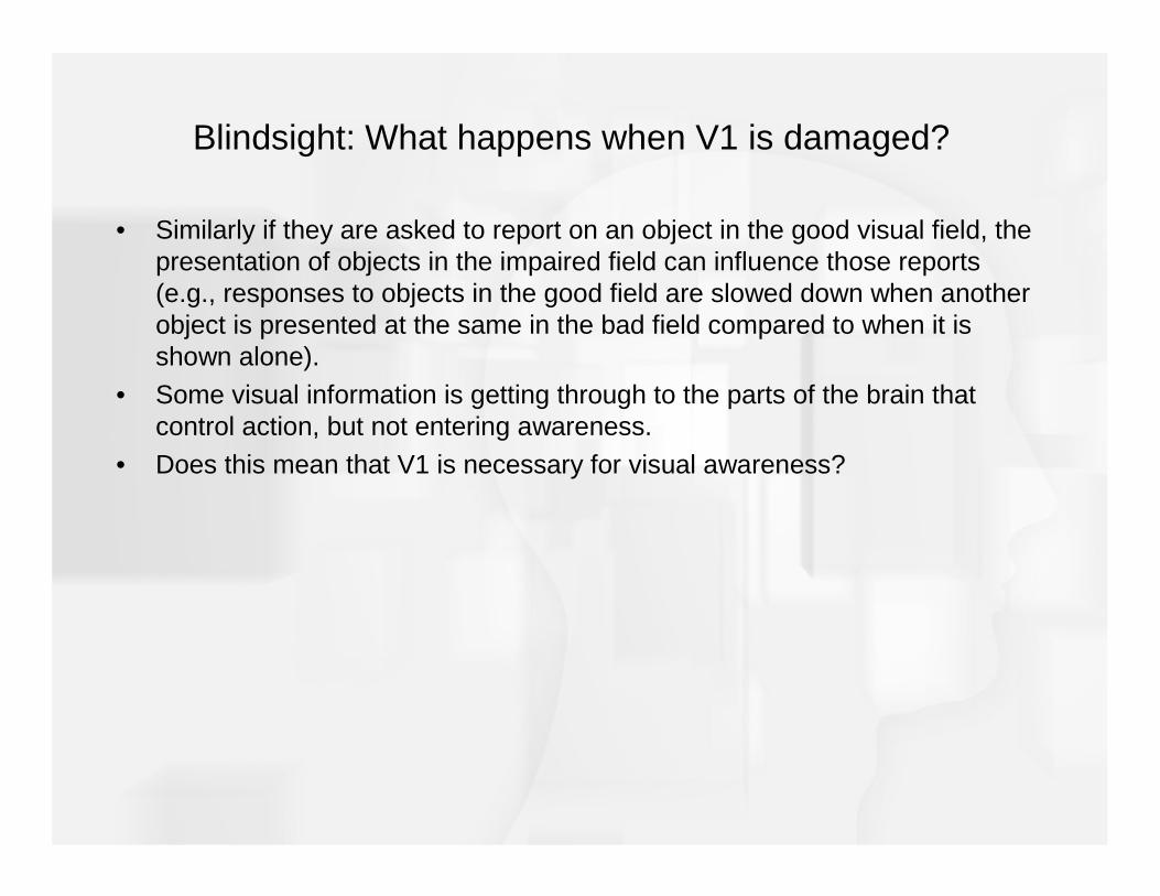
• Similarly if they are asked to report on an object in the good visual field, the presentation of objects in the impaired field can influence those reports (e.g., responses to objects in the good field are slowed down when another object is presented at the same in the bad field compared to when it is shown alone).
• Some visual information is getting through to the parts of the brain that control action, but not entering awareness.
• Does this mean that V1 is necessary for visual awareness?
Blindsight: What happens when V1 is damaged?

Blindsight patient GY has a lesion in left V1. He reports being blind to objects in the right visual field, yet he can correctly point to and grasp them.
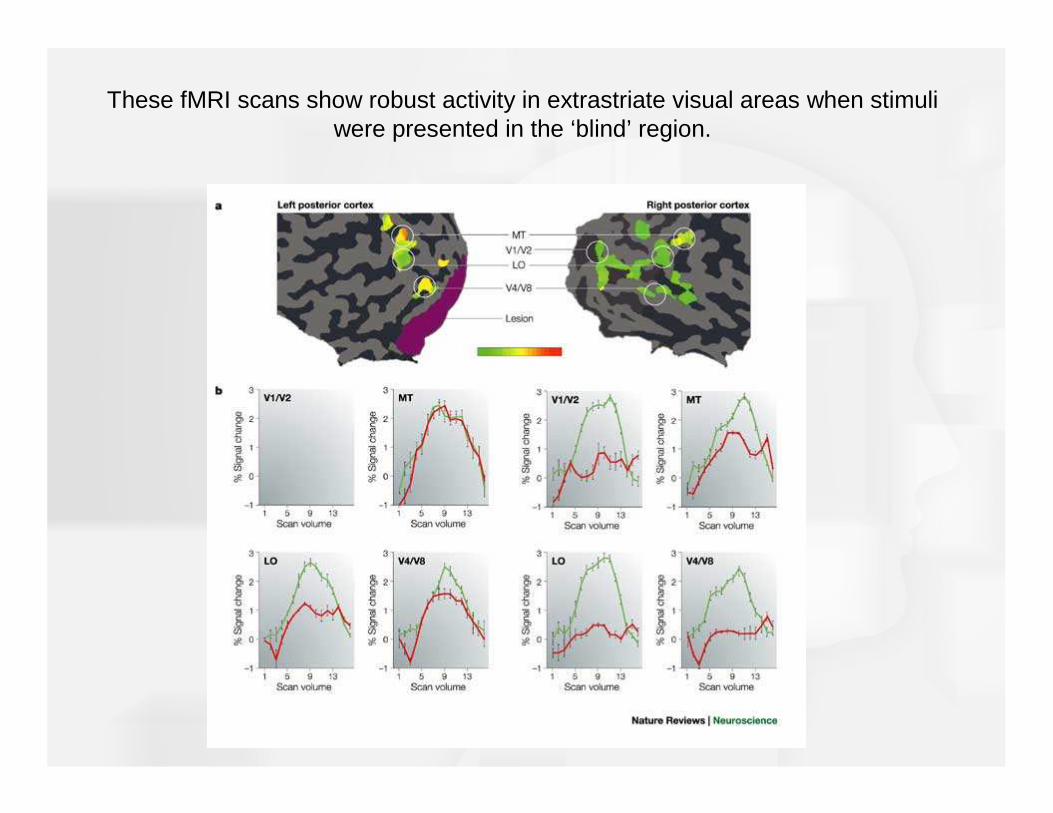
These fMRI scans show robust activity in extrastriate visual areas when stimuli were presented in the ‘blind’ region.
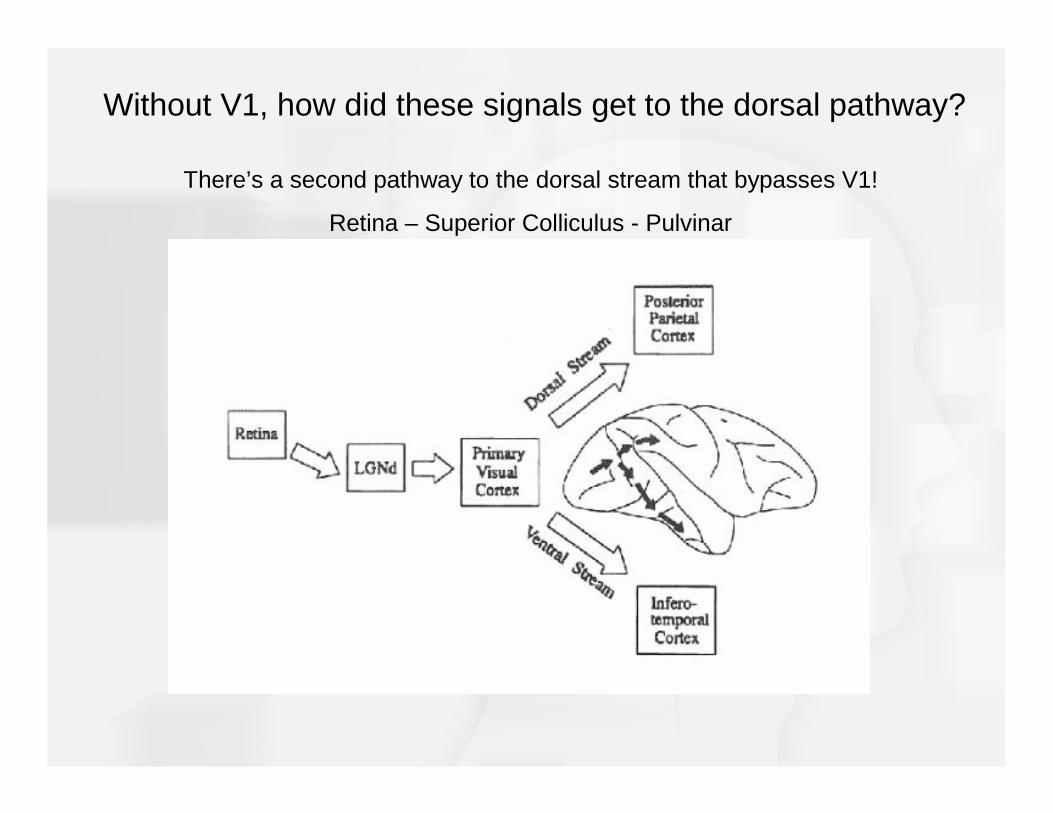
Without V1, how did these signals get to the dorsal pathway?
There’s a second pathway to the dorsal stream that bypasses V1!
Retina – Superior Colliculus - Pulvinar

So damage to V1 can lead to sight without awareness (blindsight).
But damage to the (usually right) parietal lobe can lead to the opposite problem, called anosognosia – which is when you don’t know that you don’t know something.
These patients can be blind, but insist that they can see. They’ll often confabulate stories or excuses to prevent them from demonstrating their disability.
This means that an intact V1, but a damaged parietal lobe can lead to awareness without sight!