chapter 7 acute vascular syndromes
TRANSCRIPT
CHAPTER 7
ACUTE VASCULAR SYNDROMES
7.1 ACUTE AORTIC SYNDROMES ���������������������������������������������� p.92 A. Evangelista
7.2 ACUTE PULMONARY EMBOLISM ������������������������������������������ p.102 A. Torbicki
7
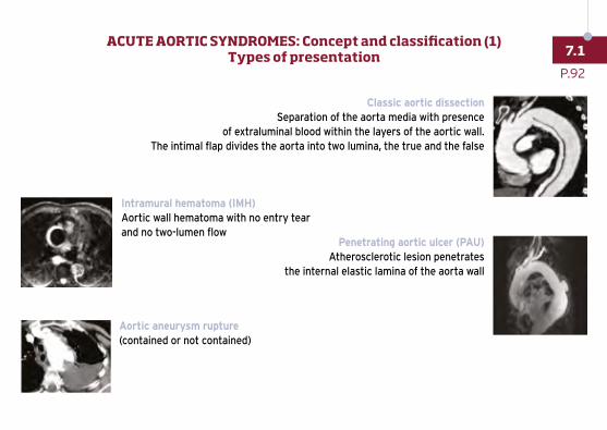
Classic aortic dissectionSeparation of the aorta media with presence
of extraluminal blood within the layers of the aortic wall.The intimal flap divides the aorta into two lumina, the true and the false
Penetrating aortic ulcer (PAU)Atherosclerotic lesion penetrates
the internal elastic lamina of the aorta wall
Aortic aneurysm rupture (contained or not contained)
Intramural hematoma (IMH)Aortic wall hematoma with no entry tear and no two-lumen flow
P.92
7.1ACUTE AORTIC SYNDROMES: Concept and classification (1)
Types of presentation
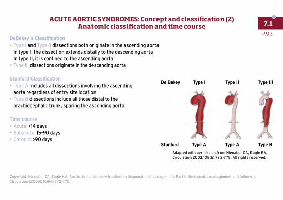
DeBakey’s Classification• Type I and Type II dissections both originate in the ascending aorta
In type I, the dissection extends distally to the descending aorta In type II, it is confined to the ascending aorta
• Type III dissections originate in the descending aorta
Stanford Classification• Type A includes all dissections involving the ascending
aorta regardless of entry site location• Type B dissections include all those distal to the
brachiocephalic trunk, sparing the ascending aorta
Time course• Acute: <14 days• Subacute: 15-90 days• Chronic: >90 days
Copyright: Nienaber CA, Eagle KA. Aortic dissection: new frontiers in diagnosis and management: Part II: therapeutic management and follow-up. Circulation (2003); 108(6),772-778.
Adapted with permission from Nienaber CA, Eagle KA, Circulation 2003;108(6):772-778. All rights reserved.
De Bakey
Stanford
Type I
Type A
Type II
Type A
Type III
Type B
P.93
7.1ACUTE AORTIC SYNDROMES: Concept and classification (2)
Anatomic classification and time course
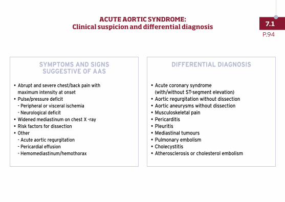
SYMPTOMS AND SIGNS SUGGESTIVE OF AAS
• Abrupt and severe chest/back pain with maximum intensity at onset
• Pulse/pressure deficit - Peripheral or visceral ischemia - Neurological deficit
• Widened mediastinum on chest X -ray• Risk factors for dissection• Other
- Acute aortic regurgitation - Pericardial effusion - Hemomediastinum/hemothorax
DIFFERENTIAL DIAGNOSIS
• Acute coronary syndrome (with/without ST-segment elevation)
• Aortic regurgitation without dissection• Aortic aneurysms without dissection• Musculoskeletal pain• Pericarditis• Pleuritis• Mediastinal tumours• Pulmonary embolism• Cholecystitis• Atherosclerosis or cholesterol embolism
P.94
7.1ACUTE AORTIC SYNDROME:
Clinical suspicion and differential diagnosis

Copyright: Hiratzka et al. 2010 Guidelines on Thoracic Aortic Disease. Circulation. (2010) ; 121: page-310 (fig 25 step 2).
Consider acute aortic dissection in all patients presenting with:
• Chest, back or abdominal pain• Syncope• Symptoms consistent with perfusion deficit (central nervous system, visceral myocardial or limb ischemia)
Pre-test risk assessment for acute aortic dissection
• Marfan’s syndrome• Connective tissue disease• Family history of aortic disease• Aortic valve disease• Thoracic aortic aneurysm
• Perfusion deficit: - Pulse deficit - SBP differential - Focal neurological deficit• Aortic regurgitation murmur• Hypotension or shock
Chest, back or abdominal paindescribed as:
Abrupt at onset, severe in intensity, and ripping/sharp or stabbing quality
High-risk conditions High-risk pain features High-risk exam features
P.95
7.1General approach to the patient
with suspected ACUTE AORTIC SYNDROME
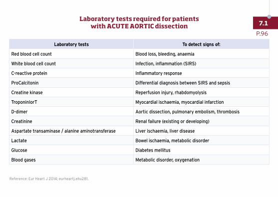
Laboratory tests To detect signs of:
Red blood cell count Blood loss, bleeding, anaemia
White blood cell count Infection, inflammation (SIRS)
C-reactive protein Inflammatory response
ProCalcitonin Differential diagnosis between SIRS and sepsis
Creatine kinase Reperfusion injury, rhabdomyolysis
TroponinIorT Myocardial ischaemia, myocardial infarction
D-dimer Aortic dissection, pulmonary embolism, thrombosis
Creatinine Renal failure (existing or developing)
Aspartate transaminase / alanine aminotransferase Liver ischaemia, liver disease
Lactate Bowel ischaemia, metabolic disorder
Glucose Diabetes mellitus
Blood gases Metabolic disorder, oxygenation
Reference: Eur Heart J 2014; eurheartj.ehu281.
P.96
7.1Laboratory tests required for patients
with ACUTE AORTIC dissection
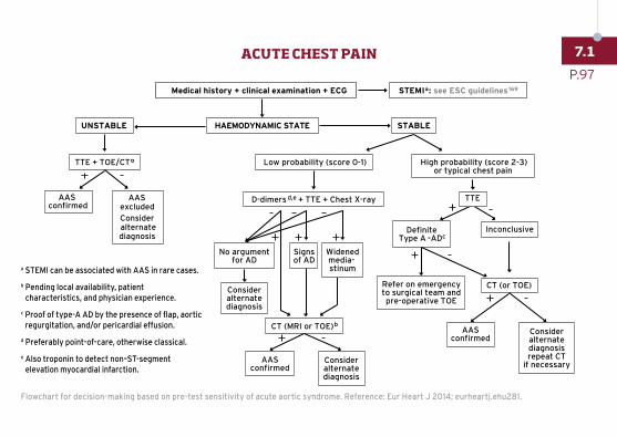
a STEMI can be associated with AAS in rare cases.
b Pending local availability, patient characteristics, and physician experience.
c Proof of type-A AD by the presence of flap, aortic regurgitation, and/or pericardial effusion.
d Preferably point-of-care, otherwise classical.
e Also troponin to detect non–ST-segment elevation myocardial infarction.
Flowchart for decision-making based on pre-test sensitivity of acute aortic syndrome. Reference: Eur Heart J 2014; eurheartj.ehu281.
ACUTE CHEST PAIN
High probability (score 2-3)or typical chest pain
Medical history + clinical examination + ECG STEMIa: see ESC guidelines169
HAEMODYNAMIC STATEUNSTABLE
Low probability (score 0-1)TTE + TOE/CT°
STABLE
AASconfirmed
AASexcludedConsideralternatediagnosis
D-dimers d,e + TTE + Chest X-ray TTE
Consideralternatediagnosis
No argumentfor AD
Signsof AD
Widenedmedia- stinum
DefiniteType A -AD c
Inconclusive
Refer on emergencyto surgical team andpre-operative TOE
CT (or TOE)
AASconfirmed
Consideralternatediagnosisrepeat CT
if necessaryAAS
confirmedConsideralternatediagnosis
CT (MRI or TOE)b
P.97
7.1ACUTE CHEST PAIN
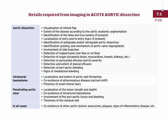
Aortic dissection • Visualization of intimal flap• Extent of the disease according to the aortic anatomic segmentation• Identification of the false and true lumens (if present)• Localization of entry and re-entry tears (if present)• Identification of antegrade and/or retrograde aortic dissection• Identification grading, and mechanism of aortic valve regurgitation• Involvement of side branches• Detection of malperfusion (low flow or no flow)• Detection of organ ischaemia (brain, myocardium, bowels, kidneys, etc.)• Detection of pericardial effusion and its severity• Detection and extent of pleural effusion• Detection of peri-aortic bleeding• Signs of mediastinal bleeding
Intramural haematoma
• Localization and extent of aortic wall thickening• Co-existence of atheromatous disease (calcium shift)• Presence of small intimal tears
Penetrating aortic ulcer
• Localization of the lesion (length and depth)• Co-existence of intramural haematoma• Involvement of the peri-aortic tissue and bleeding• Thickness of the residual wall
In all cases • Co-existence of other aortic lesions: aneurysms, plaques, signs of inflammatory disease, etc.
P.98
7.1Details required from imaging in ACUTE AORTIC dissection
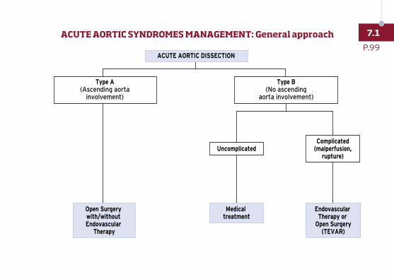
ACUTE AORTIC SYNDROMES MANAGEMENT: GENERAL APPROACH
ACUTE AORTIC DISSECTION
Type A(Ascending aorta
involvement)
Type B(No ascending
aorta involvement)
Uncomplicated
Medical treatment
Open Surgery with/without Endovascular
Therapy
Endovascular Therapy or
Open Surgery(TEVAR)
Complicated(malperfusion,
rupture)
P.99
7.1ACUTE AORTIC SYNDROMES MANAGEMENT: General approach
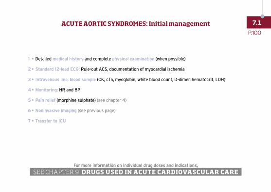
1 • Detailed medical history and complete physical examination (when possible)
2 • Standard 12-lead ECG: Rule-out ACS, documentation of myocardial ischemia
3 • Intravenous line, blood sample (CK, cTn, myoglobin, white blood count, D-dimer, hematocrit, LDH)
4 • Monitoring: HR and BP
5 • Pain relief (morphine sulphate) (see chapter 4)
6 • Noninvasive imaging (see previous page)
7 • Transfer to ICU
For more information on individual drug doses and indications,
SEE CHAPTER 9 DRUGS USED IN ACUTE CARDIOVASCULAR CARE
P.100
7.1ACUTE AORTIC SYNDROMES: Initial management
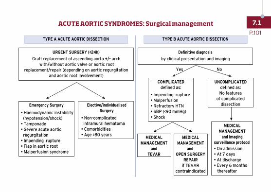
URGENT SURGERY (<24h)
Emergency Surgery
Graft replacement of ascending aorta +/- arch with/without aortic valve or aortic root
replacement/repair (depending on aortic regurgitationand aortic root involvement)
ACUTE AORTIC SYNDROMES: SURGICAL MANAGEMENT
TYPE A ACUTE AORTIC DISSECTION TYPE B ACUTE AORTIC DISSECTION
• Haemodynamic instability (hypotension/shock) • Tamponade• Severe acute aortic regurgitation• Impending rupture• Flap in aortic root• Malperfusion syndrome
Elective/individualisedSurgery
• Non-complicated intramural hematoma• Comorbidities• Age >80 years
Definitive diagnosis
COMPLICATEDdefined as:
by clinical presentation and imaging
• Impending rupture • Malperfusion • Refractory HTN • SBP (<90 mmHg) • Shock
UNCOMPLICATEDdefined as:No features
of complicateddissection
MEDICALMANAGEMENT
and imagingsurveillance protocol• On admission• At 7 days• At discharge• Every 6 months thereafter
MEDICALMANAGEMENT
andTEVAR
MEDICALMANAGEMENT
and OPEN SURGERY
REPAIRif TEVAR
contraindicated
Yes No
P.101
7.1ACUTE AORTIC SYNDROMES: Surgical management
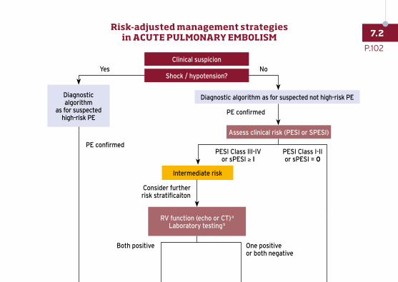
Diagnostic algorithm
as for suspected high-risk PE
Diagnostic algorithm as for suspected not high-risk PE
Assess clinical risk (PESI or SPESI)
RV function (echo or CT) a
Laboratory testing b
Intermediate risk
PE confirmed
Consider further risk stratificaiton
PESI Class III-IVor sPESI ≥ I
PESI Class I-IIor sPESI = 0
PE confirmed
Both positive One positiveor both negative
Clinical suspicion
Shock / hypotension?NoYes
P.102
7.2Risk-adjusted management strategies
in ACUTE PULMONARY EMBOLISM
Reference: Eur Heart J 2014; 35:3033-3073.
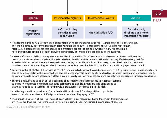
a If echocardiography has already been performed during diagnostic work-up for PE and detected RV dysfunction, or if the CT already performed for diagnostic work–up has shown RV enlargement (RV/LV (left ventricular) ratio ≥0.9, a cardiac troponin test should be performed except for cases in which primary reperfusion is not a therapeutic option (e.g. due to severe comorbidity or limited life expectancy of the patient).
b Markers of myocardial injury (e.g. elevated cardiac troponin I or T concentrations in plasma), or of heart failure as a result of (right) ventricular dysfunction (elevated natriuretic peptide concentrations in plasma). If a laboratory test for a cardiac biomarker has already been performed during initial diagnostic work-up (e.g. in the chest pain unit) and was positive, then an echocardiogram should be considered to assess RV function, or RV size should be (re)assessed on CT.
c Patients in the PESI Class I-II, or with sPESI of 0, and elevated cardiac biomarkers or signs of RV dysfunction on imaging tests, are also to be classified into the intermediate-low risk category. This might apply to situations in which imaging or biomarker results become available before calculation of the clinical severity index. These patients are probably no candidates for home treatment.
d Thrombolysis, if (and as soon as) clinical signs of haemodynamic decompensation appear; surgical pulmonary embolectomy or percutaneous catheter-directed treatment may be considered as alternative options to systemic thrombolysis, particularly if the bleeding risk is high.
e Monitoring should be considered for patients with confirmed PE and a positive troponin test, even if there is no evidence of RV dysfunction on echocardiography or CT.
f The simplified version of the PESI has not been validated in prospective home treatment trials; inclusion criteria other than the PESI were used in two single-armed (non-randomized) management studies.
Intermediate-high risk Intermediate-low risk
A/C; monitoringconsider rescue
reperfusiondHospitalization A/C e
Consider early discharge and home treatment if feasible f
Primary reperfusion
High-risk Low riskc
P.103
7.2
CARDIOVASCULARSymptoms/Signs
including but not limited to:
Shock? orSBP <90 mmHg?
orSBP fall by >40 mmHg?
persisting >15 min, otherwise unexplained
RESPIRATORYSymptoms/Signs
including but not limited to:
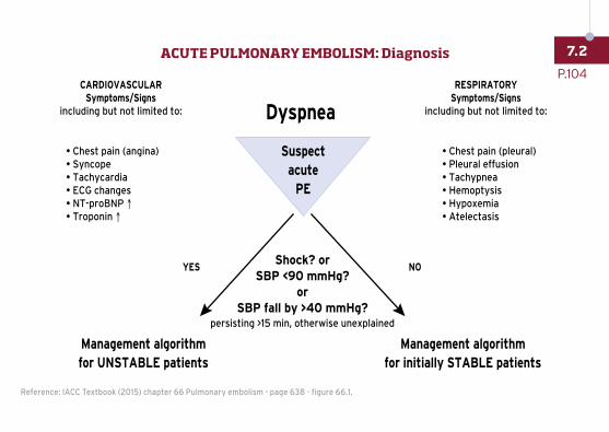
ACUTE PULMONARY EMBOLISM: DIAGNOSIS
YES NO
Dyspnea
• Chest pain (angina)• Syncope • Tachycardia• ECG changes• NT-proBNP ↑• Troponin ↑
• Chest pain (pleural)• Pleural effusion• Tachypnea• Hemoptysis• Hypoxemia• Atelectasis
Suspectacute
PE
Management algorithmfor UNSTABLE patients
Management algorithmfor initially STABLE patients
Reference: IACC Textbook (2015) chapter 66 Pulmonary embolism - page 638 - figure 66.1.
P.104
7.2ACUTE PULMONARY EMBOLISM: Diagnosis
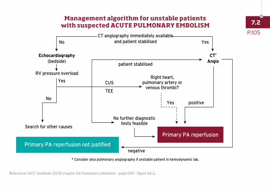
MANAGEMENT ALGORITHM FOR UNSTABLE PATIENTS WITHSUSPECTED ACUTE PULMONARY EMBOLISM
CT angiography immediately availableand patient stabilised
Primary PA reperfusion
Primary PA reperfusion not justified
patient stabilised
No further diagnostictests feasible
Right heart,pulmonary artery or
venous thrombi?
Echocardiography(bedside)
CT*
Angio
RV pressure overload
CUS
No Yes
Yes
Yes positive
negative
No
TEE
Search for other causes
* Consider also pulmonary angiography if unstable patient in hemodynamic lab.
Reference: IACC Textbook (2015) chapter 66 Pulmonary embolism - page 639 - figure 66.2.
P.105
7.2Management algorithm for unstable patients
with suspected ACUTE PULMONARY EMBOLISM

Shock or hypotension YES
Contraindications for thrombolysis
No Relative Absolute
Primary PAreperfusion strategy
Thrombolysis
Low- dose transcatheterthrombolysis /
clot fragmetation
Surgical orPercutaneous catheter
embolectomy(availability/experience)
Supportive treatment i.v. UFH, STABILISE SYSTEMIC BLOOD PRESSURE, CORRECT HYPOXEMIA
P.106
7.2ACUTE PE: Management strategy for initially unstable patients
with confirmed high risk pulmonary embolism
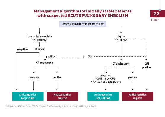
MANAGEMENT ALGORITHM FOR INITIALLY STABLE PATIENTS WITHSUSPECTED ACUTE PULMONARY EMBOLISM
Low or intermediate“PE unlikely“
positive
negative D-dimer
CT angiography
negative positivenegative
Confirm by CUSV/Q scan or angiography
positive
CT angiography
CUS
CUSpositive
High or“PE likely“
Anticoagulationnot justified
Anticoagulationrequired
Anticoagulationnot justified
Anticoagulationrequired
Asses clinical (pre-test) probalility
Reference: IACC Textbook (2015) chapter 66 Pulmonary embolism - page 640 - figure 66.3.
P.107
7.2Management algorithm for initially stable patients
with suspected ACUTE PULMONARY EMBOLISM

Markers for myocardial injury Positive Positive Negative
Markers for RV overload Positive Positive Negative
Clinical risk assessment score (PESI)
Positive (class III-V) Positive (class III-V) Negative (class I-II)
Suggested initial anticoagulation
UFH i.v./LMWH s.c.LMWH/Fonda/apixaban/
rivaroxabanapixaban/rivaroxaban
STRATEGYMonitoring (ICU)*
rescue thrombolysisHospitalisation**
(telemonitoring)Early
discharge***
* When all markers are positive. - ** When at least one marker is positive. - *** When all markers are negative.
For more information on individual drug doses and indications,
SEE CHAPTER 9 DRUGS USED IN ACUTE CARDIOVASCULAR CARE
P.108
7.2Suggested management strategy for initially stable patients
with (non-high risk) confirmed PE
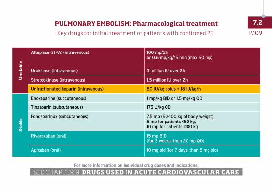
Key drugs for initial treatment of patients with confirmed PE
Uns
tabl
e
Alteplase (rtPA) (intravenous) 100 mp/2h or 0.6 mp/kg/15 min (max 50 mp)
Urokinase (intravenous) 3 million IU over 2h
Streptokinase (intravenous) 1.5 million IU over 2h
Unfractionated heparin (intravenous) 80 IU/kg bolus + 18 IU/kg/h
Sta
ble
Enoxaparine (subcutaneous) 1 mp/kg BID or 1.5 mp/kg QD
Tinzaparin (subcutaneous) 175 U/kg QD
Fondaparinux (subcutaneous) 7.5 mp (50-100 kg of body weight)5 mp for patients <50 kg, 10 mp for patients >100 kg
Rivaroxaban (oral) 15 mp BID (for 3 weeks, then 20 mp QD)
Apixaban (oral) 10 mg bid (for 7 days, than 5 mg bid)
For more information on individual drug doses and indications,
SEE CHAPTER 9 DRUGS USED IN ACUTE CARDIOVASCULAR CARE
P.109
7.2PULMONARY EMBOLISM: Pharmacological treatment
