chapter 7 cell structure and function - viggenz.com · chapter 7 cell structure and function this...
TRANSCRIPT

Prentice Hall Biology
file:///Users/viggen/Desktop/Biology/PH%20BIO%20Interactive%20Text/iText/products/0-13-115516-4/ch7/ch7_0_0.html[10/11/16, 4:01:54 PM]
Chapter 7 Cell Structure and Function
This is a transmission electron micrograph of a neutrophil, a cell found in bone marrow. Color has been added to highlight thevarious organelles (magnification: 27,500×).
What is a cell?
Procedure
1. Look through a microscope at a slide of a plant leaf or stem cross section. CAUTION: Handle themicroscope and slide carefully to avoid breaking them. Sketch one or more cells. Record a description oftheir features, such as shape and internal parts.
2. Repeat step 1 with slides of nerve cells, bacteria, and paramecia.3. Compare the cells by listing the characteristics they have in common and some of the differences among
them.
Think About It
1. Forming Operational Definitions Use your observations to write a definition of “cell.”2. Classifying Classify the cells you observed into two or more groups. Explain what characteristics you
used to put each cell in a particular group.
Prentice Hall Biology
file:///Users/viggen/Desktop/Biology/PH%20BIO%20Interactive%20Text/iText/products/0-13-115516-4/ch7/ch7_ca_1.html[10/11/16, 4:01:56 PM]
Chapter 7 Cell Structure and Function
Understanding Concepts
11. Make a table to summarize the contributions made to the cell theoryby Robert Hooke, Matthias Schleiden, Theodor Schwann, and RudolfVirchow.
12. How are prokaryotic and eukaryotic cells alike? How do they differ?
13. Draw a cell nucleus. Label and give the function of the followingstructures: chromatin, nucleolus, and nuclear envelope.
14. What is the function of a ribosome?15. What process takes place in the rough endoplasmic reticulum?16. Describe the role of the Golgi apparatus.17. Other than the nucleus, which two organelles contain their own DNA?
What explanation has Lynn Margulis proposed to account for thepresence of DNA in these organelles?
18. Name and describe the two types of structures that make up thecytoskeleton.
19. Briefly describe the structure of a cell membrane. How does the cellmembrane affect the contents of a cell?
20. What is meant by the concentration of a solution? Give a specificexample of concentration involving volume and mass.
21. Describe the process of diffusion. Name and describe the conditionthat exists when the diffusion of a particular substance is complete.
22. What is the relationship between osmosis and diffusion? By definition,what's the only substance that carries out osmosis?
23. Using the example of a cell in a sugar solution, explain what is meantby an isotonic solution.
24. Name and describe the cell structure that helps prevent damage tocertain cells when they are subjected to high osmotic pressure.
25. Use an example to describe the relationship among cells, tissues,organs, and organ systems.

Prentice Hall Biology
file:///Users/viggen/Desktop/Biology/PH%20BIO%20Interactive%20Text/iText/products/0-13-115516-4/ch7/ch7_ca_2.html[10/11/16, 4:01:56 PM]
Chapter 7 Cell Structure and Function
Critical Thinking
26. Predicting The beaker in the diagram has a selectively permeablemembrane separating two solutions. Assume that the water moleculesand salt can pass freely through the membrane. When equilibrium isreached, will the fluid levels be the same as they are now? Explainyour answer.
27. Calculating Which salt solution is more concentrated, solution A,which contains 18 g of salt in 6 L of water, or solution B, whichcontains 24 g of salt in 12 L of water? Explain.
28. Predicting What would happen to a sample of your red blood cells ifthey were placed into a hypotonic solution? Explain your prediction.
29. Inferring Would you expect skin cells to contain more or fewermitochondria than muscle cells? Explain your answer.
30. Designing Experiments You are given vegetable coloring and threebeakers. The first beaker contains water at room temperature, thesecond beaker contains ice water, and the third beaker contains hotwater. Design an experiment to determine the effects of temperatureon the rate of diffusion. Be sure to state your hypothesis and to includea control.
31. Inferring The pancreas, an organ present in certain animals, producesenzymes used elsewhere in the animals' digestive systems. Which typeof cell structure(s) might produce those enzymes? Explain youranswer.
32. Using Analogies Compare a cell to a factory, as in the chapter, or tosomething else, such as a school. (For example, a cell has a nucleus,and a school has a principal.) Use that analogy to describe the functionof different parts of the cell.
33. Applying Concepts As waste chemicals build up in a cell,homeostasis is threatened. State how diffusion helps cells maintain
Prentice Hall Biology
file:///Users/viggen/Desktop/Biology/PH%20BIO%20Interactive%20Text/iText/products/0-13-115516-4/ch7/ch7_ca_2.html[10/11/16, 4:01:56 PM]
homeostasis.
34. Comparing and Contrasting Diffusion and active transport areprocesses that are important to the maintenance of homeostasis inorganisms. Compare the two processes, including examples thatdescribe how they are important to living organisms.
35. Connecting Concepts In Chapter 2, you learned about fourcategories of carbon compounds called the “molecules of life.”Explain where some of those compounds are found in a typical cell.
Writing in ScienceDifferent beverages have different concentrations of solutes. Some beverages have low solute concentrations and canbe a source of water for body cells. Other beverages have high solute concentrations and can actually dehydrate yourbody cells. Should companies that market these high-solute beverages say that these drinks quench your thirst?

Prentice Hall Biology
file:///Users/viggen/Desktop/Biology/PH%20BIO%20Interactive%20Text/iText/products/0-13-115516-4/ch7/ch7_ca_3.html[10/11/16, 4:01:56 PM]
Chapter 7 Cell Structure and Function
Performance-Based AssessmentPrepare to Debate One day, unicellular organisms got tired of being referred to as simple organisms by themulticellular organisms. They felt that they should be recognized as complex individuals, and challenged themulticellular organisms to a debate. As a unicellular organism, what arguments would you use to defend yourposition?

Prentice Hall Biology
file:///Users/viggen/Desktop/Biology/PH%20BIO%20Interactive%20Text/iText/products/0-13-115516-4/ch7/ch7_s1_0.html[10/11/16, 4:01:58 PM]
Key Concepts
What is the cell theory?
What are the characteristicsof prokaryotes andeukaryotes?
Vocabulary cell cell theory nucleus eukaryote prokaryoteReading Strategy: FindingMain Ideas As you read, lookfor evidence to support thestatement “The cell theoryrevolutionized how biologiststhought about living things.”
7–1 Life Is Cellular
Look closely at a part of a living thing, and what do you see? Hold a blade ofgrass up against the light, and you see tiny lines running the length of theblade. Examine the tip of your finger, and you see the ridges and valleys thatmake up fingerprints. Place an insect under a microscope, and you see theintricate structures of its wings and the spikes and bristles that protect itsbody. As interesting as these close-up views may be, however, they're onlythe beginning of the story. Look closer and deeper with a more powerfulmicroscope, and you'll see that there is a common structure that makes upevery living thing—the cell.

Prentice Hall Biology
file:///Users/viggen/Desktop/Biology/PH%20BIO%20Interactive%20Text/iText/products/0-13-115516-4/ch7/ch7_s1_1.html[10/11/16, 4:01:59 PM]
7–1 Life Is Cellular (continued)The Discovery of the Cell
“Seeing is believing,” an old saying goes. It would be hard to find a better example ofthis than the discovery of the cell. Without the instruments to make them visible, cellsremained out of sight and, therefore, out of mind for most of human history. All ofthis changed with a dramatic advance in technology—the invention of the microscope.
Early Microscopes It was not until the mid-1600s that scientists began to usemicroscopes to observe living things. In 1665, Englishman Robert Hooke used anearly compound microscope to look at a thin slice of cork, a plant material. Under themicroscope, cork seemed to be made of thousands of tiny, empty chambers. Hookecalled these chambers “cells” because they reminded him of a monastery's tiny rooms,which were called cells. One of Hooke's illustrations of cells is shown in the figurebelow. The term cell is used in biology to this day. We now know, however, that cellsare not empty but contain living matter.
Cork Cells Using an early microscope, Hooke made this drawing of cork cells. In Hooke's drawings, the cells look likeempty chambers because he was looking at dead plant matter. Today, we know that living cells are made up of manystructures.
In Holland around the same time, Anton van Leeuwenhoek used a single-lensmicroscope to observe pond water and other things. To his amazement, the microscoperevealed a fantastic world of tiny living organisms that seemed to be everywhere, evenin the very water he and his neighbors drank.
The Cell Theory Soon, numerous observations made it clear that cells were the basicunits of life. In 1838, German botanist Matthias Schleiden concluded that all plantswere made of cells. The next year, German biologist Theodor Schwann stated that all
A History of the Cell

Prentice Hall Biology
file:///Users/viggen/Desktop/Biology/PH%20BIO%20Interactive%20Text/iText/products/0-13-115516-4/ch7/ch7_s1_1.html[10/11/16, 4:01:59 PM]
animals were made of cells. In 1855, the German physician Rudolf Virchowconcluded that new cells could be produced only from the division of existing cells.These discoveries, confirmed by other biologists, are summarized in the cell theory, afundamental concept of biology. The cell theory states:
All living things are composed of cells.Cells are the basic units of structure and function in living things.New cells are produced from existing cells.
For: Links on cell theoryVisit: www.SciLinks.orgWeb Code: cbn-3071
Prentice Hall Biology
file:///Users/viggen/Desktop/Biology/PH%20BIO%20Interactive%20Text/iText/products/0-13-115516-4/ch7/ch7_s1_2.html[10/11/16, 4:02:01 PM]
7–1 Life Is Cellular (continued)Exploring the Cell
Following in the footsteps of Hooke, Virchow, and others, modern biologists still usemicroscopes to explore the cell. However, today's researchers use microscopes andtechniques more powerful than the pioneers of biology could have imagined.Researchers can use fluorescent labels and light microscopy to follow moleculesmoving through the cell. Confocal light microscopy, which scans cells with a laserbeam, makes it possible to build three-dimensional images of cells and their parts.High-resolution video technology makes it easy to produce movies of cells as theygrow, divide, and develop.
These new technologies make it possible for researchers to study the structure andmovement of living cells in great detail. Unfortunately, light itself limits the detail, orresolution, of images that can be made with the light microscope. Like all forms ofradiation, light waves are diffracted, or scattered, as they pass through matter, makingit impossible to visualize tiny structures such as proteins and viruses with lightmicroscopy.
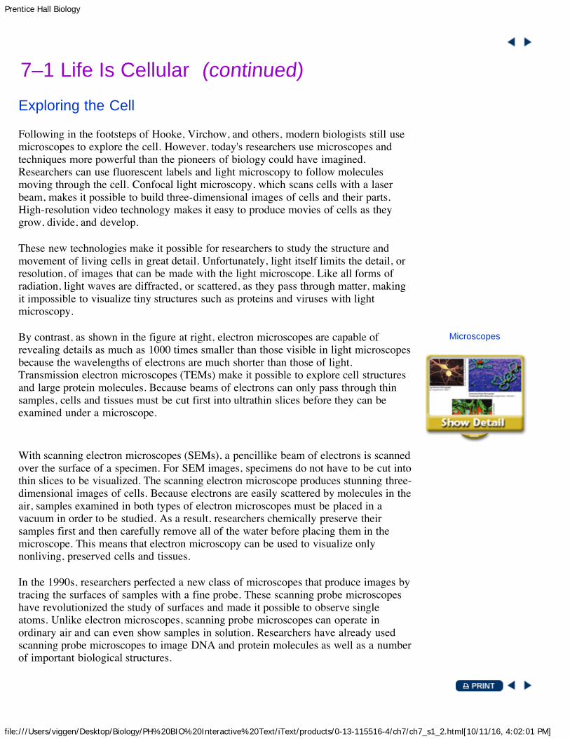
By contrast, as shown in the figure at right, electron microscopes are capable ofrevealing details as much as 1000 times smaller than those visible in light microscopesbecause the wavelengths of electrons are much shorter than those of light.Transmission electron microscopes (TEMs) make it possible to explore cell structuresand large protein molecules. Because beams of electrons can only pass through thinsamples, cells and tissues must be cut first into ultrathin slices before they can beexamined under a microscope.
Microscopes
With scanning electron microscopes (SEMs), a pencillike beam of electrons is scannedover the surface of a specimen. For SEM images, specimens do not have to be cut intothin slices to be visualized. The scanning electron microscope produces stunning three-dimensional images of cells. Because electrons are easily scattered by molecules in theair, samples examined in both types of electron microscopes must be placed in avacuum in order to be studied. As a result, researchers chemically preserve theirsamples first and then carefully remove all of the water before placing them in themicroscope. This means that electron microscopy can be used to visualize onlynonliving, preserved cells and tissues.
In the 1990s, researchers perfected a new class of microscopes that produce images bytracing the surfaces of samples with a fine probe. These scanning probe microscopeshave revolutionized the study of surfaces and made it possible to observe singleatoms. Unlike electron microscopes, scanning probe microscopes can operate inordinary air and can even show samples in solution. Researchers have already usedscanning probe microscopes to image DNA and protein molecules as well as a numberof important biological structures.

Prentice Hall Biology
file:///Users/viggen/Desktop/Biology/PH%20BIO%20Interactive%20Text/iText/products/0-13-115516-4/ch7/ch7_s1_2.html[10/11/16, 4:02:01 PM]
Prentice Hall Biology
file:///Users/viggen/Desktop/Biology/PH%20BIO%20Interactive%20Text/iText/products/0-13-115516-4/ch7/ch7_s1_3.html[10/11/16, 4:02:03 PM]
7–1 Life Is Cellular (continued)Prokaryotes and Eukaryotes
Cells come in a great variety of shapes and an amazing range of sizes. Althoughtypical cells range from 5 to 50 micrometers in diameter, the tiniest mycoplasmabacteria are only 0.2 micrometers across, so small that they are difficult to see undereven the best light microscopes. In contrast, the giant amoeba Chaos chaos may be1000 micrometers in diameter, large enough to be seen with the unaided eye as a tinyspeck in pond water. Despite their differences, all cells have two characteristics incommon. They are surrounded by a barrier called a cell membrane; and, at some pointin their lives, they contain the molecule that carries biological information—DNA.
For: Articles on cells
Cells fall into two broad categories, depending on whether they contain a nucleus. Thenucleus (plural: nuclei) is a large membrane-enclosed structure that contains the cell'sgenetic material in the form of DNA. (A membrane is a thin layer of material thatserves as a covering or lining.) The nucleus controls many of the cell's activities.Eukaryotes (yoo-KAR-ee-ohts) are cells that contain nuclei. Prokaryotes (pro-KAR-ee-ohts) are cells that do not contain nuclei. Both words derive from the Greek wordskaryon, meaning “kernel,” or nucleus, and eu, meaning “true,” or pro, meaning“before.” These words reflect the idea that prokaryotic cells evolved before nucleideveloped.
Prokaryotes Prokaryotic cells are generally smaller and simpler than eukaryotic cells,although there are many exceptions to this rule. Prokaryotic cells have geneticmaterial that is not contained in a nucleus. Some prokaryotes contain internalmembranes, but prokaryotes are generally less complicated than eukaryotes. Despitetheir simplicity, prokaryotes carry out every activity associated with living things.They grow, reproduce, respond to the environment, and some can even move bygliding along surfaces or swimming through liquids. The organisms we call bacteriaare prokaryotes.
Eukaryotes Eukaryotic cells are generally larger and more complex than prokaryoticcells. Eukaryotic cells generally contain dozens of structures and internal membranes,and many are highly specialized. Eukaryotic cells contain a nucleus in whichtheir genetic material is separated from the rest of the cell. Eukaryotes displaygreat variety. Some eukaryotes live solitary lives as single-celled organisms. Othersform large, multicellular organisms. Plants, animals, fungi, and protists are eukaryotes.

Prentice Hall Biology
file:///Users/viggen/Desktop/Biology/PH%20BIO%20Interactive%20Text/iText/products/0-13-115516-4/ch7/ch7_s1_4.html[10/11/16, 4:02:04 PM]
7–1 Life Is Cellular
1. Key Concept What three statements describe the cell theory? 2. Key Concept What are the differences between prokaryotic cells and eukaryotic cells? 3. Compare the processes used to produce a TEM and an SEM. 4. What structures do all cells have? 5. Critical Thinking Inferring How did the invention of the microscope help the development of the cell theory?
Constructing a ChartMake a three-column chart comparing prokaryotes with eukaryotes. In the first column, list the traits found in allcells. In the second column, list the features of prokaryotes. In the third column, list the features of eukaryotes.

Prentice Hall Biology
file:///Users/viggen/Desktop/Biology/PH%20BIO%20Interactive%20Text/iText/products/0-13-115516-4/ch7/ch7_s2_0.html[10/11/16, 4:02:06 PM]
Key Concept
What are the functions ofthe major cell structures?
Vocabulary organelle cytoplasm nuclear envelope chromatin chromosome nucleolus ribosome endoplasmic reticulum Golgi apparatus lysosome vacuole mitochondrion chloroplast cytoskeleton centrioleReading Strategy: BuildingVocabulary Before you read,preview the vocabulary byskimming the section andmaking a list of the boldfaceterms. Leave space to makenotes as you read.
7–2 Eukaryotic Cell Structure
At first glance, a factory is a puzzling place. A bewildering variety ofmachines buzz and clatter, people move quickly in different directions, andthe sheer diversity of so much activity can be confusing. However, if you takeyour time and watch carefully, before long you will begin to identify patterns.What might at first have seemed like chaos begins to make sense.

Prentice Hall Biology
file:///Users/viggen/Desktop/Biology/PH%20BIO%20Interactive%20Text/iText/products/0-13-115516-4/ch7/ch7_s2_1.html[10/11/16, 4:02:07 PM]
7–2 Eukaryotic Cell Structure (continued)Comparing the Cell to a Factory
In some respects, the eukaryotic cell is like a factory. The first time you look at amicroscope image of a cell, the cell seems impossibly complex. Look closely at aeukaryotic cell, however, and patterns begin to emerge. To see those patterns moreclearly, we'll look at some structures that are common to eukaryotic cells, shown in thefigure at right. Because many of these structures act as if they are specialized organs,these structures are known as organelles, literally “little organs.”
Plant and Animal Cells
Cell biologists divide the eukaryotic cell into two major parts: the nucleus and thecytoplasm. The cytoplasm is the portion of the cell outside the nucleus. As you willsee, the nucleus and cytoplasm work together in the business of life.
For: Cell Structure activityVisit: PHSchool.comWeb Code: cbp-3072

Prentice Hall Biology
file:///Users/viggen/Desktop/Biology/PH%20BIO%20Interactive%20Text/iText/products/0-13-115516-4/ch7/ch7_s2_10.html[10/11/16, 4:02:09 PM]
7–2 Eukaryotic Cell Structure
1. Key Concept Describe the functions of the endoplasmic reticulum, Golgi apparatus, chloroplast, andmitochondrion.
2. Describe the role of the nucleus in the cell. 3. What are two functions of the cytoskeleton? 4. How is a cell like a factory? 5. Critical Thinking Inferring You examine an unknown cell under the microscope and discover that the cell
contains chloroplasts. What type of organism could you infer that the cell came from?
Persuasive WritingImagine that you are Lynn Margulis. Write a persuasive letter to the editor of a magazine, explaining your idea.Your explanation should be clear to people who do not have a biology background. Hint: Review the concept ofsymbiosis in Section 4-2.

Prentice Hall Biology
file:///Users/viggen/Desktop/Biology/PH%20BIO%20Interactive%20Text/iText/products/0-13-115516-4/ch7/ch7_s2_2.html[10/11/16, 4:02:10 PM]
7–2 Eukaryotic Cell Structure (continued)Nucleus
In the same way that the main office controls a large factory, the nucleus is the controlcenter of the cell. The nucleus contains nearly all the cell's DNA and with itthe coded instructions for making proteins and other important molecules. Thestructure of the nucleus is shown in the figure at right.
The Nucleus
The nucleus is surrounded by a nuclear envelope composed of two membranes. Thenuclear envelope is dotted with thousands of nuclear pores, which allow material tomove into and out of the nucleus. Like messages, instructions, and blueprints movingin and out of a main office, a steady stream of proteins, RNA, and other moleculesmove through the nuclear pores to and from the rest of the cell.
For: Cell structure activity
The granular material you can see in the nucleus is called chromatin. Chromatinconsists of DNA bound to protein. Most of the time, chromatin is spread throughoutthe nucleus. When a cell divides, however, chromatin condenses to formchromosomes (KROH-muh-sohms). These distinct, threadlike structures contain thegenetic information that is passed from one generation of cells to the next. You willlearn more about chromosomes in later chapters.
Most nuclei also contain a small, dense region known as the nucleolus (noo-KLEE-uh-lus). The nucleolus is where the assembly of ribosomes begins.
Prentice Hall Biology
file:///Users/viggen/Desktop/Biology/PH%20BIO%20Interactive%20Text/iText/products/0-13-115516-4/ch7/ch7_s2_3.html[10/11/16, 4:02:12 PM]
7–2 Eukaryotic Cell Structure (continued)Ribosomes
One of the most important jobs carried out in the cellular “factory” is making proteins. Proteins are assembled on ribosomes. Ribosomes are small particles of RNA
and protein found throughout the cytoplasm. They produce proteins by followingcoded instructions that come from the nucleus. Each ribosome, in its own way, is likea small machine in a factory, turning out proteins on orders that come from its“boss”—the cell nucleus. Cells that are active in protein synthesis are often packedwith ribosomes.
Prentice Hall Biology
file:///Users/viggen/Desktop/Biology/PH%20BIO%20Interactive%20Text/iText/products/0-13-115516-4/ch7/ch7_s2_4.html[10/11/16, 4:02:14 PM]
7–2 Eukaryotic Cell Structure (continued)Endoplasmic Reticulum
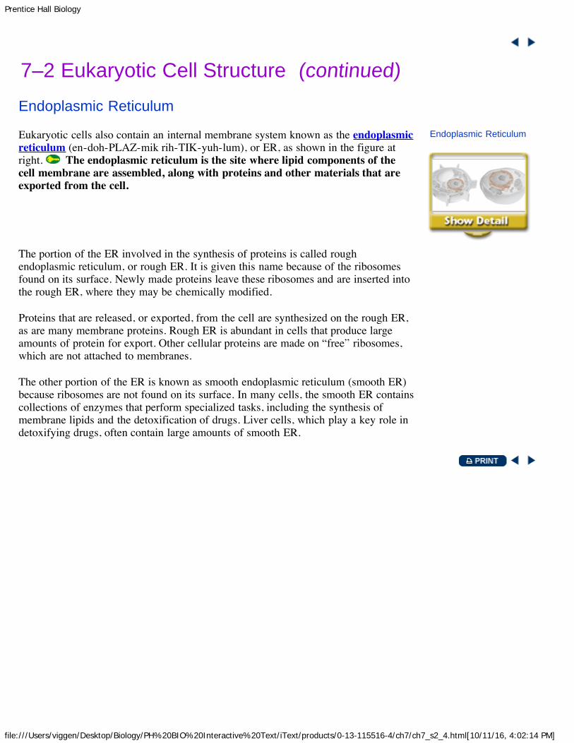
Eukaryotic cells also contain an internal membrane system known as the endoplasmicreticulum (en-doh-PLAZ-mik rih-TIK-yuh-lum), or ER, as shown in the figure atright. The endoplasmic reticulum is the site where lipid components of thecell membrane are assembled, along with proteins and other materials that areexported from the cell.
Endoplasmic Reticulum
The portion of the ER involved in the synthesis of proteins is called roughendoplasmic reticulum, or rough ER. It is given this name because of the ribosomesfound on its surface. Newly made proteins leave these ribosomes and are inserted intothe rough ER, where they may be chemically modified.
Proteins that are released, or exported, from the cell are synthesized on the rough ER,as are many membrane proteins. Rough ER is abundant in cells that produce largeamounts of protein for export. Other cellular proteins are made on “free” ribosomes,which are not attached to membranes.
The other portion of the ER is known as smooth endoplasmic reticulum (smooth ER)because ribosomes are not found on its surface. In many cells, the smooth ER containscollections of enzymes that perform specialized tasks, including the synthesis ofmembrane lipids and the detoxification of drugs. Liver cells, which play a key role indetoxifying drugs, often contain large amounts of smooth ER.

Prentice Hall Biology
file:///Users/viggen/Desktop/Biology/PH%20BIO%20Interactive%20Text/iText/products/0-13-115516-4/ch7/ch7_s2_5.html[10/11/16, 4:02:15 PM]
7–2 Eukaryotic Cell Structure (continued)Golgi Apparatus
Proteins produced in the rough ER move next into an organelle called the Golgiapparatus, discovered by the Italian scientist Camillo Golgi. As you can see in thefigure at right, Golgi appears as a stack of closely apposed membranes. Thefunction of the Golgi apparatus is to modify, sort, and package proteins and othermaterials from the endoplasmic reticulum for storage in the cell or secretionoutside the cell. The Golgi apparatus is somewhat like a customization shop, wherethe finishing touches are put on proteins before they are ready to leave the “factory.”From the Golgi apparatus, proteins are then “shipped” to their final destinationsthroughout the cell or outside of the cell.
Golgi Apparatus
Prentice Hall Biology
file:///Users/viggen/Desktop/Biology/PH%20BIO%20Interactive%20Text/iText/products/0-13-115516-4/ch7/ch7_s2_6.html[10/11/16, 4:02:17 PM]
7–2 Eukaryotic Cell Structure (continued)Lysosomes
Even the neatest, cleanest factory needs a cleanup crew, and that's what lysosomes(LY-suh-sohmz) are. Lysosomes are small organelles filled with enzymes. Onefunction of lysosomes is the digestion, or breakdown, of lipids, carbohydrates, andproteins into small molecules that can be used by the rest of the cell.
Lysosomes are also involved in breaking down organelles that have outlived theirusefulness. Lysosomes perform the vital function of removing “junk” that mightotherwise accumulate and clutter up the cell. A number of serious human diseases,including Tay-Sachs disease, can be traced to lysosomes that fail to function properly.

Prentice Hall Biology
file:///Users/viggen/Desktop/Biology/PH%20BIO%20Interactive%20Text/iText/products/0-13-115516-4/ch7/ch7_s2_7.html[10/11/16, 4:02:18 PM]
7–2 Eukaryotic Cell Structure (continued)Vacuoles
Every factory needs a place to store things, and cells contain places for storage aswell. Some kinds of cells contain saclike structures called vacuoles (VAK-yoo-ohlz)that store materials such as water, salts, proteins, and carbohydrates. In many plantcells there is a single, large central vacuole filled with liquid. The pressure of thecentral vacuole in these cells makes it possible for plants to support heavy structuressuch as leaves and flowers.
Vacuoles are also found in some single-celled organisms and in some animals. Theparamecium contains a vacuole called a contractile vacuole. By contractingrhythmically, this specialized vacuole pumps excess water out of the cell. The controlof water content within the cell is just one example of an important process known ashomeostasis. Homeostasis is the maintenance of a controlled internal environment.
Prentice Hall Biology
file:///Users/viggen/Desktop/Biology/PH%20BIO%20Interactive%20Text/iText/products/0-13-115516-4/ch7/ch7_s2_8.html[10/11/16, 4:02:20 PM]
7–2 Eukaryotic Cell Structure (continued)Mitochondria and Chloroplasts
All living things require a source of energy. Factories are hooked up to the localpower company, but what about cells? Most cells get energy in one of two ways—from food molecules or from the sun.
Mitochondria Nearly all eukaryotic cells, including plants, contain mitochondria(myt-oh-KAHN-dree-uh; singular: mitochondrion). Mitochondria are organellesthat convert the chemical energy stored in food into compounds that are moreconvenient for the cell to use. Mitochondria are enclosed by two membranes—anouter membrane and an inner membrane. The inner membrane is folded up inside theorganelle.
One of the most interesting aspects of mitochondria is the way in which they areinherited. In humans, all or nearly all of our mitochondria come from the cytoplasm ofthe ovum, or egg cell. This means that when your relatives are discussing which sideof the family should take credit for your best characteristics, you can tell them that yougot your mitchondria from Mom!
Chloroplasts Plants and some other organisms contain chloroplasts. Chloroplasts are organelles that capture the energy from sunlight and convert itinto chemical energy in a process called photosynthesis. Chloroplasts are thebiological equivalents of solar power plants. Like mitochondria, chloroplasts aresurrounded by two membranes. Inside the organelle are large stacks of othermembranes, which contain the green pigment chlorophyll.
Organelle DNA Unlike other organelles that contain no DNA, chloroplasts andmitochondria contain their own genetic information in the form of small DNAmolecules. Lynn Margulis, an American biologist, has suggested that mitochondriaand chloroplasts are actually the descendants of ancient prokaryotes. Margulissuggests that the prokaryotic ancestors of these organelles evolved a symbioticrelationship with early eukaryotes, taking up residence within the eukaryotic cell. Onegroup of prokaryotes had the ability to use oxygen to generate ATP. Theseprokaryotes evolved into mitochondria. Other prokaryotes that carried outphotosynthesis evolved into chloroplasts. This idea is called the endosymbiotic theory.
How can you make a model of a cell?
Materials variety of craft supplies, index cards

Prentice Hall Biology
file:///Users/viggen/Desktop/Biology/PH%20BIO%20Interactive%20Text/iText/products/0-13-115516-4/ch7/ch7_s2_8.html[10/11/16, 4:02:20 PM]
Procedure
1. Your class is going to make a model of a plantcell using the whole classroom. Work with apartner or in a small group to decide what cellpart or organelle you would like to model. (Usethe figure Plant and Animal Cells as a startingpoint. It will give you an idea of the relative sizesof various cell parts and their possible positions.)
2. Using materials of your choice, make a three-dimensional model of the cell part or organelle you chose.Make the model as complete and as accurate as you can.
3. Label an index card with the name of your cell part or organelle and list its main features and functions.Attach the card to your model.
4. Attach your model to an appropriate place in the room. If possible, attach your model to another relatedcell part or organelle.
Analyze and Conclude 1. Inferring What are the functions of the different organelles in plant cells?2. Calculating Assume that a typical plant cell is 50 micrometers wide. Calculate the scale of your classroom
cell model. (Hint: Divide the width of the classroom by the width of a cell, making sure to use the sameunits.)
3. Comparing and Contrasting How is your model cell part or organelle similar to the real cell part ororganelle? How is it different?
4. Evaluating Based on your work with this model, describe how you could make a better model. Specifywhat new information the improved model would demonstrate.

Prentice Hall Biology
file:///Users/viggen/Desktop/Biology/PH%20BIO%20Interactive%20Text/iText/products/0-13-115516-4/ch7/ch7_s2_9.html[10/11/16, 4:02:22 PM]
7–2 Eukaryotic Cell Structure (continued)Cytoskeleton
A supporting structure and a transportation system complete our picture of the cell asa factory. As you know, a factory building is supported by steel or cement beams andby columns that support its walls and roof. Eukaryotic cells have a structure—thecytoskeleton—that helps support the cell. The cytoskeleton is a network ofprotein filaments that helps the cell to maintain its shape. The cytoskeleton is alsoinvolved in movement. Microfilaments and microtubules are two of the principalprotein filaments that make up the cytoskeleton.
Microfilaments are threadlike structures made of a protein called actin. They formextensive networks in some cells and produce a tough, flexible framework thatsupports the cell. Microfilaments also help cells move. Microfilament assembly anddisassembly is responsible for the cytoplasmic movements that allow cells, such asamoebas, to crawl along surfaces.
Microtubules, as shown in the figure below, are hollow structures made up of proteinsknown as tubulins. In many cells, they play critical roles in maintaining cell shape.Microtubules are also important in cell division, where they form a structure known asthe mitotic spindle, which helps to separate chromosomes. In animal cells, tubulin isalso used to form a pair of structures known as centrioles. Centrioles are located nearthe nucleus and help to organize cell division. Centrioles are not found in plant cells.
The Cytoskeleton The cytoskeleton is a network of protein filaments that helps the cell to maintain its shapeand is involved in many forms of cell movement. The micrograph shows the microtubules of kidney cells. Microtubules arepart of the cytoskeleton that help maintain cell shape.
Microtubules also help to build projections from the cell surface, which are known ascilia (singular: cilium) and flagella (singular: flagellum), that enable cells to swimrapidly through liquids. Cilia and flagella can produce considerable force; and in somecells they move almost like the oars of a boat, pulling or pushing cells through thewater. You will learn more about cilia and flagella in later chapters.
Now that you have learned about all of the cell structures, try the matching activity atright.
Cell Structures

Prentice Hall Biology
file:///Users/viggen/Desktop/Biology/PH%20BIO%20Interactive%20Text/iText/products/0-13-115516-4/ch7/ch7_s2_9.html[10/11/16, 4:02:22 PM]

Prentice Hall Biology
file:///Users/viggen/Desktop/Biology/PH%20BIO%20Interactive%20Text/iText/products/0-13-115516-4/ch7/ch7_s3_0.html[10/11/16, 4:02:23 PM]
Key Concepts
What are the mainfunctions of the cellmembrane and the cellwall?
What happens duringdiffusion?
What is osmosis?
Vocabulary cell membrane cell wall lipid bilayer concentration diffusion equilibrium osmosis isotonic hypertonic hypotonic facilitated diffusion active transport endocytosis phagocytosis pinocytosis exocytosisReading Strategy:Summarizing As you read,make a list of the ways inwhich substances can movethrough the cell membrane.Write one sentence describingeach process.
7–3 Cell Boundaries
When you first study a country, you may begin by examining a map of thecountry's borders. Before you can learn anything about a nation, it's importantto understand where it begins and where it ends. The same principle appliesto cells. Among the most important parts of a cell are its borders, whichseparate the cell from its surroundings. All cells are surrounded by a thin,flexible barrier known as the cell membrane. Many cells also produce astrong supporting layer around the membrane known as a cell wall.

Prentice Hall Biology
file:///Users/viggen/Desktop/Biology/PH%20BIO%20Interactive%20Text/iText/products/0-13-115516-4/ch7/ch7_s3_1.html[10/11/16, 4:02:25 PM]
7–3 Cell Boundaries (continued)Cell Membrane
The cell membrane regulates what enters and leaves the cell and alsoprovides protection and support. The composition of nearly all cell membranes is adouble-layered sheet called a lipid bilayer. As you can see in the figure at right, thereare two layers of lipids, hence the name bilayer. The lipid bilayer gives cellmembranes a flexible structure that forms a strong barrier between the cell and itssurroundings.
Cell Membrane Structure
In addition to lipids, most cell membranes contain protein molecules that areembedded in the lipid bilayer. Carbohydrate molecules are attached to many of theseproteins. In fact, there are so many kinds of molecules in cell membranes thatscientists describe the membrane as a “mosaic” of different molecules. A mosaic is awork of art made of individual tiles or other pieces assembled to form a picture ordesign. As you will see, some of the proteins form channels and pumps that help tomove material across the cell membrane. Many of the carbohydrates act like chemicalidentification cards, allowing individual cells to identify one another.
For: Links on cell membranesVisit: www.SciLinks.orgWeb Code: cbn-3073
Prentice Hall Biology
file:///Users/viggen/Desktop/Biology/PH%20BIO%20Interactive%20Text/iText/products/0-13-115516-4/ch7/ch7_s3_2.html[10/11/16, 4:02:26 PM]
7–3 Cell Boundaries (continued)Cell Walls
Cell walls are present in many organisms, including plants, algae, fungi, and manyprokaryotes. Cell walls lie outside the cell membrane. Most cell walls are porousenough to allow water, oxygen, carbon dioxide, and certain other substances to passthrough easily. The main function of the cell wall is to provide support andprotection for the cell.
Most cell walls are made from fibers of carbohydrate and protein. These substancesare produced within the cell and then released at the surface of the cell membranewhere they are assembled to form the wall. Plant cell walls are composed mostly ofcellulose, a tough carbohydrate fiber. Cellulose is the principal component of bothwood and paper, so every time you pick up a sheet of paper, you are holding the stuffof cell walls in your hand.

Prentice Hall Biology
file:///Users/viggen/Desktop/Biology/PH%20BIO%20Interactive%20Text/iText/products/0-13-115516-4/ch7/ch7_s3_3.html[10/11/16, 4:02:28 PM]
7–3 Cell Boundaries (continued)Diffusion Through Cell Boundaries
Every living cell exists in a liquid environment that it needs to survive. It may notalways seem that way; yet even in the dust and heat of a desert, the cells of cactusplants, scorpions, and vultures are bathed in liquid. One of the most importantfunctions of the cell membrane is to regulate the movement of dissolved moleculesfrom the liquid on one side of the membrane to the liquid on the other side.
Measuring Concentration The cytoplasm of a cell contains a solution of manydifferent substances in water. Recall that a solution is a mixture of two or moresubstances. The substances dissolved in the solution are called solutes. Theconcentration of a solution is the mass of solute in a given volume of solution, ormass/volume. For example, if you dissolved 12 grams of salt in 3 liters of water, theconcentration of the solution would be 12 g/3 L, or 4 g/L (grams per liter). If you had12 grams of salt in 6 liters of water, the concentration would be 12 g/6 L, or 2 g/L.The first solution is twice as concentrated as the second solution.
Diffusion In a solution, particles move constantly. They collide with one another andtend to spread out randomly. As a result, the particles tend to move from an areawhere they are more concentrated to an area where they are less concentrated, aprocess known as diffusion (dih-FYOO-zhun). When the concentration of the soluteis the same throughout a system, the system has reached equilibrium.
Diffusion
What do diffusion and equilibrium have to do with cell membranes? Suppose asubstance is present in unequal concentrations on either side of a cell membrane, asshown in the figure at right. If the substance can cross the cell membrane, its particleswill tend to move toward the area where it is less concentrated until equilibrium isreached. At that point, the concentration of the substance on both sides of the cellmembrane will be the same.
Diffusion
Because diffusion depends upon random particle movements, substancesdiffuse across membranes without requiring the cell to use energy. Even whenequilibrium is reached, particles of a solution will continue to move across themembrane in both directions. However, because almost equal numbers of particlesmove in each direction, there is no further change in concentration.
For: Diffusion activityVisit: PHSchool.comWeb Code: cbp-3073

Prentice Hall Biology
file:///Users/viggen/Desktop/Biology/PH%20BIO%20Interactive%20Text/iText/products/0-13-115516-4/ch7/ch7_s3_3.html[10/11/16, 4:02:28 PM]

Prentice Hall Biology
file:///Users/viggen/Desktop/Biology/PH%20BIO%20Interactive%20Text/iText/products/0-13-115516-4/ch7/ch7_s3_4.html[10/11/16, 4:02:30 PM]
7–3 Cell Boundaries (continued)Osmosis
Although many substances can diffuse across biological membranes, some are toolarge or too strongly charged to cross the lipid bilayer. If a substance is able to diffuseacross a membrane, the membrane is said to be permeable to it. A membrane isimpermeable to substances that cannot pass across it. Most biological membranes areselectively permeable, meaning that some substances can pass across them and otherscannot.
Water passes quite easily across most membranes, even though many solute moleculescannot. An important process known as osmosis is the result. Osmosis is thediffusion of water through a selectively permeable membrane.
How Osmosis Works Look at the beaker on the left in the figure at right. There aremore sugar molecules on the left side of the selectively permeable membrane than onthe right side. That means that the concentration of water is lower on the left than it ison the right. The membrane is permeable to water but not to sugar. This means thatwater can cross the membrane in both directions, but sugar cannot. As a result, there isa net movement of water from the area of high concentration to the area of lowconcentration.
Osmosis
For: Osmosis activityVisit: PHSchool.comWeb Code: cbp-3075
Water will tend to move across the membrane to the left until equilibrium is reached.At that point, the concentrations of water and sugar will be the same on both sides ofthe membrane. When this happens, the two solutions will be isotonic, which means“same strength.” When the experiment began, the more concentrated sugar solutionwas hypertonic, which means “above strength,” as compared to the dilute sugarsolution. The dilute sugar solution was hypotonic, or “below strength.”
Osmotic Pressure For organisms to survive, they must have a way to balance theintake and loss of water. Osmosis exerts a pressure known as osmotic pressure on thehypertonic side of a selectively permeable membrane. Osmotic pressure can causeserious problems for a cell. Because the cell is filled with salts, sugars, proteins, andother molecules, it will almost always be hypertonic to fresh water. This means thatosmotic pressure should produce a net movement of water into a typical cell that issurrounded by fresh water. If that happens, the volume of a cell will increase until thecell becomes swollen. Eventually, the cell may burst like an overinflated balloon. Theeffects of osmosis are shown in the figure at right.
Effects of Osmosis

Prentice Hall Biology
file:///Users/viggen/Desktop/Biology/PH%20BIO%20Interactive%20Text/iText/products/0-13-115516-4/ch7/ch7_s3_4.html[10/11/16, 4:02:30 PM]
Osmosis
Fortunately, cells in large organisms are not in danger of bursting. Most cells in suchorganisms do not come in contact with fresh water. Instead, the cells are bathed influids, such as blood, that are isotonic. These isotonic fluids have concentrations ofdissolved materials roughly equal to those in the cells themselves.
Other cells, such as plant cells and bacteria, which do come into contact with freshwater, are surrounded by tough cell walls. The cell walls prevent the cells fromexpanding, even under tremendous osmotic pressure. However, the increased osmoticpressure makes the cells extremely vulnerable to injuries to their cell walls.

Prentice Hall Biology
file:///Users/viggen/Desktop/Biology/PH%20BIO%20Interactive%20Text/iText/products/0-13-115516-4/ch7/ch7_s3_5.html[10/11/16, 4:02:31 PM]
7–3 Cell Boundaries (continued)Facilitated Diffusion
A few molecules, such as the sugar glucose, seem to pass through the cell membranemuch more quickly than they should. One might think that these molecules are toolarge or too strongly charged to cross the membrane, and yet they diffuse across quiteeasily.
How does this happen? The answer is that cell membranes have protein channels thatmake it easy for certain molecules to cross the membrane. Red blood cells, forexample, have a cell membrane protein with an internal channel that allows glucose topass through it. Only glucose can pass through this channel, and it can move throughin either direction. This cell membrane protein is said to facilitate, or help, thediffusion of glucose across the membrane. The process, shown below, is known asfacilitated diffusion (fuh-SIL-uh-tayt-ud). Hundreds of different protein channelshave been found that allow particular substances to cross different membranes.
Facilitated Diffusion During facilitated diffusion, molecules, such as glucose, that cannot diffuse across the cellmembrane's lipid bilayer on their own move through protein channels instead.
Although facilitated diffusion is fast and specific, it is still diffusion. Therefore, a netmovement of molecules across a cell membrane will occur only if there is a higherconcentration of the particular molecules on one side than on the other side. Thismovement does not require the use of the cell's energy.
Passive Transport

Prentice Hall Biology
file:///Users/viggen/Desktop/Biology/PH%20BIO%20Interactive%20Text/iText/products/0-13-115516-4/ch7/ch7_s3_5.html[10/11/16, 4:02:31 PM]
How can you model permeability in cells?
Materials graduated cylinder, plastic sandwich bag, starch, twist tie, 500-mLbeaker, iodine solution
Procedure
1. Pour about 50 mL of water into a plastic sandwich bag. Add 10 mLof starch. Secure the bag with a twist tie, and shake it gently to mix in the starch.
2. Put on your goggles, plastic gloves, and apron.3. Pour 250 mL of water into a 500-mL beaker. CAUTION: Handle the beaker carefully. Add 15 drops of
iodine. CAUTION: Iodine is corrosive and irritating to the skin and can stain skin and clothing. Becareful not to spill it on yourself.
4. Place the sandwich bag of water and starch into the beaker of water and iodine.5. After 20 minutes, look at the sandwich bag in the beaker. Observe and record any changes that occurred.
Analyze and Conclude 1. Using Models What cell structure does the sandwich bag represent?2. Observing What did you see inside the sandwich bag? Outside the sandwich bag?3. Inferring Iodine turns blue-black in the presence of starch. What process do you think occurred that caused
the results you observed? Explain your answer.
Prentice Hall Biology
file:///Users/viggen/Desktop/Biology/PH%20BIO%20Interactive%20Text/iText/products/0-13-115516-4/ch7/ch7_s3_6.html[10/11/16, 4:02:33 PM]
7–3 Cell Boundaries (continued)Active Transport
As powerful as diffusion is, cells sometimes must move materials in the oppositedirection—against a concentration difference. This is accomplished by a processknown as active transport. As its name implies, active transport requires energy. Theactive transport of small molecules or ions across a cell membrane is generally carriedout by transport proteins or “pumps” that are found in the membrane itself. Largermolecules and clumps of material can also be actively transported across the cellmembrane by processes known as endocytosis and exocytosis. The transport of theselarger materials sometimes involves changes in the shape of the cell membrane.
Active Transport
Molecular Transport Small molecules and ions are carried across membranes byproteins in the membrane that act like energy-requiring pumps. Many cells use suchproteins to move calcium, potassium, and sodium ions across cell membranes.Changes in protein shape, as shown in the figure at right, seem to play an importantrole in the pumping process. A considerable portion of the energy used by cells intheir daily activities is devoted to providing the energy to keep this form of activetransport working. The use of energy in these systems enables cells to concentratesubstances in a particular location, even when the forces of diffusion might tend tomove these substances in the opposite direction.
Molecular Transport
Endocytosis and Exocytosis Larger molecules and even solid clumps of materialmay be transported by movements of the cell membrane. One of these movements iscalled endocytosis (en-doh-sy-TOH-sis). Endocytosis is the process of taking materialinto the cell by means of infoldings, or pockets, of the cell membrane. The pocket thatresults breaks loose from the outer portion of the cell membrane and forms a vacuolewithin the cytoplasm. Large molecules, clumps of food, and even whole cells can betaken up in this way. Two examples of endocytosis are phagocytosis (fag-oh-sy-TOH-sis) and pinocytosis (py-nuh-sy-TOH-sis).
Endocytosis and Exocytosis
For: Active Transport activityVisit: PHSchool.comWeb Code: cbp-3076
Phagocytosis means “cell eating.” In phagocytosis, extensions of cytoplasm surrounda particle and package it within a food vacuole. The cell then engulfs it. Amoebas usethis method of taking in food. Engulfing material in this way requires a considerableamount of energy and, therefore, is correctly considered a form of active transport.
In a process similar to endocytosis, many cells take up liquid from the surroundingenvironment. Tiny pockets form along the cell membrane, fill with liquid, and pinchoff to form vacuoles within the cell. This process is known as pinocytosis.
Many cells also release large amounts of material from the cell, a process known as

Prentice Hall Biology
file:///Users/viggen/Desktop/Biology/PH%20BIO%20Interactive%20Text/iText/products/0-13-115516-4/ch7/ch7_s3_6.html[10/11/16, 4:02:33 PM]
exocytosis (ek-soh-sy-TOH-sis). During exocytosis, the membrane of the vacuolesurrounding the material fuses with the cell membrane, forcing the contents out of thecell. The removal of water by means of a contractile vacuole is one example of thiskind of active transport.
Crossing the Cell MembraneThe cell membrane regulates what enters and leaves the celland also provides protection and support. The core of nearly allcell membranes is a double-layered sheet called a lipid bilayer.Most materials entering the cell pass across this membrane bydiffusion. The graph shows the sizes of several molecules thatcan diffuse across a lipid bilayer.
1. Predicting Which substances do you think will diffuseacross the lipid bilayer most quickly? Most slowly?Explain your answers.
2. Formulating Hypotheses Formulate a hypothesis aboutthe relationship between molecule size and rate ofdiffusion.
3. Designing Experiments Design an experiment to testyour hypothesis.

Prentice Hall Biology
file:///Users/viggen/Desktop/Biology/PH%20BIO%20Interactive%20Text/iText/products/0-13-115516-4/ch7/ch7_s3_7.html[10/11/16, 4:02:35 PM]
7–3 Cell Boundaries
1. Key Concept Describe the functions of the cell membrane and cell wall. 2. Key Concept What happens during diffusion? 3. Key Concept Describe how water moves during osmosis. 4. What is the basic structure of a cell membrane? 5. What is the difference between phagocytosis and pinocytosis? 6. Critical Thinking Comparing and Contrasting What is the main way that active transport differs from
diffusion?
HomeostasisWhat is the relationship between active transport and homeostasis? Give one example of active transport in anorganism, and explain how the organism uses energy to maintain homeostasis. You may want to refer to Section1-3.

Prentice Hall Biology
file:///Users/viggen/Desktop/Biology/PH%20BIO%20Interactive%20Text/iText/products/0-13-115516-4/ch7/ch7_s4_0.html[10/11/16, 4:02:36 PM]
Key Concepts
What is cell specialization?
What are the four levels oforganization inmulticellular organisms?
Vocabulary cell specialization tissue organ organ systemReading Strategy: UsingVisuals Before you read,preview Levels ofOrganization. As you read,note the different levels oforganization in the body.
7–4 The Diversity of Cellular Life
Earth is sometimes called a living planet, and for good reason. From itssimple beginnings, life has spread to every corner of the globe, penetratingdeep into the earth and far beneath the surface of the seas. The diversity oflife is so great that you might have to remind yourself that all living thingsare composed of cells, use the same basic chemistry, follow the same geneticcode, and even contain the same kinds of organelles. This does not mean thatall living things are the same. It does mean that their differences arise fromthe ways in which cells are specialized to perform certain tasks and the waysin which cells associate with each other to form multicellular organisms.

Prentice Hall Biology
file:///Users/viggen/Desktop/Biology/PH%20BIO%20Interactive%20Text/iText/products/0-13-115516-4/ch7/ch7_s4_1.html[10/11/16, 4:02:38 PM]
7–4 The Diversity of Cellular Life (continued)Unicellular Organisms
Cells are the basic living units of all organisms, but sometimes a single cell is a littlemore than that. Sometimes, a cell is the organism. A single-celled organism is alsocalled a unicellular organism. Unicellular organisms do everything that you wouldexpect a living thing to do. They grow, respond to the environment, transform energy,and reproduce. In terms of their numbers, unicellular organisms dominate life onEarth. One example of a unicellular organism is the bacterium shown below.
A Unicellular Organism The unicellular spiral-shaped bacterium Leptospira interrogans causes a seriousdisease in humans.

Prentice Hall Biology
file:///Users/viggen/Desktop/Biology/PH%20BIO%20Interactive%20Text/iText/products/0-13-115516-4/ch7/ch7_s4_2.html[10/11/16, 4:02:39 PM]
7–4 The Diversity of Cellular Life (continued)Multicellular Organisms
Organisms that are made up of many cells are called multicellular. There is a greatvariety among multicellular organisms. However, all multicellular organisms dependon communication and cooperation among specialized cells. Cells throughout anorganism can develop in different ways to perform different tasks. This process iscalled cell specialization. Some examples of specialized cells are shown at right.
Cell Specialization
Specialized Animal Cells Animal cells are specialized in many ways. Red bloodcells are specialized to transport oxygen. Red blood cells contain a protein that bindsto oxygen in the lungs and transports the oxygen throughout the body where it isreleased. Cells specialized to produce proteins, for example, are found in the pancreas.The pancreas is a gland that produces enzymes that make it possible to digest food. Asyou might expect, pancreatic cells are packed with ribosomes and rough ER, whichare where proteins are produced. Pancreatic cells also possess large amounts of otherorganelles needed for protein export, including a well-developed Golgi apparatus andclusters of storage vacuoles loaded with enzymes.
Cell Specialization
The human ability to move is result of the specialized structures of muscle cells.These cells generate force by using a dramatically overdeveloped cytoskeleton.Skeletal muscle cells are packed with fibers arranged in a tight, regular pattern. Thosefibers are actin microfilaments and a cytoskeletal protein called myosin. When theycontract, muscle cells use chemical energy to pull these fibers past each other,generating force. Whether your muscles are large or small, your muscle cellsthemselves are “bulked up” with these specialized cytoskeletal proteins to a degreethat makes them the body's undisputed heavy-lifting champions.
Specialized Plant Cells A plant basking in the sunlight may seem quiet and passive,but it is actually interacting with the environment at every moment. It rapidlyexchanges carbon dioxide, oxygen, water vapor, and other gases through tiny openingscalled stomata on the undersides of leaves. Highly specialized cells, known as guardcells, regulate this exchange. Guard cells monitor the plant's internal conditions,changing their shape according to those conditions. For example, when the plant canbenefit from gas exchange, the stomata open. The stomata close tightly when theplant's internal conditions change.
HistotechnologistJob Description: work in a hospital laboratory, research institution,industrial laboratory, or government agency to prepare slides of body

Prentice Hall Biology
file:///Users/viggen/Desktop/Biology/PH%20BIO%20Interactive%20Text/iText/products/0-13-115516-4/ch7/ch7_s4_2.html[10/11/16, 4:02:39 PM]
tissues for microscopic examination using special dyes and moreadvanced techniques, such as electron microscopy
Education: a bachelor's degree from a certified histotechnologyprogram or a bachelor's degree with emphasis in biology and chemistryand one year's experience under a board-certified pathologist tobecome eligible for national certification exam, leading to ahistotechnologist (HTL) certification
Skills: background in biology, anatomy, pathology, and/or chemistry;manual dexterity; attention to detail; good organizational skills; strongwriting skills; computer literacy
For: Career links
Prentice Hall Biology
file:///Users/viggen/Desktop/Biology/PH%20BIO%20Interactive%20Text/iText/products/0-13-115516-4/ch7/ch7_s4_3.html[10/11/16, 4:02:41 PM]
7–4 The Diversity of Cellular Life (continued)Levels of Organization
Biologists have identified levels of organization that make it easier to describe thecells within a multicellular organism. The levels of organization in amulticellular organism are individual cells, tissues, organs, and organ systems.These levels of organization are shown at right.
Levels of Organization
Tissues In multicellular organisms, cells are the first level of organization. Similarcells are grouped into units called tissues. A tissue is a group of similar cells thatperform a particular function. The collection of cells that produce digestive enzymesin the pancreas makes up one kind of tissue. Most animals have four main types oftissue: muscle, epithelial, nervous, and connective tissue. You will read about thesetissues in later chapters.
Organs Many tasks within the body are too complicated to be carried out by just onetype of tissue. In these cases, many groups of tissues work together as an organ. Forexample, each muscle in your body is an individual organ. Within a muscle, however,there is much more than muscle tissue. There are nerve tissues and connective tissues.Each type of tissue performs an essential task to help the organ function.
Organ Systems In most cases, an organ completes a series of specialized tasks. Agroup of organs that work together to perform a specific function is called an organsystem.
The organization of the body's cells into tissues, organs, and organ systems creates adivision of labor among those cells that makes multicellular life possible. Specializedcells such as nerve and muscle cells are able to function precisely because other cellsare specialized to obtain the food and oxygen needed by those cells. This overallspecialization and interdependence is one of the remarkable attributes of living things.Appreciating this characteristic is an important step in understanding the nature ofliving things.

Prentice Hall Biology
file:///Users/viggen/Desktop/Biology/PH%20BIO%20Interactive%20Text/iText/products/0-13-115516-4/ch7/ch7_s4_4.html[10/11/16, 4:02:43 PM]
7–4 The Diversity of Cellular Life
1. Key Concept In what kinds of organisms is cell specialization a characteristic? 2. Key Concept List the levels of biological organization in multicellular organisms from most simple to
most complex. 3. How are unicellular organisms similar to multicellular organisms?4. Critical Thinking Predicting Using what you know about the ways muscle moves, predict which organelles
would be most common in muscle cells.
Using AnalogiesUse an organized area in your life—such as school, sports, or extracurricular activities—to construct an analogyto explain how the levels of organization in that chosen area can be compared with those of living organisms.

Prentice Hall Biology
file:///Users/viggen/Desktop/Biology/PH%20BIO%20Interactive%20Text/iText/products/0-13-115516-4/ch7/ch7_sg_0.html[10/11/16, 4:02:44 PM]
Chapter 7 Cell Structure and Function
Click on a Key Concept to link to the page where the concept is explained.
7–1 Life Is Cellular Key Concepts
The cell theory states that all living things arecomposed of cells, cells are the basic units ofstructure and function in living things, and new cellsare produced from existing cells.Prokaryotic cells have genetic material that is notcontained in a nucleus. Eukaryotic cells contain anucleus in which their genetic material is separatedfrom the rest of the cell.
Vocabulary
cell cell theory nucleus eukaryote prokaryote
7–2 Eukaryotic Cell Structure Key Concepts
The nucleus contains nearly all the cell's DNA andthe coded instructions for making proteins and otherimportant molecules.Proteins are assembled on ribosomes.One type of endoplasmic reticulum makesmembranes and secretory proteins. The other typeof ER makes lipids and helps to detoxify, or removeharmful substances.The Golgi apparatus modifies, sorts, and packagesproteins and other materials from the endoplasmicreticulum for storage or secretion outside the cell.Mitochondria convert the chemical energy stored in
Vocabulary
organelle cytoplasm nuclear envelope chromatin chromosome nucleolus ribosome endoplasmic reticulum Golgi apparatus lysosome vacuole mitochondrion chloroplast cytoskeleton

Prentice Hall Biology
file:///Users/viggen/Desktop/Biology/PH%20BIO%20Interactive%20Text/iText/products/0-13-115516-4/ch7/ch7_sg_0.html[10/11/16, 4:02:44 PM]
Thinking Visually Movement Into and Out of a Cell
food into compounds that are more convenient forthe cell to use.Chloroplasts capture the energy from sunlight andconvert it into chemical energy.The cytoskeleton is a network of protein filamentsthat helps the cell to maintain its shape. Thecytoskeleton is also involved in movement ofmaterials within and outside the cell.
centriole
7–3 Cell Boundaries Key Concepts
All cells have a cell membrane. The cell membraneregulates what enters and leaves the cell and alsoprovides protection and support. Some cells alsohave cell walls. Cell walls provide additional supportand protection.Diffusion causes many substances to move acrossa cell membrane but does not require the cell to useenergy.Osmosis is the diffusion of water through aselectively permeable membrane.
Vocabulary
cell membrane cell wall lipid bilayer concentration diffusion equilibrium osmosis isotonic hypertonic hypotonic facilitated diffusion active transport endocytosis phagocytosis pinocytosis exocytosis
7–4 The Diversity of Cellular Life Key Concepts
Cells in multicellular organisms develop in differentways to perform particular functions within theorganism.The levels of organization in a multicellular organismare individual cells, tissues, organs, and organsystems.
Vocabulary
cell specialization tissue organ organ system

Prentice Hall Biology
file:///Users/viggen/Desktop/Biology/PH%20BIO%20Interactive%20Text/iText/products/0-13-115516-4/ch7/ch7_sg_0.html[10/11/16, 4:02:44 PM]