chapter 7: lipid diversity and its implications for...
TRANSCRIPT

125
Lipid Diversity and Its Implications for Membrane Organization
Jianjun Pan, Norbert Kučerka, Mu-Ping Nieh, Frederick A. Heberle, Paul Drazba, and John Katsaras
7.1 INTRODUCTION
Biological membranes are complex, mesoscopic assemblies that possess functions, which are far more elaborate than a simple permeability barrier (or a passive matrix) in which proteins reside and carry out their associated functions. Instead, biomembranes are complex, highly functional dynamic machines that are central to a host of biological processes, including the transport of mate-rials, cell defense, recognition, adhesion, and signaling. Although the exact composition of biomem-branes varies among different types of organisms—and even within individual organisms—all cell membranes share a generic lipid bilayer architecture.
Membrane lipid molecules contain a polar (hydrophilic) headgroup and typically, two long nonpolar (hydrophobic) hydrocarbon fatty acid chains (Figure 7.1). In a lipid bilayer, two sheets (leaflets) of lipids align to form a pseudoplanar structure with their polar headgroups exposed to water, and their hydrocarbon chains packed in an oil-like phase. For membrane models, bilay-ers can be prepared as vesicles (Iqbal et al. 2011), which are closed, spherical systems—rang-ing from nanometers to micrometers in diameter—or as supported bilayers (Katsaras 1997, 1998), which are planar and therefore, highly suitable for experimental techniques such as x-ray and neutron diffraction (both in-plane and out-of-plane), and different types of microscopies. Monolayers prepared at the air–water interface using a Langmuir–Blodgett trough are simpli-fied models of bilayers, and are useful experimental platforms all by themselves (Abraham et al. 2008).
7
CONTENTS
7.1 Introduction .......................................................................................................................... 1257.2 Models of Biological Membranes ......................................................................................... 1267.3 Lipid Bilayer Structure and SDP Model ............................................................................... 1267.4 Nonuniversal Behavior of Cholesterol’s Effect on Bilayer Bending Modulus ..................... 1287.5 Lipid Composition Determines Cholesterol’s Orientation in Model Membranes ................ 1297.6 Alignable Model Membranes by External Fields and Some of Their Applications............. 1317.7 Lipid Bilayer Determines Antimicrobial Peptide Organization ........................................... 1347.8 Ion-Specific Effects in Bacterial Membranes ....................................................................... 1357.9 Conclusions ........................................................................................................................... 135References ...................................................................................................................................... 137

126 Liposomes, Lipid Bilayers and Model Membranes
7.2 MODELS OF BIOLOGICAL MEMBRANES
Danielli and Davson developed what is arguably the first widely accepted model of a biological membrane (Danielli and Davson 1935). In that model of the membrane, the lipid bilayer proposed by Gorter and Grendel (1925) was coated with a layer of protein, which was used to address Danielli’s observations regarding the surface tension of lipid bilayers. However, the Danielli–Davson model proved to be inadequate (Hendler 1971), and was later modified by Danielli to address some of its obvious shortcomings (Danielli 1975). Later, using electron microscopy data, J. D. Robertson pro-posed a variant of the Danielli–Davson model of the membrane (Robertson 1957). More importantly, however, was his proposal that all biological membranes share a common three-layered structure (two protein layers adsorbed to the lipid bilayer), and are about 7.5-nm thick (Robertson 1959). This notion of universality was, and is a significant contribution to membrane biology.
On the basis of three-dimensional (3D) x-ray crystallographic structures of water-soluble pro-teins, electron micrographs of membranes, the role of hydrophobic amino acids, and their own stud-ies of proteins with a high α-helical content, Lenard and Singer proposed the possibility that some proteins may be capable of spanning the lipid bilayer (Lenard and Singer 1966). Eventually, this notion was refined into the now well-known Singer–Nicolson or “fluid mosaic” model of the mem-brane (Singer and Nicolson 1972). Basically, this model retained the core lipid bilayer structure, but instead of proteins coating it, they were now permitted to reside within the two-dimensional (2D) fluid matrix, with lipids and proteins diffusing freely. Of course, no model is perfect and neither is the fluid mosaic model of the membrane. Since 1972, there have been a number of new ideas that have been added to it. For example, free diffusion of lipids and proteins within the membrane is often limited to a few tens of nanometers because of interactions with the cytoskeleton (Marguet et al. 2006). Moreover, functional domains (the so-called “rafts”) may also exist within membranes. These domains may limit the diffusion of biomolecules and impart functionality. Although this topic of functional domains has been well scrutinized for more than three decades (Lingwood and Simons 2010), many questions still remain.
7.3 LIPID BILAYER STRUCTURE AND SDP MODEL
Currently, many individual lipid species have been identified along with their synthetic pathways and physiological relevance (Shevchenko and Simons 2010)—a few of the commonly studied lipids are shown in Figure 7.1. Lipids display a great range of chemical diversity, and the human plasma membrane, alone, is made up of thousands of lipid species (Quehenberger et al. 2010). As a result, fundamental questions regarding the origins of such lipid diversity have been the focus of much research (van Meer et al. 2008).
POPE POPC SM POPA POPG POPS
FIGURE 7.1 Molecular structures of common lipid species with different headgroup and backbone moieties.
Dow
nloa
ded
by [
Jian
jun
Pan]
at 1
1:40
21
Mar
ch 2
014
127Lipid Diversity and Its Implications for Membrane Organization
Lipid bilayers are supramolecular assemblies that can affect the functionalities of various mem-brane-associated proteins, either through specific chemical recognition or regulation of their bulk physical properties (e.g., hydrophobic thickness, curvature, and lateral pressure) (Phillips et al. 2009). Thus, the need to accurately quantify the structural properties of lipid bilayers is clear. In this regard, the scattering density profile (SDP) model that was developed by Kučerka et al. (2008a, 2012) using multiple contrast scattering data sets (i.e., different contrast small-angle neutron (SANS) and x-ray (SAXS) scattering data), in conjunction with molecular dynamics (MD) simulations, has proven especially useful in achieving this goal. Compared to traditional Fourier reconstruction of quasi-Bragg peaks, or model analysis using a single data set (i.e., neutron or x-ray), the data obtained from SDP model analysis have resulted in robust structural parameters for a number of different lipid bilayers (Heberle et al. 2012).
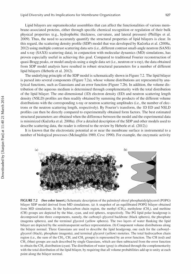
The underlying principle of the SDP model is schematically shown in Figure 7.2. The lipid bilayer is parsed into several components (Figure 7.2a), whose volume distributions are represented by ana-lytical functions, such as Gaussians and an error function (Figure 7.2b). In addition, the volume dis-tribution of the aqueous medium is determined through complementarity with the total distribution of the lipid bilayer. The one-dimensional (1D) electron density (ED) and neutron scattering length density (NSLD) profiles are then readily obtained by summing the products of the different volume distributions with the corresponding x-ray or neutron scattering amplitudes (i.e., the number of elec-trons or the neutron scattering length, respectively). By Fourier’s transform, the 1D ED and NSLD profiles can then be directly compared to experimentally obtained form factors. The best estimated structural parameters are obtained when the difference between the model and the experimental data is minimized (Kučerka et al. 2008a). (For a detailed description of the SDP and other models used to describe scattering data, the reader is referred to the review by Heberle et al. (2012).)
It is known that the electrostatic potential at or near the membrane surface is instrumental to a number of biological processes (Mclaughlin 1989, Cevc 1990). For example, the enzymatic activity
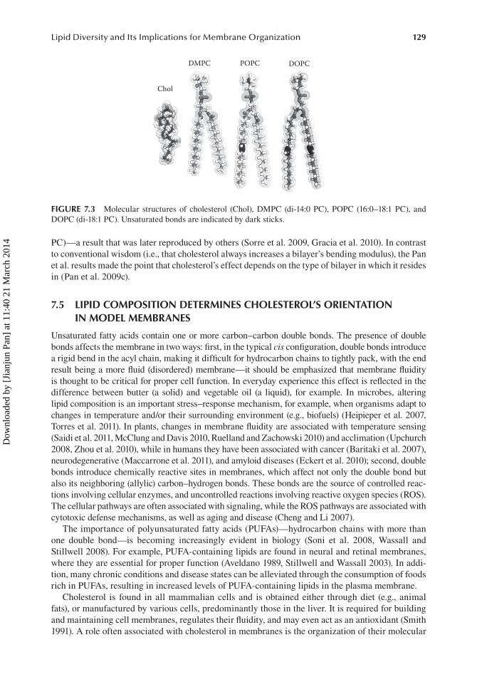
FIGURE 7.2 (See color insert.) Schematic description of the palmitoyl oleoyl phosphatidylglycerol (POPG) bilayer SDP model derived from MD simulations. (a) A snapshot of an equilibrated POPG bilayer obtained from MD simulations. In the hydrocarbon chain region, the methyl (CH3), methylene (CH2), and methine (CH) groups are depicted by the blue, cyan, and red spheres, respectively. The PG lipid polar headgroup is decomposed into three components, namely, the carbonyl–glycerol backbone (black spheres), the phosphate (magenta spheres), and the terminal glycerol (yellow spheres). The two layers of water flanking the lipid bilayer are depicted by the semitransparent surface representation. (b) Component volume distributions along the bilayer normal. Three Gaussians are used to describe the lipid headgroup, one each for the carbonyl– glycerol (black), phosphate (magenta), and terminal glycerol (yellow) moieties. The total hydrocarbon chain region (i.e., the sum of the CH2, CH, and CH3 groups) is represented by an error function. The CH (red) and CH3 (blue) groups are each described by single Gaussians, which are then subtracted from the error function to obtain the CH2 distribution (cyan). The distribution of water (gray) is obtained through the complementarity with the total distribution of the lipid bilayer, by requiring that all volume probabilities add up to unity at each point along the bilayer normal.
Dow
nloa
ded
by [
Jian
jun
Pan]
at 1
1:40
21
Mar
ch 2
014
128 Liposomes, Lipid Bilayers and Model Membranes
of Ca2+ lipid-dependent protein kinase C is strictly associated with the lipid’s headgroup moiety (Newton 1993). Moreover, the orientation and topology of membrane proteins are found to depend on the total amount of anionic lipids present in a membrane (van Klompenburg et al. 1997). Other examples include the role of anionic lipids in the activation of the adenosine triphosphate (ATP)-sensitive potassium channel (Fan and Makielski 1997), in providing the most energetically favor-able environment for voltage-sensing proteins (Schmidt et al. 2006), and in promoting peptide fibril formation (Olofsson et al. 2007). Suffice it to say, the charge state of the lipid headgroup is crucial in maintaining proper membrane function.
To systematically compare the specialized structural and functional properties of neutral and charged lipids, Kučerka et al. used the SDP model to look at the commonly studied phosphatidylcho-line (PC, neutral) and phosphatidylglycerol (PG, monoanionic) lipids in their biologically relevant fluid phase (Kučerka et al. 2008a, Pan et al. 2012). They found that the area per lipid—a key struc-tural parameter obtained from SDP model analysis, and which is used extensively in the validation of MD simulations—decreased with increasing hydrocarbon chain length, primarily the result of enhanced attractive van-der-Waals interactions between neighboring hydrocarbon chains. However, the introduction of an unsaturated bond in PC bilayers disrupted the packing of the hydrocarbon chains, resulting in increased lipid areas. An unexpected result was that despite their smaller head-group volume, monoanionic PG lipids possessed larger areas per lipid than their zwitterionic PC counterparts, implying that the long-range electrostatic interactions between charged headgroups play a prominent role in governing lipid lateral packing. By extrapolating to infinite chain length, Pan et al. discovered that areas per lipid of equivalent PC and PG lipids differed very little between the two different headgroup lipids, suggesting that the same steric interactions are experienced by both (Pan et al. 2012). In fact, the data indicated that the glycerol–carbonyl backbone is primarily responsible for the lateral packing of lipids at infinite chain length. This notion is particularly infor-mative when one considers the broad spectrum of membrane properties that are most likely affected by the glycerol backbone, a moiety that effectively delineates the membrane–water interface.
7.4 NONUNIVERSAL BEHAVIOR OF CHOLESTEROL’S EFFECT ON BILAYER BENDING MODULUS
A mechanical property that is essential for proper membrane function (e.g., cell endocytosis and exo-cytosis) is the membrane’s bending modulus, which is defined as the energy required to deform a membrane from its intrinsic curvature. Spontaneous, highly curved membranes are often formed dur-ing viral infection (e.g., stalk formation) and during the initial steps of cell division. To facilitate and maintain these highly curved structures, modifications to the membrane’s bending modulus are made.
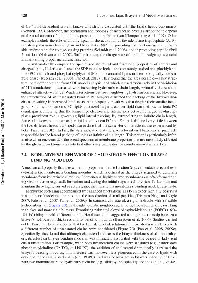
Membrane softening accompanied by enhanced fluctuations has been experimentally observed in a number of model membranes upon the introduction of small peptides (Tristram-Nagle and Nagle 2007, Pabst et al. 2007, Pan et al. 2009a). In contrast, cholesterol, a rigid molecule with a flexible hydrocarbon tail (Figure 7.3), is thought to order neighboring, fluid hydrocarbon chains, resulting in thicker and more rigid bilayers. Examining palmitoyl oleyol phosphatidylcholine (POPC) (16:0–18:1 PC) bilayers with different sterols, Henriksen et al. suggested a simple relationship between a bilayer’s hydrocarbon thickness and its bending modulus (Henriksen et al. 2006). Studies carried out by Pan et al., however, found that the Henriksen et al. relationship broke down when lipids with a different number of unsaturated chains were considered (Figure 7.3) (Pan et al. 2008, 2009c). Specifically, they found that although cholesterol increases the bilayer thickness of all fluid bilay-ers, its effect on bilayer bending modulus was intimately associated with the degree of fatty acid chain unsaturation. For example, when both hydrocarbon chains were saturated (e.g., dimyristoyl phosphatidylcholine (DMPC), di-14:0 PC), the addition of cholesterol dramatically increased the bilayer’s bending modulus. This increase was, however, less pronounced in the case of lipids with only one monounsaturated chain (e.g., POPC), and was nonexistent in bilayers made up of lipids with two monounsaturated hydrocarbon chains (e.g., dioleoyl phosphatidylcholine (DOPC), di-18:1
Dow
nloa
ded
by [
Jian
jun
Pan]
at 1
1:40
21
Mar
ch 2
014
129Lipid Diversity and Its Implications for Membrane Organization
PC)—a result that was later reproduced by others (Sorre et al. 2009, Gracia et al. 2010). In contrast to conventional wisdom (i.e., that cholesterol always increases a bilayer’s bending modulus), the Pan et al. results made the point that cholesterol’s effect depends on the type of bilayer in which it resides in (Pan et al. 2009c).
7.5 LIPID COMPOSITION DETERMINES CHOLESTEROL’S ORIENTATION IN MODEL MEMBRANES
Unsaturated fatty acids contain one or more carbon–carbon double bonds. The presence of double bonds affects the membrane in two ways: first, in the typical cis configuration, double bonds introduce a rigid bend in the acyl chain, making it difficult for hydrocarbon chains to tightly pack, with the end result being a more fluid (disordered) membrane—it should be emphasized that membrane fluidity is thought to be critical for proper cell function. In everyday experience this effect is reflected in the difference between butter (a solid) and vegetable oil (a liquid), for example. In microbes, altering lipid composition is an important stress–response mechanism, for example, when organisms adapt to changes in temperature and/or their surrounding environment (e.g., biofuels) (Heipieper et al. 2007, Torres et al. 2011). In plants, changes in membrane fluidity are associated with temperature sensing (Saidi et al. 2011, McClung and Davis 2010, Ruelland and Zachowski 2010) and acclimation (Upchurch 2008, Zhou et al. 2010), while in humans they have been associated with cancer (Baritaki et al. 2007), neurodegenerative (Maccarrone et al. 2011), and amyloid diseases (Eckert et al. 2010); second, double bonds introduce chemically reactive sites in membranes, which affect not only the double bond but also its neighboring (allylic) carbon–hydrogen bonds. These bonds are the source of controlled reac-tions involving cellular enzymes, and uncontrolled reactions involving reactive oxygen species (ROS). The cellular pathways are often associated with signaling, while the ROS pathways are associated with cytotoxic defense mechanisms, as well as aging and disease (Cheng and Li 2007).
The importance of polyunsaturated fatty acids (PUFAs)—hydrocarbon chains with more than one double bond—is becoming increasingly evident in biology (Soni et al. 2008, Wassall and Stillwell 2008). For example, PUFA-containing lipids are found in neural and retinal membranes, where they are essential for proper function (Aveldano 1989, Stillwell and Wassall 2003). In addi-tion, many chronic conditions and disease states can be alleviated through the consumption of foods rich in PUFAs, resulting in increased levels of PUFA-containing lipids in the plasma membrane.
Cholesterol is found in all mammalian cells and is obtained either through diet (e.g., animal fats), or manufactured by various cells, predominantly those in the liver. It is required for building and maintaining cell membranes, regulates their fluidity, and may even act as an antioxidant (Smith 1991). A role often associated with cholesterol in membranes is the organization of their molecular
Chol
DMPC POPC DOPC
FIGURE 7.3 Molecular structures of cholesterol (Chol), DMPC (di-14:0 PC), POPC (16:0–18:1 PC), and DOPC (di-18:1 PC). Unsaturated bonds are indicated by dark sticks.
Dow
nloa
ded
by [
Jian
jun
Pan]
at 1
1:40
21
Mar
ch 2
014

130 Liposomes, Lipid Bilayers and Model Membranes
structure. It is well known that the introduction of cholesterol to gel phase disaturated PC bilayers (e.g., di-16:0 PC, DPPC) disrupts the regular packing of their fatty acid chains, but restricts their reorientation in liquid crystalline bilayers (Vist and Davis 1990). Nominally, in membranes, cho-lesterol is oriented in its understood upright orientation, with its 3β-hydroxyl group locating just below the lipid–water interface (Leonard et al. 2001). In biological membranes, dynamic, functional domains (i.e., rafts) are thought to exist, and cholesterol has been identified as a key component of lipid rafts in mammalian cell membranes (Silvius 2003).
Although it has been shown that cholesterol interacts differentially with a number of membrane lipids—for example, interacting vigorously with high-transition temperature sphingolipids and phos-pholipids, and less so with lipids containing unsaturated fatty acid chains (McMullen and McElhaney 1996, Brown and London 2000)—cholesterol’s interaction with PUFA-containing phospholipids is less well understood. Over the past few years, Harroun et al. (2006b, 2008) and Kučerka et al. (2009, 2010) have studied the orientation of cholesterol in different PC bilayers, including those composed of PUFAs. Using neutron scattering in conjunction with “headgroup” and “tail” deuterated cholesterol, the studies by Harroun et al. demonstrated that although cholesterol assumes its nominal upright orientation (Leonard et al. 2001) in POPC (16:0–18:1 PC), DOPC (di-18:1 PC), and 18:0–20:4 PC bilayers (Figure 7.4a), it sequesters itself into the middle of PUFA (di-20:4 PC) bilayers (Figure 7.4b) (Harroun et al. 2006b, 2008). This differential behavior by cholesterol in the different PC bilayers was rationalized in terms of the high degree of disorder commonly associated with PUFAs, whereby they created an environment that was not amenable to cholesterol’s rigid steroid moiety to remain near the lipid–water interface. Subsequent coarse-grained MD simulations confirmed the Harroun et al. result, albeit with a slight twist (Marrink et al. 2008). Namely, while the neutron scattering stud-ies located cholesterol exclusively in its nominal upright orientation in all bilayers—the exception being the PUFA bilayer, where cholesterol sequesters itself into the bilayer’s center (i.e., flat orienta-tion)—the MD simulations noted that cholesterol experienced flip-flop rates in all bilayers, ranging from nanoseconds to microseconds in PUFA and POPC bilayers, respectively (Marrink et al. 2008).
Kučerka et al. (2009, 2010) followed up on the Harroun et al. (2006b, 2008) studies by doping cholesterol-containing PUFA bilayers with increasing amounts of either POPC or DMPC (di-14:0 PC). They reported that it took the addition of almost 50 mol% of POPC into cholesterol-containing PUFA bilayers to induce cholesterol to flip from its location in the bilayer center to its upright orientation. Conversely, only 5 mol% of DMPC achieved the same result (Kučerka et al. 2009). Moreover, MD simulations performed on similar systems predicted the formation of DMPC-rich domains in which cholesterol preferentially locates in its upright orientation, while in DMPC-depleted domains, cholesterol was found predominantly in its flat orientation (Kučerka et al. 2010). These experimental and simulation studies (Harroun et al. 2006b, 2008, Kučerka et al. 2009, 2010)
FIGURE 7.4 Two orientations of cholesterol in a lipid bilayer. (a) The nominal upright orientation. (b) The flat orientation where cholesterol sequesters itself into the middle of the bilayer.
Dow
nloa
ded
by [
Jian
jun
Pan]
at 1
1:40
21
Mar
ch 2
014
131Lipid Diversity and Its Implications for Membrane Organization
are a clear evidence that a biological membrane’s lipid composition not only affects the membrane’s in-plane organization (i.e., domains), but that a molecule’s orientation (i.e., function) in a membrane may depend on the domain in which it resides, highlighting the importance of lipid diversity as it pertains to the proper functioning of biological membranes.
7.6 ALIGNABLE MODEL MEMBRANES BY EXTERNAL FIELDS AND SOME OF THEIR APPLICATIONS
Lipid heterogeneity affects not only the conformation of biomolecules in membranes, but also the aggregate morphology in which the lipids find themselves in. An example of this is the long- and short-chain lipid mixture, known as “bicelles” (bilayered micelles), and first referred to as such by Sanders and Landis (1995). For the most part, bicellar mixtures have been commonly used for the structural determination of membrane-associated proteins (Ujwal and Bowie 2011, Faham and Bowie 2002, Andersson and Mäler 2002, Zandomeneghi et al. 2003, Seddon et al. 2004, Marcotte and Auger 2005, Prosser et al. 2006, Diller et al. 2009, Halskau et al. 2009, Matsumori and Murata 2010, Warschawski et al. 2011), and there are several unique properties which make bicelles attractive for studying such pro-teins. For example, they can be uniformly dispersed in water at a much higher total lipid concentration, Clp (>30 wt.%) than pure long-chain lipid solutions (Ram and Prestegard 1988, Sanders and Prestegard 1990), thus greatly enhancing the concentration of membrane proteins. Moreover, since a biological membrane’s underlying structure is a lipid bilayer, bicellar mixtures lend themselves as a better mem-brane mimic for membrane-associated proteins than commonly used detergent-based substrates.
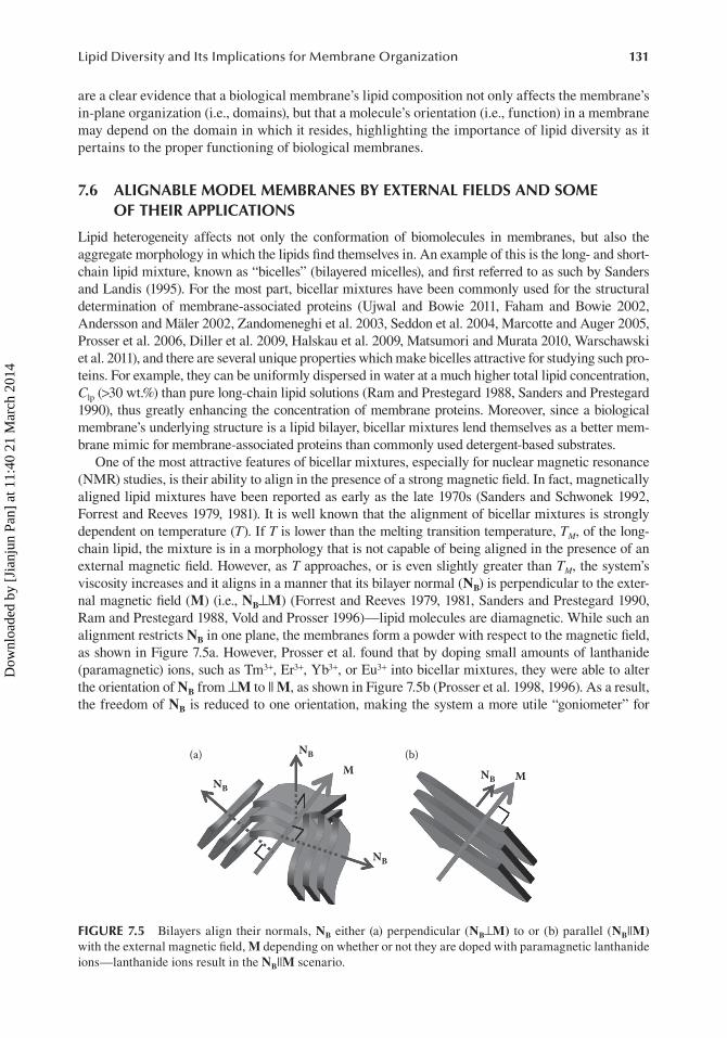
One of the most attractive features of bicellar mixtures, especially for nuclear magnetic resonance (NMR) studies, is their ability to align in the presence of a strong magnetic field. In fact, magnetically aligned lipid mixtures have been reported as early as the late 1970s (Sanders and Schwonek 1992, Forrest and Reeves 1979, 1981). It is well known that the alignment of bicellar mixtures is strongly dependent on temperature (T). If T is lower than the melting transition temperature, TM, of the long-chain lipid, the mixture is in a morphology that is not capable of being aligned in the presence of an external magnetic field. However, as T approaches, or is even slightly greater than TM, the system’s viscosity increases and it aligns in a manner that its bilayer normal (NB) is perpendicular to the exter-nal magnetic field (M) (i.e., NB⊥M) (Forrest and Reeves 1979, 1981, Sanders and Prestegard 1990, Ram and Prestegard 1988, Vold and Prosser 1996)—lipid molecules are diamagnetic. While such an alignment restricts NB in one plane, the membranes form a powder with respect to the magnetic field, as shown in Figure 7.5a. However, Prosser et al. found that by doping small amounts of lanthanide (paramagnetic) ions, such as Tm3+, Er3+, Yb3+, or Eu3+ into bicellar mixtures, they were able to alter the orientation of NB from ⊥M to || M, as shown in Figure 7.5b (Prosser et al. 1998, 1996). As a result, the freedom of NB is reduced to one orientation, making the system a more utile “goniometer” for
NB
(a) (b)
NB
NB
NBM M
FIGURE 7.5 Bilayers align their normals, NB either (a) perpendicular (NB⊥M) to or (b) parallel (NB||M) with the external magnetic field, M depending on whether or not they are doped with paramagnetic lanthanide ions—lanthanide ions result in the NB||M scenario.
Dow
nloa
ded
by [
Jian
jun
Pan]
at 1
1:40
21
Mar
ch 2
014
132 Liposomes, Lipid Bilayers and Model Membranes
structural studies of membrane-associated proteins. It has been reported that the temperature range in which these lipid mixtures are magnetically alignable depends on the charge density of the membrane, that is, mixtures with a higher molar ratio of charged lipids or paramagnetic ions (Nieh et al. 2002).
Compared with solid substrate aligned bilayers, magnetically aligned membranes provide a more biomimetic membrane environment for integral proteins. However, the requisite strong magnetic fields can only be realistically implemented using only a few characterization techniques (e.g., NMR and SANS). (It should be mentioned that a 0.9 T fixed field device was developed by Harroun et al. for use with standard optical microscopes (Harroun et al. 2006a).) Nieh et al. also examined a lantha-nide-free charged bicellar mixture and found that the presence of an oscillating shear flow induced good alignment in membranes, with their NB aligning parallel to the direction of the shear (Nieh et al. 2003). Importantly, alignment persisted for a period of hours after shear flow ceased, thus greatly simplifying the apparatus needed for inducing alignment. In doing so, this method of alignment may enable a number of other physical techniques to interrogate these interesting and versatile systems.
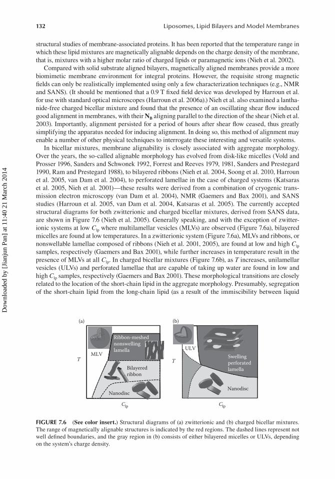
In bicellar mixtures, membrane alignability is closely associated with aggregate morphology. Over the years, the so-called alignable morphology has evolved from disk-like micelles (Vold and Prosser 1996, Sanders and Schwonek 1992, Forrest and Reeves 1979, 1981, Sanders and Prestegard 1990, Ram and Prestegard 1988), to bilayered ribbons (Nieh et al. 2004, Soong et al. 2010, Harroun et al. 2005, van Dam et al. 2004), to perforated lamellae in the case of charged systems (Katsaras et al. 2005, Nieh et al. 2001)—these results were derived from a combination of cryogenic trans-mission electron microscopy (van Dam et al. 2004), NMR (Gaemers and Bax 2001), and SANS studies (Harroun et al. 2005, van Dam et al. 2004, Katsaras et al. 2005). The currently accepted structural diagrams for both zwitterionic and charged bicellar mixtures, derived from SANS data, are shown in Figure 7.6 (Nieh et al. 2005). Generally speaking, and with the exception of zwitter-ionic systems at low Clp where multilamellar vesicles (MLVs) are observed (Figure 7.6a), bilayered micelles are found at low temperatures. In a zwitterionic system (Figure 7.6a), MLVs and ribbons, or nonswellable lamellae composed of ribbons (Nieh et al. 2001, 2005), are found at low and high Clp samples, respectively (Gaemers and Bax 2001), while further increases in temperature result in the presence of MLVs at all Clp. In charged bicellar mixtures (Figure 7.6b), as T increases, unilamellar vesicles (ULVs) and perforated lamellae that are capable of taking up water are found in low and high Clp samples, respectively (Gaemers and Bax 2001). These morphological transitions are closely related to the location of the short-chain lipid in the aggregate morphology. Presumably, segregation of the short-chain lipid from the long-chain lipid (as a result of the immiscibility between liquid
Ribbon-meshednonswellinglamella
MLV
(a) (b)
Bilayeredribbon
Swellingperforatedlamella
ULV
TT
NanodiscNanodisc
Clp Clp
FIGURE 7.6 (See color insert.) Structural diagrams of (a) zwitterionic and (b) charged bicellar mixtures. The range of magnetically alignable structures is indicated by the red regions. The dashed lines represent not well defined boundaries, and the gray region in (b) consists of either bilayered micelles or ULVs, depending on the system’s charge density.
Dow
nloa
ded
by [
Jian
jun
Pan]
at 1
1:40
21
Mar
ch 2
014

133Lipid Diversity and Its Implications for Membrane Organization
disordered and gel phases) favors structures with a larger total circumference, as the short-chain lipid, which has a larger spontaneous curvature, is able to stabilize the high curvature edge.
Besides aligning membrane proteins, bicellar mixtures have also been used as substrates to crystallize membrane-associated proteins. For example, Fahama and Bowie successfully used a bicellar mixture to crystallize bacteriorhodopsin extracted from Halobacterium salinarum. They took advantage of the inherent low-viscosity solution formed by bilayered micelles at low T, which allowed the application of general screening methods (Faham and Bowie 2002). For further details regarding this method of crystallizing proteins, the reader is referred to the review by Ujwal and Bowie (2011), while other applications of bicellar mixtures, as studied by NMR and other spectro-scopic techniques, are summarized in Table 7.1.
TABLE 7.1Composition of Bicellar Mixtures and Their Applications
Long Chain/Short Chain Applications References
DMPC/DHPC Structural determination of membrane-associated molecules by NMR
Andersson and Mäler (2002), Zandomeneghi et al. (2003), Marcotte and Auger (2005), Prosser et al. (2006)
Aligning water-soluble proteins Ottiger and Bax (1998a, b, 1999), Martin-Pastor and Bush (2000)
Protein crystallization Faham and Bowie (2002), Ujwal and Bowie (2011)
Separation and sensor devices Mills and Holland (2004), Pappas and Holland (2008), Luo et al. (2010)
Application to skin Barbosa-Barros et al. (2008a), Rodríguez et al. (2010, 2011)
DMPC/CHAPS Structural determination of membrane-associated molecules
Booth et al. (1997), Sugiyama et al. (1999), Kim et al. (2001), Renthal and Velasquez (2002), Andersson et al. (2007), McKibbin et al. (2007), Gayen and Mukhopadhyay (2008), Krishnamani et al. (2012)
DMPC/CHAPSO Structural determination of membrane-associated molecules
Sanders and Prestegard (1990, 1991), Aubin et al. (1993), Salvatore et al. (1996), Chen and Gouaux (1999), Kawaguchi et al. (2003), Wang et al. (2012)
Protein crystallization Faham et al. (2005)
DPPC/DHPC Structural determination of membrane-associated molecules
Lind et al. (2008)
Protein/drug carrier Nieh et al. (2006), Rubio et al. (2011)
Application to skin Barbosa-Barros et al. (2008b, 2009), Rodríguez et al. (2010, 2011)
Carbon nanotube assembly Wallace and Sansom (2009)
DLPC/DHPC Structural determination of membrane-associated molecules
Lind et al. (2008)
DLPC/CHAPSO Wang et al. (1998)
POPC/DHPC Chou et al. (2004), Wang et al. (2004)
DPC/SDS Baek et al. (2011)
DMLPC/DHPC Structural characterization of bicellar mixtures
Aussenac et al. (2005)
DIOMPC/DIOHPC Evanics and Prosser (2005)
SM/DHPC Yamaguchi et al. (2012)
TBBPC/DHPC Loudet et al. (2010)
Dow
nloa
ded
by [
Jian
jun
Pan]
at 1
1:40
21
Mar
ch 2
014

134 Liposomes, Lipid Bilayers and Model Membranes
7.7 LIPID BILAYER DETERMINES ANTIMICROBIAL PEPTIDE ORGANIZATION
Antimicrobial peptides (AMPs) are a class of small molecules which are capable of disrupting biological membranes through a number of different mechanisms. However, their ability to differ-entiate between eukaryotic and prokaryotic membranes makes them promising therapeutic agents against certain pathogens importantly, without inducing drug resistance—an often occurring prob-lem with drugs targeting a specific protein or gene.
Alamethicin (Alm) is a small AMP that spontaneously aggregates to form a membrane-spanning bundle (Figure 7.7). To compensate for the hydrophobic mismatch between the bilayer’s hydrophobic core and the protein’s embedded hydrophobic domain, the membrane is deformed—this is because the peptide is stiffer than the bilayer. The energy cost associated with such deformation depends on the membrane’s thickness, its bending and area stretch moduli, and its intrinsic curvature.
Pan et al. studied how the Alm bundle structure behaves in two lipid bilayers, namely, di-18:1 PC and di-22:1 PC (Pan et al. 2009b). These bilayers have similar physical properties, except that the di-22:1 PC bilayer is about 7 Å thicker than di-18:1 PC. It was found that Alm forms a hexametric bundle in di-18:1 PC, while a nonamer structure was discovered in di-22:1 PC. The smaller Alm bundle in di-18:1 PC was the result of hydrophobic thickness matching between di-18:1 PC bilayers and Alm—as mentioned, the hydrophobic thickness of di-22:1 PC bilayers is 7 Å larger. This notion is consistent with the well-known functional cutoff effect (Balgavý and Devínsky 1996) observed, for example, in Ca2+-transporting ATPase incorporated in lipid bilayers (Karlovská et al. 2006). The proper function of a membrane protein in a biological membrane, thus depends on the structural and dynamical properties of the underlying lipid matrix.
The close interplay between the lipid matrix and associated AMPs has also emerged from other studies. Sani et al. reported that lipid composition is an important regulator in controlling maculatin 1.1’s conformation and orientation (Sani et al. 2012). In the case of zwitterionic PC lipid bilayers, the peptide’s helical content—a good indicator of the peptide’s interaction potential with lipid bilay-ers—was found to depend on lipid hydrocarbon chain length and degree of unsaturation, while in anionic lipid bilayers, maculatin 1.1 interacted strongly and oriented orthogonal to the bilayer nor-mal. In another study involving a PC/PG mixture and the cationic AMP, aurein, Cheng et al. found that AMP–membrane interactions were affected by the amount of charged PG lipid present and the hydrophobic thickness of the lipid bilayer (Cheng et al. 2009). More recently, MD simulations of gramicidin A in different lipid bilayers have shown a radial dependence of lipid bilayer perturba-tion, induced by the addition of gramicidin A (Kim et al. 2012).
From these studies, one can surmise that AMP organization and function are to a great extent regulated by the host lipid bilayer through a variety of chemical and physical interactions. In-depth studies of AMP interactions with model membranes are paving the way in deciphering how AMPs
FIGURE 7.7 The Alm hexameric structure in a lipid bilayer. (a) Side view and (b) top view.
Dow
nloa
ded
by [
Jian
jun
Pan]
at 1
1:40
21
Mar
ch 2
014
135Lipid Diversity and Its Implications for Membrane Organization
interact with the different lipid species that make up biological membranes. We are of the belief that such knowledge will prove to be invaluable when designing and developing more effective peptide-based antibiotics.
7.8 ION-SPECIFIC EFFECTS IN BACTERIAL MEMBRANES
In addition to lipid–peptide (and lipid–protein) interactions, the significance of the aqueous phase for the proper function of biological membranes cannot be overestimated. Biological membranes are surrounded by an electrolytic liquid containing Na+, K+, Ca2+, Mg2+, and Cl− ions. Their inter-actions with cell membranes are understood to influence, for example, the gating of ion channels, membrane fusion, and membrane fluidity, to name a few. Over the years, there have been copious amounts of biophysical reports demonstrating that ions affect the physical properties of lipid bilay-ers (see, e.g., Pabst et al. 2010, for a recent review).
The effect of Ca2+ cations was recently reported in bacterial mimetic membranes composed of lipopolysaccharides (LPSs) (Kučerka et al. 2008b). LPSs are the major lipid component mak-ing up the outermost leaflet of the asymmetric outer membrane (OM) of Gram-negative bacteria (Wilkinson 1996, Nikaido 2003). It contributes to the OM’s structural integrity and also protects the bacteria from a variety of toxic molecules, such as certain antibiotics (e.g., penicillin), diges-tive enzymes (e.g., lysozyme), detergents, heavy metals, bile salts, and some dyes. The passage of nucleotides, disaccharides, amino acids, vitamins, and iron for nutritional growth are usually transported through the OM by porin proteins, but it is LPS that provides the bacteria with its remarkable selectively permeable membrane that is resistant to a variety of deleterious agents. In particular, Pseudomonas aeruginosa is well noted for its recalcitrance to conventional antibiotic therapy, partly as a result of its unique surface chemistry (Rocchetta et al. 1999). For this reason, and also due to the ubiquity of P. aeruginosa and its impact upon health as both an opportunistic and nosocomial pathogen, this organism represents an attractive candidate for medical and phar-macological studies.
Although LPS molecules are structurally diverse, they share a common architecture composed of three basic units. The first is a lipid A moiety that anchors the LPS molecule into the hydrophobic domain of the OM. It consists of two phosphorylated glucosamine units that are typically acylated with four to six fatty acids and is considered to be responsible for most of the toxicity associated with LPS. Second, the LPS’ core oligosaccharide is made up of 8–12 monosaccharide units, and is connected to lipid A by a 2-keto-3-deoxyoctonoic acid (Kdo). Finally, the third part is formed by repetitive monosaccharide subunits (i.e., O-side chain), which are responsible for much of the bacterium’s immunospecificity (Caroff and Karibian 2003). However, recent experiments revealed a determining effect of counterions involved in the system.
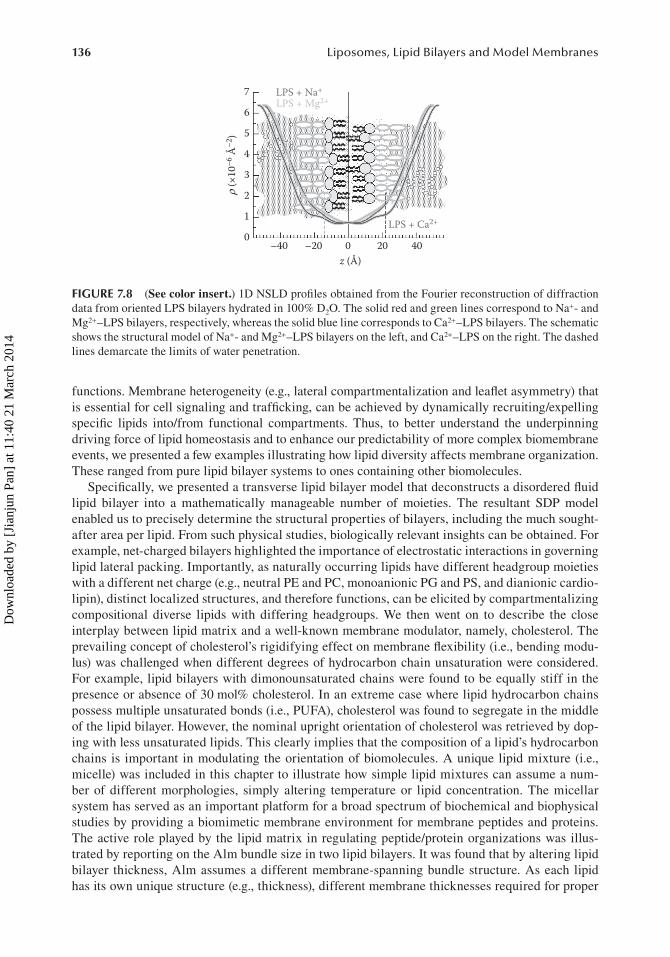
Small-angle neutron diffraction (SAND) data showed that water penetrates Ca2+–LPS bilayers to a lesser extent than Na+- and Mg2+–LPS bilayers (Kučerka et al. 2008b). While Ca2+ cations make LPS bilayers more compact and less permeable to water, a significant amount of water penetrates deep into Mg2+–LPS and Na+–LPS bilayers, including the bilayer’s hydrophobic core (Figure 7.8). It is believed that such increased levels of hydration could be associated with enhanced biological activity in these bacterial membranes. As such, a more accurate determination of the membrane’s structure may allow for a better understanding of membrane function. For example, differences in a bilayer’s permeability to water could have implications with regard to how small molecules permeate through the OM of Gram-negative bacteria, aiding in the development of more effective antibiotics.
7.9 CONCLUSIONS
Cell membranes possess a ubiquitous bilayer architecture that is vital for proper biological func-tion. Lipids make up the underlying membrane scaffold enabling proteins to carry out their various
Dow
nloa
ded
by [
Jian
jun
Pan]
at 1
1:40
21
Mar
ch 2
014
136 Liposomes, Lipid Bilayers and Model Membranes
functions. Membrane heterogeneity (e.g., lateral compartmentalization and leaflet asymmetry) that is essential for cell signaling and trafficking, can be achieved by dynamically recruiting/expelling specific lipids into/from functional compartments. Thus, to better understand the underpinning driving force of lipid homeostasis and to enhance our predictability of more complex biomembrane events, we presented a few examples illustrating how lipid diversity affects membrane organization. These ranged from pure lipid bilayer systems to ones containing other biomolecules.
Specifically, we presented a transverse lipid bilayer model that deconstructs a disordered fluid lipid bilayer into a mathematically manageable number of moieties. The resultant SDP model enabled us to precisely determine the structural properties of bilayers, including the much sought-after area per lipid. From such physical studies, biologically relevant insights can be obtained. For example, net-charged bilayers highlighted the importance of electrostatic interactions in governing lipid lateral packing. Importantly, as naturally occurring lipids have different headgroup moieties with a different net charge (e.g., neutral PE and PC, monoanionic PG and PS, and dianionic cardio-lipin), distinct localized structures, and therefore functions, can be elicited by compartmentalizing compositional diverse lipids with differing headgroups. We then went on to describe the close interplay between lipid matrix and a well-known membrane modulator, namely, cholesterol. The prevailing concept of cholesterol’s rigidifying effect on membrane flexibility (i.e., bending modu-lus) was challenged when different degrees of hydrocarbon chain unsaturation were considered. For example, lipid bilayers with dimonounsaturated chains were found to be equally stiff in the presence or absence of 30 mol% cholesterol. In an extreme case where lipid hydrocarbon chains possess multiple unsaturated bonds (i.e., PUFA), cholesterol was found to segregate in the middle of the lipid bilayer. However, the nominal upright orientation of cholesterol was retrieved by dop-ing with less unsaturated lipids. This clearly implies that the composition of a lipid’s hydrocarbon chains is important in modulating the orientation of biomolecules. A unique lipid mixture (i.e., micelle) was included in this chapter to illustrate how simple lipid mixtures can assume a num-ber of different morphologies, simply altering temperature or lipid concentration. The micellar system has served as an important platform for a broad spectrum of biochemical and biophysical studies by providing a biomimetic membrane environment for membrane peptides and proteins. The active role played by the lipid matrix in regulating peptide/protein organizations was illus-trated by reporting on the Alm bundle size in two lipid bilayers. It was found that by altering lipid bilayer thickness, Alm assumes a different membrane-spanning bundle structure. As each lipid has its own unique structure (e.g., thickness), different membrane thicknesses required for proper
7
6
5
4
3
2
1
0–40 –20 0 20 40
z (Å)
ρ (×
10–6
Å–2
)LPS + Ca2+
LPS + Na+
LPS + Mg2+
FIGURE 7.8 (See color insert.) 1D NSLD profiles obtained from the Fourier reconstruction of diffraction data from oriented LPS bilayers hydrated in 100% D2O. The solid red and green lines correspond to Na+- and Mg2+–LPS bilayers, respectively, whereas the solid blue line corresponds to Ca2+–LPS bilayers. The schematic shows the structural model of Na+- and Mg2+–LPS bilayers on the left, and Ca2+–LPS on the right. The dashed lines demarcate the limits of water penetration.
Dow
nloa
ded
by [
Jian
jun
Pan]
at 1
1:40
21
Mar
ch 2
014

137Lipid Diversity and Its Implications for Membrane Organization
membrane function can be achieved by varying lipid composition. Finally, we showed that not only the membrane, but also ions in the aqueous medium surrounding the biomembrane play an important role in modulating membrane structure and function.
REFERENCES
Abraham, T. et al. 2008. Monolayer film behavior of lipopolysaccharide from Pseudomonas aeruginosa at the air–water interface. Biomacromolecules, 9(10), 2799–2804.
Andersson, A. et al. 2007. The membrane-induced structure of melittin is correlated with the fluidity of the lipids. Biochimica et Biophysica Acta—Biomembranes, 1768(1), 115–121.
Andersson, A. and Mäler, L. 2002. NMR solution structure and dynamics of motilin in isotropic phospholipid bicellar solution. Journal of Biomolecular NMR, 24(2), 103–112.
Aubin, Y. et al. 1993. Structure and dynamics of the sialic acid moiety of GM3-ganglioside at the surface of a magnetically oriented membrane. Biochemistry, 32(49), 13405–13413.
Aussenac, F., Lavigne, B., and Dufourc, E. J. 2005. Toward bicelle stability with ether-linked phospholipids: Temperature, composition, and hydration diagrams by 2H and 31P solid-state NMR. Langmuir, 21(16), 7129–7135.
Aveldano, M. I. 1989. Dipolyunsaturated species of retina phospholipids and their fatty-acids. Biomembranes and Nutrition, 195, 87–96.
Baek, S. B. et al. 2011. An NMR study on the conformation of substance P in acidic bicelles. Bulletin of the Korean Chemical Society, 32(10), 3702–3706.
Balgavý, P. and Devínsky, F. 1996. Cut-off effects in biological activities of surfactants. Advances in Colloid and Interface Science, 66, 23–63.
Barbosa-Barros, L. et al. 2008a. Effect of bicellar systems on skin properties. International Journal of Pharmaceutics, 352(1–2), 263–272.
Barbosa-Barros, L. et al. 2008b. Penetration and growth of DPPC/DHPC bicelles inside the stratum corneum of the skin. Langmuir, 24(11), 5700–5706.
Barbosa-Barros, L. et al. 2009. Lipid nanostructures: Self-assembly and effect on skin properties. Molecular Pharmaceutics, 6(4), 1237–1245.
Baritaki, S. et al. 2007. Reversal of tumor resistance to apoptotic stimuli by alteration of membrane fluidity: Therapeutic implications. Advances in Cancer Research, 98, 149–190.
Booth, P. J. et al. 1997. Evidence that bilayer bending rigidity affects membrane protein folding. Biochemistry, 36(1), 197–203.
Brown, D. A. and London, E. 2000. Structure and function of sphingolipid- and cholesterol-rich membrane rafts. Journal of Biological Chemistry, 275(23), 17221–17224.
Caroff, M. and Karibian, D. 2003. Structure of bacterial lipopolysaccharides. Carbohydrate Research, 338(23), 2431–2447.
Cevc, G. 1990. Membrane electrostatics. Biochimica et Biophysica Acta, 1031(3), 311–382.Chen, G. Q. and Gouaux, E. 1999. Probing the folding and unfolding of wild-type and mutant forms of bacte-
riorhodopsin in micellar solutions: Evaluation of reversible unfolding conditions. Biochemistry, 38(46), 15380–15387.
Cheng, J. T. J. et al. 2009. Effect of membrane composition on antimicrobial peptides aurein 2.2 and 2.3 from Australian southern bell frogs. Biophysical Journal, 96(2), 552–565.
Cheng, Z. Y. and Li, Y. Z. 2007. What is responsible for the initiating chemistry of iron-mediated lipid peroxida-tion: An update. Chemical Reviews, 107(5), 2165–2165.
Chou, J. J., Baber, J. L., and Bax, A. 2004. Characterization of phospholipid mixed micelles by translational diffusion. Journal of Biomolecular NMR, 29(3), 299–308.
Danielli, J. F. 1975. The bilayer hypothesis of membrane structure. In: Weissmann, G. and Clairborne, R. eds. Cell Membranes: Biochemistry, Cell Biology, and Pathology. New York: Hospital Practice, 3–11.
Danielli, J. F. and Davson, H. 1935. A contribution to the theory of permeability of thin films. Journal of Cellular and Comparative Physiology, 5(4), 495–508.
Diller, A. et al. 2009. Bicelles: A natural molecular goniometer for structural, dynamical and topological stud-ies of molecules in membranes. Biochimie, 91(6), 744–751.
Eckert, G. P., Wood, W. G., and Muller, W. E. 2010. Lipid membranes and beta-amyloid: A harmful connection. Current Protein and Peptide Science, 11(5), 319–325.
Evanics, F. and Prosser, R. S. 2005. Discriminating binding and positioning of amphiphiles to lipid bilayers by 1H NMR. Analytica Chimica Acta, 534(1), 21–29.
Dow
nloa
ded
by [
Jian
jun
Pan]
at 1
1:40
21
Mar
ch 2
014

138 Liposomes, Lipid Bilayers and Model Membranes
Faham, S. et al. 2005. Crystallization of bacteriorhodopsin from bicelle formulations at room temperature. Protein Science, 14(3), 836–840.
Faham, S. and Bowie, J. U. 2002. Bicelle crystallization: A new method for crystallizing membrane proteins yields a monomeric bacteriorhodopsin structure. Journal of Molecular Biology, 316(1), 1–6.
Fan, Z. and Makielski, J. C. 1997. Anionic phospholipids activate ATP-sensitive potassium channels. Journal of Biological Chemistry, 272(9), 5388–5395.
Forrest, B. J. and Reeves, L. W. 1979. Studies in membrane processes X: A deuterium magnetic resonance study of dipalmitoyl lecithin and palmitic acid guests in magnetically-oriented hexadecyltrimethyl-ammonium bromide liquid crystalline system. Chemistry and Physics of Lipids, 24(2), 183–192.
Forrest, B. J. and Reeves, L. W. 1981. New lyotropic liquid crystals composed of finite nonspherical micelles. Chemical Reviews, 81(1), 1–14.
Gaemers, S. and Bax, A. 2001. Morphology of three lyotropic liquid crystalline biological NMR media studied by translational diffusion anisotropy. Journal of the American Chemical Society, 123(49), 12343–12352.
Gayen, A. and Mukhopadhyay, C. 2008. Evidence for effect of GM1 on opioid peptide conformation: NMR study on leucine enkephalin in ganglioside-containing isotropic phospholipid bicelles. Langmuir, 24(10), 5422–5432.
Gorter, E. and Grendel, F. 1925. On bimolecular layers of lipoids on the chromocytes of the blood. Journal of Experimental Medicine, 41(4), 439–443.
Gracia, R. S. et al. 2010. Effect of cholesterol on the rigidity of saturated and unsaturated membranes: Fluctuation and electrodeformation analysis of giant vesicles. Soft Matter, 6(7), 1472–1482.
Halskau, Ø., Muga, A., and Martínez, A. 2009. Linking new paradigms in protein chemistry to reversible mem-brane protein interactions. Current Protein and Peptide Science, 10(4), 339–359.
Harroun, T. A. et al. 2006a. 0.9 T static magnetic field and temperature-controlled specimen environment for use with general-purpose optical microscopes. Review of Scientific Instruments, 77(1), 014102.
Harroun, T. A., Katsaras, J., and Wassall, S. R. 2006b. Cholesterol hydroxyl group is found to reside in the center of a polyunsaturated lipid membrane. Biochemistry, 45(4), 1227–1233.
Harroun, T. A., Katsaras, J., and Wassall, S. R. 2008. Cholesterol is found to reside in the center of a polyun-saturated lipid membrane. Biochemistry, 47(27), 7090–7096.
Harroun, T. A. et al. 2005. Comprehensive examination of mesophases formed by DMPC and DHPC mixtures. Langmuir, 21(12), 5356–5361.
Heberle, F. A. et al. 2012. Model-based approaches for the determination of lipid bilayer structure from small-angle neutron and x-ray scattering data. European Biophysics Journal, 41, 875–890.
Heipieper, H. J. et al. 2007. Solvent-tolerant bacteria for biotransformations in two-phase fermentation sys-tems. Applied Microbiology and Biotechnology, 74(5), 961–973.
Hendler, R. W. 1971. Biological membrane ultrastructure. Physiological Reviews, 51(1), 66–97.Henriksen, J. et al. 2006. Universal behavior of membranes with sterols. Biophysical Journal, 90(5), 1639–1649.Iqbal, U. et al. 2011. Small unilamellar vesicles: A platform technology for molecular imaging of brain tumors.
Nanotechnology, 22, 195102(1)–195102(15).Karlovská, J. et al. 2006. Influence of N-dodecyl-N,N-dimethylamine N-oxide on the activity of sarcoplasmic
reticulum Ca(2+)-transporting ATPase reconstituted into diacylphosphatidylcholine vesicles: Effects of bilayer physical parameters. Biophysical Chemistry, 119(1), 69–77.
Katsaras, J. 1997. Highly aligned lipid membrane systems in the physiologically relevant “excess water” condi-tion. Biophysical Journal, 73(6), 2924–2929.
Katsaras, J. 1998. Adsorbed to a rigid substrate, dimyristoylphosphatidylcholine multibilayers attain full hydra-tion in all mesophases. Biophysical Journal, 75(5), 2157–2162.
Katsaras, J. et al. 2005. Bicellar lipid mixtures as used in biochemical and biophysical studies. Naturwissenschaften, 92(8), 355–366.
Kawaguchi, K., Kimura, K., and Asakura, T. 2003. Direct observations of high resolution 1H NMR in liquid phase for peptides bound to bicelles. Kobunshi Ronbunshu, 60(4), 199–202.
Kim, J. M. et al. 2001. Structure and function in bacteriorhodopsin: The role of the interhelical loops in the folding and stability of bacteriorhodopsin. Journal of Molecular Biology, 308(2), 409–422.
Kim, L. et al. 2012. Influence of hydrophobic mismatch on structures and dynamics of gramicidin A and lipid bilayers. Biophysical Journal, 102(7), 1551–1560.
Krishnamani, V. et al. 2012. Secondary and tertiary structure of bacteriorhodopsin in the SDS denatured state. Biochemistry, 51(6), 1051–1060.
Kučerka, N. et al. 2012. Scattering density profile model of POPG bilayers as determined by molecular dynam-ics simulations and small-angle neutron and x-ray scattering experiments. Journal of Physical Chemistry B, 116(1), 232–239.
Dow
nloa
ded
by [
Jian
jun
Pan]
at 1
1:40
21
Mar
ch 2
014

139Lipid Diversity and Its Implications for Membrane Organization
Kučerka, N. et al. 2010. Cholesterol in bilayers with PUFA chains: Doping with DMPC or POPC results in sterol reorientation and membrane-domain formation. Biochemistry, 49(35), 7485–7493.
Kučerka, N. et al. 2009. The functional significance of lipid diversity: Orientation of cholesterol in bilayers is determined by lipid species. Journal of the American Chemical Society, 131(45), 16358–16359.
Kučerka, N. et al. 2008a. Lipid bilayer structure determined by the simultaneous analysis of neutron and x-ray scattering data. Biophysical Journal, 95(5), 2356–2367.
Kučerka, N. et al. 2008b. Effect of cations on the structure of bilayers formed by lipopolysaccharides isolated from Pseudomonas aeruginosa PAO1. Journal of Physical Chemistry B, 112(27), 8057–8062.
Lenard, J. and Singer, S. J. 1966. Protein conformation in cell membrane preparations as studied by opti-cal rotatory dispersion and circular dichroism. Proceedings of the National Academy of Sciences of the United States of America, 56(6), 1828–1835.
Leonard, A. et al. 2001. Location of cholesterol in DMPC membranes. A comparative study by neutron diffrac-tion and molecular mechanics simulation. Langmuir, 17(6), 2019–2030.
Lind, J., Nordin, J., and Mäler, L. 2008. Lipid dynamics in fast-tumbling bicelles with varying bilayer thickness: Effect of model transmembrane peptides. Biochimica et Biophysica Acta—Biomembranes, 1778(11), 2526–2534.
Lingwood, D. and Simons, K. 2010. Lipid rafts as a membrane-organizing principle. Science, 327(5961), 46–50.
Loudet, C. et al. 2010. Biphenyl phosphatidylcholine: A promoter of liposome deformation and bicelle collec-tive orientation by magnetic fields. Progress in Lipid Research, 49(3), 289–297.
Luo, R. J., Archer-Hartmann, S. A., and Holland, L. A. 2010. Transformable capillary electrophoresis for oligo-saccharide separations using phospholipid additives. Analytical Chemistry, 82(4), 1228–1233.
Maccarrone, M. et al. 2011. Cannabinoid receptor signalling in neurodegenerative diseases: A potential role for membrane fluidity disturbance. British Journal of Pharmacology, 163(7), 1379–1390.
Marcotte, I. and Auger, M. 2005. Bicelles as model membranes for solid- and solution-state NMR studies of membrane peptides and proteins. Concepts in Magnetic Resonance Part A: Bridging Education and Research, 24(1), 17–37.
Marguet, D. et al. 2006. Dynamics in the plasma membrane: How to combine fluidity and order. EMBO Journal, 25(15), 3446–3457.
Marrink, S. J. et al. 2008. Cholesterol shows preference for the interior of polyunsaturated lipid. Journal of the American Chemical Society, 130(1), 10–11.
Martin-Pastor, M. and Bush, C. A. 2000. Conformational studies of human milk oligosaccharides using H-1-C-13 one-bond NMR residual dipolar couplings. Biochemistry, 39(16), 4674–4683.
Matsumori, N. and Murata, M. 2010. 3D structures of membrane-associated small molecules as determined in isotropic bicelles. Natural Product Reports, 27(10), 1480–1492.
McClung, C. R. and Davis, S. J. 2010. Ambient thermometers in plants: From physiological outputs towards mechanisms of thermal sensing. Current Biology, 20(24), R1086–R1092.
McKibbin, C. et al. 2007. Opsin stability and folding: Modulation by phospholipid bicelles. Journal of Molecular Biology, 374(5), 1319–1332.
Mclaughlin, S. 1989. The electrostatic properties of membranes. Annual Review of Biophysics and Biophysical Chemistry, 18, 113–136.
McMullen, T. P. W. and McElhaney, R. N. 1996. Physical studies of cholesterol–phospholipid interactions. Current Opinion in Colloid and Interface Science, 1(1), 83–90.
Mills, J. O. and Holland, L. A. 2004. Membrane-mediated capillary electrophoresis: Interaction of cationic peptides with bicelles. Electrophoresis, 25(9), 1237–1242.
Newton, A. C. 1993. Interaction of proteins with lipid headgroups—Lessons from protein-kinase-C. Annual Review of Biophysics and Biomolecular Structure, 22, 1–25.
Nieh, M.-P. et al. 2001. SANS study of the structural phases of magnetically alignable lanthanide-doped phos-pholipid mixtures. Langmuir, 17(9), 2629–2638.
Nieh, M.-P. et al. 2002. SANS study on the effect of lanthanide ions and charged lipids on the morphology of phospholipid mixtures. Biophysical Journal, 82(5), 2487–2498.
Nieh, M.-P. et al. 2006. Spontaneously forming ellipsoidal phospholipid unilamellar vesicles and their interac-tions with helical domains of saposin C. Langmuir, 22(26), 11028–11033.
Nieh, M.-P. et al. 2004. Magnetically alignable phase of phospholipid bicelle mixtures is a chiral nematic made up of wormlike micelles. Langmuir, 20(19), 7893–7897.
Nieh, M.-P. et al. 2005. Spontaneously formed unilamellar vesicles with path-dependent size distribution. Langmuir, 21(15), 6656–6661.
Dow
nloa
ded
by [
Jian
jun
Pan]
at 1
1:40
21
Mar
ch 2
014

140 Liposomes, Lipid Bilayers and Model Membranes
Nieh, M.-P. et al. 2003. Highly aligned lamellar lipid domains induced by macroscopic confinement. Langmuir, 19(17), 6936–6941.
Nikaido, H. 2003. Molecular basis of bacterial outer membrane permeability revisited. Microbiology and Molecular Biology Reviews, 67(4), 593–656.
Olofsson, A. et al. 2007. Negatively charged phospholipid membranes induce amyloid formation of medin via an alpha-helical intermediate. Journal of Molecular Biology, 374(1), 186–194.
Ottiger, M. and Bax, A. 1998a. Characterization of magnetically oriented phospholipid micelles for measure-ment of dipolar couplings in macromolecules. Journal of Biomolecular NMR, 12(3), 361–372.
Ottiger, M. and Bax, A. 1998b. Determination of relative N–H(N) N–C′, C(α)–C′, and C(α)–H(α) effective bond lengths in a protein by NMR in a dilute liquid crystalline phase. Journal of the American Chemical Society, 120(47), 12334–12341.
Ottiger, M. and Bax, A. 1999. How tetrahedral are methyl groups in proteins? A liquid crystal NMR study. Journal of the American Chemical Society, 121(19), 4690–4695.
Pabst, G. et al. 2007. Entropy-driven softening of fluid lipid bilayers by alamethicin. Langmuir, 23(23), 11705–11711.
Pabst, G. et al. 2010. Applications of neutron and x-ray scattering to the study of biologically relevant model membranes. Chemistry and Physics of Lipids, 163(6), 460–479.
Pan, J. et al. 2012. Molecular structures of fluid phase phosphatidylglycerol bilayers as determined by small angle neutron and x-ray scattering. Biochimica et Biophysica Acta—Biomembranes, 1818(9), 2135–2148.
Pan, J. J. et al. 2008. Cholesterol perturbs lipid bilayers nonuniversally. Physical Review Letters, 100(19), 198103.
Pan, J. J. et al. 2009a. Alamethicin in lipid bilayers: Combined use of x-ray scattering and MD simulations. Biochimica et Biophysica Acta—Biomembranes, 1788(6), 1387–1397.
Pan, J. J., Tristram-Nagle, S., and Nagle, J. F. 2009b. Alamethicin aggregation in lipid membranes. Journal of Membrane Biology, 231(1), 11–27.
Pan, J. J., Tristram-Nagle, S., and Nagle, J. F. 2009c. Effect of cholesterol on structural and mechanical proper-ties of membranes depends on lipid chain saturation. Physical Review E, 80(2), 021931.
Pappas, T. J. and Holland, L. A. 2008. Fluid steering in a microfluidic chip by means of thermally responsive phospholipids. Sensors and Actuators B—Chemical, 128(2), 427–434.
Phillips, R. et al. 2009. Emerging roles for lipids in shaping membrane–protein function. Nature, 459(7245), 379–385.
Prosser, R. S. et al. 2006. Current applications of bicelles in NMR studies of membrane-associated amphiphiles and proteins. Biochemistry, 45(28), 8453–8465.
Prosser, R. S. et al. 1996. Magnetically aligned membrane model systems with positive order parameter: Switching the sign of Szz with paramagnetic ions. Journal of the American Chemical Society, 118(1), 269–270.
Prosser, R. S., Hwang, J. S., and Vold, R. R. 1998. Magnetically aligned phospholipid bilayers with positive ordering: A new model membrane system. Biophysical Journal, 74(5), 2405–2418.
Quehenberger, O. et al. 2010. Lipidomics reveals a remarkable diversity of lipids in human plasma. Journal of Lipid Research, 51(11), 3299–3305.
Ram, P. and Prestegard, J. H. 1988. Magnetic field induced ordering of bile/salt phospholipid micelles: New media for NMR structural investigations. Biochimica et Biophysica Acta, 940(2), 289–294.
Renthal, R. and Velasquez, D. 2002. Self-association of helical peptides in a lipid environment. Journal of Protein Chemistry, 21(4), 255–264.
Robertson, J. D. 1957. New observations on the ultrastructure of the membranes of frog peripheral nerve fibers. Journal of Biophysical and Biochemical Cytology, 3(6), 1043–1047.
Robertson, J. D. 1959. The ultrastructure of cell membranes and their derivatives. Biochemical Society Symposia, 16, 3–43.
Rocchetta, H. L., Burrows, L. L., and Lam, J. S. 1999. Genetics of O-antigen biosynthesis in Pseudomonas aeruginosa. Microbiology and Molecular Biology Reviews, 63(3), 523–553.
Rodríguez, G. et al. 2011. Bicellar systems as modifiers of skin lipid structure. Colloids and Surfaces B—Biointerfaces, 84(2), 390–394.
Rodríguez, G. et al. 2010. Application of bicellar systems on skin: Diffusion and molecular organization effects. Langmuir, 26(13), 10578–10584.
Rubio, L. et al. 2011. Structural effects of flufenamic acid in DPPC/DHPC bicellar systems. Soft Matter, 7(18), 8488–8497.
Ruelland, E. and Zachowski, A. 2010. How plants sense temperature. Environmental and Experimental Botany, 69(3), 225–232.
Dow
nloa
ded
by [
Jian
jun
Pan]
at 1
1:40
21
Mar
ch 2
014

141Lipid Diversity and Its Implications for Membrane Organization
Saidi, Y., Finka, A., and Goloubinoff, P. 2011. Heat perception and signalling in plants: A tortuous path to ther-motolerance. New Phytologist, 190(3), 556–565.
Salvatore, B. A., Ghose, R., and Prestegard, J. H. 1996. NMR studies of a 13C,15N-labeled GM4-lactam gly-colipid at an oriented model–membrane interface. Journal of the American Chemical Society, 118(17), 4001–4008.
Sanders, C. R. and Landis, G. C. 1995. Reconstitution of membrane-proteins into lipid-rich bilayered mixed micelles for NMR-studies. Biochemistry, 34(12), 4030–4040.
Sanders, C. R. and Prestegard, J. H. 1990. Magnetically orientable phospholipid bilayers containing small amounts of a bile salt analog, CHAPSO. Biophysical Journal, 58(2), 447–460.
Sanders, C. R. and Prestegard, J. H. 1991. Orientation and dynamics of β-dodecyl glucopyranoside in phos-pholipid-bilayers by oriented sample NMR and order matrix analysis. Journal of the American Chemical Society, 113(6), 1987–1996.
Sanders, C. R. and Schwonek, J. P. 1992. Characterization of magnetically orientable bilayers in mixtures of dihexanoylphosphatidylcholine and dimyristoylphosphatidylcholine by solid-state NMR. Biochemistry, 31(37), 8898–8905.
Sani, M. A., Whitwell, T. C., and Separovic, F. 2012. Lipid composition regulates the conformation and inser-tion of the antimicrobial peptide maculatin 1.1. Biochimica et Biophysica Acta—Biomembranes, 1818(2), 205–211.
Schmidt, D., Jiang, Q. X., and MacKinnon, R. 2006. Phospholipids and the origin of cationic gating charges in voltage sensors. Nature, 444(7120), 775–779.
Seddon, A. M., Curnow, P., and Booth, P. J. 2004. Membrane proteins, lipids and detergents: Not just a soap opera. Biochimica et Biophysica Acta—Biomembranes, 1666(1–2), 105–117.
Shevchenko, A. and Simons, K. 2010. Lipidomics: Coming to grips with lipid diversity. Nature Reviews Molecular Cell Biology, 11(8), 593–598.
Silvius, J. R. 2003. Role of cholesterol in lipid raft formation: Lessons from lipid model systems. Biochimica et Biophysica Acta—Biomembranes, 1610(2), 174–183.
Singer, S. J. and Nicolson, G. L. 1972. The fluid mosaic model of the structure of cell membranes. Science, 175(4023), 720–731.
Smith, L. L. 1991. Another cholesterol hypothesis—Cholesterol as antioxidant. Free Radical Biology and Medicine, 11(1), 47–61.
Soni, S. P. et al. 2008. Docosahexaenoic acid enhances segregation of lipids between raft and nonraft domains: H-2-NMR study. Biophysical Journal, 95(1), 203–214.
Soong, R. et al. 2010. Bicellar mixtures containing pluronic F68: Morphology and lateral diffusion from com-bined SANS and PFG NMR studies. Langmuir, 26(4), 2630–2638.
Sorre, B. et al. 2009. Curvature-driven lipid sorting needs proximity to a demixing point and is aided by proteins. Proceedings of the National Academy of Sciences of the United States of America, 106(14), 5622–5626.
Stillwell, W. and Wassall, S. R. 2003. Docosahexaenoic acid: Membrane properties of a unique fatty acid. Chemistry and Physics of Lipids, 126(1), 1–27.
Sugiyama, Y., Fujii, K., and Mukohata, Y. 1999. The effect of carboxyl group modification on the chromophore regeneration of archaeopsin-1 and bacterioopsin. Journal of Biochemistry, 125(6), 1144–1150.
Torres, S., Pandey, A., and Castro, G. R. 2011. Organic solvent adaptation of Gram positive bacteria: Applications and biotechnological potentials. Biotechnology Advances, 29(4), 442–452.
Tristram-Nagle, S. and Nagle, J. F. 2007. HIV-1 fusion peptide decreases bending energy and promotes curved fusion intermediates. Biophysical Journal, 93(6), 2048–2055.
Ujwal, R. and Bowie, J. U. 2011. Crystallizing membrane proteins using lipidic bicelles. Methods, 55(4), 337–341.
Upchurch, R. G. 2008. Fatty acid unsaturation, mobilization, and regulation in the response of plants to stress. Biotechnology Letters, 30(6), 967–977.
van Dam, L., Karlsson, G., and Edwards, K. 2004. Direct observation and characterization of DMPC/DHPC aggregates under conditions relevant for biological solution NMR. Biochimica et Biophysica Acta—Biomembranes, 1664(2), 241–256.
van Klompenburg, W. et al. 1997. Anionic phospholipids are determinants of membrane protein topology. EMBO Journal, 16(14), 4261–4266.
van Meer, G., Voelker, D. R., and Feigenson, G. W. 2008. Membrane lipids: Where they are and how they behave. Nature Reviews Molecular Cell Biology, 9(2), 112–124.
Vist, M. R. and Davis, J. H. 1990. Phase-equilibria of cholesterol dipalmitoylphosphatidylcholine mixtures—H-2 nuclear magnetic-resonance and differential scanning calorimetry. Biochemistry, 29(2), 451–464.
Dow
nloa
ded
by [
Jian
jun
Pan]
at 1
1:40
21
Mar
ch 2
014
142 Liposomes, Lipid Bilayers and Model Membranes
Vold, R. R. and Prosser, R. S. 1996. Magnetically oriented phospholipid bilayered micelles for structural studies of polypeptides. Does the ideal bicelle exist? Journal of Magnetic Resonance Series B, 113(3), 267–271.
Wallace, E. J. and Sansom, M. S. P. 2009. Carbon nanotube self-assembly with lipids and detergent: A molecu-lar dynamics study. Nanotechnology, 20(4), 045101.
Wang, H. et al. 1998. A liquid crystalline medium for measuring residual dipolar couplings over a wide range of temperatures. Journal of Biomolecular NMR, 12(3), 443–446.
Wang, H., Elferich, J., and Gouaux, E. 2012. Structures of LeuT in bicelles define conformation and substrate binding in a membrane-like context. Nature Structural and Molecular Biology, 19(2), 212–219.
Wang, J. F., Schnell, J. R., and Chou, J. J. 2004. Amantadine partition and localization in phospholipid mem-brane: A solution NMR study. Biochemical and Biophysical Research Communications, 324(1), 212–217.
Warschawski, D. E. et al. 2011. Choosing membrane mimetics for NMR structural studies of transmembrane proteins. Biochimica et Biophysica Acta—Biomembranes, 1808(8), 1957–1974.
Wassall, S. R. and Stillwell, W. 2008. Docosahexaenoic acid domains: The ultimate non-raft membrane domain. Chemistry and Physics of Lipids, 153(1), 57–63.
Wilkinson, S. G. 1996. Bacterial lipopolysaccharides—Themes and variations. Progress in Lipid Research, 35(3), 283–343.
Yamaguchi, T. et al. 2012. NMR-based conformational analysis of sphingomyelin in bicelles. Bioorganic and Medicinal Chemistry, 20(1), 270–278.
Zandomeneghi, G. et al. 2003. Switched-angle spinning applied to bicelles containing phospholipid-associated peptides. Journal of Biomolecular NMR, 25(2), 125–132.
Zhou, Z. et al. 2010. Changes in freezing tolerance in hybrid poplar caused by up- and down-regulation of PtFAD2 gene expression. Transgenic Research, 19(4), 647–654.
Dow
nloa
ded
by [
Jian
jun
Pan]
at 1
1:40
21
Mar
ch 2
014