chapter 8 *lecture outline copyright © the mcgraw-hill companies, inc. permission required for...
TRANSCRIPT

Chapter 8
*Lecture Outline
Copyright © The McGraw-Hill Companies, Inc. Permission required for reproduction or display.
*See separate FlexArt PowerPoint slides for all figures and tables pre-inserted into PowerPoint
without notes.

Chapter 8 Outline
• Pectoral Girdle
• Upper Limb
• Pelvic Girdle
• Lower Limb
• Aging of the Appendicular Skeleton
• Development of the Appendicular Skeleton

Appendicular Skeleton
Figure 8.1

Pectoral Girdle
• Clavicle
• Scapula
Figure 8.2

Clavicle• S-shaped• Articulations
– medially with manubrium of sternum– laterally with acromion of scapula
Figure 8.2

Scapula• Broad, flat triangle
– three borders, three angles
• Articulations– Lateral: glenoid cavity for head of
humerus
• Other features– Posterior: bony ridge = spine– Lateral: acromion process– Anterior projection: coracoid process

Scapula
Figure 8.3

Upper Limb• 30 bones per “arm”
–Humerus in brachium (upper arm)
–Radius and ulna in antebrachium (forearm)
–8 carpal bones in wrist–5 metacarpals in palm–14 phalanges in fingers

HumerusProximal features:
• Head: articulates with scapula
• Anatomical and surgical necks
• Greater and lesser tubercles: for muscle attachment
• Intertubercular sulcus: for biceps brachii tendon

HumerusAnterior View
Figure 8.4
Anatomical neck
Head
HeadGreatertubercle
Lessertubercle
Intertubercularsulcus
Surgical neck
Deltoidtuberosity
Shaft
Coronoid fossa
Radial fossa
Coronoid fossa
(a) Right humerus, anterior view
CapitulumCapitulum Trochlea
Medialepicondyle
Lateral epicondyle
Trochlea
Copyright © The McGraw-Hill Companies, Inc. Permission required for reproduction or display.
right: © The McGraw-Hill Companies, Inc./Photo by Christine Eckel

Humerus• Distal features:
– Shaft– Deltoid tuberosity for attachment
of deltoid
– Medial and lateral epicondyles for muscle attachments
– Capitulum: round lateral articulation for radius
– Trochlea: spool-like medial articulation for ulna

HumerusDistal fossae (depressions):• Anterior
– Radial: lateral depression for radius
– Coronoid: medial, for anterior ulna
• Posterior– Olecranon: largest, for posterior
ulna

Humerus – Posterior View
Figure 8.4
Copyright © The McGraw-Hill Companies, Inc. Permission required for reproduction or display.
Head
Greatertubercle
Anatomicalneck
Surgicalneck
Deltoidtuberosity
Radialgroove
Olecranonfossa
Lateralepicondyle
(d) Right humerus, posterior viewTrochlea
Lateralepicondyle
Medialepicondyle
Trochlea
Medialepicondyle
Olecranonfossa
(right): © The McGraw-Hill Companies, Inc./Photo by Christine Eckel

Radius and Ulna
• Antebrachial bones–parallel to each other
–in anatomical position,
radius is lateral to ulna
Figure 8.5
Copyright © The McGraw-Hill Companies, Inc. Permission required for reproduction or display.
Olecranon Olecranon
Trochlearnotch
Coronoid process
Radialtuberosity
Neck
Proximalradioulnar joint
Head
Neck
Radius
UlnaRadius
Shaft
Ulna
Interosseousmembrane
Interosseousborders
Styloidprocess
(a) Right radius and ulna, anterior view
Styloidprocess
Distal
Styloid process
Head
radioulnar joint
Head
Tuberosity of ulna
a(right): © The McGraw-Hill Companies, Inc./Photo by Christine Eckel

Radius• Proximal features:
– Head: articulates with capitulum of humerus
– Neck: narrowest region– Radial tuberosity: for biceps brachii
muscle• Shaft• Distal features:
– Styloid process: lateral “wrist bump”– Ulnar notch: medial dent for head of ulna

Ulna• Proximal features:
– Trochlear notch: for trochlea of humerus– Olecranon: posterior “elbow bump” for
triceps brachii muscle– Coronoid process: anterior tip of
trochlear notch– Radial notch: lateral, for head of radius
• Distal features:– Head: knoblike end– Styloid process: posteromedial “wrist
bump”

Radius and Ulna
Figure 8.5
Copyright © The McGraw-Hill Companies, Inc. Permission required for reproduction or display.
OlecranonOlecranon
Head
Neck
Proximalradioulnar joint
RadiusUlna
Shaft
Radius
Interosseousmembrane
Interosseousborders
(f) Right ulna and radius, posterior view
Styloid processes
Head
Distalradioulnar joint
Styloid processes
Head
Ulna
(right): © The McGraw-Hill Companies, Inc./Photo by Christine Eckel
Posterior View

Radius and Ulna(proximal and distal features)
Figure 8.5

Carpus• 8 “wrist” bones
– Two rows (1 proximal and 1 distal) of four
Figure 8.6

Carpals
Proximal Row
(lateral to medial)1. Scaphoid
2. Lunate
3. Triquetrum
4. Pisiform
Distal Row
(lateral to medial)5. Trapezium
6. Trapezoid
7. Capitate
8. Hamate

Metacarpals• 5 in palm
– named by Roman numerals I–V from medial to lateral
Figure 8.6

Phalanges• 14 per hand
– 3 per finger #2–5• Proximal, middle, and
distal– 2 in pollex (thumb)
• Proximal and distal
Figure 8.6

Pelvic Girdle• Girdle = right and left ossa coxae
– with sacrum and coccyx = the pelvis
Figure 8.7

Os Coxae• The “hip bone”
– fusion of ilium, ischium, and pubis at 13–15 years of age
• Articulations:– anteriorly with other os coxae– posteriorly with the sacrum– laterally with femur at acetabulum
• all three bones of the os coxae contribute to the acetabulum

Acetabulum
Figure 8.9

Ilium• Largest of the three fused bones• Superior portion of os coxae and acetabulum• Features:
– Ala: wide, fan-shaped portion– Arcuate line: ridge along inferior border of
the ala– Iliac fossa: large depression on medial
surface– Anterior, posterior, and inferior gluteal
lines: lateral site of muscle attachments

Ilium
Figure 8.9
Copyright © The McGraw-Hill Companies, Inc. Permission required for reproduction or display.
(bott):© The McGraw-Hill Companies, Inc./Photo by Christine Eckel
Iliac crest
Ala
Anterior gluteal line
Posterior superior iliac spine
Posterior gluteal line
Posterior inferior iliac spine
Greater sciatic notch
Body of ischium
Ischial spine
Lesser sciatic notch
Ischial tuberosity
Ilium
Anterior
Pubis
Posterior
Ischium
Lateral view
Anterior gluteal line
Posterior gluteal line
Posterior superior iliac spine
Posterior inferior iliac spine
Greater sciatic notch
Body of ischium
Ischial spine
Lesser sciatic notch
Ischial tuberosity
(a) Right os coxae, lateral view
Ramus of ischium
Obturator foramen
Superior pubic ramus
Pubic crest
Pubic tubercleInferior pubic ramus
Lunate surface
Acetabulum
Anterior superior iliac spine
Anterior inferior iliac spine
Inferior glutealline
Iliaccrest
Ala
Ramus of ischium
Obturator foramen
Superior pubic ramus
Inferior pubic ramus
Pubic crest
Pubic tubercle
Anterior super ioriliac spine
Inferior gluteal line
Anterior inferior iliac spine
Lunate surface
Acetabulum
Iliac crest
Iliac fossa
Anterior superior iliac spine
Anterior inferior iliac spine
Arcuate line
Posterior superiorIliac spine
Auricular surface
Greater sciatic notch
Posterior inferior iliac spine
Ischial spine
Lesser sciatic notch
Body of ischium
Ischial tuberosity
Ramus of ischium
Iliac crest
Posterior superioriliac spine
Iliac fossa
Auricular surface
Posterior inferioriliac spine
Ramus of ischium
Greater sciaticnotch
Ischial spine
Lesser sciaticnotch
Body of ischium
(b) Right os coxae, medial view
Pectineal line
Superior pubicramus
Symphysial surface
Obturator foramen
Inferior pubic ramusIlium
Anterior Posterior
IschiumPubis
Medial view
Anterior superior iliac spine
Anterior inferior iliac spine
Arcuate line
Inferior pubicramus
Symphysial surface
Obturator foramen
Pectineal line
Superior pubicramus
Pubic tubercle
Ischial tuberosity
Pubic tubercle

Ilium• Additional features:
– Iliac crest: superior ridge
– Anterior and posterior, superior and inferior iliac spines: projections along iliac crest
– Greater sciatic notch: for sciatic nerve entering lower limb
– Auricular surface: medial articulation with sacrum

Ischium• Superior/posterior margin of
acetabulum
• Features:– Ischial spine: prominent medial
process
– Ischial tuberosity: rough inferior region that supports weight of body when seated
– Ischial ramus: bridge from tuberosity to pubis

Ischium
Figure 8.9
Copyright © The McGraw-Hill Companies, Inc. Permission required for reproduction or display.
Iliac crest
Ala
Anterior gluteal line
Posterior superior iliac spine
Posterior gluteal line
Posterior inferior iliac spineGreater sciatic notch
Body of ischiumIschial spine
Lesser sciatic notch
Ischial tuberosity
Ilium
Anterior
Pubis
Posterior
Ischium
Lateral view
Anterior gluteal linePosterior gluteal linePosterior superior iliac spine
Posterior inferior iliac spineGreater sciatic notch
Body of ischiumIschial spine
Lesser sciatic notch
Ischial tuberosity
(a) Right os coxae, lateral view
Ramus of ischiumObturator foramen
Superior pubic ramus
Pubic crestPubic tubercleInferior pubic ramus
Lunate surface
Acetabulum
Anterior superior iliac spine
Anterior inferior iliac spineInferior glutealline
Iliaccrest
Ala
Ramus of ischiumObturator foramen
Superior pubic ramus
Inferior pubic ramus
Pubic crestPubic tubercle
Anterior super ioriliac spine
Inferior gluteal line
Anterior inferior iliac spine
Lunate surface
Acetabulum
(bottom): © The McGraw-Hill Companies, Inc./Photo by Christine Eckel
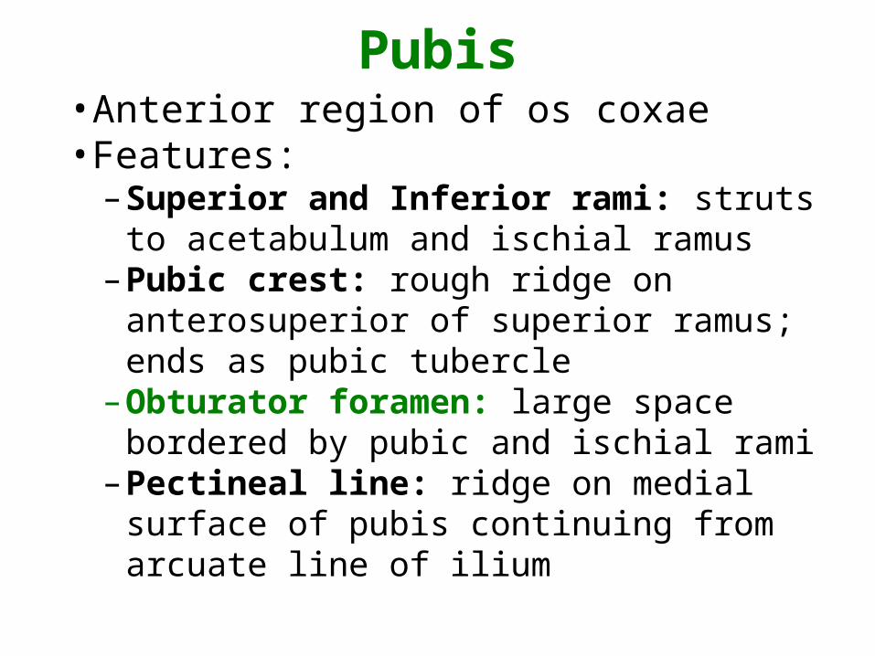
Pubis• Anterior region of os coxae• Features:
– Superior and Inferior rami: struts to acetabulum and ischial ramus
– Pubic crest: rough ridge on anterosuperior of superior ramus; ends as pubic tubercle
– Obturator foramen: large space bordered by pubic and ischial rami
– Pectineal line: ridge on medial surface of pubis continuing from arcuate line of ilium

Pubis
Figure 8.7

True vs. False Pelvis
• True pelvis: bony basin inferior to pelvic brim containing pelvic organs
• False pelvis: superior to pelvic brim bound by ilia laterally and abdominal wall anteriorly– Pelvic brim: continuous oval ridge formed by pubic
crest, pectineal line, arcuate line, and sacral promontory
• Pelvic inlet: superior entrance to true pelvis, at pelvic brim
• Pelvic outlet: exit of true pelvis, defined by coccyx, ischial tuberosities, and inferior border of pubic symphysis

Features of the Pelvis
Figure 8.10
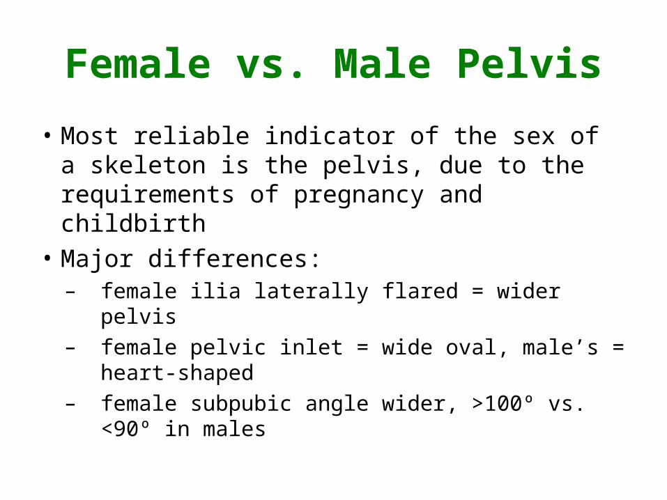
Female vs. Male Pelvis
• Most reliable indicator of the sex of a skeleton is the pelvis, due to the requirements of pregnancy and childbirth
• Major differences:– female ilia laterally flared = wider pelvis– female pelvic inlet = wide oval, male’s =
heart-shaped– female subpubic angle wider, >100º vs. <90º
in males

Female vs. Male PelvisCopyright © The McGraw-Hill Companies, Inc. Permission required for reproduction or display.
Anterior View
Features Female Characteristic Male Characteristic
Table 8.1 Sex Differences Between the Female and Male Pelves
Male PelvisFemale PelvisView
Medial View
General Appearance
General Width
Superior Inlet
Acetabulum
Greater Sciatic Notch
Ilium
Obturator Foramen
Subpubic Angle
Body of Pubis
Preauricular Sulcus
Sacrum
Coccyx
Tilt of Pelvis
Ischiopubic Ramus
Ischial Spine Rarely projects into pelvic outlet
Narrow and sharp
Frequently rotated inward, projects into pelvic outlet
Broad and fl at
Anterior tilt to superior end of pelvis
Narrow greatersciatic notch
Triangularpubic body
Large, ovalobturatorforamen
Narrow subpubic angle
Rectangularpubic body
Triangularobturatorforamen
Preauricularsulcus
Wide greatersciatic notch
Wide subpubic angle
More massive; more robust processes, more prominent musclemarkings
Less massive; gracile processes, less prominent musclemarkings
Hips are wider, more flared
Spacious, wide, and oval
Smaller
Wide and shallow
Shallow: Does not project far above sacroiliac joint
Smaller and triangular
Broader, more convex, usually greater than 100 degrees
Longer, more rectangular
Usually present
Shorter and wider; flatter sacral curvature
Posterior tilt
Hips are narrower and more vertically oriented, less flared
Heart-shaped
Larger
Narrow and U-shaped, deep
Deep: Projects farther above sacroiliac joint
Larger and oval
Narrow, V-shaped, usually less than 90 degrees
Shorter, triangular
Usually absent
Narrower and longer; more curved (greater sacral curvature)
Vertical
Superior end of pelvis relatively vertical
a-b: © David Hunt/ Smithsonian Institution; c-d: © L. Bassett/ Visuals Unlimited

Lower Limb• 30 bones per “leg”
– femur in the femoral region (thigh)
– patella (kneecap) in the patellar region
– tibia and fibula in the crural region (leg)
– 7 tarsals in ankle and proximal foot
– 5 metatarsals in sole of foot
– 14 phalanges in the toes

Femur• Longest, strongest, and heaviest
bone in the body
• Proximal features:– Head: articulates with os coxae at
acetabulum– Fovea: dent in head for ligament to
acetabulum– Neck: constricted region just distal to head– Greater and lesser trochanters: massive
processes for attachment of powerful hip and thigh muscles

Femur
Figure 8.11
Copyright © The McGraw-Hill Companies, Inc. Permission required for reproduction or display.
Greatertrochanter
Neck Head
Fovea
Greatertrochanter
Lesser trochanter
Intertrochanteric line
Head
Fovea
Neck
Shaft
(b) Right femoral head, medial view
Medialcondyle
(c) Right femur, inferior view
Lateralcondyle
Intercondylarfossa
Patellarsurface
Shaft
Medialcondyle
Medialepicondyle
Adductortubercle
Lateralepicondyle
Lateralepicondyle
Lateralcondyle
(a) Right femur, anterior view
Medialcondyle
Patellarsurface Patellar
surface
Lateralcondyle
Medialepicondyle
Adductortubercle
Head
Greatertrochanter
Neck
Intertrochantericcrest
Lesser trochanter
Shaft
a(right), b,c: © The McGraw-Hill Companies, Inc./Photo by Christine Eckel
Anterior View

Femur• Additional features:
– Intertrochanteric line: anterior between trochanters marking the distal edge of the hip capsule
– Gluteal tuberosity: posterior rough region for attachment of the gluteus maximus muscle
– Linea aspera: ridge on posterior shaft for attachment of many thigh muscles
– Distally, linea aspera splits into medial and lateral supracondylar lines

Femur
Figure 8.11
Posterior view

Femur• Distal features:
– Medial and lateral condyles: smooth, rounded articular surfaces
– Medial and lateral epicondyles: projections just superior to the condyles
– Intercondylar fossa: deep posterior depression that separates the condyles
– Patellar surface: smooth anterior region between condyles where patella articulates with the femur

Patella• The “kneecap”
– Triangular with broad superior border and inferiorly pointed apex
– Articulates with patellar surface of femur
Figure 8.12 Figure 8.13

Tibia and Fibula
• 2 bones in the leg–parallel to each
other
–tibia is medial to fibula
Figure 8.13
Copyright © The McGraw-Hill Companies, Inc. Permission required for reproduction or display.
Tibia
Medialmalleolus
Inferior articularsurface
(a) Right tibia and fibula, anterior view
Lateralmalleolus
Fibula
Articularfacet
Head
Neck
Medialmalleolus
Anterior border
Tibia Fibula
Shaft
Interosseousborders
Neck
Tibial tuberosity
Medial condyleLateral condyle
Superiortibiofibularjoint
Head
Lateralcondyle
Intercondylareminence
Medialcondyle
Lateralmalleolus
Inferior articular surface
Inferiortibiofibularjoint
a(right): © The McGraw-Hill Companies, Inc./Photo by Christine Eckel

Tibia• Medial bone in crural region
• Proximal features:– Medial and lateral condyles:
smooth surfaces for articulation with femur
– Fibular articular facet: articulation site for head of fibula under lateral condyle

Tibia- Posterior View
Figure 8.13
(e) Right knee joint, posterior view
Intercondylareminence
Intercondylarfossa
Lateralcondyles
FibulaTibia
Medialcondyles
Femur
© The McGraw-Hill Companies, Inc./Photo by Christine Eckel
Copyright © The McGraw-Hill Companies, Inc. Permission required for reproduction or display.
Copyright © The McGraw-Hill Companies, Inc. Permission required for reproduction or display.
(d) Right tibia and fibula, posterior view
Lateral malleolus
Medialmalleolus
Fibularnotch
Inferiortibiofibularjoint
Medialmalleolus
Lateral malleolus
Tibia Tibia
Intercondylareminence
Lateralcondyle
Medialcondyle
Superiortibiofibularjoint
Head
Neck
Fibula
Interosseousborders
Shaft
Medialcondyle
Intercondylareminence
Lateralcondyle
Fibular articularfacet
(right): © The McGraw-Hill Companies, Inc./Photo by Christine Eckel

Tibia• Other features:
– Tibial tuberosity: rough anterior projection inferior to condyles; can be palpated just inferior to the patella; for attachment of patellar ligament
– Tibial border: ridge along anterior surface extending from tuberosity distally; the “shin”
– Medial malleolus: inferiormost prominent medial process; “ankle bump”
– Articular surface: inferior surface articulates with the talus

Tibia- Posterior View
Figure 8.13
Copyright © The McGraw-Hill Companies, Inc. Permission required for reproduction or display.
(d) Right tibia and fibula, posterior view
Lateral malleolus
Medialmalleolus
Fibularnotch
Inferiortibiofibularjoint
Medialmalleolus
Lateral malleolus
Tibia Tibia
Intercondylareminence
Lateralcondyle
Medialcondyle
Superiortibiofibularjoint
Head
Neck
Fibula
Interosseousborders
Shaft
Medialcondyle
Intercondylareminence
Lateralcondyle
Fibular articularfacet
(right): © The McGraw-Hill Companies, Inc./Photo by Christine Eckel

Fibula• Long, thin, lateral crural bone
– Not weight-bearing
• Features:– proximal head with flat articular facet
for articulation with the tibia
– narrow neck and slender shaft
– distal end expands into lateral malleolus

Fibula
Figure 8.13
Copyright © The McGraw-Hill Companies, Inc. Permission required for reproduction or display.
(d) Right tibia and fibula, posterior view
Lateral malleolus
Medialmalleolus
Fibularnotch
Inferiortibiofibularjoint
Medialmalleolus
Lateral malleolus
Tibia Tibia
Intercondylareminence
Lateralcondyle
Medialcondyle
Superiortibiofibularjoint
Head
Neck
Fibula
Interosseousborders
Shaft
Medialcondyle
Intercondylareminence
Lateralcondyle
Fibular articularfacet
(right): © The McGraw-Hill Companies, Inc./Photo by Christine Eckel

Articulation of Head of Fibula with Tibia
Figure 8.13
Posterior View

Tarsus• 7 bones form ankle and
proximal foot–Calcaneus: largest; forms the heel
–Talus: superior-most; weight-bearing; articulates with tibia
–Navicular
–Cuneiforms: medial, intermediate and lateral
–Cuboid

Tarsals
Figure 8.14
Copyright © The McGraw-Hill Companies, Inc. Permission required for reproduction or display.
I
V
I
V
I
V
I
V
Distal phalanxof hallux
Proximal phalanxof hallux(great toe)
Distal phalanx
Middle phalanx
II IIIIV
II IIIIV
IIIIIIV
IVIII
II
(b) Right foot, inferior view
Calcaneus
Tarsals
Phalanges
Medial cuneiform
Navicular
Intermediatecuneiform
Talus
Tarsals
Metatarsals
Cuboid
Lateralcuneiform
Calcaneus
Lateral cuneiform
Cuboid
Talus
Navicular
Medialcuneiform
Intermediatecuneiform
(Sesamoid bonesfor flexor hallucisbrevis tendons)
Phalanges
(a) Right foot, superior view
Calcaneus
Distal phalanx
Middle phalanx
Proximal phalanx
Metatarsals
Distal phalanx
Middle phalanx
Proximalphalanx
Calcaneus
TalusTarsals
Talus Tarsals
Cuboid
Navicular
Medial cuneiform
Navicular
Intermediatecuneiform
Cuboid
Lateralcuneiform
Metatarsals
Distal phalanxof hallux
Phalanges
Proximal phalanx
Distal phalanx
Middle phalanx
Proximal phalanxPhalanges
Proximal phalanxof hallux(great toe)
Metatarsals
Intermediatecuneiform
Medialcuneiform
Lateral cuneiform
a(right), b(right): © The McGraw-Hill Companies, Inc./Photo by Christine Eckel

Metatarsals• 5 bones in sole of foot
• Articulations:
–proximally with tarsals
–distally with phalanges
• Identified by Roman numerals I–V from medial to lateral

Phalanges• 14 bones per foot
–3 phalanges per toes 2–5
•Proximal, middle, and distal
–Great toe (hallux) only 2
•Proximal and distal

Foot Bones
Figure 8.14
Copyright © The McGraw-Hill Companies, Inc. Permission required for reproduction or display.
I
V
I
V
I
V
I
V
Distal phalanxof hallux
Proximal phalanxof hallux(great toe)
Distal phalanx
Middle phalanx
II IIIIV
II IIIIV
IIIIIIV
IVIII
II
(b) Right foot, inferior view
Calcaneus
Tarsals
Phalanges
Medial cuneiform
Navicular
Intermediatecuneiform
Talus
Tarsals
Metatarsals
Cuboid
Lateralcuneiform
Calcaneus
Lateral cuneiform
Cuboid
Talus
Navicular
Medialcuneiform
Intermediatecuneiform
(Sesamoid bonesfor flexor hallucisbrevis tendons)
Phalanges
(a) Right foot, superior view
Calcaneus
Distal phalanx
Middle phalanx
Proximal phalanx
Metatarsals
Distal phalanx
Middle phalanx
Proximalphalanx
Calcaneus
TalusTarsals
Talus Tarsals
Cuboid
Navicular
Medial cuneiform
Navicular
Intermediatecuneiform
Cuboid
Lateralcuneiform
Metatarsals
Distal phalanxof hallux
Phalanges
Proximal phalanx
Distal phalanx
Middle phalanx
Proximal phalanxPhalanges
Proximal phalanxof hallux(great toe)
Metatarsals
Intermediatecuneiform
Medialcuneiform
Lateral cuneiform
a(right), b(right): © The McGraw-Hill Companies, Inc./Photo by Christine Eckel

Foot Arches
• To prevent pinching of muscles, nerves, and blood vessels feet do not rest flat on floor
• Three major arches:– Medial: from heel to hallux; highest arch– Lateral: from heel to 5th toe; lowest arch– Transverse: perpendicular to other
arches; along distal row of tarsals

Foot Arches
Figure 8.15