chapter 9 – ears, nose, throat and mouth · pdf filepediatric clinical practice...
TRANSCRIPT
Pediatric Clinical Practice Guidelines for Nurses in Primary Care 2010
CHAPTER 9 – EARS, NOSE, THROAT AND MOUTH
First Nations and Inuit Health Branch (FNIHB) Pediatric Clinical Practice Guidelines for Nurses in Primary Care. The section on Pharyngotonsillitis, Bacterial has been updated as of August 2016. The remaining content of this chapter was reviewed in December 2009.
Table of contents
ASSESSMENT OF THE EARS, NOSE, THROAT AND MOUTH .............................9–1
History of Present Illness and Review of Systems .............................................9–1
Physical Examination .........................................................................................9–2
COMMON PROBLEMS OF THE EARS, NOSE AND THROAT ...............................9–3
Ceruminosis (Impacted Cerumen) .....................................................................9–3
Foreign Body in the Nose ..................................................................................9–3
Otitis Externa .....................................................................................................9–4
Otitis Media, Acute (AOM) .................................................................................9–4
Otitis Media, Chronic Suppurative .....................................................................9–7
Otitis Media, Serous (Otitis Media with Effusion) ...............................................9–7
Pharyngotonsillitis ..............................................................................................9–8
Pharyngotonsillitis, Bacterial ..............................................................................9–9
Pharyngotonsillitis, Viral ...................................................................................9–17
Rhinitis .............................................................................................................9–18
Rhinosinusitis ...................................................................................................9–20
COMMON PROBLEMS OF THE MOUTH .............................................................9–21
Absence of Teeth, Congenital (Anodontia).......................................................9–21
Absence of Teeth, Partial (Oligodontia or “congenitally missing teeth”) ...........9–21
Ankyloglossia (Tongue-Tie) ..............................................................................9–21
Common Malocclusions ...................................................................................9–21
Dental Abscess – Permanent Tooth .................................................................9–21
Dental Abscess – Primary Tooth ......................................................................9–22
Dental Decay ...................................................................................................9–23
Early Childhood Dental Decay .....................................................................9–23
Childhood and Adolescent Dental Decay .....................................................9–24
Prevention of Dental Decay ...........................................................................................................................9–24
Discoloured (non-vital) Permanent Tooth .........................................................9–26
Discoloured (non-vital) Anterior Primary Tooth .................................................9–26
Epstein Pearls ..................................................................................................9–27
Pediatric Clinical Practice Guidelines for Nurses in Primary Care2010
Ears, Nose, Throat and Mouth
Eruption Cyst ...................................................................................................9–27
Impacted Tooth .................................................................................................9–27
Intruded Tooth ..................................................................................................9–27
Migratory Glossitis (Geographic Tongue) .........................................................9–28
Neonatal Teeth .................................................................................................9–28
Normal Tooth Development ..............................................................................9–28
Stomatitis .........................................................................................................9–29
Toothache ........................................................................................................9–32
Thumb-sucking ................................................................................................9–32
EMERGENCY PROBLEMS OF THE NOSE, THROAT AND MOUTH ...................9–33
Avulsed Tooth ...................................................................................................9–33
Epistaxis ...........................................................................................................9–33
Fractured Tooth ...............................................................................................9–35
Mastoiditis ........................................................................................................9–35
Oral Trauma .....................................................................................................9–36
Peritonsillar Abscess ........................................................................................9–36
Retropharyngeal Abscess ................................................................................9–37
SOURCES ..............................................................................................................9–39
Pediatric Clinical Practice Guidelines for Nurses in Primary Care 2010
Ears, Nose, Throat and Mouth 9–1
HISTORY OF PRESENT ILLNESS AND REVIEW OF SYSTEMS
The following characteristics of each symptom should be elicited and explored:
– Onset (sudden or gradual)
– Chronology
– Current situation (improving or deteriorating)
– Location
– Radiation
– Quality
– Timing (frequency, duration)
– Severity
– Precipitating and aggravating factors
– Relieving factors
– Associated symptoms
– Effects on daily activities
– Previous diagnosis of similar episodes
– Previous treatments
– Efficacy of previous treatments
CARDINAL SYMPTOMS
Characteristics of specific symptoms should be elicited, as follows.
Ears
– Recent changes in hearing
– Itching
– Earache
– Discharge
– Tinnitus
– Vertigo
– Ear trauma, including Q-tip use
– Pain
Nose
– Nasal discharge or postnasal drip
– Epistaxis
– Obstruction of airflow
– Sinus pain, pressure
– Itching
– Nasal trauma
Mouth and Throat
– Dental status
– Pain
– Oral lesions
– Bleeding gums
– Sore throat
– Dysphagia (difficulty swallowing)
– Hoarseness or recent voice change
Neck
– Pain
– Swelling
– Enlargement of glands
Other Associated Symptoms
– Fever
– Malaise
– Nausea and vomiting
PAST MEDICAL HISTORY (SPECIFIC TO ENT)
– Seasonal allergies, allergies
– Frequent ear or throat infections
– Rhinosinusitis
– Trauma to head or ENT area
– ENT surgery
– Audiometric screening results indicating hearing loss
– Prescription or over-the-counter medications used regularly
ASSESSMENT OF THE EARS, NOSE, THROAT AND MOUTH
For more information on the history and physical examination of the ears, nose and throat in older children and adolescents see the chapter, “Ears, Nose and Throat” in the adult clinical guidelines.

Pediatric Clinical Practice Guidelines for Nurses in Primary Care2010
Ears, Nose, Throat and Mouth9–2
FAMILY HISTORY (SPECIFIC TO ENT)
– Others at home with similar symptoms
– Seasonal allergies
– Asthma
– Hearing loss
PERSONAL AND SOCIAL HISTORY (SPECIFIC TO ENT)
– Feeding methods (breast or bottle), bottle propping
– Frequent exposure to water (swimmer’s ear)
– Use of foreign object to clean ear
– Insertion of foreign body in ear
– Crowded living conditions
– Poor personal hygiene
– Dental hygiene habits
– Exposure to cigarette smoke, wood smoke or other respiratory toxins
– Recent air travel
REVIEW OF SYSTEMS
Obtain a history about other relevant systems for the presenting concern. This may include information about the eyes, central nervous system, gastrointestinal system and/or respiratory system.
PHYSICAL EXAMINATION
– Apparent state of health (for example, appearance of acute illness)
– Hydration status
– Degree of comfort or distress
– Colour (flushed or pale)
– Character of cry (in infants < 6 months old)
– Activity level (spontaneous activity or lethargy)
– Mental status (whether alert and active)
– Degree of cooperation, consolability
– Emotional reaction to parent (or caregiver) and examiner
– Hygiene
– Posture
– Difficulty with gait or balance
– Nutritional status (obese or emaciated)
SAFETY TIP
For examination, it may be necessary to restrain a struggling child. For example, lay the child in a supine position and have the parent or caregiver hold the child’s arms extended, in a position close to the sides of the head. This will limit side-to-side movements while you are examining ENT structures. Brace the otoscope, and guard against sudden head movements.
EARS
Inspection
– External ear: position (in relation to eyes) – low-set or small, deformed auricles may indicate associated congenital defects, especially renal agenesis
– Pinna: lesions, abnormal appearance or position
– Canal: discharge, swelling, redness, odour, wax, foreign bodies
– Eardrum: colour, light reflex, landmarks, bulging or retraction, perforation, scarring, air bubbles, fluid level
– Check mobility of the eardrum using a pneumatic otoscope (if available); decrease may indicate acute otitis media (see “Guidelines for Pneumatic Otoscopy”).
– Estimate hearing by producing a loud noise (for example, by clapping hands) for an infant or young child (which should elicit a blink response) or by performing a watch or whisper test for an older child.
Perform tympanometry (if equipment available).
Clinical tip: For the best view of the eardrum in an infant or a child < 6 years old, pull the outer ear downward, outward and backward.
Palpation
– Tenderness over tragus or mastoid process
– Tenderness on manipulation of the pinna
NOSE
Inspection
– External: inflammation, deformity, discharge, bleeding
– Internal: colour of mucosa, edema, deviated septum, polyps, bleeding points
– Transilluminate sinuses to check for dulling of light reflex in children > 6 years
– Nasal vs. mouth breathing
Pediatric Clinical Practice Guidelines for Nurses in Primary Care 2010
Ears, Nose, Throat and Mouth 9–3
Palpation
– Check for sinus and nasal tenderness (only in older children who can cooperate and provide a response)
Percussion
– Check for sinus and nasal tenderness (only in older children who can cooperate and provide a response)
MOUTH AND THROAT
Inspection
– Lips: colour uniformity (light to dark pink), lesions, symmetry of lips
– Oral mucosa and tongue: breath odour, colour, lesions of buccal mucosa, palate, tongue
– Gums: redness, swelling, caries
– Teeth: caries, fractures
– Throat: colour, tonsillar enlargement, exudates, uvula midline
NECK
Inspection
– Symmetry
– Swelling
– Masses
– Redness
– Enlargement of thyroid
– Active range of motion
Palpation
– Tenderness, enlargement, mobility, contour and consistency of masses
– Thyroid: size, consistency, contour, position, tenderness
LYMPH NODES OF THE HEAD AND NECK
Palpation
Tenderness, enlargement, mobility, contour and consistency of nodes.
– Pre- and post-auricular nodes
– Anterior and posterior cervical nodes
– Tonsillar
– Submaxillary
– Submandibular
– Occipital
COMMON PROBLEMS OF THE EARS, NOSE AND THROAT
CERUMINOSIS (IMPACTED CERUMEN)
The diagnosis and management of ceruminosis in children is the same as in adults (see “Ceruminosis” in the adult clinical guidelines).
FOREIGN BODY IN THE NOSE
Children frequently put foreign bodies in their nostrils. Occasionally, the foreign body (anything from a pea to a small bead or toy part) obstructs the airway or becomes embedded, possibly causing significant infection.
HISTORY
– Generally unilateral
– History of purulent rhinorrhea and difficulty with breathing through the affected nostril
– Typically, the parent or caregiver relates that a very foul smell is emanating from the child
– Fever and other systemic features uncommon
PHYSICAL FINDINGS
– Obvious mucopurulent discharge, generally unilateral
– Nasal blockage may be so severe that adequate visualization of the foreign body is impossible
– Suction may be necessary to visualize the foreign body
It is important to explore the opposite nostril and ears for other foreign bodies.

Pediatric Clinical Practice Guidelines for Nurses in Primary Care2010
Ears, Nose, Throat and Mouth9–4
DIFFERENTIAL DIAGNOSIS
– Sinusitis
– Rhinitis
COMPLICATIONS
– Sinus infection
– Epistaxis
– Other ENT infections
DIAGNOSTIC TESTS
– None
MANAGEMENT
Goals of Treatment
– Remove foreign body
– Prevent recurrence
Nonpharmacologic Interventions1,2
One must be cautious to not displace the foreign body posteriorly or into the airway.
It is not recommended to attempt removal of a foreign body beyond that dictated by common sense. The child will become increasingly frightened and the procedure increasingly difficult.
Attempt to remove clearly visible foreign bodies and do not attempt to remove foreign bodies that cannot be seen. Visible foreign bodies can be removed by:
– Using a suction catheter
– Using a cerumen loop (curette)
– Using a nasal speculum and forceps, ask the child to exhale forcibly through the nostril containing the foreign body while the opposite nostril is occluded. This technique may be difficult for the very young patient.
– Providing oral positive pressure. Have the child sit or stand, depending upon their preference. Occlude the unaffected side of the nose and instruct the parent to firmly seal their mouth over the child’s mouth and give a short, sharp puff of air into the child’s mouth. This technique has the advantage that it does not require physical restraint.
If a foreign body is embedded with granulation tissue, consultation with an ENT specialist and removal under general anesthesia may be necessary.
Educate the parents or caregiver about the problems associated with foreign bodies, particularly the risk of aspiration and the need to remove foreign bodies under general anesthetic.
OTITIS EXTERNA
For otitis externa, the clinical presentation and management are the same in adults and children.
OTITIS MEDIA, ACUTE (AOM)
Acute suppurative infection of the middle ear, often preceded by a viral upper respiratory tract infection.
CAUSES
Often, AOM is of mixed pathogenesis, virus and bacteria.3
Viral Organisms
– Respiratory syncytial virus
– Influenza A virus
– Coxsackievirus
– Adenovirus
– Parainfluenza virus
Common Bacterial Organisms
This is most common in bilateral AOM.
– Streptococcus pneumoniae
– Hemophilus influenzae
– Moraxella catarrhalis
Less Common Organisms
– Mycoplasma
– Chlamydia
Other Miscellaneous Causes
– Immunoreactivity
– Allergic rhinitis
Pediatric Clinical Practice Guidelines for Nurses in Primary Care 2010
Ears, Nose, Throat and Mouth 9–5
Risk Factors3
Occurs more frequently in the following groups and situations:
– Children with cleft palate, allergic rhinitis, Down’s syndrome or any change in anatomy of the skull and eustachian tube
– Daycare attendance
– Children of Aboriginal origin
– Possibly bottle-fed children, if the child is propped up for feeding or goes to sleep with a bottle of milk at night
– Children who use pacifiers
– Children 6–18 months of age; peaks again at school entry age to 7 years of age
– During fall and winter months
– Children who are not breastfed for at least 3 months
– Children exposed to cigarette smoke
– Family history of acute otitis media
– Male gender
HISTORY
– Otalgia (pain is absent in 20% of children)
– Fever
– Cold and cough symptoms
– Irritability (in infants)
– Hearing loss
– Diffuse mild peri-umbilical pain
– Vomiting or diarrhea may be present
– Nonspecific sensation of tugging at ears
– Restless sleep
PHYSICAL FINDINGS
– Fever may be present
– May appear acutely ill
– Conjunctivitis may also be present (this is more common when child is < 2 years of age)
Inspection of the tympanic membrane is the key to diagnosis:
– Light reflex and bony landmarks usually disappear in acute otitis media
– Tympanic membrane appears dull, red and bulging in acute otitis media
– Reduction in or lack of movement of the tympanic membrane on pneumatic otoscopy (see description below)
For a diagnosis of AOM, the tympanic membrane must be both red and be bulging or have acute inflammation present with decreased tympanic membrane movement (as demonstrated by pneumatic otoscopy).
Wax and other debris should be removed from the ear canal to allow a clear view of the tympanic membrane.
Redness of the tympanic membrane in the absence of other signs may be due to crying, agitation, a common cold, aggressive examination or manipulation of the external ear canal, or serous otitis media with effusion (see “Serous Otitis Media [Otitis Media with Effusion]”).
Guidelines for Pneumatic Otoscopy
Anyone can learn pneumatic otoscopy, but practice is needed. This method consists of applying air pressure to the tympanic membrane and watching the resultant movement.
– Tools: a battery-operated, bright light with a well-charged battery and a hermetically sealed otoscope with pneumatic attachment
– Client must remain still during the examination (it may be necessary to restrain a child)
– Apply positive pressure (by squeezing a full bulb) and negative pressure (by releasing the bulb), and observe any movement of the eardrum
– Lack of movement implies the presence of fluid in the middle ear or chronic stiffness of the tympanic membrane
DIFFERENTIAL DIAGNOSIS
– Acute otitis externa
– Pharyngitis or tonsillitis
– Noninfectious middle ear effusion
– Trauma to or foreign body in ear canal
– Referred pain from dental abscess
– Mastoiditis (rare)
– Eustachian tube disorders
COMPLICATIONS
– Perforated tympanic membrane
– Hearing loss leading to speech impairment and cognitive impairment
– Serous otitis media
– Meningitis
– Mastoiditis (rare)

Pediatric Clinical Practice Guidelines for Nurses in Primary Care2010
Ears, Nose, Throat and Mouth9–6
DIAGNOSTIC TESTS
– If ear is draining, swab for culture and sensitivity
– Most cases are caused by the most common organisms
MANAGEMENT
Goals of Treatment
– Control pain and fever
– Relieve infection
– Prevent complications
– Avoid antibiotic resistance
Appropriate Consultation
Usually not necessary if condition is uncomplicated.
Nonpharmacologic Interventions
Client Education
– Recommend increased rest in the acute febrile phase
– Counsel parents or caregiver about appropriate use of medications (dosage, compliance, follow-up)
– Explain disease course and expected outcome
– Recommend avoidance of flying until symptoms have resolved
Pharmacologic Interventions
Antipyretic and analgesic for fever and pain:
acetaminophen (Tylenol), 10–15 mg/kg/dose PO q4–6h prn
It appears prudent to consider all cases of AOM candidates for antimicrobial therapy in order to minimize the likelihood of complications. However, some experts recommend watchful waiting for 48–72 hours before initiating antibiotic therapy for children aged 2 and above presenting with no risk factors.4 This approach may be feasible in mildly unwell children over 2 years of age if good follow-up can be assured and the child does not have any of the following risk factors:
– Recent antibiotic use
– Daycare attendance
– Recent episode of AOM
– Treatment failure or early recurrence
Antibiotic therapy, first-line drug:
amoxicillin (Amoxil), 80–90 mg/kg/day, divided bid or tid, PO for 5–7 days
For children < 2 years old or with a perforated ear drum, treat for 10 days with amoxicillin.
For penicillin/beta-lactam allergy or known beta-lactamase resistance in the community:
azithromycin 10 mg/kg/day first day then 5 mg/kg/day PO for the remaining 4 days
Consider second-line antibiotic therapy under the following conditions:
– Penicillin allergy
– Acute otitis media unresponsive in 48–72 hours to a trial of amoxicillin and accompanied by persistent fever, irritability or pain
– Early recurrence of otitis media (< 2 months after initial bout), which is often due to bacteria that produce ß-lactamase and are thus resistant to amoxicillin, pneumococci with reduced susceptibility to penicillins or cephalosporin, or organisms resistant to sulfamethoxazole-trimethoprim
– Immunocompromised patients (for example, leukemia)
– Infection in infants < 2 months old
Second-line choices:
amoxicillin/clavulanic acid (Clavulin), 40 mg/kg/day of the amoxicillin component, divided bid-tid
Because clavulanic acid commonly causes diarrhea, if high dose amoxicillin is to be given with clavulanic acid, dosage is better given as two prescriptions: one for regular amoxicillin and one for amoxicillin/clavulanic acid (Clavulin).
cefuroxime axetil (Ceftin), 30 mg/kg/day divided bid for 10 days
Drug choice should be based on efficacy, cost and acceptability to the child.
Antihistamines and decongestants have no proven efficacy in the treatment of acute otitis media and should be avoided. For children under 6 years, there is no evidence that cough and cold medicines are of benefit and are not to be administered.
Pediatric Clinical Practice Guidelines for Nurses in Primary Care 2010
Ears, Nose, Throat and Mouth 9–7
Monitoring and Follow-Up
Instruct parents or caregiver to bring the child back to the clinic in 3 days if symptoms do not diminish or if symptoms progress despite therapy.
Otherwise, follow up in 14 days:
– If ear is normal, do not give any treatment
– If ear is still dull but asymptomatic (no pain or hearing loss), follow up again in 6 weeks
– If condition is unresolved, consider treatment with a second-line antibiotic or consult
– Assess hearing 1 month after treatment is complete or when effusion is no longer present
– If fluid remains present beyond 6 weeks, consult a physician
In 70% to 80% of patients, effusion persists after 2 weeks, and 10% still have effusion at 3 months and may exhibit conductive loss of hearing (see “Serous Otitis Media [Otitis Media with Effusion]”).
Referral
Not necessary if condition is uncomplicated. Refer to a physician if effusion persists beyond 3 months.
OTITIS MEDIA, RECURRENT ACUTE4
Recurrence of this condition is very common in children. Recurrent otitis media is defined as 3 or more episodes of acute otitis media over the preceding 6 months, or 4 or more episodes in the last year.
– If infection recurs less than 2 months after the previous infection, use one of the second-line antibiotics
– If infection recurs more than 2 months after the previous infection, treat as acute otitis media with amoxicillin (Amoxil)
Monitoring and Follow-Up
– Assess compliance with medication for treatment of acute episode and for prophylaxis
– Observe closely for acute recurrent episodes
– Assess hearing periodically
– Some physicians may choose to use prophylaxis antibiotics for recurrent OM
Referral
Refer to a physician any child with: otitis media with an effusion for > 3 months with bilateral hearing loss; a retracted tympanic membrane; cleft palate or craniofacial malformations; multiple episodes of acute otitis media (more than 4 episodes in a single year; more than 3 episodes in 6 months).
An ears, nose and throat (ENT) consultation is advisable. Myringotomy with insertion of T-tubes (plus adenoidectomy) may be indicated.
OTITIS MEDIA, CHRONIC SUPPURATIVE
Persistent (longer than 6 weeks) or recurrent purulent drainage through a perforated tympanic membrane and persistent inflammation in the middle ear or mastoid cavity.5
The diagnosis and management of chronic otitis media in children is the same as in adults (see “Otitis Media, Chronic Suppurative” in the adult clinical guidelines).
Referral6
Refer to a physician any child with: otitis media with an effusion for > 3 months with bilateral hearing loss; a retracted tympanic membrane; cleft palate or craniofacial malformations; multiple episodes of acute otitis media (more than 4 episodes in a single year; more than 3 episodes in 6 months).
An ears, nose and throat (ENT) consultation is advisable. Myringotomy with insertion of T-tubes (plus adenoidectomy) may be indicated.
OTITIS MEDIA, SEROUS (OTITIS MEDIA WITH EFFUSION)
An accumulation of serous fluid in the middle ear, with no signs or symptoms of acute infection. This is common after acute otitis media.
CAUSES
– Unclear
– Bacteria are isolated from a significant proportion of middle-ear aspirates
Pediatric Clinical Practice Guidelines for Nurses in Primary Care2010
Ears, Nose, Throat and Mouth9–8
HISTORY
– Previous asymptomatic otitis media – Feeling of fullness in the ear – Tinnitus (uncommon) – Hearing reduced (as indicated by hearing
examination)
PHYSICAL FINDINGS
– Tympanic membrane dull, translucent or bulging; landmarks diminished or absent
– Reduction of mobility of tympanic membrane, indicated by pneumatic otoscopy (for description of technique, see “Otitis Media, Acute”)
DIFFERENTIAL DIAGNOSIS
– Acute otitis media – Dysfunction of eustachian tube
COMPLICATIONS
– Secondary infection – Chronic serous otitis media – Hearing loss
Complicating factors, such as nasal allergy, submucous clefts and nasopharyngeal tumors, must be excluded.
DIAGNOSTIC TESTS
– Tympanography (if available) may support the diagnosis of effusion
MANAGEMENT
Goals of Treatment
– Prevent hearing loss
Nonpharmacologic Interventions
– Observation for 2–3 months is appropriate – Ensure appropriate seating at school (for example,
close to front of classroom) – Encourage compliance and routine follow-up – Encourage parents or caregiver to speak clearly and
directly to child – Measure hearing by audiology if effusion persists
at 2–3 months after acute otitis media
Pharmacologic Interventions
– None
Antihistamines, decongestants and steroids have no proven efficacy.
Monitoring and Follow-Up
– Check ears and hearing every 2 weeks
– In a young child, follow for language development while effusion persists with a speech language pathologist
Referral
Refer to a physician if the effusion persists for more than 3 months, hearing loss is suspected, or retraction of the tympanic membrane is present. An ENT consultation regarding surgical management may be indicated.
General indications for myringotomy and T-tubes:
– Persistent effusion for more than 3 months, with associated hearing loss
– Recurrent middle ear infections (6 per year or 4 in 6 months)
– Retraction of the eardrum
– Possibly, poor language development
PHARYNGOTONSILLITIS
A painful condition of the oropharynx associated with infection and inflammation of the mucous membranes of the pharynx and palatine tonsils. The condition may be caused by a bacterium or virus, and it may be difficult to differentiate between these two forms clinically. Viral infections are the most common cause of pharyngotonsillitis in younger children; bacterial pharyngotonsillitis is very rare in children < 3 years old, but its prevalence increases with age.
Pharyngitis may also be caused by non-infectious causes such as:
– Allergic rhinitis
– Sinusitis with postnasal drip
– Mouth breathing
– Trauma
– Gastroesophageal reflux disease
The next two sections describe bacterial and viral pharyngotonsillitis in detail.
Pediatric Clinical Practice Guidelines for Nurses in Primary Care
Ears, Nose, Throat and Mouth 9–9
2016
PHARYNGOTONSILLITIS, BACTERIAL
OVERVIEW
Please refer to provincial/territorial guidelines for bacterial pharyngotonsillitis where available.
Pharyngotonsillitis is a painful condition of the oropharynx associated with infection and inflammation of the mucous membranes of the pharynx and palatine tonsils. The condition may be caused by a bacterium or virus, and a clinician cannot definitively differentiate between these two forms clinically. Viral infections are the most common cause of pharyngotonsillitis in younger children; bacterial pharyngotonsillitis is rare in children less than 3 years old, but its prevalence increases with age1.
CAUSES
– Group A Streptococci (GAS) is the most common cause of bacterial pharyngitis, accounting for 20% to 30% of cases of acute pharyngotonsillitis in children2
– Group C and Group G Streptococci2
– Anaerobic organisms of the mouth (including Arcanobacterium)2
– Neisseria gonorrhoeae for those engaging in oral sex2,3
– Mycoplasma pneumoniae (M. pneumoniae)2
– Diphtheria for those with inadequate diphtheria immunization2
Incubation, transmission and communicability vary depending on the cause of bacterial pharyngitis. The following sections focus on the transmission, incubation and communicability of GAS pharyngitis.
TRANSMISSION
– Person-to-person spread by respiratory droplets is the most common method of transmission of GAS pharyngitis4.
– Direct contact with infected individuals or carriers4.
– Foodborne outbreaks of GAS pharyngitis occur rarely and are a consequence of human contamination of food by infected or colonized food handlers in conjunction with improper food preparation or refrigeration procedures5.
INCUBATION PERIOD
– The incubation period for GAS pharyngitis is one to three days after exposure6.
COMMUNICABILITY
– If untreated, a client with GAS pharyngitis is usually infectious during the acute phase of the illness, typically 7 to 10 days, and much less infectious one week after the acute phase. If antibiotics are used, the infectious period is reduced to 24 hours4.
– The bacterium can remain in the body in its carrier state without causing illness in the host for weeks or months and is transmissible in this state4.
ASSESSMENT
Medication Review: Review current medications including over-the-counter, complementary and alternative medicines, as well as chemical or substance intake which may impact management. Allergy History: Screen for medication, latex, environmental or other allergies including identifying approximately when and what type of reaction occurred.
RISK FACTORS
– Age. GAS pharyngitis occurs predominantly in school-age children 5 to 15 years of age (although it can occur in both younger and older individuals)2
– Overcrowding7
– Previous episodes of GAS pharyngitis2
– A history of GAS pharyngitis in the household, community, neighborhood, or school8
HISTORY OF PRESENT ILLNESS
The general history for bacterial pharyngotonsillitis may vary depending on the bacterial etiology. The optimal approach for differentiating among various causes of pharyngitis requires a problem-focused history, a physical examination and appropriate lab testing9.
– There is a broad overlap between the signs and symptoms of GAS and non-GAS pharyngitis2.
Pediatric Clinical Practice Guidelines for Nurses in Primary Care
Ears, Nose, Throat and Mouth9–10
2016
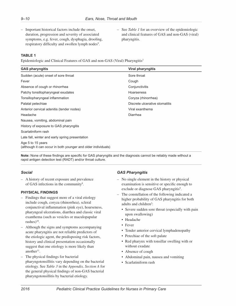
– Important historical factors include the onset, duration, progression and severity of associated symptoms, e.g. fever, cough, dysphagia, drooling, respiratory difficulty and swollen lymph nodes9.
– See Table 1 for an overview of the epidemiologic and clinical features of GAS and non-GAS (viral) pharyngitis.
TABLE 1
Epidemiologic and Clinical Features of GAS and non-GAS (Viral) Pharyngitis2
GAS pharyngitis Viral pharyngitis
Sudden (acute) onset of sore throat
Fever
Absence of cough or rhinorrhea
Patchy tonsillopharyngeal exudates
Tonsillopharyngeal inflammation
Palatal petechiae
Anterior cervical adenitis (tender nodes)
Headache
Nausea, vomiting, abdominal pain
History of exposure to GAS pharyngitis
Scarlatiniform rash
Late fall, winter and early spring presentation
Age 5 to 15 years (although it can occur in both younger and older individuals)
Sore throat
Cough
Conjunctivitis
Hoarseness
Coryza (rhinorrhea)
Discrete ulcerative stomatitis
Viral exanthema
Diarrhea
Note: None of these findings are specific for GAS pharyngitis and the diagnosis cannot be reliably made without a rapid antigen detection test (RADT) and/or throat culture.
Social
– A history of recent exposure and prevalence of GAS infections in the community8.
PHYSICAL FINDINGS
– Findings that suggest more of a viral etiology include cough, coryza (rhinorrhea), scleral conjunctival inflammation (pink eye), hoarseness, pharyngeal ulcerations, diarrhea and classic viral exanthema (such as vesicles or maculopapular rashes)10.
– Although the signs and symptoms accompanying acute pharyngitis are not reliable predictors of the etiologic agent, the predisposing risk factors, history and clinical presentation occasionally suggest that one etiology is more likely than another11.
– The physical findings for bacterial pharyngotonsillitis vary depending on the bacterial etiology. See Table 3 in the Appendix, Section A for the general physical findings of non-GAS bacterial pharyngotonsillitis by bacterial etiology.
GAS Pharyngitis
– No single element in the history or physical examination is sensitive or specific enough to exclude or diagnose GAS pharyngitis9.
– The constellation of the following indicated a higher probability of GAS pharyngitis for both adults and children2:
• Severe sudden sore throat (especially with pain upon swallowing)
• Headache
• Fever
• Tender anterior cervical lymphadenopathy
• Petechiae of the soft palate
• Red pharynx with tonsillar swelling with or without exudate
• Absence of cough
• Abdominal pain, nausea and vomiting
• Scarlatiniform rash
Pediatric Clinical Practice Guidelines for Nurses in Primary Care
Ears, Nose, Throat and Mouth 9–11
2016
DIFFERENTIAL DIAGNOSIS
Consult physician/nurse practitioner when practice is outside legislated scope and without authorized delegation.
– Viral pharyngotonsillitis (common cold, influenza, enterorvirus, adenovirus and infectious mononucleosis)2
– Epiglottitis. Note: Consult physician/nurse practitioner immediately if epiglottitis is suspected. See Epiglottitis in adult Chapter 10, Respiratory System
– Gonococcal pharyngitis in sexually-active individuals2,3
– Diphtheria2
– Non-infectious causes of pharyngitis, e.g. gastroesophageal reflux, postnasal drip, thyroiditis, allergies, foreign body9
COMPLICATIONS
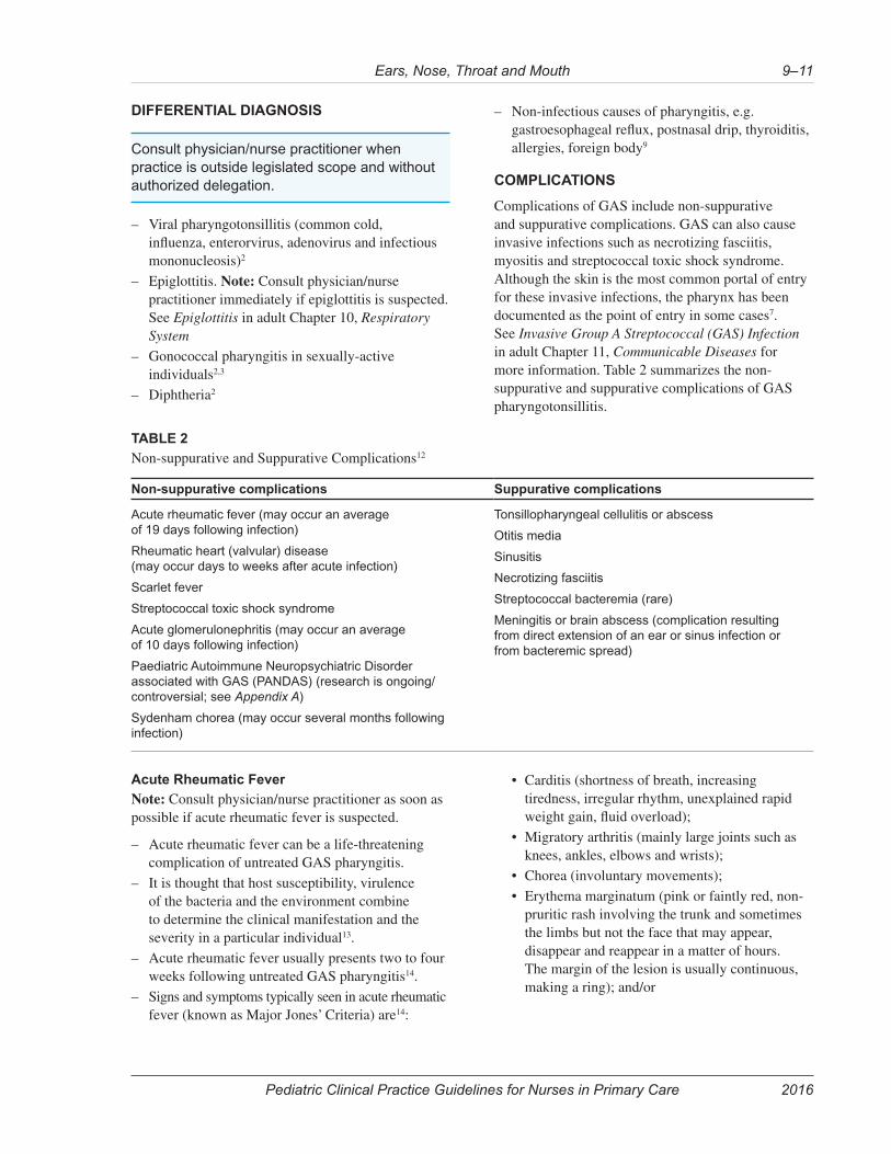
Complications of GAS include non-suppurative and suppurative complications. GAS can also cause invasive infections such as necrotizing fasciitis, myositis and streptococcal toxic shock syndrome. Although the skin is the most common portal of entry for these invasive infections, the pharynx has been documented as the point of entry in some cases7. See Invasive Group A Streptococcal (GAS) Infection in adult Chapter 11, Communicable Diseases for more information. Table 2 summarizes the non-suppurative and suppurative complications of GAS pharyngotonsillitis.
TABLE 2
Non-suppurative and Suppurative Complications12
Non-suppurative complications Suppurative complications
Acute rheumatic fever (may occur an average of 19 days following infection)
Rheumatic heart (valvular) disease (may occur days to weeks after acute infection)
Scarlet fever
Streptococcal toxic shock syndrome
Acute glomerulonephritis (may occur an average of 10 days following infection)
Paediatric Autoimmune Neuropsychiatric Disorder associated with GAS (PANDAS) (research is ongoing/controversial; see Appendix A)
Sydenham chorea (may occur several months following infection)
Tonsillopharyngeal cellulitis or abscess
Otitis media
Sinusitis
Necrotizing fasciitis
Streptococcal bacteremia (rare)
Meningitis or brain abscess (complication resulting from direct extension of an ear or sinus infection or from bacteremic spread)
Acute Rheumatic Fever
Note: Consult physician/nurse practitioner as soon as possible if acute rheumatic fever is suspected.
– Acute rheumatic fever can be a life-threatening complication of untreated GAS pharyngitis.
– It is thought that host susceptibility, virulence of the bacteria and the environment combine to determine the clinical manifestation and the severity in a particular individual13.
– Acute rheumatic fever usually presents two to four weeks following untreated GAS pharyngitis14.
– Signs and symptoms typically seen in acute rheumatic fever (known as Major Jones’ Criteria) are14:
• Carditis (shortness of breath, increasing tiredness, irregular rhythm, unexplained rapid weight gain, fluid overload);
• Migratory arthritis (mainly large joints such as knees, ankles, elbows and wrists);
• Chorea (involuntary movements);
• Erythema marginatum (pink or faintly red, non-pruritic rash involving the trunk and sometimes the limbs but not the face that may appear, disappear and reappear in a matter of hours. The margin of the lesion is usually continuous, making a ring); and/or
Pediatric Clinical Practice Guidelines for Nurses in Primary Care
Ears, Nose, Throat and Mouth9–12
2016
• Erythema nodosum (small, round, firm, non-inflammatory, sometimes painless subcutaneous lesions. The lesions may range from a few millimeters to 2 cm in size and are usually located over a bony surface or prominence or near tendons).
– For more information, see Rheumatic Fever (Carditis) in Chapter 11, Cardiovascular System.
– There are three circumstances when the nurse should be highly suspicious of acute rheumatic fever and in which immediate consultation with a physician/nurse practitioner is required14:
• Chorea is the only manifestation
• Carditis of insidious onset and slow progression is the only manifestation in clients who present months after an acute GAS pharyngitis infection
• Recurrent rheumatic fever in clients with a history of rheumatic fever or rheumatic heart disease
Note: Strict adherence to the Jones’ criteria in areas of high prevalence may result in under-diagnosis14.
High-risk Clients
Those at increased risk for acute rheumatic fever include10:
– Past history or concurrent diagnosis of acute rheumatic fever, especially with carditis or valvular disease.
– Household contact with someone having a history of rheumatic fever.
DIAGNOSTIC TESTS
Consult physician/nurse practitioner when practice is outside legislated scope and without authorized delegation.
– Diagnostic testing is based on client history, risk factors, physical examination findings and test availability. Testing should be carried out as per provincial/territorial policies and procedures.
– Laboratory diagnosis of GAS pharyngitis is important because it is most often impossible to distinguish clinically between bacterial and viral pharyngitis.
Laboratory
– Rapid Antigen Detection Test (RADT) (if available). A positive RADT is considered definitive for GAS2.
– Throat swab for C+S (if RADT is negative or unavailable)2.
Sampling Technique
– Correct swabbing of the oropharynx is of paramount importance. Both tonsillar fauci and posterior oropharynx must be vigorously swabbed. False negative cultures may result from an inadequate specimen collection process10.
– Proper technique includes sampling of the tonsils and peritonsillar pillars, as cultures of saliva and buccal mucosa often yield a negative result1. Sample any purulent, ulcerated or inflamed areas in the back of the throat. Do not touch the teeth, cheeks, gums, or tongue when inserting or removing the swab15.
– See Throat Swabs at www.nlm.nih.gov/medlineplus/ency/imagepages/9950.htm for a throat culture diagram and additional information16.
Pharyngitis in Children Less Than Three Years of Age
– GAS infection in children less than three years of age is often associated with fever, mucopurulent rhinitis, excoriated nares and diffuse adenopapthy; exudative pharyngitis is rare in this age group2.
– Lab testing for GAS is not routinely indicated for children less than three years of age. If, however, there is high clinical suspicion in a high-prevalence area, GAS pharyngitis work-up may be considered2. Note: The prevalence of GAS pharyngitis and the risk of developing acute rheumatic fever are low in children less than three years of age2.
– Lab testing of symptomatic children under the age of three years may be considered in the following circumstances2:
• If there is any household contact, including contact with a school-aged child with documented GAS pharyngitis or acute rheumatic fever.
• If a child is in a day care or another setting with a high rate of cases of GAS infections.
Lab Testing of Close Contacts
Routine testing of, or treatment of asymptomatic close contacts of patients with GAS pharyngitis is not warranted2. However, lab testing asymptomatic close contacts should occur in the following high-risk circumstances1:
Pediatric Clinical Practice Guidelines for Nurses in Primary Care
Ears, Nose, Throat and Mouth 9–13
2016
– Client has had three or more episodes of GAS pharyngitis in the last one year.
– Client has a family or household member with rheumatic fever or post-streptococcal glomerulonephritis.
– During an outbreak of rheumatic fever.
– Repeat transmission within families.
– In an outbreak of GAS pharyngitis in a closed or semi-closed setting, e.g. a classroom or school, consider consultation with public health physician to determine if wider testing is required beyond the family.
Note: Treat all close contacts who test positive for GAS pharyngitis if any of the above high-risk circumstances are present.
MANAGEMENT
Consult physician/nurse practitioner when practice is outside legislated scope and without authorized delegation.
GOALS OF TREATMENT
– Prevent rheumatic fever and suppurative complications2
– Prevent spread of GAS infection to others2
– Relieve symptoms2
NON-PHARMACOLOGICAL INTERVENTIONS
Interventions
– Appropriate monitoring of individuals in the community with respect to complications of rheumatic fever.
– Appropriate surveillance of the community for prevalence of rheumatic fever.
Client Education
– Encourage rest.
– Encourage fluid intake in adequate amounts to maintain hydration.
– To minimize the risk of transmission, advise parent/caregiver or client to: wash hands regularly; not to share eating or drinking utensils; use tissues to cover the mouth and nose if coughing or sneezing; dispose used tissues immediately after use to prevent contamination17 and discard toothbrush 24 hours after antibiotic initiation.
– Counsel parents/caregiver or client about appropriate use of medications; dose, frequency, importance of adherence, potential side effects and interactions.
– Advise parents/caregiver that the child must complete the entire course of antibiotics, even if symptoms resolve.
– Advise parents/caregiver or client that the client should not return to school or daycare until the first 24 hours of antibiotic therapy is complete5.
– Emphasize the importance of observing for the warning signs and symptoms of complications of GAS pharyngitis. Advise the parents/caregiver to promptly bring the client back to the clinic for re-assessment if child has any of the warning signs and symptoms of complications at any point during the course of the illness.
– If a client with confirmed GAS pharyngitis remains symptomatic on appropriate antibiotic therapy after 48 hours, the client should be reassessed for such factors as acute complications of GAS pharyngitis, e.g. peritonsillar abscess, concurrent viral infections and antibiotic adherence or antibiotic failure.
PHARMACOLOGICAL INTERVENTIONS
In addition to consulting a physician/nurse practitioner, review the drug monograph and the FNIHB Nursing Station Formulary or provincial/territorial formulary, before initiating treatment.
Antibiotic Therapy
GAS pharyngitis is the only commonly-occurring form of acute pharyngitis for which antibiotic therapy is definitely indicated2.
Indications for Empiric Therapy
– For those at high risk of acute rheumatic fever, consult with a physician/nurse practitioner to initiate antibiotic treatment immediately while awaiting culture results. In consultation with the physician/nurse practitioner, discontinuation of empiric therapy may be appropriate if the throat culture is available and yields no growth10.
– Other indications to start antibiotics empirically include:
• Client appears acutely ill
• Client is symptomatic and has had contact with a documented case of GAS pharyngitis
• Client has pharyngitis complications, e.g. early peritonsillar abscess

Pediatric Clinical Practice Guidelines for Nurses in Primary Care
Ears, Nose, Throat and Mouth9–14
2016
Indications to Delay Therapy Pending Culture Results
For populations at low risk for acute rheumatic fever, and in the absence of other indications for empiric therapy, delaying antibiotic therapy is unlikely to increase the risk of acute rheumatic fever as long as treatment of GAS pharyngitis is initiated within 9 days of onset of illness. This approach also minimizes the number of clients being treated unnecessarily before the test results are available1.
Note: In some exceptional circumstances, however, where it may be very difficult to contact the client for follow-up, it may be appropriate to initiate antibiotic therapy.
Note: If RADT is positive (if available) treat client immediately.
Preferred Treatment
Consider one of the following:
– Child less than/equal to 27 kg: penicillin V 300 mg PO BID for 10 days18
– Child greater than 27 kg: penicillin V 600 mg PO BID for 10 days18
– amoxicillin 50 mg/kg/dose PO daily for 10 days; maximum 1,000 mg in 24 hours2
– amoxicillin 25 mg/kg/dose PO BID for 10 days; maximum 500 mg/dose2
Note: Amoxicillin should not be used prior to a confirmatory diagnosis of GAS pharyngitis because it can induce rash with some viral infections.
Alternate Treatment: If Known or Suspected Non-Anaphylactic Allergy to Penicillin2
cephalexin 20 mg/kg/dose PO BID for 10 days; maximum 500 mg/dose
Alternate Treatment: If Known or Suspected Anaphylactic Allergy to Penicillin or Cephalosporin2
clindamycin 7 mg/kg/dose PO TID for 10 days; maximum 300 mg/dose
Alternate Treatment: If Medication Compliance or Follow-up is a Concern2
If medication compliance or follow-up is a concern, benzathine penicillin G IM for one dose may be given. Benzathine penicillin G may be obtained through the Non-Insured Health Benefits Program, if not available through provincial/territorial formulary. It is not listed in the FNIHB Nursing Station Formulary.
Recurrent Infection
A client with a recurrence of GAS pharyngitis shortly after completing a course of an oral antimicrobial agent can be re-treated with the same agent or given an alternative oral medication10 in consultation with the physician/nurse practitioner.
Fever and/or Pain Management
Acetaminophen
acetaminophen 10 to 15 mg/kg/dose PO q4-6h PRN
Maximum from all sources: acetaminophen 75 mg/kg in 24 hours or 4,000 mg in 24 hours, whichever is less19.
Ibuprofen for 6 Months to 12 Years of Age
ibuprofen 5 to 10 mg/kg/dose PO q6-8h PRN; maximum 400 mg/dose20
Ibuprofen for Greater than 12 Years of Age
ibuprofen 200 to 400 mg PO q4-6h PRN; maximum 400 mg/dose
Maximum from all sources: ibuprofen 40 mg/kg in 24 hours or 2,400 mg in 24 hours, whichever is less20.
MONITORING AND FOLLOW-UP
Consult physician/nurse practitioner when practice is outside legislated scope and without authorized delegation.
MONITORING
– If administering an agent with risk of anaphylaxis, monitor the client closely for 30 minutes
– Monitor vital signs as indicated by client’s condition
– Monitor for symptoms of airway distress or airway obstruction, tripod positioning, stridor, dysphagia, drooling or anxiety
FOLLOW-UP
The client diagnosed with GAS pharyngitis will be assessed as follows to monitor response to therapy and to monitor for complications:
For All Clients
Follow up should occur:
– At any time if the client is getting worse.
Pediatric Clinical Practice Guidelines for Nurses in Primary Care
Ears, Nose, Throat and Mouth 9–15
2016
– In two to three days to monitor for medication adherence and clinical response to therapy, or to check for throat C+S test result.
– If client is identified as being at increased risk of any complications.
– Following a course of antimicrobial therapy if there is a recurrence of symptoms compatible with GAS pharyngitis.
Note: Clinical response to appropriate antimicrobial treatment is usually evident within 24-48 hours. Persistence of high fever and severe symptoms beyond this period indicates the need for reassessment and is suggestive of the development of complication(s) or another underlying disease. Antibiotic failure is also a possibility.
Follow-up for Clients at High Risk for Acute Rheumatic Fever
In addition to the above, follow-up throat cultures are recommended after a course of appropriate antibiotic treatment for clients at high risk of acute rheumatic fever.
Note: Acute rheumatic fever presents days to weeks after an acute GAS pharyngitis.
Referrals
– Arrange for medical evacuation if clinically indicated.
– Children who have had recurrent, documented episodes of tonsillitis (including, but not limited to recurrent infections caused by GAS) should be referred to a physician/nurse practitioner regarding the need for an ENT consultation.
APPENDIX FOR BACTERIAL PHARYNGOTONSILLITIS
SECTION A: SUPPLEMENTAL CLINICAL MANAGEMENT INFORMATION
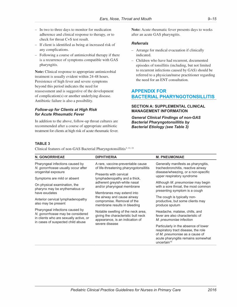
General Clinical Findings of non-GAS Bacterial Pharyngotonsillitis by Bacterial Etiology (see Table 3)
TABLE 3
Clinical features of non-GAS Bacterial Pharyngotonsillitis3; 11; 21
N. GONORRHEAE DIPHTHERIA M. PNEUMONIAE
Pharyngeal infections caused by N. gonorrhoeae usually occur after orogenital exposure
Symptoms are mild or absent
On physical examination, the pharynx may be erythematous or have exudates
Anterior cervical lymphadenopathy also may be present
Pharyngeal infections caused by N. gonorrhoeae may be considered in clients who are sexually active, or in cases of suspected child abuse
A rare, vaccine-preventable cause of life-threatening pharyngotonsillitis
Presents with cervical lymphadenopathy and a thick, adherent greyish-white nasal and/or pharyngeal membrane
Membranes may extend into the airway and cause airway compromise. Removal of the membrane results in bleeding
Notable swelling of the neck area, giving the characteristic bull neck appearance, is an indication of severe disease
Generally manifests as pharyngitis, tracheobronchitis, reactive airway disease/wheezing, or a non-specific upper respiratory syndrome
Although M. pneumoniae may begin with a sore throat, the most common presenting symptom is a cough
The cough is typically non-productive, but some clients may produce sputum
Headache, malaise, chills, and fever are also characteristic of M. pneumoniae infection
Particularly in the absence of lower respiratory tract disease, the role of M. pneumoniae as a cause of acute pharyngitis remains somewhat uncertain10
Pediatric Clinical Practice Guidelines for Nurses in Primary Care
Ears, Nose, Throat and Mouth9–16
2016
Paediatric Autoimmune Neuropscyhiatric Disorder (PANDAS)8; 22
PANDAS has been characterized as the abrupt, dramatic onset of obsessive-compulsive disorder (including severely-restricted food intake) or tics in some children as an autoimmune response following a GAS infection. The concept of PANDAS as a distinct disease entity is controversial and research is ongoing. At present, there is insufficient evidence to support routine testing for GAS in children with neuropsychiatric symptoms, or to support long-term prophylaxis or immune-modifying therapies in children with neuropsychiatric symptoms. Any child presenting with acute-onset obsessive-compulsive disorder/eating disorders must have a thorough medical evaluation.
BIBLIOGRAPHY FOR BACTERIAL PHARYNGOTONSILLITIS
The following References and Other Sources have informed the updating of this Clinical Practice Guideline
REFERENCES
1. Toward Optimized Practice. (2008). Guideline for the diagnosis and management of acute pharyngitis. Alberta Clinical Practice Guidelines.
2. Shulman, S. T., Bisno, A. L., Clegg, H. W., Gerber, M. A., Kaplan, E. L., … Van Beneden, C. (2012). Clinical practice guideline for the diagnosis and management of group a streptococcal pharyngitis: 2012 update by the infectious diseases society of America. Clinical Infectious Diseases, 55(10), 1–17. doi:10.1093/cid/cis629
3. Miller, K. E. (2006). Diagnosis and treatment of Neisseria gonorrhoeae infections. American Family Physician. Retrieved July 28, 2015, from http://www.aafp.org/afp/2006/0515/p1779.pdf
4. Public Health Agency of Canada. (2010). Streptococcus pyogenes - Pathogen Safety Data Sheets. Retrieved May 12, 2015, from http://www.phac-aspc.gc.ca/lab-bio/res/psds-ftss/strep-pyogenes-eng.php
5. American Academy of Pediatrics. Group A Streptococcal Infections. In: Kimberlin, D.W., Brady, M.T., Jackson, M.A., Long, S.S., editors. Red Book, 2015 Report on the Committee on Infectious Diseases. 30th ed. Elk Grove Village, IL: American Academy of Pediatrics; 2015. p.732-44.
6. Choby, B. A. (2009). Diagnosis and treatment of streptococcal pharyngitis. American Family Physician, 79(5), 383–390.
7. Hayes, C., Williamson Jr., H. (2001). Management of group A beta-hemolytic Streptococcal pharyngitis. American Family Physician. Retrieved May 14, 2015, from http://www.aafp.org/afp/2001/0415/p1557.pdf
8. Armstrong, C. (2010). AHA guidelines on prevention of rheumatic fever and diagnosis and treatment of acute streptococcal pharyngitis. American Family Physician. Retrieved December 16, 2015, from http://www.nimh.nih.gov/labs-at-nimh/research-areas/clinics-and-labs/pdnb/web.shtml
9. Vincent, M.T., Celestin, N., Hussain, A. N. (2004). Pharyngitis. American Family Physician. Retrieved June 22, 2015, from http://www.aafp.org/afp/2004/0315/p1465.pdf
10. Quality Management Program University of Michigan. (2013). Pharyngitis. Guidelines for Clnicial Care Ambulatory.
11. Rubin, M.A., Ford, L.C., Gonzales, R. (2012). Pharyngitis, sinusitis, otitis, and other upper respiratory tract infections. In J. Longo, D.L., Kasper, D.L., Jameson, J.L., Fauci, A.S., Hauser, S.L., Loscalzo (Ed.), Harrison’s Principles of Internal Medicine (18th ed., pp. 255–267). New York: McGraw Hill Medical.
12. Pichichero, M. E. (2015). Complications of streptococcal tonsillopharyngitis. UptoDate. Retrieved December 15, 2015, from www.uptodate.com
13. Knott, L. (2014). Rheumatic Fever. Retrieved June 11, 2015, from http://patient.info/pdf/2731.pdf
14. Gibofsky, A. (2013). Clinical manifestations and diagnosis of acute rheumatic fever. UptoDate [Intranet].
15. Alina Health Laboratory. (2012). How to collect a specimen for a throat culture. Retrieved November 26, 2015, from https://ww5.allinahealth.org/ahs/allinalabs.nsf/page/ThroatCultureCollect.pdf/$FILE/ThroatCultureCollect.pdf
16. Throat swabs: MedlinePlus Medical Encyclopedia Image. (2014). Retrieved October 14, 2015, from https://www.nlm.nih.gov/medlineplus/ency/imagepages/9950.htm
17. Ray, S. (2014). Managing outbreaks of scarlet fever. Nursing Times, 110(39), 23–24.
18. The Hospital for Sick Children. (2014). 2015 Drug Handbook and Formulary. (E. Chen, J., Lau, Ed.). Hudson, OH: Lexicomp.
Ears, Nose, Throat and Mouth 9–17
Pediatric Clinical Practice Guidelines for Nurses in Primary Care 2016
19. Lexicomp. (2015). Acetaminophen (Pediatric). Lexicomp Online [Intranet]. Retrieved May 22, 2015, from http://online.lexi.com
20. Lexicomp. (2015). Ibuprofen (Pediatric). Lexicomp Online. Retrieved May 22, 2015, from http://online.lexi.com
21. Public Health Agency of Canada. (2014). Diphtheria. Retrieved July 6, 2015, from http://www.phac-aspc.gc.ca/im/vpd-mev/diphtheria-diphterie-eng.php
22. National Institute of Mental Health. (n.d.). Information About PANDAS. Retrieved December 16, 2015, from http://www.nimh.nih.gov/labs-at-nimh/research-areas/clinics-and-labs/pdnb/web.shtml
OTHER SOURCES
Health Canada. (2014). First Nations and Inuit Health Branch (FNIHB) Nursing Station Formulary and Drug Classification System January 2014
Canadian Pharmacists Association. (2012). Penicillin V. e-Therapeutics [Intranet]. Retrieved May 6th, 2015, from https://www.e-therapeutics.ca
Lexicomp. (2015). Penicillin V. Lexicomp Online [Intranet]. Retrieved July 6, 2015, from http://online.lexi.com
Canadian Pharmacists Association. (2012). Amoxicillin (Amoxicillin). e-Therapeutics [Intranet]. Retrieved July 6th, 2015, from https://www.e-therapeutics.ca/cps
Lexicomp. (2015). Amoxicillin. Lexicomp Online [Intranet]. Retrieved July 6, 2015, from http://online.lexi.com
Canadian Pharmacists Association. (2009). Cephalexin (Cephalexin). e-Therapeutics [Intranet]. Retrieved July 6th, 2015, from https://www.e-therapeutics.ca/cps
Lexicomp. (2015). Cephalexin. Lexicomp Online [Intranet]. Retrieved July 6, 2015, from http://online.lexi.com
Canadian Pharmacists Association. (2014). Clindamycin (Dalacin C). e-Therapeutics [Intranet]. Retrieved July 6th, 2015, from https://www.e-therapeutics.ca
Lexicomp. (2015). Clindamycin. Lexicomp Online [Intranet]. Retrieved July 6, 2015, from http://online.lexi.com
Canadian Pharmacists Association. (2012). Penicillin G. e-Therapeutics [Intranet]. Retrieved May 6th, 2015, from https://www.e-therapeutics.ca
Lexicomp. (2015). Penicillin G. Lexicomp Online [Intranet]. Retrieved July 6, 2015, from http://online.lexi.com
PHARYNGOTONSILLITIS, VIRAL
CAUSES
– Adenovirus or enterovirus (the latter is more common in children < 3 years old)
– Influenza virus
– Parainfluenza virus
– Coxsackievirus
– Echovirus
– Epstein-Barr virus (mononucleosis)
– Herpes simplex virus
HISTORY
– Acute sore throat combined with symptoms consistent with a viral upper respiratory tract infection (rhinorrhea, cough and often hoarseness)
PHYSICAL FINDINGS
– Fever (low-grade to significant)
– Tachycardia
– Pharyngeal and tonsillar erythema and swelling
– Petechiae of soft palate
– Tonsillar exudate similar to that occurring with bacterial infection may be present, particularly in adenovirus pharyngotonsillitis
– Anterior cervical lymphadenopathy
– Vesicles and ulcers may be present with coxsackievirus infection (for example, hand, foot and mouth ulcers occur with coxsackievirus A-16 infection [usually in the area of the soft palate]) or herpes infection (usually in the anterior portion of the mouth)
DIFFERENTIAL DIAGNOSIS
– Bacterial pharyngotonsillitis
– Epiglottitis
COMPLICATIONS
– Secondary bacterial infection
Pediatric Clinical Practice Guidelines for Nurses in Primary Care2010
Ears, Nose, Throat and Mouth9–18
DIAGNOSTIC TESTS
– None
– Collect a swab for culture and sensitivity only if it is unclear whether the pharyngotonsillitis is viral or bacterial
MANAGEMENT
Goals of Treatment
– Supportive care to relieve symptoms
Nonpharmacologic Interventions
– Rest and reassurance
– Increase oral fluids during febrile phase
– Avoidance of irritants (for example, smoke)
– Warm saline gargles qid (for older children)
Pharmacologic Interventions
Antipyretic and analgesic for fever and pain:
acetaminophen (Tylenol), 10–15 mg/kg PO or PR q4–6h prn
Occasionally, children are unable to drink secondary to the pain of pharyngotonsillitis caused by some viral infections, particularly coxsackievirus and herpesvirus. In such situations, admission to hospital may be required for IV administration of fluids (to prevent dehydration).
RHINITIS
Inflammation of the mucosal lining of the nasal cavity leading to nasal congestion and rhinorrhea (runny nose). The 3 commonest types of rhinitis to consider in the differential diagnosis of rhinitis are:
– Allergic rhinitis: Reactive inflammation of the nasal mucosa
– Vasomotor rhinitis: Perennial inflammation of the nasal mucosa, which represents a hyperreactive state of the nasal mucosa (nonallergic)
– Viral rhinitis (infection of upper respiratory tract): Viral infection confined to the upper respiratory tract. Usually mild and self-limiting.
CAUSES
Allergic Rhinitis
Sensitivity to inhaled allergens (pollens, grasses, ragweed, dust, molds, animal dander, smoke).
Vasomotor Rhinitis
– Unknown; symptoms do not correlate with exposure to specific allergens
– Atrophic mucosa (in the elderly)
– Attacks may be triggered by abrupt changes in temperature or barometric pressure, odours, emotional stress or exercise
Viral Rhinitis (Infection of Upper Respiratory Tract)
Numerous viral agents.
HISTORY
Allergic Rhinitis
– Seasonal or perennial symptoms
– History of familial allergies
– Asthma or eczema may be present
– Paroxysmal sneezing
– Itchy nose
– Nasal congestion
– Excessive, continuous, clear, watery nasal discharge
– Eyes may be itchy or watery
– Ears may be itchy
– General malaise and headache may be present
– Symptoms worst in the morning and least during the day, worsening again during the night
– Postnasal drip
– Breathing through the mouth
– Snoring and dry cough at night may be present
Vasomotor Rhinitis
– Sudden onset of nasal congestion
– Perennial symptoms
– Persistent postnasal drip
– Intermittent throat irritation
– No response to environmental controls and medications
– Sensation of constantly needing to clear throat
– Changes in acuity of hearing or smell
– Snoring at night
– Fatigue
Pediatric Clinical Practice Guidelines for Nurses in Primary Care 2010
Ears, Nose, Throat and Mouth 9–19
Viral Rhinitis (Infection of Upper Respiratory Tract)
– Nonproductive cough or cough that produces clear sputum
– Low-grade fever
– Nasal congestion with clear nasal discharge
– Sneezing
– Postnasal drip
– Scratchy throat
– Mild headache and general malaise
– Pressure in ears
PHYSICAL FINDINGS
Allergic Rhinitis
– Injected conjunctiva may be present
– Eyes may tear
– Edema of the eyelids and periorbital area may be present
– Pale, edematous nasal mucosa is pink, with clear thin secretions
– Nasal polyps may be present
– Skin around nose may be irritated
– “Allergic salute” may be present
– Sinuses may feel tender if symptoms are severe
– Mouth breathing
Vasomotor Rhinitis
– Vital signs usually normal
– Nasal mucosa red and swollen
– Nasal turbinates enlarged
– Throat may be slightly reddened because of irritation from postnasal drip
– Tonsils and adenoids may be enlarged
– Sinuses may feel tender if symptoms are severe
Viral Rhinitis (Infection of Upper Respiratory Tract)
– Temperature may be slightly elevated
– Client appears mildly ill
– Clear nasal discharge
– Skin around nares slightly irritated
– Ears may have transient middle-ear sterile effusion
– Throat may have mild erythema, but otherwise is normal
– Sinuses may feel tender if symptoms are severe
DIFFERENTIAL DIAGNOSIS (ALL TYPES OF RHINITIS)
– Acute or chronic sinusitis
– Abuse of nose drops
– Abuse of drugs or solvents (for example, cocaine, gas, glue)
– Foreign body in nares
– Nasal polyps
– Deviated septum
– Hypothyroidism as a cause of the nasal congestion
– Nasal congestion induced by pregnancy or use of oral contraceptives
COMPLICATIONS (ALL TYPES OF RHINITIS)
– Otitis media
– Nasal polyps
– Epistaxis
– Enlargement of tonsils and adenoids
– Sinusitis
DIAGNOSTIC TESTS (ALL TYPES OF RHINITIS)
Consider skin testing for allergies.
MANAGEMENT (ALL TYPES OF RHINITIS)
Goals of Treatment
– Relieve and suppress symptoms
– Identify the underlying allergen(s)
– Prevent complications
Appropriate Consultation
Consultation with a physician is not usually required.
Nonpharmacologic Interventions
Environmental control is important. Eliminate or reduce known allergen(s) in the environment wherever possible, or avoid them altogether.
Pediatric Clinical Practice Guidelines for Nurses in Primary Care2010
Ears, Nose, Throat and Mouth9–20
Client Education
– Recommend increasing fluid intake to improve hydration
– Counsel client about appropriate use of medications (dose, frequency, side effects, avoidance of overuse)
– Recommend avoidance of caffeine
– Recommend avoidance of known allergens (client should keep living area clear of dust, avoid going outside when pollen count is high and use synthetic fibres in bedding and clothing) and removal of pets (to eliminate animal dander)
– Counsel client about preventing spread of viral rhinitis to other household members
– Recommend frequent hand-washing, appropriate disposal of used facial tissues and covering of mouth and nose when coughing or sneezing
Pharmacologic Interventions
Allergic and Vasomotor Rhinitis
Normal saline nasal drops/salinex nasal spray, prn, to wash out mucus and any inhaled allergen.
Oral antihistamines to treat acute symptoms of runny nose, sneezing, itch and conjunctival symptoms (but these will not help nasal congestion):
cetirizine (Reactine) dosing (available as an oral liquid):
Children age 6–12 months: cetirizine 2. 5 mg PO once daily
Children age 12–23 months: 2.5 mg PO daily or 2.5 mg PO bid
Children age 2–6 years: cetirizine 5 mg PO daily or 2.5 mg PO bid
Children > 6 years to adult: cetirizine 5–10 mg PO daily or divided bid
Cetirizine can cause some drowsiness but to a lesser extent than that caused by first-generation antihistamines.
There is some experience using intranasal corticosteroids in children over 4 years of age. Some nasal corticosteroids may temporarily affect growth but it is unknown if there is a long-term effect on height. Consult a physician who may prescribe an intranasal corticosteroid if antihistamines are ineffective. For example:
Children > 4 years: fluticasone (Flonase), 1 spray to each nostril daily
Viral Rhinitis
Antihistamines have little proven benefit in the treatment of the common cold.
Manage fever:
acetaminophen (Tylenol), 10–15 mg/kg/dose PO q4–6h prn
Monitoring and Follow-Up
Instruct client to return for further assessment if fever develops or if symptoms have not resolved within 14 days.
Referral
Refer to a physician if symptoms of rhinitis are not controlled with initial treatment. Allergy testing, sinus radiography or other medications may be required.
RHINOSINUSITIS
Rhinosinusitis is uncommon in young children (< 10–12 years).
See “Rhinosinusitis, Acute” in the adult clinical guidelines, as the clinical presentation is the same in adults and children. The pediatric management of acute rhinosinusitis is presented below.
Pharmacologic Interventions
Decongestants are generally not recommended for children with rhinosinusitis. The use of saline drops/spray is recommended.
If antibiotics are required:
amoxicillin (Amoxil), 40 mg/kg/day, divided tid, PO for 10 days12
A higher dose of amoxicillin should be used in high-risk children (for example, recent [< 3 months] antibiotic exposure and/or daycare centre attendance [extrapolated from acute otitis media data]).
For penicillin/beta-lactam allergy or known beta-lactamase resistance in the community:
azithromycin 10 mg/kg/day PO first day then 5 mg/kg/day PO for the remaining 4 days
Referral
Consult physician should chronic rhinosinusitis develop.
Pediatric Clinical Practice Guidelines for Nurses in Primary Care 2010
Ears, Nose, Throat and Mouth 9–21
COMMON PROBLEMS OF THE MOUTH
ABSENCE OF TEETH, CONGENITAL (ANODONTIA)
Very rare. Teeth usually begin to erupt by 6 months, but may be delayed until up to 12 months.
ABSENCE OF TEETH, PARTIAL (OLIGODONTIA OR “CONGENITALLY MISSING TEETH”)
It is unlikely that the primary care nurse will detect or identify missing permanent teeth (because the primary tooth is usually retained); however, the parent might ask about it. This condition is more common with the permanent dentition, particularly the third molars, the mandibular second bicuspids, the maxillary lateral incisors and the maxillary second bicuspids. Three percent of the general population has one or more missing permanent teeth. Absence of most permanent teeth is called anodontia. This condition is rare and is usually associated with syndromes such as ectodermal dysplasia.
MANAGEMENT
Referral
Appropriate dental referral should be made.
ANKYLOGLOSSIA (TONGUE-TIE)
A condition in which a short lingual frenum attaches the tongue to the floor of the mouth, interfering with protrusion of the tongue and occasionally affecting speech and in rare instances breastfeeding.
MANAGEMENT
No treatment is warranted if the tongue can be protruded beyond the lips. In 95% of cases, reassurance is all that is required.
Referral
On occasion, a thick fibrous band of tissue interferes with the tongue’s protrusion beyond the lips. In such cases, consultation with an ears, nose and throat (ENT) specialist is suggested with a view to possible surgical release.
COMMON MALOCCLUSIONS
Crooked teeth result from a number of causes.
CAUSES
– Delayed eruption
– Rotation of incisors
– Crowded teeth
– Supplemental teeth (extra teeth)
– Large space between maxillary central incisors
– Anterior open bite (front teeth do not meet when teeth are closed)
– Protrusion of the upper or lower teeth
– Crossbite – one or more top teeth positioned behind the bottom teeth
MANAGEMENT
Early identification and referral for any of the above causes might enable preventive or interceptive interventions that can prevent more serious malocclusions from occurring.
Referral
– Children should be assessed by a dentist by age 7–10 years if any of these common abnormalities have presented
DENTAL ABSCESS – PERMANENT TOOTH
Infection of the soft tissue surrounding tooth or gums due to infection of a permanent tooth or the structures supporting the tooth.
CAUSES
– Progressive dental decay causing pulpitis from gram-positive anaerobes and Bacteroides
– Predisposing factors: deep caries, poor dental hygiene, dental trauma
HISTORY
– Localized tooth pain
– Constant, deep, throbbing pain
– Pain worsens with mastication or exposure to extreme temperatures
Pediatric Clinical Practice Guidelines for Nurses in Primary Care2010
Ears, Nose, Throat and Mouth9–22
PHYSICAL FINDINGS
– Fever (rare but possible)
– Facial swelling may be present
– Carious tooth
– Gingival edema and erythema
– Tooth mobility
– Localized tenderness over affected area of jaw
– Anterior cervical nodes enlarged and tender
– Localized tooth pain
DIFFERENTIAL DIAGNOSIS
– Disease of the salivary gland (for example, mumps)
– Sinusitis
– Cellulitis
COMPLICATIONS
– Cellulitis
– Recurrent abscess formation
DIAGNOSTIC TESTS
None.
MANAGEMENT
Goals of Treatment
– Relieve symptoms
– Prevent spread of infection
Appropriate Consultation
– Consult a physician if a large fluctuant abscess is present, if client is acutely ill or if the infection has spread to the soft tissues of the neck
Nonpharmacologic Interventions
– Warm saline oral rinses qid
Client Education
– Counsel client/parent about appropriate use of medications (dosage and side effects)
– Recommend dietary modifications (liquids or soft diet)
– Recommend improvements to dental hygiene
Pharmacologic Interventions
Oral antibiotics dosing for adolescents (for a child, see “Dental Abscess – Primary Tooth”):
penicillin V potassium (Penicillin V), 300 mg PO qid for 7–10 days
For adolescents with penicillin allergy:
clindamycin, 300 mg PO tid-qid for 10 days
Adolescent doses of simple analgesics for mild to moderate dental pain:
ibuprofen (Motrin), 200 mg, 1–2 tabs PO q4h prn
or
acetaminophen (Tylenol), 325 mg, 1–2 tabs PO q4–6h prn
Monitoring and Follow-Up
Follow up in 48–72 hours, if there is not a dentist available.
Referral
Refer to a dentist for definitive therapy.
DENTAL ABSCESS – PRIMARY TOOTH
Infection of the soft tissue surrounding tooth or gums due to infection of a primary (baby) tooth or the structures supporting the tooth.
CAUSES
– Progressive dental decay causing pulpitis from gram-positive anaerobes and Bacteroides
– Predisposing factors: deep caries, poor dental hygiene, dental trauma
HISTORY
– Localized tooth pain
– Constant, deep, throbbing pain
– Tooth may be mobile
– Gingival or facial swelling (or both) may be present
– Fistula on the gum above the tooth
Pediatric Clinical Practice Guidelines for Nurses in Primary Care 2010
Ears, Nose, Throat and Mouth 9–23
PHYSICAL FINDINGS
– A primary tooth, more so than a permanent tooth, when it abscesses will form a draining fistula (observed as a bubble in the gum above the tooth), and if so will be less subject to pain
– Mobility of the tooth, compared to its counterpart on the opposite side
– Decay or a large existing restoration
– Fever (rare but possible)
– Facial swelling may be present
– Gingival edema and erythema
– Localized tooth pain
– Carious tooth
– Localized tenderness over affected area of jaw
– Anterior cervical and/or sub-mandibular lymph nodes enlarged and tender
– Localized tooth pain
DIFFERENTIAL DIAGNOSIS
– Disease of the salivary gland (for example, mumps)
– Sinusitis
– Cellulitis
COMPLICATIONS
– Cellulitis
– Recurrent abscess formation
DIAGNOSTIC TESTS
– None
MANAGEMENT
Goals of Treatment
– Relieve symptoms
– Prevent spread of infection
Appropriate Consultation
– Consult a physician if a large fluctuant abscess is present, if client is acutely ill or if the infection has spread to the soft tissues of the neck
Nonpharmacologic Interventions
Client/Parent Education
– Recommend improvements to dental hygiene
– Warm saline oral rinses qid
– Counsel client/parent about appropriate use of medications (dosage and side effects)
– Recommend dietary modifications (liquids or soft diet)
Pharmacologic Interventions
If the abscessed tooth has developed a draining fistula, antibiotics are not necessary.
To relieve pain and fever:
acetaminophen (Tylenol),10–15 mg/kg/dose PO q4–6h prn
or
ibuprofen (Motrin), 5–10 mg/kg/dose PO q6–8h prn (maximum: 40 mg/kg/day)
Antibiotic therapy:
Oral antibiotics (only if there is facial swelling and no fistula present):
penicillin V (Pen-Vee), 25–50 mg/kg/day PO divided bid for 10 days
For clients with penicillin allergy:
clindamycin 8–16 mg/kg/day PO divided tid-qid for 10 days (maximum: 1.8 g/day)
Monitoring and Follow-Up
Follow up in 48–72 hours, if there is not a dentist available.
Referral
Refer to a dentist for definitive therapy.
DENTAL DECAY
See also “Dental Decay” in the adult clinical guidelines, for details about the pathology, progression and clinical presentation of dental decay.
EARLY CHILDHOOD DENTAL DECAY
Caries of the deciduous teeth, most commonly the maxillary incisors, maxillary primary first molar and mandibular molars. Also called milk caries or “baby bottle tooth decay.” May be severe enough to cause dental abscess.
Very common in Aboriginal groups in Canada, often requiring extraction of the affected teeth and resulting in problems with permanent teeth (tooth development problems, crooked teeth).
CAUSES
– Secondary to prolonged nursing (either bottle or breast) at bedtime. Liquid pools around the child’s teeth, causing significant caries, particularly in the maxillary incisors
Pediatric Clinical Practice Guidelines for Nurses in Primary Care2010
Ears, Nose, Throat and Mouth9–24
MANAGEMENT
Prevention
Preventive educational interventions with parents are of primary importance:
– Encourage breastfeeding
– Discourage bottle propping
– Discourage use of sweet fluids in bottle
– A nighttime bottle should not contain anything other than water
– Encourage drinking from a cup by 1 year and weaning by 18–24 months
– Encourage the mother to “lift the lip” routinely to inspect the teeth
– Encourage good oral hygiene, conducted by the parent: cleaning of teeth with gauze as soon as they erupt and cleaning of toddlers’ teeth with a soft toothbrush; to ensure effective brushing, an adult must supervise the child until 6 years of age
– Encourage parents or caregiver to take children for their first dental assessment by 3 years of age, or at the first signs of white spots or decay beginning to show
Referral
Appropriate management includes referral to a dental practitioner for preventive services and dental fillings. The repair procedure may require a general anesthetic, particularly for severe early childhood caries. Fillings last for 8–10 years.
CHILDHOOD AND ADOLESCENT DENTAL DECAY
Dental decay is an infectious disease.
CAUSES
Streptococcus mutans is the primary bacteria involved. It is not present in a newborn’s mouth, but is generally acquired from caregivers (usually the mother) through contact by testing temperature of spoons or bottle nipples, or through shared toothbrushes, kissing, etc. Colonization of Strep mutans occurs between the ages of 12–24 months. Once established in the mouth, it is there to stay; however, good oral hygiene practices can reduce the levels of bacteria present.
Bacterial colonies (dental plaque) convert the sugar in carbohydrates to an acid, which causes demineralization of the tooth enamel and dentine.
Relative acidity of foods and liquids (pop, fruit drinks, energy drinks) also plays a role in demineralization of enamel and dentine.
Between acid attacks on the enamel, protective factors in the saliva help the enamel to remineralize. The relative balance between demineralization and remineralization determines the rate of dental decay.
MANAGEMENT
Prevention
Preventive interventions to slow and sometimes halt progression of dental decay:
– Regular twice-daily toothbrushing with a fluoride toothpaste
– Rinsing mouth with fluoridated mouth rinse for children who will not swallow it
– Application of fluoride varnish
See also the section “Prevention of Dental Decay.”
Referral
Appropriate management includes referral to a dental practitioner for preventive and restorative services.
PREVENTION OF DENTAL DECAY13
Dental decay is a multifactorial disease. In general, bacterial colonies (dental plaque) convert the sugar in fermentable carbohydrates into an acid that demineralizes the dental enamel. When demineralization is not occurring, protective factors such as from the saliva or fluoride exposures result in remineralization of the enamel. Decay occurs when the balance tilts toward demineralization exceeding remineralization over an extended period of time. In the early stages of decay, the enamel takes on a dull white appearance; however, the decay can still be halted or reversed at this stage. It is usually asymptomatic. If demineralization is allowed to continue, eventually the enamel breaks down and cavitation occurs, at which time the process becomes less reversible.
With the introduction of fluoride into the drinking water of some urban and rural communities and most toothpastes, and with increased attention to dental health, there has been a decrease in the prevalence of pediatric dental caries in most southern populations.
Environmental factors (such as hygiene and diet), particularly as influenced by the parents or caregiver, play a significant role in childhood dental problems.
Pediatric Clinical Practice Guidelines for Nurses in Primary Care 2010
Ears, Nose, Throat and Mouth 9–25
MANAGEMENT
Prevention
Patient/parent Counselling
– Fluoridated toothpaste14 should be used twice per day, ideally after breakfast and before bed. Do not rinse after brushing. Children under 8 years of age should be supervised during brushing and should only use a small amount (for example, a pea-sized portion) of toothpaste. Children under 5 years of age should have their teeth brushed by an adult using only a smear of toothpaste from the time the first tooth erupts. Swallowing of toothpaste should be avoided as much as possible
– Flossing from the time the child reaches school age should be encouraged
– Encourage reduction in the frequency of sugar consumption
Use of Fluoride
– Fluoride varnish – Fluoride varnish shows good evidence of benefit.15 Preschool children particularly would benefit from fluoride varnish application, because they are rarely seen by dental personnel until they are of school age. The application of fluoride varnish in primary care settings has been shown to be effective.16 Fluoride varnish should be applied twice and up to 4 times per year on children who are at high risk of dental decay (any previous or current decay). Children can be referred to the Children’s Oral Health Initiative (COHI) worker. If there is no COHI worker in the community, nurses can advocate for the program to be introduced. Additionally, the COHI worker could be invited to well-child clinics or visits to ensure preschool children benefit from the application
– Fluoride mouth rinses17 are an effective preventive measure for at-risk individuals and should be used according to the specific needs of the individual. Fluoride mouth rinsing is not recommended for children under 6 years of age. Since compliance levels are very low, fluoride mouth rinses are best suited to school-based rinsing programs
– Fluoride Supplements – Fluoride supplements are available in the form of chewable tablets, lozenges or drops, with chewable forms being the more effective because the fluoride stays on the teeth longer. Their use is advised only for high dental caries risk patients (any previous or current decay) and not advised if the patient is receiving adequate fluoride from other sources such as fluoridated water or twice-daily brushing with a fluoride toothpaste. If fluoride supplements are recommended to a parent, follow-up is necessary because compliance rates are generally very low. Daily dose for preschool children (3–6 years of age) is 0.5 mg and for school-aged children (6–16 years of age) is 1 mg18
Advocate for School-based Dental Preventive Services
School-based preventive services provided by a dental therapist or dental hygienist are the most effective and efficient method of delivery of preventive services for school-aged children. Other school-based programs shown to be effective include:
– Daily toothbrushing – Children do supervised toothbrushing using a fluoride toothpaste. It is most effective/efficient when it is conducted after breakfast or lunch programs. It requires coordinated efforts by teachers and an overall monitoring supervisor (to assure that it is sustained)
– Fluoride varnish program19
– Fluoride rinse program17 – If a fluoride varnish program cannot be conducted, the next best fluoride regimen is a daily or weekly fluoride rinse program
– Dental sealants20 – Dental sealants (organic polymers) that bond to the enamel are intended for teeth with deep developmental grooves. They prevent decay on the biting surface of back teeth by sealing off the grooves from bacteria and fermentable carbohydrates. They are applied by qualified licensed personnel – dentists, dental hygienists or dental therapists
Advocate for Community Water Fluoridation21
Fluoridation of community water supplies is a safe and effective public health measure that can reduce dental decay by 25–40% in high-risk communities.
Pediatric Clinical Practice Guidelines for Nurses in Primary Care2010
Ears, Nose, Throat and Mouth9–26
Nutrition Counselling
Some important messages for the prevention of dental disease are:
– Breastfeeding of infants is better than bottle-feeding in terms of dental decay
– Baby bottles should only be used for milk at feeding times during the day and should not be propped. A nighttime bottle should not contain anything other than water
– Frequency of exposure to fermentable carbohydrates (most foods) should be reduced to as few times as possible during the day. Each time carbohydrates are taken into the mouth the demineralization of tooth enamel occurs for about half an hour after. Frequent snacking increases the potential decaying time significantly. Between meals, parents should encourage noncarbohydrate snacks
– Follow Canada’s Food Guide. Good nutrition for the teeth is the same
– If gum is being chewed it should be of a nonsugar type. Chewing gum sweetened with xylitol has a mild anti-cariogenic effect
– Children who have a good breakfast generally have lower decay rates
– Pop and fruit drinks should be used in moderation. Energy drinks are particularly cariogenic
DISCOLOURED (NON-VITAL) PERMANENT TOOTH
A permanent tooth that has been injured, but not fractured, might turn dark, compared to other teeth around it. The darkening is from the blood supply being cut off, and red blood cells breaking down within the pulp. The tooth becomes non-vital and may eventually cause an abscess. In some cases the pulp becomes completely occluded, but the tooth does not abscess.
CAUSES
– Trauma
HISTORY
– Recent trauma
PHYSICAL FINDINGS
– The tooth takes on a dull appearance or turns dark
COMPLICATIONS
– The tooth may abscess at some point
MANAGEMENT
Pharmacologic Interventions
– None, unless there are signs of the tooth abscessing
Referral
– Refer to a dentist for monitoring and definitive treatment
DISCOLOURED (NON-VITAL) ANTERIOR PRIMARY TOOTH
A primary anterior tooth that is non-vital due to trauma could turn dark in colour. It might or might not abscess. In general, if it is not abscessed no treatment is necessary
CAUSES
– Trauma. The parent might not be aware of the actual traumatic event
HISTORY
– Trauma, though it may not have been severe or noticed
PHYSICAL FINDINGS
– A front primary tooth that is dark in colour, compared to its counterparts
– Signs of abscess are a fistula on the gum above the tooth or mobility compared to its counterpart
COMPLICATIONS
– Most non-vital primary anterior teeth exfoliate normally and at normal times and do not have complications
– If the primary tooth is abscessed at a very young age it could disrupt development of the permanent successor
– It might not exfoliate normally and, if so, could cause the permanent successor to deflect and erupt crooked
MANAGEMENT
Goals of Treatment
– Normal eruption of the successor tooth
Pediatric Clinical Practice Guidelines for Nurses in Primary Care 2010
Ears, Nose, Throat and Mouth 9–27
Pharmacologic Interventions
– None
Referral
– Refer to a dentist discoloured anterior primary teeth for definitive treatment after age 6. Earlier referral may be needed, for example, at age 3 if advanced discolouration of the tooth is present
– Treat if signs of abscess (see “Dental Abscess – Permanent Tooth”) are present (for example, fistula on the gum above the tooth, or tooth is mobile compared to its counterpart)
EPSTEIN PEARLS
Small, white, keratinized lesions along gums and roof of the mouth in a newborn. Occurs only in the newborn and seen in approximately 80% of newborns. The condition is harmless, although it sometimes worries new mothers.
CAUSES
Protein-filled cysts that eventually resolve.
MANAGEMENT
Reassure parents or caregiver that this condition will resolve on its own and needs no treatment.
ERUPTION CYST
Small white, grey or bluish translucent eruptions on crest of maxilla or mandible.
CAUSES
Remnants of dental lamina, which are usually shed after birth.
MANAGEMENT
Reassure parents or caregiver that this condition will resolve on its own and needs no treatment.
IMPACTED TOOTH
An impacted tooth is one that is unable to erupt into the mouth. The tooth could be completely impacted or partially impacted. Although any tooth could be impacted, the most commonly impacted teeth are the wisdom teeth (third molars) and the maxillary permanent cuspids (eye teeth).
CAUSES
Teeth may become impacted because there is insufficient room in the arch for them to erupt or because the path of eruption is off course.
PHYSICAL FINDINGS
– Unlikely to be identified unless they are causing pain
– Totally impacted teeth very rarely cause pain
– Wisdom teeth usually erupt between the ages of 18 and 21. If there is insufficient space, they might be partially erupted. Partially erupted teeth are more subject to decay or pericoronal infection.
MANAGEMENT
For partially impacted teeth with pain:
– Pericoronal infection (pericoronitis) does not require antibiotics (see also pericoronitis)
– Advise the patient to rinse the area with warm saline water 4 times daily for 4–7 days, and to adhere to meticulous toothbrushing on other teeth
Pharmacologic Interventions
For pain, acetaminophen or ibuprofen can be used.
Referral
Refer to a dentist for follow-up.
INTRUDED TOOTH
Due to trauma, a tooth may be intruded apically into the socket. Intrusion injuries occur when the tooth is driven further into the jaw along its long axis, making the tooth appear shorter. A primary tooth might be completely intruded, while a permanent tooth is more likely to be partially intruded.
CAUSES
– Trauma
HISTORY
– Trauma
PHYSICAL FINDINGS
– Primary tooth at a lower level than its counterparts, or not visible
– Permanent tooth at a lower level than its counterparts
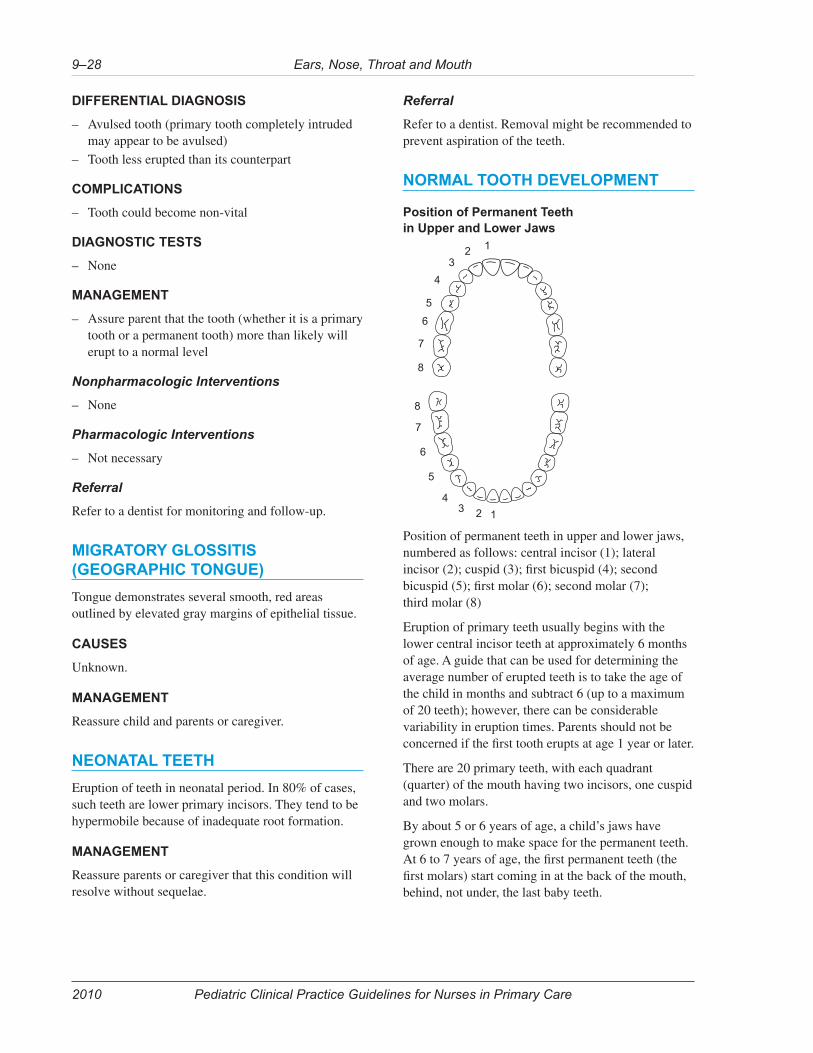
Pediatric Clinical Practice Guidelines for Nurses in Primary Care2010
Ears, Nose, Throat and Mouth9–28
DIFFERENTIAL DIAGNOSIS
– Avulsed tooth (primary tooth completely intruded may appear to be avulsed)
– Tooth less erupted than its counterpart
COMPLICATIONS
– Tooth could become non-vital
DIAGNOSTIC TESTS
– None
MANAGEMENT
– Assure parent that the tooth (whether it is a primary tooth or a permanent tooth) more than likely will erupt to a normal level
Nonpharmacologic Interventions
– None
Pharmacologic Interventions
– Not necessary
Referral
Refer to a dentist for monitoring and follow-up.
MIGRATORY GLOSSITIS (GEOGRAPHIC TONGUE)
Tongue demonstrates several smooth, red areas outlined by elevated gray margins of epithelial tissue.
CAUSES
Unknown.
MANAGEMENT
Reassure child and parents or caregiver.
NEONATAL TEETH
Eruption of teeth in neonatal period. In 80% of cases, such teeth are lower primary incisors. They tend to be hypermobile because of inadequate root formation.
MANAGEMENT
Reassure parents or caregiver that this condition will resolve without sequelae.
Referral
Refer to a dentist. Removal might be recommended to prevent aspiration of the teeth.
NORMAL TOOTH DEVELOPMENT
Position of Permanent Teeth in Upper and Lower Jaws
123
4
5
6
7
8
1234
5
6
7
8
Position of permanent teeth in upper and lower jaws, numbered as follows: central incisor (1); lateral incisor (2); cuspid (3); first bicuspid (4); second bicuspid (5); first molar (6); second molar (7); third molar (8)
Eruption of primary teeth usually begins with the lower central incisor teeth at approximately 6 months of age. A guide that can be used for determining the average number of erupted teeth is to take the age of the child in months and subtract 6 (up to a maximum of 20 teeth); however, there can be considerable variability in eruption times. Parents should not be concerned if the first tooth erupts at age 1 year or later.
There are 20 primary teeth, with each quadrant (quarter) of the mouth having two incisors, one cuspid and two molars.
By about 5 or 6 years of age, a child’s jaws have grown enough to make space for the permanent teeth. At 6 to 7 years of age, the first permanent teeth (the first molars) start coming in at the back of the mouth, behind, not under, the last baby teeth.
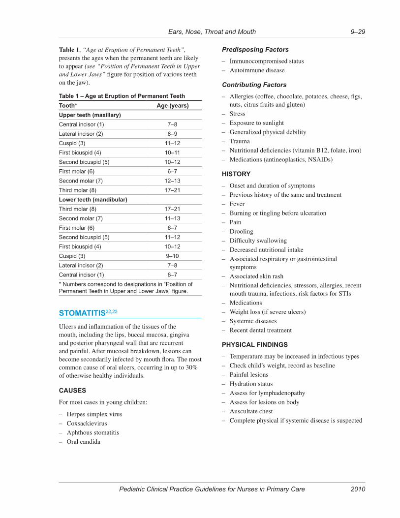
Pediatric Clinical Practice Guidelines for Nurses in Primary Care 2010
Ears, Nose, Throat and Mouth 9–29
Table 1, “Age at Eruption of Permanent Teeth”, presents the ages when the permanent teeth are likely to appear (see “Position of Permanent Teeth in Upper and Lower Jaws” figure for position of various teeth on the jaw).
Table 1 – Age at Eruption of Permanent Teeth
Tooth* Age (years)
Upper teeth (maxillary)
Central incisor (1) 7–8
Lateral incisor (2) 8–9
Cuspid (3) 11–12
First bicuspid (4) 10–11
Second bicuspid (5) 10–12
First molar (6) 6–7
Second molar (7) 12–13
Third molar (8) 17–21
Lower teeth (mandibular)
Third molar (8) 17–21
Second molar (7) 11–13
First molar (6) 6–7
Second bicuspid (5) 11–12
First bicuspid (4) 10–12
Cuspid (3) 9–10
Lateral incisor (2) 7–8
Central incisor (1) 6–7
* Numbers correspond to designations in “Position of Permanent Teeth in Upper and Lower Jaws” figure.
STOMATITIS22,23
Ulcers and inflammation of the tissues of the mouth, including the lips, buccal mucosa, gingiva and posterior pharyngeal wall that are recurrent and painful. After mucosal breakdown, lesions can become secondarily infected by mouth flora. The most common cause of oral ulcers, occurring in up to 30% of otherwise healthy individuals.
CAUSES
For most cases in young children:
– Herpes simplex virus
– Coxsackievirus
– Aphthous stomatitis
– Oral candida
Predisposing Factors
– Immunocompromised status
– Autoimmune disease
Contributing Factors
– Allergies (coffee, chocolate, potatoes, cheese, figs, nuts, citrus fruits and gluten)
– Stress
– Exposure to sunlight
– Generalized physical debility
– Trauma
– Nutritional deficiencies (vitamin B12, folate, iron)
– Medications (antineoplastics, NSAIDs)
HISTORY
– Onset and duration of symptoms
– Previous history of the same and treatment
– Fever
– Burning or tingling before ulceration
– Pain
– Drooling
– Difficulty swallowing
– Decreased nutritional intake
– Associated respiratory or gastrointestinal symptoms
– Associated skin rash
– Nutritional deficiencies, stressors, allergies, recent mouth trauma, infections, risk factors for STIs
– Medications
– Weight loss (if severe ulcers)
– Systemic diseases
– Recent dental treatment
PHYSICAL FINDINGS
– Temperature may be increased in infectious types
– Check child’s weight, record as baseline
– Painful lesions
– Hydration status
– Assess for lymphadenopathy
– Assess for lesions on body
– Auscultate chest
– Complete physical if systemic disease is suspected
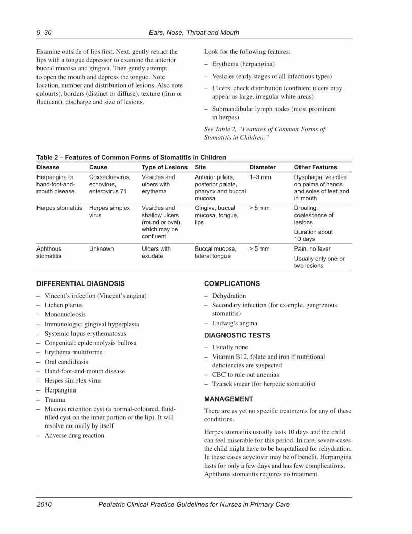
Pediatric Clinical Practice Guidelines for Nurses in Primary Care2010
Ears, Nose, Throat and Mouth9–30
Examine outside of lips first. Next, gently retract the lips with a tongue depressor to examine the anterior buccal mucosa and gingiva. Then gently attempt to open the mouth and depress the tongue. Note location, number and distribution of lesions. Also note colour(s), borders (distinct or diffuse), texture (firm or fluctuant), discharge and size of lesions.
Look for the following features:
– Erythema (herpangina)
– Vesicles (early stages of all infectious types)
– Ulcers: check distribution (confluent ulcers may appear as large, irregular white areas)
– Submandibular lymph nodes (most prominent in herpes)
See Table 2, “Features of Common Forms of Stomatitis in Children.”
Table 2 – Features of Common Forms of Stomatitis in Children
Disease Cause Type of Lesions Site Diameter Other Features
Herpangina or hand-foot-and-mouth disease
Coxsackievirus, echovirus, enterovirus 71
Vesicles and ulcers with erythema
Anterior pillars, posterior palate, pharynx and buccal mucosa
1–3 mm Dysphagia, vesicles on palms of hands and soles of feet and in mouth
Herpes stomatitis Herpes simplex virus
Vesicles and shallow ulcers (round or oval), which may be confluent
Gingiva, buccal mucosa, tongue, lips
> 5 mm Drooling, coalescence of lesions
Duration about 10 days
Aphthous stomatitis
Unknown Ulcers with exudate
Buccal mucosa, lateral tongue
> 5 mm Pain, no fever
Usually only one or two lesions
DIFFERENTIAL DIAGNOSIS
– Vincent’s infection (Vincent’s angina)
– Lichen planus
– Mononucleosis
– Immunologic: gingival hyperplasia
– Systemic lupus erythematosus
– Congenital: epidermolysis bullosa
– Erythema multiforme
– Oral candidiasis
– Hand-foot-and-mouth disease
– Herpes simplex virus
– Herpangina
– Trauma
– Mucous retention cyst (a normal-coloured, fluid-filled cyst on the inner portion of the lip). It will resolve normally by itself
– Adverse drug reaction
COMPLICATIONS
– Dehydration
– Secondary infection (for example, gangrenous stomatitis)
– Ludwig’s angina
DIAGNOSTIC TESTS
– Usually none
– Vitamin B12, folate and iron if nutritional deficiencies are suspected
– CBC to rule out anemias
– Tzanck smear (for herpetic stomatitis)
MANAGEMENT
There are as yet no specific treatments for any of these conditions.
Herpes stomatitis usually lasts 10 days and the child can feel miserable for this period. In rare, severe cases the child might have to be hospitalized for rehydration. In these cases acyclovir may be of benefit. Herpangina lasts for only a few days and has few complications. Aphthous stomatitis requires no treatment.
Pediatric Clinical Practice Guidelines for Nurses in Primary Care 2010
Ears, Nose, Throat and Mouth 9–31
Goals of Treatment
– Relieve symptoms
– Prevent complications
Appropriate Consultation
The disease is self-limiting, so consultation is usually unnecessary, unless there are complications.
Nonpharmacologic Interventions
– Maintenance of hydration is important
– Increase oral intake of fluids (that is, maintenance requirements + fluid deficits caused by fever)
– Maintain oral hygiene with a soft-bristled toothbrush
Client Education
– Counsel parents or caregiver about the expected duration of this illness and the signs and symptoms of dehydration
– Recommend dietary adjustments: bland, non-acidic fluids (such as milk and water); older children may eat popsicles, ice cream and similar food items; avoid citrus foods, such as orange juice
– Recommend local mouthwashes for older children only (1:1 hydrogen peroxide and water), especially after eating
– Recommend warm saline rinse 4 times daily for traumatic or viral ulcers
– To prevent spread of infection, recommend avoidance of direct contact with infected individuals (for example, kissing, sharing glasses and utensils, hand contact)
– Provide support to parents or caregiver to help them cope with a “cranky” child
– Educate parents and patients that the herpes virus can spread even when sores are not present
– If candidiasis is present and an infant is breastfeeding, consider assessing the mother’s nipples
Pharmacologic Interventions
Antipyretic and analgesic for fever and pain:
acetaminophen (Tylenol), 10–15 mg/kg PO or PR q4h prn
Oral anesthetic rinses may make the child more comfortable.
Diphenhydramine (Benadryl) elixir or syrup can be mixed in a one-to-one solution with either Kaolin pectin (Kaopectate) or magnesia-alumina (Maalox) to be used as a topical anesthetic every 2 hours or before meals; the child should be instructed to rinse his or her mouth and then spit out the solution.24
A topical anesthetic containing benzocaine (for example, Anbesol) can be obtained from a retail pharmacy.
Do not treat this condition with antibiotics, as they are not indicated and are not helpful.
Herpetic Lesions on the Lips
If the lesions are herpetic, consult a physician who may suggest oral antiviral therapy depending on severity/recurrence. Topical antivirals such as acyclovir (for example, Zovirax) are sometimes used but must be started before lesions appear.25
Oral Candidiasis
Antifungal (nystatin):
Neonates: nystatin 100,000 units (1 mL) applied qid
Infants: nystatin 200,000 units (2 mL) applied qid
Children: nystatin 500,000 units (5 mL) swish and swallow qid26
If large (> 1 cm), persistent and painful lesions interfere with nutrition, consult a physician for further management options.
Monitoring and Follow-Up
– Reassess the young child (< 2 years of age) in 24–48 hours to ensure maintenance of hydration
– Check weight
– If lesions are severe, follow up in 2–3 days
– For lesions of unknown origin, follow up in 7 days
– Educate parents to return with client if lesions persist after 3 weeks despite treatment, or the client is unable to eat or is losing weight
Referral
Refer to a physician for lesions that are not resolving after 3 weeks.
Pediatric Clinical Practice Guidelines for Nurses in Primary Care2010
Ears, Nose, Throat and Mouth9–32
TOOTHACHE
If a patient presents with a sore tooth, the signs, symptoms and tests will determine whether the tooth can be saved by relatively simple procedures or if it requires extensive treatment or extraction.
CAUSES
– Dental decay
– Injury or previous injury
– Crack in tooth
HISTORY
– Onset and duration of pain
– Constant or intermittent pain (constant throbbing pain is more indicative of a dental abscess)
– Degree of pain – mild, moderate or severe
– Pain keeping the client awake at night (more indicative of a dental abscess)
– Sensitivity to hot or cold (sensitive to hot is more indicative of a dental abscess)
– Pain on pressure
PHYSICAL FINDINGS
– Decay on the tooth in question
– Decay on other teeth on the same side (there can be referred pain)
– Large restoration on the tooth in question may be present
– Mobility of the tooth may be present
– Possible discolouration of the tooth, compared to adjacent teeth
– Also see “Dental Abscess – Primary Tooth” and “Dental Abscess – Permanent Tooth”
DIFFERENTIAL DIAGNOSIS
– Sinus infection
– Crack in tooth
COMPLICATIONS
– Dental abscess, if it is not already
MANAGEMENT
Goals of Treatment
– Relieve pain
– Prevent complications
Nonpharmacologic Interventions
Client Education
– Recommend dietary modifications (liquids or soft diet, avoid sugars)
– Recommend improvements to dental hygiene
Pharmacologic Interventions
– Antibiotics, only if there are signs of dental abscess (see“Dental Abscess – Primary Tooth” and “Dental Abscess – Permanent Tooth”)
– For pain relief: ibuprofen, acetaminophen
Monitoring and Follow-Up
– Follow up in 48–72 hours if there is not a dentist available
Referral
– Refer to a dentist for definitive therapy
THUMB-SUCKING
This generally benign activity may result in protrusion of the maxillary incisors and anterior open bite. However, most children suffer no effects to their dentition.
MANAGEMENT
– Reassure the parents or caregiver. Children entering school generally stop sucking the thumb as a result of peer pressure
– Thumb-sucking in a young child is common and is not considered abnormal
– Parents can be advised to ignore the habit. Excessive attempts to stop the habit often reinforce it by giving the child attention
Referral
In rare cases, the older child with a severe thumb-sucking problem may need referral to a dentist and close follow-up for anterior open bite.
Pediatric Clinical Practice Guidelines for Nurses in Primary Care 2010
Ears, Nose, Throat and Mouth 9–33
EMERGENCY PROBLEMS OF THE NOSE, THROAT AND MOUTH
AVULSED TOOTH
Lost tooth or teeth due to traumatic injury.
CAUSES
– Trauma
PHYSICAL FINDINGS
– Tooth completely missing due to trauma
DIFFERENTIAL DIAGNOSIS
– It is possible that a portion of the root of the tooth might be retained
– A primary tooth might appear to be avulsed, but is intruded instead
COMPLICATIONS
– Malocclusion can occur due to adjacent teeth drifting into the space
MANAGEMENT
Nonpharmacologic Interventions
– Advise the client to transport the tooth to the nursing station in warm milk or water
– The tooth can be rinsed off with warm water and re-implanted in its socket by a nurse if it is within 20–30 minutes of the accident
– Advise the client that there is a 50% chance that the re-implanted tooth may not take hold, even it if it re-implanted within 20–30 minutes. After this period the success rate diminishes significantly, so there is little value in trying to re-implant it
Pharmacologic Interventions
– Not necessary – pain or infection are not likely
Monitoring and Follow-Up
– None
Referral
– Refer to a dentist for definitive treatment during their next regular visit to the community
EPISTAXIS
Bleeding from the nostril. Very common in childhood, and often associated with acute upper respiratory tract infection and allergic rhinitis.
CAUSES
– Mechanical dysfunction of the nose secondary to mucosal drying (for example, from wood heat or dry air), trauma or inflammation
– Bleeding from the anterior nasal septum (Little’s area or Kiesselbach’s plexus) is most common
– Posterior bleeding (usually from the sphenopalatine artery) is much less common in childhood
– Uncommon causes (for example, tumor, foreign body, leukemia, thrombocytopenia, rheumatic fever, high blood pressure and bleeding disorders such as von Willebrand’s disease) must always be considered, but are rare in childhood
HISTORY
– Bleeding may range from a mild trickling of blood to significant bleeding because of trauma or neoplasm
– Usually, bleeding is almost entirely from the anterior nostril
– In posterior epistaxis, bleeding tends to be more brisk and severe, and blood flows into the nasopharynx and mouth even when the child is in a sitting position
– Ask about possibility of trauma, nose-picking, or blood noticed on pillow or bedding
– Rule out possibility of underlying bleeding disorder, ingestion of acetylsalicylic acid (ASA) or other factors that might increase risk of bleeding
– Ask about level of humidity in the house
Pediatric Clinical Practice Guidelines for Nurses in Primary Care2010
Ears, Nose, Throat and Mouth9–34
PHYSICAL EXAMINATION
Examine child sitting up and leaning forward so that the blood will flow forward. Good illumination is essential; you will need an appropriate flashlight, as well as suction to remove the blood and secretions; topical vasoconstrictors may be helpful for visualization (see “Pharmacologic Interventions”).
– Assess ABCs and vital signs, and stabilize as required
– Blood pressure normal, unless bleeding is severe enough to cause loss of volume
– Heart rate may be elevated because of fear or if bleeding is severe enough to cause loss of volume
– Obvious deformity or displacement may be present
– Bleeding from anterior portion of septum may be present
– Inspect throat for posterior bleeding
– Sinuses may feel tender
– Septum may be deviated
– Try to ensure that there is no foreign body, polyp or tumor
DIFFERENTIAL DIAGNOSIS
– Mild infection of nasal mucosa
– Dryness and irritation of nasal mucosa
– Nasal fracture
– Foreign body
– Malignant lesion
– Tuberculosis
– Blood dyscrasias
DIAGNOSTIC TESTS
– None
MANAGEMENT
Goals of Treatment
– Stop loss of blood
– Prevent further episodes
Appropriate Consultation
Consult with a physician if:
– The above measures fail to control bleeding
– More severe bleeding occurs
– The bleeding is suspected to be coming from the posterior nasal area
– The epistaxis is recurrent and there is concern about an underlying problem
Nonpharmacologic Interventions
Most bleeding will be stopped by application of pressure to both sides of the nose, with firm pressure against the nasal septum for 5–15 minutes.
Client Education
– Recommend increasing room humidity with a humidifier
– Recommend trying humidification of the nasal mucosa with saline drops applied 2–4 times daily27
– Counsel parents or caregiver about appropriate use of medication, including dosage and side effects, as well as avoidance of overuse
– Recommend avoidance of known irritants and local trauma (for example, nose-picking, forceful nose-blowing)
– Instruct parents or caregiver (and the child, if of an appropriate age) about first-aid control of recurrent epistaxis. The child should sit up and lean forward, applying firm, direct pressure to nasal cartilage (not bones) for at least 5 minutes before checking if bleeding has stopped28
– Advise parents or caregiver to keep the child’s fingernails trimmed to avoid trauma from nose-picking
Pharmacologic Interventions
If direct pressure alone is insufficient to stop the bleeding, use a vasoconstricting nose drop:
xylometazoline 0.1% drops (Otrivin)28
Soak a cotton ball with the solution. Place the medicated cotton ball in the anterior portion of the nose. Press firmly against the bleeding nasal septum for 10 minutes.
For older children (≥ 12 years of age), use procedures presented in “Anterior Epistaxis” in the adult clinical guidelines.
Provide appropriate analgesia for pain (for example, acetaminophen).
Monitoring and Follow-Up
– Monitor ABCs if significant bleeding has occurred or is still occurring
– Follow up as necessary if current bleeding resolves with first-line treatment
Pediatric Clinical Practice Guidelines for Nurses in Primary Care 2010
Ears, Nose, Throat and Mouth 9–35
Referral
In rare cases, a child may require evacuation for consultation with an ENT specialist, with a view to arterial ligation, but only if all three steps above (pressure, application of medicated cotton ball and packing) have failed to control the bleeding.
A telephone consultation with a physician is mandatory before transporting any child with epistaxis.
If there has been trauma, it is important to rule out septal hematoma. Hematoma of the nasal septum must be managed surgically, and medevac is necessary.
If the problem is recurrent, refer the child to a physician for evaluation.
FRACTURED TOOTH
A cracked or broken tooth or teeth.
CAUSES
– Trauma
PHYSICAL FINDINGS
– One or more teeth may have portions of the crown fractured off
– Possible pulp exposure
– Tooth/teeth may be displaced
COMPLICATIONS
– Tooth abscess, especially if the pulp is exposed
MANAGEMENT
Nonpharmacologic Interventions
Client Education
– Reassure parent that treatment can likely be done by a dentist to restore the tooth
– Emphasize the need to keep the tooth clean through regular oral hygiene so that the gum will be healthy when definitive treatment is performed
Pharmacologic Interventions
– Not necessary, unless follow-up indicates dental abscess
Monitoring and Follow-Up
– Follow-up re-assessment in 7 days, if there is not a dentist available
Referral
– Refer to a dentist for definitive treatment. If the tooth is displaced, it can be repositioned and splinted by a dentist
MASTOIDITIS
Suppurative (bacterial) inflammation/infection of mastoid antrum and air cells. It can be acute or chronic. It mostly affects young children, and peaks in children aged 6–13 months.28
CAUSES
– Acute mastoiditis is a rare complication of acute otitis media
– Chronic mastoiditis is more commonly associated with chronic suppurative otitis media (tympanic perforation with chronic drainage)
– Most common organisms: group A Streptococcus, Streptococcus pneumonia, Hemophilus influenzae
Risk Factors
– Recurrent otitis media
– Immunocompromised status
HISTORY
– Ear or mastoid pain
– Recent or recurrent otitis media
– Spiking fever
– Tinnitus
– Otorrhea if ear drum is perforated
– In infants, poor feeding, irritability, diarrhea
PHYSICAL FINDINGS29
– Temperature moderately to severely elevated
– Client appears moderately ill
– Posterior auricular swelling and erythema
– Pinna may be displaced anteriorly or protruding if edema severe
– Manipulation of pinna and otoscopic exam of the ear causes acute pain
– Erythematous, bulging tympanic membrane
– Purulent drainage if tympanic membrane ruptured
– Posterior auricular warmth
– Erythema, swelling or tenderness over mastoid process
– Anterior cervical and peri-auricular nodes enlarged and tender

Pediatric Clinical Practice Guidelines for Nurses in Primary Care2010
Ears, Nose, Throat and Mouth9–36
DIFFERENTIAL DIAGNOSIS
– Severe otitis externa
– Posterior auricular cellulitis
– Benign or malignant neoplasm
– Infection of deep neck space (Ludwig’s angina)
– Parotitis
COMPLICATIONS
– Residual hearing loss
– Meningitis
– Intracranial abscess
– Subperiosteal abscess
DIAGNOSTIC TESTS
Swab for culture and sensitivity if ear is draining.
MANAGEMENT
Goals of Treatment
– Relieve pain and swelling
– Prevent spread of infection
Appropriate Consultation
Consult a physician concerning referral to ENT and intravenous (IV) antibiotic therapy.
Adjuvant Therapy
Start IV therapy with normal saline. Adjust rate according to state of hydration.
Pharmacologic Interventions
Analgesics for pain and fever:
acetaminophen (Tylenol), 10–15 mg/kg/dose PO q4–6h prn
Antibiotics (a physician must be consulted before initiating intravenous therapies). Polymicrobial coverage is necessary, for example, cefuroxime (Zinacef) 150 mg/kg/day IV/IM divided q8h for ≥ 14 days29 (maximum 6 g/day)
Referral
Medevac to hospital as soon as possible; client will need an urgent ENT consultation. Client may need several days of IV drug therapy and possibly surgery.
ORAL TRAUMA
With trauma, a tooth may fracture, become displaced or become non-vital (and abscess) or oral mucosa may be damaged or ulcerated.
MANAGEMENT
Nonpharmacologic Interventions
– Warm saline rinse 4 times daily for traumatic ulcers
Referral
Any problems resulting from trauma should be referred to a dentist for monitoring and/or treatment.
PERITONSILLAR ABSCESS
A collection of pus between the tonsil capsule and either the anterior or posterior tonsillar pillar.
CAUSES
May be viewed as a complication of bacterial pharyngotonsillitis.
– Infection spreads from superior pole of the infected tonsil
HISTORY
– Much more common in adolescents than in younger children
– Previous history of sore throat often present
– Fever prominent
– Pain, drooling and dysphagia
– Trismus (difficulty opening mouth) may be present
– Breathing may be difficult
PHYSICAL FINDINGS
Before examining the pharynx, consider the diagnosis of epiglottitis. If epiglottitis is suspected, do not examine the throat due to the high risk of causing respiratory obstruction.
– Inspection reveals unilateral swelling of the anterior or posterior tonsillar pillar
– Tonsils displaced, with uvula shifted to the opposite side from the infection
– May be difficult to examine children because of trismus
Pediatric Clinical Practice Guidelines for Nurses in Primary Care 2010
Ears, Nose, Throat and Mouth 9–37
DIFFERENTIAL DIAGNOSIS
– Epiglottitis (if there is stridor, drooling and fever)
– Severe tonsillopharyngitis (for example, diphtheria, coxsackievirus [herpangina])
– Mononucleosis
– Retropharyngeal abcess
COMPLICATIONS
– Airway obstruction
– Parapharyngeal abscess
– Aspiration (if abscess ruptures)
DIAGNOSTIC TESTS
– None
MANAGEMENT
Goals of Treatment
– Relieve symptoms
– Prevent complications
Appropriate Consultation
Consult with a physician immediately. Referral to hospital and an ears, nose and throat (ENT) specialist is in order.
Intravenous (IV) antibiotic treatment may be instituted while awaiting transfer, especially if the transfer is expected to take many hours.
Peritonsillar cellulitis in an older child may be treated on an outpatient basis,30 but only on the advice of a physician.
Adjuvant Therapy
– Start IV therapy with normal saline, at a rate adequate to maintain hydration (rate depends on size and hydration status of the child)
Nonpharmacologic Interventions
– Bed rest
– If child is drooling, give nothing by mouth
– Give sips of cold liquids only if the child is able to swallow saliva
Pharmacologic Interventions
Antibiotics:
clindamycin (Dalacin C), 25–40 mg/kg/day IV/IM divided q6–8h, max. 3.6 g/day31
or
cefotaxime 100–150 mg/kg/day IV/IM divided q6–8h, max. 6–10 g/day
Provide appropriate analgesia for pain (for example, acetaminophen).32
Monitoring and Follow-Up
Monitor child closely to ensure that an adequate airway is maintained.
Referral
Medevac to hospital. Consultation with an ENT specialist is usually necessary, and the condition may require surgical intervention.
RETROPHARYNGEAL ABSCESS
A collection of pus in the retropharyngeal space.
CAUSES
May be viewed as a complication of bacterial pharyngotonsillitis.
– Trauma to the oropharynx (for example, dental trauma, attempted intubation)
HISTORY
– More common in young children than adolescents
– Fever, drooling and refusal to swallow
– May present with stridor
– Rule out trauma to the oropharynx
PHYSICAL FINDINGS
Before examining the pharynx, consider the diagnosis of epiglottitis. If epiglottitis is suspected, do not examine the throat due to the high risk of causing respiratory obstruction.
– Child appears acutely ill
– Stiffness of the neck and possibly refusal to flex the neck
– Obvious redness and swelling on inspection of the posterior pharynx
– Exudate may be seen on the tonsils
– Cervical lymphadenopathy generally present
Pediatric Clinical Practice Guidelines for Nurses in Primary Care2010
Ears, Nose, Throat and Mouth9–38
DIFFERENTIAL DIAGNOSIS
– Epiglottitis (if there is stridor, drooling and fever)
– Severe tonsillopharyngitis (for example, diphtheria, coxsackievirus [herpangina])
– Mononucleosis
– Peritonsillar abcess
COMPLICATIONS
– Airway obstruction
– Parapharyngeal abscess
– Aspiration (if abscess ruptures)
DIAGNOSTIC TESTS
– None
MANAGEMENT
Goals of Treatment
– Relieve symptoms
– Prevent complications
Appropriate Consultation
Consult with a physician immediately. Referral to hospital and an ears, nose and throat (ENT) specialist is in order.
Intravenous (IV) antibiotic treatment may be instituted while awaiting transfer, especially if the transfer is expected to take many hours.
Adjuvant Therapy
– Start IV therapy with normal saline, at a rate adequate to maintain hydration (rate depends on size and hydration status of the child)
Nonpharmacologic Interventions
– Bed rest
– If child is drooling, give nothing by mouth
– Give sips of cold liquids only if the child is able to swallow saliva
Pharmacologic Interventions
Antibiotics:
clindamycin (Dalacin C), 25–40 mg/kg/day IV/IM divided q6–8h, max. 3.6 g/day32
or
cefotaxime 100–150 mg/kg/day IV/IM divided q6–8h, max. 6–10 g/day
Provide appropriate analgesia for pain (for example, acetaminophen).33
Monitoring and Follow-Up
Monitor child closely to ensure that an adequate airway is maintained.
Referral
Medevac to hospital. Consultation with an ENT specialist is usually necessary, and the condition may require surgical intervention.
Pediatric Clinical Practice Guidelines for Nurses in Primary Care 2010
Ears, Nose, Throat and Mouth 9–39
SOURCES
Internet addresses are valid as of June 2010.
BOOKS AND MONOGRAPHS
Alberta Medical Association. January 2008. Guideline for the diagnosis and management of acute otitis media.
Behrman RE, Kliegman R, Jenson HB. Nelson’s essentials of pediatrics. 17th ed. Philadelphia, PA: W.B. Saunders; 2001.
Benenson A (Editor). Control of communicable diseases manual. 16th ed. Washington, DC: American Public Health Association; 1995.
Berkowitz CD. Pediatrics: A primary care approach. Philadelphia, PA: W.B. Saunders; 2000.
Bickley LS. Bates’ pocket guide to physical examination and history taking. 4th ed. Baltimore, MD: Lippincott Williams & Wilkins; 2004.
Cash JC, Glass CA. Family practice guidelines. Philadelphia, PA: Lippincott Williams & Wilkins; 1999.
Cheng A, Williams B, Sivarajan B (Editors). The Hospital for Sick Children handbook of pediatrics. 10th ed. Toronto, ON: Elsevier Canada; 2003.
Edmunds M, Mayhew M. Procedures for primary care practitioners. Baltimore, MD: Mosby; 1996.
First Nations and Inuit Health Branch. 2000. Clinical practice guidelines for primary care nurses. Health Canada, Ottawa, ON. Cat. No. H34-109/2000E.
Gray J (Editor-in-chief). Therapeutic choices. 5th ed. Ottawa, ON: Canadian Pharmacists Association; 2007.
Hay WW, Hayward AR, Sondheimer JM. Current pediatric diagnosis and treatment. New York, NY: McGraw-Hill; 2000.
Hazinski MF (Sr editor). PALS provider manual. Dallas, TX: American Heart Association; 2002.
Health Canada. Canadian immunization guide. 5th ed. Ottawa, ON: Public Works and Government Services Canada; 1998.
Karch AM. Lippincott’s 2002 nursing drug guide. Philadelphia, PA: Lippincott; 2002.
Pagna K, Pagna T. Diagnostic testing and nursing implications. 5th ed. St. Louis, MO: Mosby; 1999.
Prateek L, Waddell A. Toronto notes – MCCQE 2003 review notes. 19th ed. Toronto, ON: University of Toronto, Faculty of Medicine; 2003.
Robinson DL, Kidd P, Rogers KM. Primary care across the lifespan. St. Louis, MO: Mosby; 2000.
Rosser WW, Pennie RA, Pilla NJ and the anti-infective review panel. Anti-infective guidelines for community acquired infections. Toronto, ON: MUMS Guidelines Clearing House; 2005.
Rudolph CD, et al. Rudolph’s pediatrics. 21st ed. McGraw-Hill; 2003.
Strange GR (Editor). APLS – The pediatric emergency medicine course manual. 3rd ed. Elk Grove Village, IL: American College of Emergency Physicians and American Academy of Pediatrics; 1998.
Tierney LM Jr., McPhee SJ, Papadakis MA. Current medical diagnosis and treatment 2001. 40th ed. New York, NY: Lange Medical Books – McGraw-Hill; 2001.
Uphold CR, Graham MV. Clinical guidelines in family practice. 3rd ed. Gainesville, FL: Barmarrae Books; 1998.
INTERNET GUIDELINES, STATEMENTS AND OTHER DOCUMENTS
Towards Optimizing Practice: Guidelines. Available at: http://www.topalbertadoctors.org)
Blondell-Hill E, Fryters S. Bugs and drugs 2006. Edmonton, AB: Capital Health; 2006. Available at: http://www.bugsanddrugs.ca
SUGGESTED READING
Canadian Oral Health Strategy. Available at: http://www.fptdwg.ca/English/e-cohs.html
Where there is no dentist. Available at: http://www.hesperian.org/publications_download_dentist.php
ENDNOTES
1 Ojo A. (2009, May). Foreign bodies: Ear and nose. UpToDate Online 17.2. Available at: http://www.utdol.com
2 Heim SW, Maughan KL. Foreign bodies in the ear, nose and throat. American Family Physician 2007;76:1185-89. Available at: http://www.aafp.org/afp/20071015/1185.html
3 O’Klein J, Pelton S. (2009, May). Acute otitis media in children: Epidemiology, pathogenesis, clinical manifestations, and complications. Up toDate Online 17.2. Available at: http://www.utdol.com
Pediatric Clinical Practice Guidelines for Nurses in Primary Care2010
Ears, Nose, Throat and Mouth9–40
4 Towards Optimized Practice. (2008, January). Guideline for the diagnosis and treatment of acute otitis media in children. Available at: http://www.topalbertadoctors.org/informed_practice/cpgs/acute_otitis_media.html
5 Ramakrishnan K, Sparks RA, Berryhill WE. (2007). Diagnosis and treatment of otitis media. American Family Physician 2007;76(11):1650-58.
6 Canadian Society of Otolaryngology. (2009). Myringotomy and tubes. Available at: http://www.entcanada.org/public2/patient10.asp
7 Ebell MH. (2003). Point-of-care guide: Strep throat. American Family Physician. Available at: http://www.aafp.org/afp/20030901/poc.html
8 Gray J (Editor in chief). Therapeutic choices. 5th Ed. Ottawa, ON: Canadian Pharmacists Association; 2007. p.1198-1205.
9 Blondell-Hill E, Fryters S. Bugs and drugs 2006. Edmonton, AB: Capital Health; 2006. p. 107-10. Available at: http://www.bugsanddrugs.ca
10 Pilla NJ, et al. Anti-infective guidelines for community-acquired infections. Toronto, ON: MUMS Guideline Clearinghouse; 2001. p. 4.
11 Canadian Society of Otolaryngology. (2009). Tonsillectomy. Available at: http://www.entcanada.org/public2/patient9.asp
12 Blondell-Hill E, Fryters S. Bugs and drugs 2006. Edmonton, AB: Capital Health; 2006. p. 122. Available at: http://www.bugsanddrugs.ca
13 Dental disorders (n.d.). MD Guidelines. Available at: http://www.mdguidelines.com/dental-disorders
14 Marinho VCC, Higgins JPT, Logan S, Sheiham A. (2003). Fluoride toothpastes for preventing dental caries in children and adolescents. Available at: http://www.cochrane.org/reviews/en/ab002278.html
15 Marinho VCC, Higgins JPT, Logan S, Sheiham A. (2002). Fluoride varnishes for preventing dental caries in children and adolescents. Available at: http://www.cochrane.org/reviews/en/ab002279.html
16 Bader JD, Rozier RG, Lohr KN, Frame PS. Physicians’ roles in preventing dental caries in preschool children: A summary of the evidence for the U.S. preventive services task force. Am J Prev Med 2004; 26(4):315-25.
17 Oral Health Services Research Centre (OHSRC). (2005). Topical fluorides: Guidance on the use of topical fluorides for caries prevention in children and adolescents in Ireland. p. 25-35. Available at: http://ohsrc.ucc.ie/html/guidelines.html
18 Canadian Paediatric Society. The use of fluoride in infants and children. (2009). Available at: http://www.cps.ca/english/statements/n/n02-01.htm
19 Azarpazhooh A, Main PA. Fluoride varnish in the prevention of dental caries in children and adolescents: A systematic review. J Can Dent Assoc 2008;74(1):73-9.
20 Association of State and Territorial Dental Directors. (2003). Best Practice Approach. School-based dental sealant programs. p. 6-7. Available at: http://www.astdd.org/docs/BPASchoolSealantPrograms.pdf
21 McDonagh MS, Whiting PF, Wilson PM, et al. Systematic review of water fluoridation. BMJ 2000;321(7265):855-9.
22 Uphold CR, Graham MV. Clinical guidelines in family practice. 3rd ed. Gainesville, FL: Barmarrae Books; 1998. p. 382-84.
23 Wolff K, Johnson RA. Fitzpatrick’s color atlas and synopsis of clinical dermatology. 6th ed. Toronto, ON: McGraw-Hill; 2009. p. 1034-35.
24 Delaney JE, Keels MA. (2009). Soft tissue lesions of the oral cavity in children. Available at: http://www.utdol.com
25 Goldstein BG, Goldstein AO. (2009, May). Oral lesions. Available at: http://www.utdol.com
26 Taketomo CK, Hodding JH, Kraus DM. Pediatric dosage handbook. 14th ed. Lexi-Comp. p. 1156-57.
27 Messner AH. Management of epistaxis in children. (2008 October). Available at: http://www.utdol.com
28 Fontenette DC, Doty CI. (2008, September 12). Mastoiditis, eMedicine. Available at: eMedicine.medscape.com/article/784176-overview
29 Blondel-Hill E, Fryters S. Bugs and drugs. Edmonton, AB: Capital Health; 2006. p. 112-17.
30 Wald ER. (2009, May). Peritonsillar cellulitis and abscess in children and adolescents. UpToDate Online 17.2. Available at: http://www.utdol.com
31 Lau E (Editor). SickKids Drug Handbook and Formulary. Toronto, ON; 2008. p. 84.
32 Rosser WW, Pennie RA, Pilla NJ and the anti-infective review panel. Anti-infective guidelines for community-acquired infections. Toronto, ON: MUMS Guideline Clearinghouse; 2005.