chapter enzymes and biochemical pathways - wiley · enzymes and biochemical pathways chapter figure...
TRANSCRIPT
kEy kNOWLEdgE
This chapter is designed to enable students to: ■ recognise that enzymes play an essential role as biological catalysts ■ develop awareness of the distinguishing characteristics of enzymes ■ distinguish between monomeric and oligomeric enzymes ■ understand the mode of operation of enzymes ■ recognise the factors that affect enzyme activity ■ gain knowledge and understanding of the concept of enzyme inhibition, both reversible and irreversible
■ become familiar with the role of cofactors, coenzymes and prosthetic groups in enzyme activity.
3 Enzymes and biochemical pathways
chAPTER
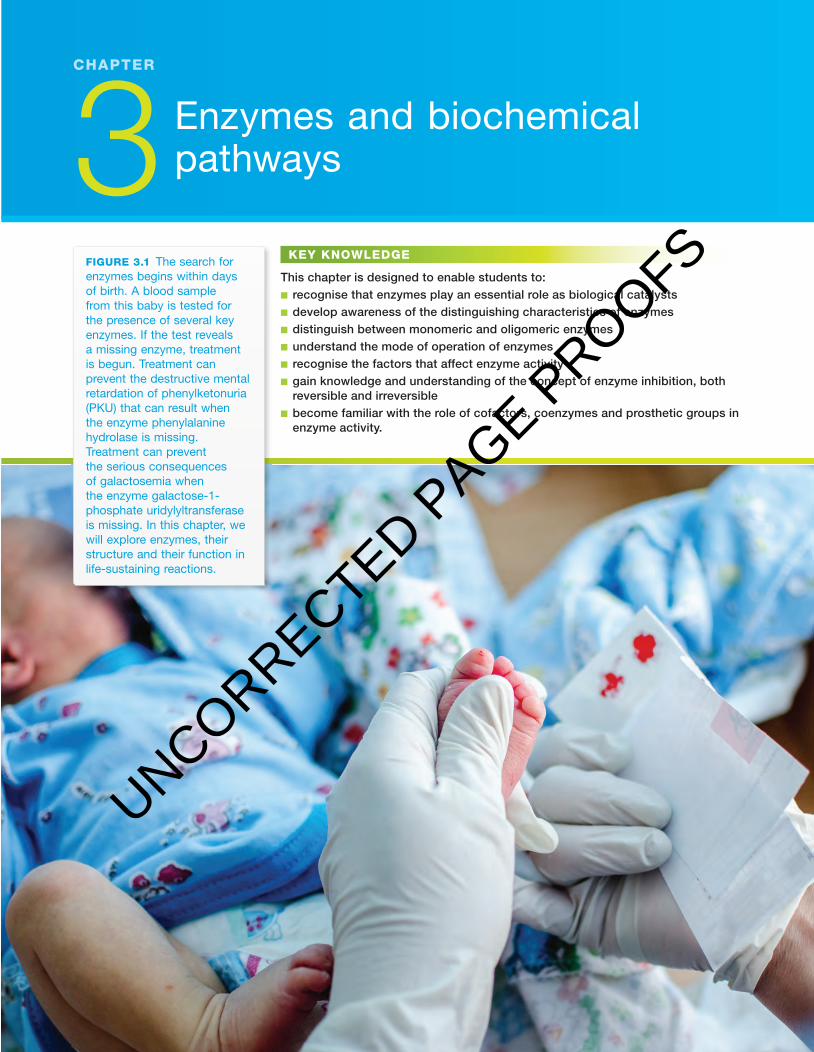
figuRE 3.1 The search for enzymes begins within days of birth. A blood sample from this baby is tested for the presence of several key enzymes. If the test reveals a missing enzyme, treatment is begun. Treatment can prevent the destructive mental retardation of phenylketonuria (PKU) that can result when the enzyme phenylalanine hydrolase is missing. Treatment can prevent the serious consequences of galactosemia when the enzyme galactose-1-phosphate uridylyltransferase is missing. In this chapter, we will explore enzymes, their structure and their function in life-sustaining reactions.
c03EnzymesAndBiochemicalPathways 77 24 October 2016 10:44 AM
UNCORRECTED PAGE P
ROOFS

Nature of biology 278
c03EnzymesAndBiochemicalPathways 78 24 October 2016 10:44 AM
the plague of the seaAs the long ocean voyage continued with no landfalls, the majority of the ship’s crew became increasingly ill and exhibited a range of unpleasant symptoms — their gums became swollen and purple in colour, teeth became loose in their sockets, skin discoloured and spontaneous haemorrhages occurred. Th e suf-fering of the sailors was compounded because even minor wounds failed to heal. Th en followed swelling, ulceration and, eventually, death. Th is illness described as ‘the plague of the sea’ came into prominence in the fi fteenth century when explorers fi rst ventured on extended ocean voyages or on expeditions to remote terrestrial regions in high latitudes. We now know this disease as scurvy.
Read a description of scurvy written by Jacques Cartier, an expedition leader who sailed from France in 1535 to present-day Canada and travelled up the Saint Lawrence River: ‘Some could not stand on their feet. Others also had their skins spotted with spots of blood of a purple colour then it did ascend up their ankles, knees, thighs, shoulders, arms and necks. Th eir mouths became stinking, their gums so rotten, that all their fl esh did fall off , even to the roots of their teeth which did also almost all fall out.’
What is scurvy?Scurvy is a defi ciency disease, resulting from a lack of vitamin C (ascorbic acid).
However, most animals can produce their own supplies of vitamin C by a multistep biochemical pathway that converts glucose to ascorbic acid. In con-trast, human beings and some other primates, guinea pigs and a few other animals cannot synthesise their own supplies of vitamin C. Instead, these animals must acquire vitamin C through their dietary intake. So, while the sailors on diets of dry beef and biscuits on long voyages during the fi fteenth to the eight-eenth centuries were at extreme risk of death by scurvy, the rats on these ships were scurvy-free because they could synthesise their own supplies of vitamin C.
Figure 3.2 shows the multistep pathway from glucose to ascorbic acid that occurs in the liver of almost all mammals (and in the kidneys of birds).
CHO
H
C OHH
C OHHO
C OHH
CH
CH2OH
C H
CO
C OHH
OH
C OH
OC
glucose glucuronate
gulonate
gulono-1,4-lactone
ascorbic acid(vitamin C)
E1 E2 E3
E4
E5
E6
GULO enzymeCH2OH
figuRE 3.2 Multistep biochemical pathway from glucose to vitamin C (ascorbic acid). Each step in this pathway is catalysed by a specifi c enzyme. Almost all mammals can synthesise vitamin C by this pathway, except for human beings and some other primates, guinea pigs, and bats of the genus Pteropus.
Odd fAcT
In 1520, Ferdinand Magellan lost more than 80 per cent of his crew while crossing the Pacifi c. In the 1740s, Commodore George Anson lost 1300 men from the crews of his fl eet, the majority of them dying from scurvy.
Odd fAcT
All primates except the lemurs, lorises and aye-ayes have lost the functional enzyme that catalyses the last step in the synthesis of vitamin C. So, people, apes, monkeys and tarsiers cannot manufacture their own vitamin C.
UNCORRECTED PAGE P
ROOFS

79CHaPter 3 Enzymes and biochemical pathways
c03EnzymesAndBiochemicalPathways 79 24 October 2016 10:44 AM
Th ose few mammals that cannot syn-thesise their own vitamin C are missing the terminal enzyme in the biosynthetic pathway of glucose to ascorbic acid. Th e gene encoding this enzyme, gulono-lactone oxidase, has undergone substan-tial mutation so that it no longer produces a protein that can function as an enzyme. Th is missing enzyme is the reason that sailors on long sea voyages during the fi fteenth to the eighteenth century suff ered from and were at risk of death from scurvy.
James Lind (1716–1794), a Scottish physician, was the fi rst to recognise that scurvy could be prevented by the juice of citrus fruits. In his Treatise of the Scurvy, published in 1753, Lind recommended
that citrus juice be distributed to sailors to prevent scurvy. In 1795, when the Royal Navy fi nally adopted the practice of issuing lime juice to sailors, scurvy disappeared from the crews of all their ships.
Many fresh fruits and vegetables are rich sources of vitamin C, as, for example, cantaloupe, cherries, kiwi fruit, and cabbage, caulifl ower, bean sprouts, red and green peppers, and potatoes (see fi gure 3.3).
the missing enzyme in scurvyTh ose few mammals, including humans, that cannot synthesise their own vitamin C are unable to produce an enzyme called gulono-lactone oxidase (GULO). Th is enzyme is needed to catalyse the last step in the pathway that manufac-tures ascorbic acid from glucose. In humans, the GULO gene, located on the number-8 chromosome, is defective.
When the defective GULO gene is compared to that of the rat, several major diff erences are seen: the functional GULO gene of the rat consists of a long sequence of DNA that includes 12 exons. In contrast, the homologous GULO gene in humans is missing most of these exons; it has only fi ve. Not surpris-ingly, the major deletions of DNA coding sequences mean that the protein product translated from this gene is non-functional as a GULO enzyme. Figure 3.4 shows a diagrammatic representation of the rat GULO gene and the corresponding gene in humans. In addition, compared to the rat GULO gene, the DNA of the remaining fi ve exons in the human gene shows other mutations including two single nucleotide deletions, one triple nucleotide deletion, and one single nucleotide insertion.
I(a)
(b)
II III IV V VI VII VIII IX X XI XII
figuRE 3.4 Diagram showing a comparison of (a) the gulo gene that encodes the active GULO enzyme in rats, and (b) the defective gulo gene of humans, which encodes a non-functional protein. The loss of exons in humans is also seen in the genome of most primates.
figuRE 3.3 Examples of foods that are rich sources of vitamin C (ascorbic acid).
UNCORRECTED PAGE P
ROOFS

Nature of biology 280
c03EnzymesAndBiochemicalPathways 80 24 October 2016 10:44 AM
Scurvy is just one example of the critical role of enzymes in maintaining the life of an organism. Every living organism has a myriad of enzymes that operate continuously to ensure that the cellular processes essential for life occur at rates sufficiently fast to maintain the living state.
At every moment, enzymes are jump-starting essential chemical reactions in cells and organisms, as, for example, the chemical reactions that:• enable some microbes to convert nitrogen in the air to essential amino acids• enable plants to convert the energy of sunlight to sugars• supply the energy needs of all cells, through cellular respiration• break down food of animals and fungi to forms that can be absorbed and
utilised through digestion• synthesise macromolecules that are essential to the structure and function
of mammals, including: – proteins, such as contractile proteins that enable movement, antibody
proteins that provide immunity against infectious diseases, signalling pro-teins that enable communication between cells, proteins that transport oxygen, and proteins that form the structure of bones, cartilage and con-nective tissue
– nucleic acids that determine aspects of heredity.Table 3.1 gives some examples of enzymes essential to these life-maintaining
processes.
TABLE 3.1 Examples of some enzymes involved in various biological processes. Processes involve multistep biochemical pathways, with each step catalysed by a specific enzyme.
Process example of enzyme
cellular respiration
glycolysis hexokinase, phosphofructokinase
Krebs cycle succinic dehyrogenase
electron transport chain cytochrome c oxidase
nucleic acid functions
DNA replication DNA polymerase
transcription of mRNA from DNA RNA polymerase
mRNA translation amino acyl tRNA synthetase
joining DNA segments ligase
cutting DNA endonucleases
digestion
breakdown of starch amylase
breakdown of proteins trypsin, pepsin
breakdown of lipids lipase
breakdown of galactose galactose-1-phosphate uridylyltransferase
breakdown of lactose lactase
metabolic pathways
formation of lactose lactose synthase
formation of uric acid (as in gout) xanthine oxidase
formation of melanin (pigment) tyrosinase
formation of collagen lysyl oxidase
formation of ascorbic acid (vit C) gulono-lactone oxidase
UNCORRECTED PAGE P
ROOFS
81CHaPter 3 Enzymes and biochemical pathways
c03EnzymesAndBiochemicalPathways 81 24 October 2016 10:44 AM
What is an enzyme?Th e key features of enzymes may be summarised as follows:• Enzymes are biological catalysts: Catalysts are substances that speed up
the rate of chemical reactions without themselves being used up in the reactions. Th e basic function of enzymes is to act as catalysts by increasing the rate of almost all the chemical reactions in living organisms, and to do this within the prevailing conditions of temperature and pH within cells. Later in this chapter (see page 88), we will explore how enzymes manage to speed up the rates of chemical reactions in cells.
Why is there a need to speed up chemical reactions in living organisms? You cut your hand on a piece of jagged metal and start bleeding. Enzymes are immediately activated, causing the blood in the damaged region to clot, plugging the wound and stopping the bleeding. Without the action of these enzymes, bleeding could continue, creating a life-threatening situation. Th e steak that you recently ate was broken down in the acidic conditions of your stomach at body temperature over a period of several hours. Th is breakdown was the result of the catalytic action of digestive enzymes such as pepsin and trypsin acting on the protein of the steak. To do the same without enzymes also requires acidic conditions, but would require a few days (not hours) and a temperature of 100 °C (not body temperature). Th ese two examples give a clue to the importance of enzymes in maintaining life. Without enzymes, the speed of biochemical reactions would be far too slow to sustain the living state.
• Enzymes are proteins: Almost without exception, enzymes are soluble globular proteins (in contrast to insoluble � brous protein, such as col-lagen and silk). Globular proteins are spherical or globe-like in overall shape, and are typically water-soluble. Water solubility results from the fact that, in a globular protein, the amino acids with polar side chains are exposed at the surface of the protein where they can interact with nearby polar water molecules and dissolve. In contrast, amino acids with nonpolar side chains tend to point inwards. (Refer to the Appendix to check out the diff erent side chains of amino acids.)
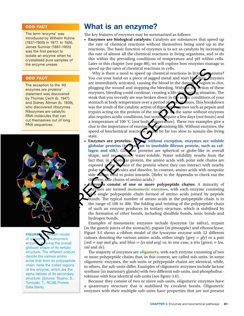
• Enzymes consist of one or more polypeptide chains: A minority of enzymes are termed monomeric enzymes, with each enzyme consisting of a single polypeptide chain formed of amino acids joined by peptide bonds. Th e typical number of amino acids in the polypeptide chain is in the range of 100 to 300. Th e folding and twisting of the polypeptide chain of such an enzyme produces its tertiary structure, which is stabilised by the formation of other bonds, including disulfi de bonds, ionic bonds and hydrogen bonds.Examples of monomeric enzymes include lysozyme (in saliva), trypsin
(in the gastric juices of the stomach), papain (in pineapple) and ribonuclease. Figure 3.5 shows a ribbon model of the lysozyme enzyme with 12 diff erent colours denoting the various amino acids, either singly (grey = gly) or a pair (red = asp and glu, and blue = lys and arg) or, in one case, a trio (green = leu, val and ile).
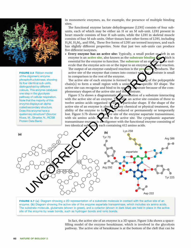
Th e majority of enzymes are oligomers, with each enzyme consisting of two or more polypeptide chains that, in this context, are called sub-units. In some oligomeric enzymes, the sub-units or polypeptide chains are identical, while, in others, the sub-units diff er. Examples of oligomeric enzymes include lactose synthase (in mammary glands) with two diff erent sub-units, and phosphofruc-tokinase with four identical sub-units (see fi gure 3.6).
Because they consist of two or more sub-units, oligomeric enzymes have a quaternary structure that is stabilised by covalent bonds. Oligomeric enzymes with their multiple sub-units have properties that are not present
Odd fAcT
The term ‘enzyme’ was introduced by Wilhelm Kuhne (1837–1900) in 1877. In 1926, James Sumner (1887–1955) was the fi rst person to isolate an enzyme when he crystallised pure samples of the enzyme urease.
Odd fAcT
The exception to the ‘All enzymes are proteins’ statement was discovered by Thomas Cech (b. 1947) and Sidney Altman (b. 1939), who discovered ribozymes. Ribozymes are catalytic RNA molecules that can cut themselves out of long RNA sequences.
figuRE 3.5 Ribbon model of lysozyme, a monomeric enzyme, showing the overall globular shape of its tertiary structure. The different colours denote the various amino acids that form its polypeptide chain. Note the coiled regions of the enzyme, which are the alpha helixes of its secondary structure. (Source: Tsujino, S., Tomizaki, T., RCSB Protein Data Bank)
UNCORRECTED PAGE P
ROOFS
Nature of biology 282
c03EnzymesAndBiochemicalPathways 82 24 October 2016 10:44 AM
seen on the left-hand side of the model. Figure 3.8b shows a stylised diagram of an active site of an enzyme with its substrate identifying the weak bonds that hold the substrate and the active site together. Th ese weak bonds are hydrogen bonds (H—H), and ionic bonds between groups on the substrate and groups on the active site that carry opposite charges. Th ese weak bonds are suffi cient to stabilise the enzyme-substrate (E-S) complex, but they can easily be broken, allowing the products of the reaction to be released, and freeing the active site to be occupied by another substrate molecule. (Later in this chapter we will see that, if a molecule forms a strong covalent bond with a group that is part of the active site of an enzyme, the enzyme will be inactivated.)
figuRE 3.8 (a) Space-fi lling model of a hexokinase enzyme with its active site at the base of the cleft shown at the left-hand side of the model. Different colours denote the various amino acids that form the single polypeptide chain of this enzyme. (Source: Feng, J., Zhao, S. and Liu, L., RCSB Protein Data Bank) (b) Schematic diagram showing the weak bonds, such as hydrogen bonds and ionic bonds, that temporarily stabilise the enzyme-substrate (E-S) complex at the active site of an enzyme.
(a)
H
RR
Active site
RH
−
+
+
−
Substrate
(b)
• Each enzyme has a specifi c activity: Th e specifi city of enzymes is demon-strated in various ways, including:
– absolute or substrate specifi city, meaning that an enzyme can act on one substrate only; for example, the enzyme lactase can only hydrolyse the milk sugar lactose to form glucose and galactose.
– bond specifi city, meaning that an enzyme can act on one kind of chemical bond; for example, peptidase enzymes act specifi cally on the peptide bonds between any two amino acids, and the enzyme amylase acts specifi cally on the 1,4 glycosidic bonds that link the glucose mono-mers of starch and glycogen.
– group specifi city, meaning that an enzyme can only act on molecules with particular functional group(s) surrounding a bond; for example, the digestive enzyme trypsin can only act on peptide bonds that are adjacent to amino acids with basic side chains, namely lysine and arginine.
a time and place for enzyme actionWhere and when does enzyme action occur? Most enzymes act on substrates in solution within cells of particular human tissues. So, most enzymes are intracellular.
However, some enzymes take action in extracellular locations, such as lysozyme in tears, which are part of the body’s fi rst line of defence against microbial infection (see chapter 7, page 287), the digestive enzymes such as amylase, which are produced in salivary glands, and the clotting enzyme thrombin. Th e digestive enzymes of the stomach, the pancreas and the small intestine are also examples of extracellular enzymes.
unit 3 Chemical nature of enzymesSummary screen and practice questions
AOs 1
Topic 5
concept 3
unit 3
see moreStructure and speci� city of enzymes
AOs 1
Topic 5
concept 3
in monomeric enzymes, as, for example, the presence of multiple binding sites.
Th e functional enzyme lactate dehydrogenase (LDH) consists of four sub-units, each of which may be either an H or an M sub-unit. LDH present in heart muscle consists of four H sub-units, while the LDH in skeletal muscle consists of four M sub-units. Other tissues have other forms of LDH, including H3M, H2M2, and HM3. Th ese fi ve forms of LDH are termed isozymes, and each has slightly diff erent properties. Note that just two sub-units can produce fi ve diff erent isozymes.• Every enzyme has an active site: Typically, a small pocket or cleft in an
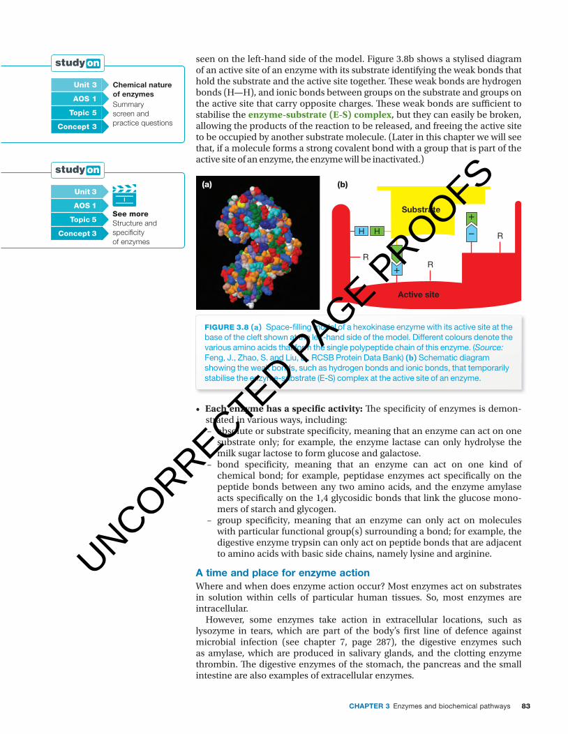
enzyme is an active site, also known as the substrate binding site, which is essential for the enzyme to function. Th e substrate of an enzyme is the mol-ecule that the enzyme acts on or the input to an enzyme-catalysed reaction. Th e output of an enzyme-catalysed reaction is the product or products. Th e active site of the enzyme that comes into contact with the substrate is small in comparison to the rest of the enzyme.Th e active site of each enzyme is formed by the folding of the polypeptide
chain(s) to form a small region with a unique stereospecifi c 3D shape. Th e active site can recognise and bind to its specifi c substrate because of the com-plementary shapes of the active site and the substrate.
Figure 3.7a shows a diagrammatic representation of a substrate interacting with the active site of an enzyme. Typically, an active site consists of three to twelve amino acids organised to form a particular shape. If the shape of the active site of an enzyme is altered by any chemical or physical treatment, the ability of the enzyme to function is reduced or permanently or temporarily lost. Figure 3.7b shows the active site of the enzyme aspartate transaminase, with six amino acids involved in the active site. Th e cytoplasmic aspartate transaminase enzyme is an oligomer with the functional enzyme consisting of two identical sub-units, each containing 412 amino acids.
figuRE 3.7 (a) Diagram showing a 2D representation of a substrate molecule in contact with the active site of an enzyme. (b) Diagram showing the active site of the enzyme aspartate transaminase, which includes six amino acids. The substrate molecule, glutamate (shown in green), and a cofactor (shown in dark blue) are held in place in the active site of the enzyme by weak bonds, such as hydrogen bonds and ionic bonds.
(a)
34
5
20
substrate
24 25 26
43
(b)
Arg 386
Lys 258
Tyr 70
Tyr 225
aspartate
Arg 292
Asp 222
In fact, the active site of an enzyme is a 3D space. Figure 3.8a shows a space-fi lling model of the enzyme hexokinase, which is involved in the glycolysis pathway. Th e active site of hexokinase is at the bottom of the cleft that can be
figuRE 3.6 Ribbon model of the oligomeric enzyme phosphofructokinase, showing its four identical sub-units, distinguished by different colours. This enzyme catalyses one step in the glycolysis pathway of cellular respiration. Note that the majority of this enzyme displays an alpha-coiled secondary structure. Does this enzyme have a quaternary structure? (Source: Kloos, M., Straeter, N., RCSB Protein Data Bank)
UNCORRECTED PAGE P
ROOFS
83CHaPter 3 Enzymes and biochemical pathways
c03EnzymesAndBiochemicalPathways 83 24 October 2016 10:44 AM
seen on the left-hand side of the model. Figure 3.8b shows a stylised diagram of an active site of an enzyme with its substrate identifying the weak bonds that hold the substrate and the active site together. Th ese weak bonds are hydrogen bonds (H—H), and ionic bonds between groups on the substrate and groups on the active site that carry opposite charges. Th ese weak bonds are suffi cient to stabilise the enzyme-substrate (E-S) complex, but they can easily be broken, allowing the products of the reaction to be released, and freeing the active site to be occupied by another substrate molecule. (Later in this chapter we will see that, if a molecule forms a strong covalent bond with a group that is part of the active site of an enzyme, the enzyme will be inactivated.)
figuRE 3.8 (a) Space-fi lling model of a hexokinase enzyme with its active site at the base of the cleft shown at the left-hand side of the model. Different colours denote the various amino acids that form the single polypeptide chain of this enzyme. (Source: Feng, J., Zhao, S. and Liu, L., RCSB Protein Data Bank) (b) Schematic diagram showing the weak bonds, such as hydrogen bonds and ionic bonds, that temporarily stabilise the enzyme-substrate (E-S) complex at the active site of an enzyme.
(a)
H
RR
Active site
RH
−
+
+
−
Substrate
(b)
• Each enzyme has a specifi c activity: Th e specifi city of enzymes is demon-strated in various ways, including:
– absolute or substrate specifi city, meaning that an enzyme can act on one substrate only; for example, the enzyme lactase can only hydrolyse the milk sugar lactose to form glucose and galactose.
– bond specifi city, meaning that an enzyme can act on one kind of chemical bond; for example, peptidase enzymes act specifi cally on the peptide bonds between any two amino acids, and the enzyme amylase acts specifi cally on the 1,4 glycosidic bonds that link the glucose mono-mers of starch and glycogen.
– group specifi city, meaning that an enzyme can only act on molecules with particular functional group(s) surrounding a bond; for example, the digestive enzyme trypsin can only act on peptide bonds that are adjacent to amino acids with basic side chains, namely lysine and arginine.
a time and place for enzyme actionWhere and when does enzyme action occur? Most enzymes act on substrates in solution within cells of particular human tissues. So, most enzymes are intracellular.
However, some enzymes take action in extracellular locations, such as lysozyme in tears, which are part of the body’s fi rst line of defence against microbial infection (see chapter 7, page 287), the digestive enzymes such as amylase, which are produced in salivary glands, and the clotting enzyme thrombin. Th e digestive enzymes of the stomach, the pancreas and the small intestine are also examples of extracellular enzymes.
unit 3 Chemical nature of enzymesSummary screen and practice questions
AOs 1
Topic 5
concept 3
unit 3
see moreStructure and speci� city of enzymes
AOs 1
Topic 5
concept 3
UNCORRECTED PAGE P
ROOFS
Nature of biology 284
c03EnzymesAndBiochemicalPathways 84 24 October 2016 10:44 AM
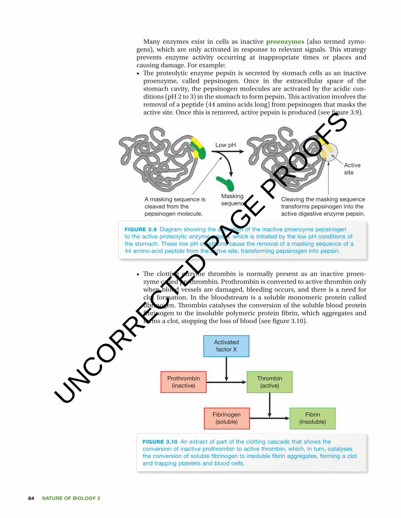
Many enzymes exist in cells as inactive proenzymes (also termed zymo-gens), which are only activated in response to relevant signals. Th is strategy prevents enzyme activity occurring at inappropriate times or places and causing damage. For example:• Th e proteolytic enzyme pepsin is secreted by stomach cells as an inactive
proenzyme, called pepsinogen. Once in the extracellular space of the stomach cavity, the pepsinogen molecules are activated by the acidic con-ditions (pH 2 to 3) in the stomach to form pepsin. Th is activation involves the removal of a peptide (44 amino acids long) from pepsinogen that masks the active site. Once this is removed, active pepsin is produced (see fi gure 3.9).
Low pH
Activesite
A masking sequence iscleaved from thepepsinogen molecule.
Cleaving the masking sequencetransforms pepsinogen into theactive digestive enzyme pepsin.
Maskingsequence
figuRE 3.9 Diagram showing the activation of the inactive proenzyme pepsinogen to the active proteolytic enzyme pepsin, which is initiated by the low pH conditions of the stomach. These low pH conditions cause the removal of a masking sequence of a 44 amino-acid peptide from the active site, transforming pepsinogen into pepsin.
• Th e clotting enzyme thrombin is normally present as an inactive proen-zyme called prothrombin. Prothrombin is converted to active thrombin only when blood vessels are damaged, bleeding occurs, and there is a need for clot formation. In the bloodstream is a soluble monomeric protein called fi brinogen. Th rombin catalyses the conversion of the soluble blood protein fi brinogen to the insoluble polymeric protein fi brin, which aggregates and forms a clot, stopping the loss of blood (see fi gure 3.10).
Activatedfactor X
Prothrombin(inactive)
Thrombin(active)
Fibrinogen(soluble)
Fibrin(insoluble)
figuRE 3.10 An extract of part of the clotting cascade that shows the conversion of inactive prothrombin to active thrombin, which, in turn, catalyses the conversion of soluble fi brinogen to insoluble fi brin aggregates, forming a clot and trapping platelets and blood cells.
UNCORRECTED PAGE P
ROOFS
85CHaPter 3 Enzymes and biochemical pathways
c03EnzymesAndBiochemicalPathways 85 24 October 2016 10:44 AM
NAmiNg ENzymEs
Usually an enzyme has two names, as follows:1. a recommended or trivial name that is short,
often ending in –ase; in many cases, the sub-strate of the enzyme is included in this name: for example, tyrosinase, lactase, sucrase, creatine kinase. (Recommended names are the enzyme names that are used throughout this chapter.) What reaction do you think the enzyme succinic dehydrogenase carries out?
2. a systematic name that is long and identifies the exact reaction that is catalysed by the enzyme; for example:• amylase, present in human saliva, has the system-
atic name 4-alpha-D-glucan glucanohydrolase• glucose oxidase has the systematic name
β-D-glucose:oxygen 1-oxidoreductase• aspartate aminotransferase has the systematic
name L-aspartate:2-oxoglutarate aminotransferase
• tyrosinase has the systematic name L-tyrosine, L-dopa:oxygen oxidoreductase.
In addition, each enzyme has an EC (Enzyme Commission) number. The EC numbers group enzymes into one of six groups identifying the types of enzyme-catalysed reaction that are carried out. Table 3.2 shows these six groups. Each enzyme has an EC identifier that consists of four numbers, such as:
Tyrosinase is identified as EC 1.14.18.1; glucose isomerase is identified as EC 5.3.1.5; aspartate aminotransferase is identified as EC 2.6.1.1.
The first number identifies the major reaction type that the enzyme can catalyse. So, any enzyme with the first EC number of 2 must be a transferase enzyme, which catalyses a particular transferase reaction.
TABLE 3.2 The Enzyme Commission (EC) groups. Every enzyme belongs to one of these six groups, which identify the types of chemical reactions that are catalysed by enzymes.
group reaction type catalysed example of enzyme
EC 1Oxidoreductases
oxidise/reduce molecules by transfer of O and H atoms succinic dehydrogenase
EC 2Transferases
transfer a group from substrate to product aspartate transaminase
EC 3 Hydrolases
cleave chemical bonds by hydrolysis amylase
EC 4Lyases
cleave bonds in a substrate (not by hydrolysis) aldolase
EC 5Isomerases
change arrangement of atoms in a molecule glucose isomerase
EC 6Ligases
join two molecules by forming a new bond glutamate-cysteine ligase
kEy idEAs
■ Enzymes are biological catalysts composed of protein. ■ Catalysts speed up the rate of chemical reactions. ■ Some enzymes are monomeric and consist of a single polypeptide chain, while most are oligomers.
■ The active site of an enzyme has a 3D shape that is complementary to the shape of its specific substrate.
Odd fAcT
The enzyme known as pepsin (EC 3.4.23.2) was given its name in 1836 by Theodor Schwann (1810–1882), who, along with Matthias Schleiden (1804–1881), formulated the Cell Theory in 1839.
UNCORRECTED PAGE P
ROOFS
Nature of biology 286
c03EnzymesAndBiochemicalPathways 86 24 October 2016 10:44 AM
Quick chEck
1 Briefly explain the meaning of the following statement: Enzymes are biological catalysts.
2 Identify a key difference between the members of the following pairs:a monomeric enzyme and oligomeric enzymeb proenzyme and enzymec substrate specificity and bond specificityd hydrolase and transferase.
3 Identify the following statements as true or false:a The typical shape of an enzyme is globular.b The enzyme lysozyme consists of a single polypeptide chain.c Most of the structure of an enzyme forms part of its active site.d Most enzymes are oligomers.e Enzymes speed up the rate of chemical reactions in the body.f Each enzyme can catalyse just one specific reaction involving a single
substrate.g The E-S complex is stabilised by strong covalent bonds.
explaining enzyme specificityLet’s now look at why enzymes have a high level of specificity for one substrate, or for a functional group or side chain, or for a bond between particular groups. To do this, let’s use models. What is a model? Models are used to represent biological entities, such as biological processes, in a way that makes them conceptually easier to understand or enables predictions to be made about their operation. A model may be an analogy, such as the lock-and-key model of enzyme-substrate interactions described below. A model may be a math-ematical representation, such as the Hardy-Weinberg equation to represent a population gene pool.
lock and key or induced fit?The essential prelude to the catalytic action of an enzyme is the binding of the substrate(s) to the active site of the enzyme. Two models have been put forward in an attempt to explain this specificity:1. the lock-and-key model2. the induced fit model.
The earliest explanation for the specificity of enzymes for particular sub-strates was proposed by Emil Fischer (1852–1919) in 1894. Fischer explained the specificity of enzymes using the analogy of a lock and key. In this lock-and-key model, the enzyme acts as the lock and the substrate as the key. Only the cor-rectly shaped key (substrate) can fit the lock (the active site of the enzyme). This model correctly identified the importance of the complementary shapes of the active site of an enzyme and its specific substrate and recognised that enzyme specificity is determined by the active site of the enzyme. The lock-and-key model assumes that the active site of an enzyme (the lock) is rigid and fixed.
A second model was premised on the active site of an enzyme having a defined shape, but one that has a degree of flexibility. This is the induced fit model, which was proposed by Daniel Koshland in 1958. Koshland proposed that, just as a glove changes its shape when a hand slips into it, so does the active site change its shape to fit tightly around its substrate. In this model, the binding site of an enzyme is not viewed as being exactly complementary to the sub-strate. When the reaction is complete, the empty active site returns to its relaxed resting state.
Figure 3.11 shows the difference between these two models.
UNCORRECTED PAGE P
ROOFS

87CHaPter 3 Enzymes and biochemical pathways
c03EnzymesAndBiochemicalPathways 87 24 October 2016 10:44 AM
(a) Lock-and-key model
SubstrateProducts
Enzyme–substratecomplex
EnzymeEnzyme
(b) Induced-fit model
SubstrateProducts
Enzyme–substratecomplex
EnzymeEnzyme
1 23
1 23 1 2
3
figuRE 3.11 Two models to explain enzyme specifi city are shown. (a) The ‘lock-and-key’ model and (b) the ‘induced fi t’ model. Examine these fi gures carefully. In which model is the shape of the active site (shown in red) exactly complementary to the shape of the substrate? In which model does the shape of the active site change slightly when it binds to the substrate, making a tight fi t?
Mode of action of enzymesAt any minute, hundreds of chemical reactions are taking place in living organisms, and this multitude of linked chemical reactions comprises the metabolism of an organism. Most of these chemical reactions occur inside cells, but a few are extracellular in location. Almost without exception, every step in a metabolic pathway is catalysed by a specifi c enzyme. In a person, enzyme-managed reactions are involved in growth, repair, cellular respiration, reproduction, digestion, breathing, clotting of blood, and immunity, with several thousand diff erent enzymes involved.
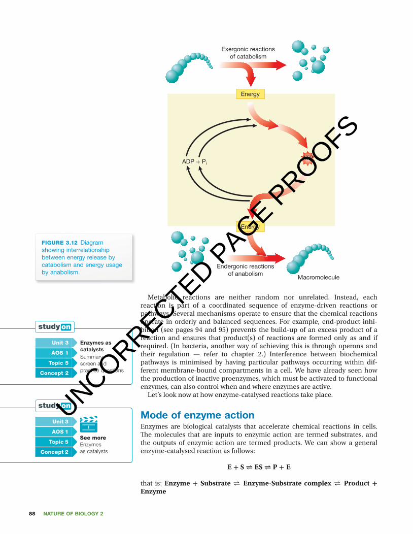
In metabolism, some compounds are broken down with an associated release of energy, while other compounds that are needed by cells are built up or synthesised in a process that requires an input of energy. Th ese two aspects of metabolism are recognised in the following classifi cation:• catabolism = series of cellular reactions that releases useful energy from the
breakdown of complex molecules• anabolism = series of cellular reactions in which the complex molecules
required by cells are synthesised from simpler building blocks, a process that requires an input of energy.Metabolic reactions that release energy are said to be exergonic, while
reactions that require energy are termed endergonic. Figure 3.12 shows how the energy released in catabolic reactions drives the energy-requiring anabolic reactions. Th is illustrates the principle of coupling of exergonic and ender-gonic reactions
unit 3 Metabolic reactions Summary screen and practice questions
AOs 1
Topic 5
concept 1
UNCORRECTED PAGE P
ROOFS
Nature of biology 288
c03EnzymesAndBiochemicalPathways 88 24 October 2016 10:44 AM
Energy
ADP + PiATP
Exergonic reactionsof catabolism
Endergonic reactionsof anabolism
Macromolecule
Energy
figuRE 3.12 Diagram showing interrelationship between energy release by catabolism and energy usage by anabolism.
Metabolic reactions are neither random nor unrelated. Instead, each reaction is part of a coordinated sequence of enzyme-driven reactions or pathways. Several mechanisms operate to ensure that the chemical reactions operate in orderly and balanced sequences. For example, end-product inhi-bition (see pages 94 and 95) prevents the build-up of an excess product of a reaction and ensures that product(s) of reactions are formed only as and if required. (In bacteria, another way of achieving this is through operons and their regulation — refer to chapter 2.) Interference between biochemical pathways is minimised by having particular pathways occurring within dif-ferent membrane-bound compartments in a cell. We have already seen how the production of inactive proenzymes, which must be activated to functional enzymes, can also control when and where enzymes are active.
Let’s look now at how enzyme-catalysed reactions take place.
Mode of enzyme actionEnzymes are biological catalysts that accelerate chemical reactions in cells. Th e molecules that are inputs to enzymic action are termed substrates, and the outputs of enzymic action are termed products. We can show a general enzyme-catalysed reaction as follows:
E + S ⇌ ES ⇌ P + E
that is: Enzyme + Substrate ⇌ Enzyme-Substrate complex ⇌ Product + Enzyme
unit 3 enzymes as catalystsSummary screen and practice questions
AOs 1
Topic 5
concept 2
unit 3
see moreEnzymes as catalysts
AOs 1
Topic 5
concept 2
UNCORRECTED PAGE P
ROOFS
89CHaPter 3 Enzymes and biochemical pathways
c03EnzymesAndBiochemicalPathways 89 24 October 2016 10:44 AM
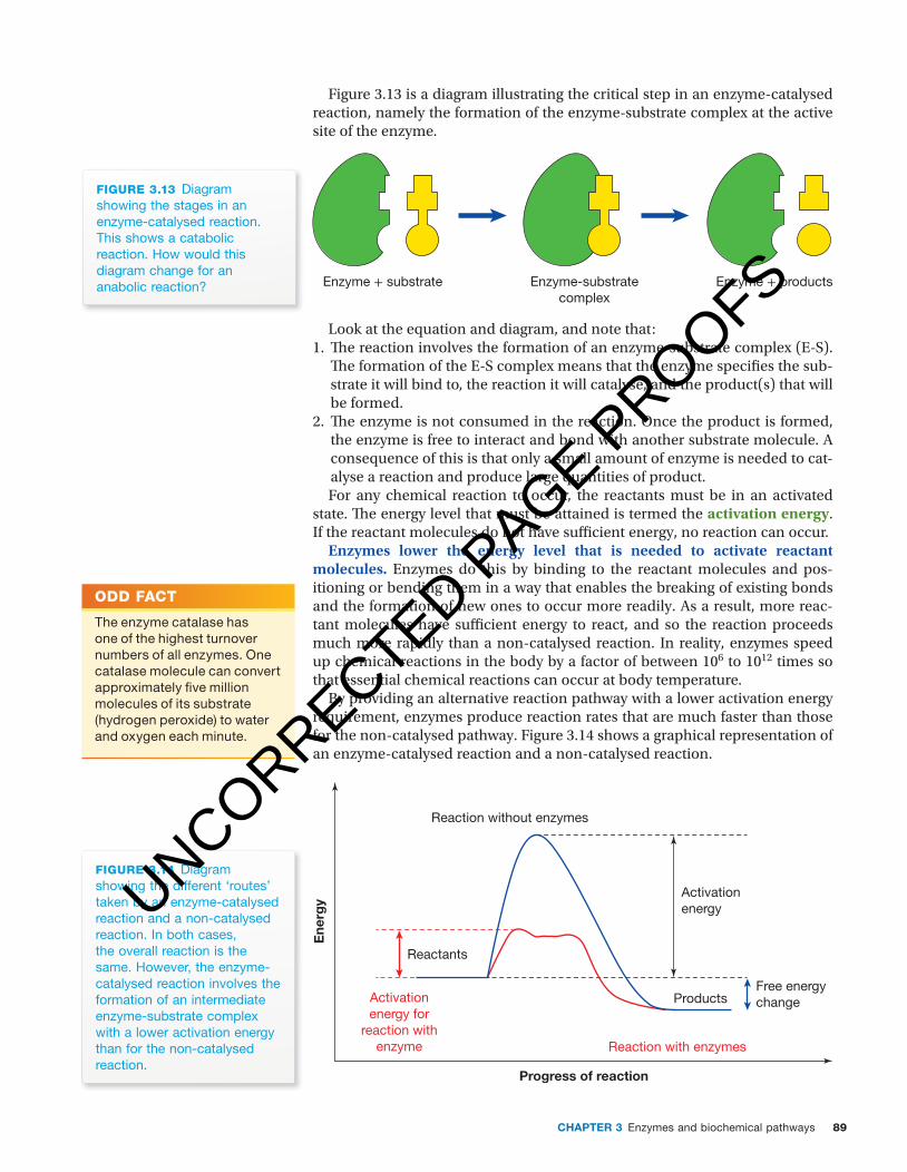
Figure 3.13 is a diagram illustrating the critical step in an enzyme-catalysed reaction, namely the formation of the enzyme-substrate complex at the active site of the enzyme.
Enzyme + productsEnzyme-substratecomplex
Enzyme + substrate
figuRE 3.13 Diagram showing the stages in an enzyme-catalysed reaction. This shows a catabolic reaction. How would this diagram change for an anabolic reaction?
Look at the equation and diagram, and note that:1. Th e reaction involves the formation of an enzyme-substrate complex (E-S).
Th e formation of the E-S complex means that the enzyme specifi es the sub-strate it will bind to, the reaction it will catalyse, and the product(s) that will be formed.
2. Th e enzyme is not consumed in the reaction. Once the product is formed, the enzyme is free to interact and bond with another substrate molecule. A consequence of this is that only a small amount of enzyme is needed to cat-alyse a reaction and produce large quantities of product.For any chemical reaction to occur, the reactants must be in an activated
state. Th e energy level that must be attained is termed the activation energy. If the reactant molecules do not have suffi cient energy, no reaction can occur.
Enzymes lower the energy level that is needed to activate reactant molecules. Enzymes do this by binding to the reactant molecules and pos-itioning or bending them in a way that enables the breaking of existing bonds and the formation of new ones to occur more readily. As a result, more reac-tant molecules have suffi cient energy to react, and so the reaction proceeds much more rapidly than a non-catalysed reaction. In reality, enzymes speed up chemical reactions in the body by a factor of between 106 to 1012 times so that essential chemical reactions can occur at body temperature.
By providing an alternative reaction pathway with a lower activation energy requirement, enzymes produce reaction rates that are much faster than those for the non-catalysed pathway. Figure 3.14 shows a graphical representation of an enzyme-catalysed reaction and a non-catalysed reaction.
figuRE 3.14 Diagram showing the different ‘routes’ taken by an enzyme-catalysed reaction and a non-catalysed reaction. In both cases, the overall reaction is the same. However, the enzyme-catalysed reaction involves the formation of an intermediate enzyme-substrate complex with a lower activation energy than for the non-catalysed reaction.
Ene
rgy
Progress of reaction
Activationenergy
ProductsFree energychange
Reaction with enzymes
Activationenergy for
reaction withenzyme
Reaction without enzymes
Reactants
Odd fAcT
The enzyme catalase has one of the highest turnover numbers of all enzymes. One catalase molecule can convert approximately fi ve million molecules of its substrate (hydrogen peroxide) to water and oxygen each minute.
UNCORRECTED PAGE P
ROOFS
Nature of biology 290
c03EnzymesAndBiochemicalPathways 90 24 October 2016 10:44 AM
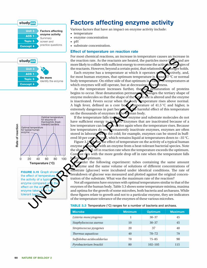
factors affecting enzyme activityVarious factors that have an impact on enzyme activity include:• temperature• enzyme concentration• pH• substrate concentration.
effect of temperature on reaction rateFor most chemical reactions, an increase in temperature causes an increase in the reaction rate. As the reactants are heated, the particles move faster and are more likely to collide with suffi cient energy to overcome the activation energies of the reactants. However, beyond a certain point, that relationship no longer holds.
Each enzyme has a temperature at which it operates most eff ectively, and, for most human enzymes, that optimum temperature is about 37 °C or normal body temperature. On either side of that optimum is a range of temperatures at which enzymes will still operate, but at decreasing eff ectiveness.
As the temperature increases further, thermal denaturation of proteins begins to occur. Heat denaturation permanently changes the tertiary shape of enzyme molecules so that the shape of the active site is altered and the enzyme is inactivated. Fevers occur when the body temperature rises above normal. A high fever, defi ned as a core body temperature of 41.5 °C and higher, is extremely dangerous in part because of the harmful eff ect of this temperature on the thousands of enzymes in the human body.
If the temperature falls too low, the enzyme and substrate molecules do not have suffi cient energy to interact. Enzymes that are inactivated because of a low temperature can become active again when the temperature rises. Because low temperatures do not permanently inactivate enzymes, enzymes are often stored in laboratories in the cold; for example, enzymes can be stored in buff -ered 50 per cent glycerol, which remains liquid at temperatures down to –35 °C.
Figure 3.15 shows the eff ect of temperature on the activity of a typical human enzyme compared with an enzyme from a heat-tolerant bacterial species. Note the sharper fall off in reaction rate when the temperature exceeds the optimum. Contrast this with the more gentle drop off in rate when the temperature falls below the optimum.
Consider the following experiment: tubes containing the same amount of enzyme and the same volume of solutions of diff erent concentrations of substrate (glucose) were incubated under identical conditions. Th e rate of breakdown of glucose was measured and plotted against the original concen-tration of the substrate. What was the maximum rate of the reaction?
Not all organisms have enzymes with optimal temperatures similar to that of the enzymes of the human body. Table 3.3 shows some temperature minima, maxima and optima for the growth of some microbes, both bacteria and archaeans. While these fi gures relate to growth and not to a particular enzyme, they are indicators of the temperature tolerance of the enzymes of these various microbes.
TABLE 3.3 Temperature (°C) ranges for a number of bacteria and archaea.
Microbe Minimum optimum Maximum
Listeria moncytogenes 1 30–37 45
Staphylococcus aureus 10 30–37 45
Streptococcus pyogenes 20 37 40
Th ermus aquaticus 40 70–72 79
Sulfolobus acidocaldarius 70 75–85 90
Pyrobacterium brackii 80 102–105 115
unit 3 factors affecting enzyme activity Summary screen and practice questions
AOs 1
Topic 5
concept 4
unit 3
do moreIdentify the enzyme
AOs 1
Topic 5
concept 4
figuRE 3.15 Graph showing the effect of temperature on the activity of a typical human enzyme compared with the effect on the activity of an enzyme taken from heat-tolerant bacteria.
Temperature (°C)
Rat
e o
f re
acti
on
1000 20 40 60 80
Optimal temperaturefor typical human enzyme
Optimal temperaturefor enzyme of thermophilic (heat-tolerant) bacteria
0%1 2 3 4 5
Carbonicanhydrase
Pepsin
Trypsin
6 7 8 9 10 11
25%
50%
Rat
e o
f re
acti
on
pH
Enzyme activity vs pH
75%
100%
figuRE 3.16 Graph showing the effect of pH on the reaction rate of three human enzymes. Can you suggest why the pH optima of these enzymes are so different from one another?
UNCORRECTED PAGE P
ROOFS

91CHaPter 3 Enzymes and biochemical pathways
c03EnzymesAndBiochemicalPathways 91 24 October 2016 10:44 AM
Enzymes synthesised by bacteria and archaea with optimal growth temperatures greater than 80 °C are termed hyperther-mophilic enzymes. In contrast to the enzymes of most organisms, these enzymes are resistant to irreversible denaturation at high temperatures, and are said to be thermostable.
effect of pH on reaction rateTh e pH (hydrogen ion concentration) at which enzymes can operate varies, typically according to the environment in which the enzyme normally operates (see fi gure 3.16). Unlike the normal human core body, which is regulated within a narrow range, various compartments of the human body have very dif-ferent prevailing pH values, such as the stomach with a pH of 2 to 3, and the small intestine with a pH around 5.
Enzymes can operate as catalysts within a range that is defi ned by a maximum pH and a minimum pH, with the highest
activity (in terms of substrate molecules catalysed per unit time) occurring at the optimal pH for the enzyme concerned. What is the pH optimum for the enzyme alkaline phosphatase?
Usually, an enzyme works within quite a narrow pH range. At the optimal pH, the reaction rate is greatest. Changes in pH can alter intra- and intermo-lecular bonds, changing the shape of the enzyme and aff ecting its effi ciency
Th e pH of a solution is a measure of the concentration of hydrogen ions per litre of solution. Th e pH of a neutral solution, such as pure water, is 7.0. A pH value below 7 indicates an acid — the lower the number, the more acidic the solution. A pH value above 7 indicates a basic solution. Most biological fl uids have a pH between 6 and 8; for example, human blood is maintained at about pH 7.4. Th ere are a few extremes beyond this range, such as gastric juice in the stomach, which has a pH of about 2.
Each enzyme acts best at a particular pH, which is known as the optimal pH for the enzyme (see fi gure 3.16). Pepsin, which is secreted in the human stomach, has an optimal pH of 2. Trypsin, secreted in the human small intestine, has an optimal pH of about 8.0. Carbonic anhydrase, an enzyme found in human blood, has an optimal pH of 7.4. Enzymes that act within human cells generally have an optimal pH of about 7.6. A change in pH from the optimum can change the shape of an enzyme and aff ect its ability to combine with its substrate. Because the enzyme is less able to combine with its substrate, it is unable to act and the rate of the metabolic reaction declines. An enzyme becomes less effi cient if the variable value is greater or less than the optimal.
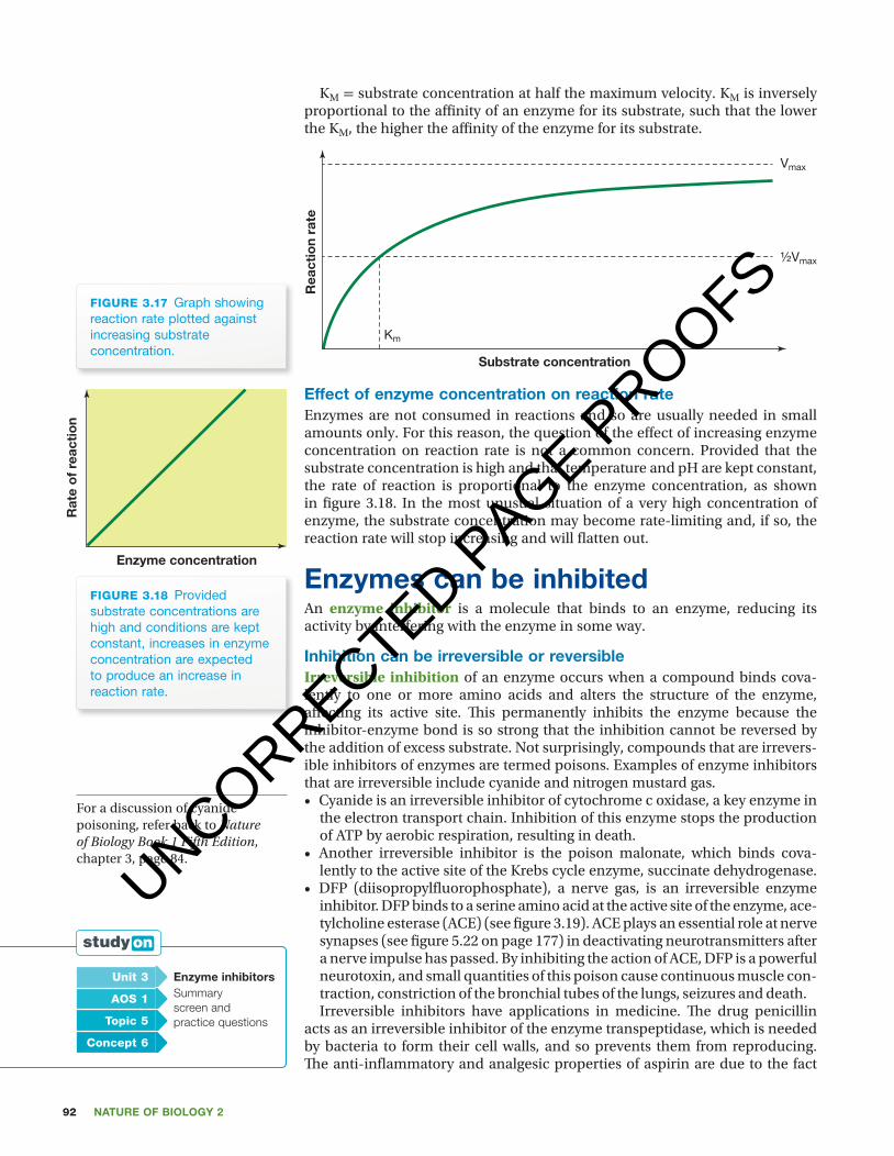
effect of substrate concentration on reaction rateTh e addition of more substrate to an enzyme solution initially increases the rate of the reaction, provided that some active sites of the enzyme are not yet occupied. However, a set amount of enzyme molecules is present, and the rate of the reaction tapers off once all the active sites of the enzyme molecules become occupied.
So, for a given enzyme concentration, the rate of reaction increases with increasing substrate concentration. But only up to a point. Beyond this, any further increase in substrate concentration produces no signifi cant change in reaction rate (see fi gure 3.17). At this point, all the active sites of the enzyme molecules at any given moment are occupied by substrate molecules. Th e E-S complexes (enzyme/substrate complexes) have to dissociate before the active sites are free to accommodate more substrate.
On such a graph, two values are typically noted:Vmax = maximum reaction rate or velocity. Vmax is reached when all the active
sites of the enzyme are saturated with substrate molecules. Vmax is directly pro-portional to enzyme concentration.
unit 3 the rate of a reactionSummary screen and practice questions
AOs 1
Topic 5
concept 5
factors affecting enzyme activityVarious factors that have an impact on enzyme activity include:• temperature• enzyme concentration• pH• substrate concentration.
effect of temperature on reaction rateFor most chemical reactions, an increase in temperature causes an increase in the reaction rate. As the reactants are heated, the particles move faster and are more likely to collide with suffi cient energy to overcome the activation energies of the reactants. However, beyond a certain point, that relationship no longer holds.
Each enzyme has a temperature at which it operates most eff ectively, and, for most human enzymes, that optimum temperature is about 37 °C or normal body temperature. On either side of that optimum is a range of temperatures at which enzymes will still operate, but at decreasing eff ectiveness.
As the temperature increases further, thermal denaturation of proteins begins to occur. Heat denaturation permanently changes the tertiary shape of enzyme molecules so that the shape of the active site is altered and the enzyme is inactivated. Fevers occur when the body temperature rises above normal. A high fever, defi ned as a core body temperature of 41.5 °C and higher, is extremely dangerous in part because of the harmful eff ect of this temperature on the thousands of enzymes in the human body.
If the temperature falls too low, the enzyme and substrate molecules do not have suffi cient energy to interact. Enzymes that are inactivated because of a low temperature can become active again when the temperature rises. Because low temperatures do not permanently inactivate enzymes, enzymes are often stored in laboratories in the cold; for example, enzymes can be stored in buff -ered 50 per cent glycerol, which remains liquid at temperatures down to –35 °C.
Figure 3.15 shows the eff ect of temperature on the activity of a typical human enzyme compared with an enzyme from a heat-tolerant bacterial species. Note the sharper fall off in reaction rate when the temperature exceeds the optimum. Contrast this with the more gentle drop off in rate when the temperature falls below the optimum.
Consider the following experiment: tubes containing the same amount of enzyme and the same volume of solutions of diff erent concentrations of substrate (glucose) were incubated under identical conditions. Th e rate of breakdown of glucose was measured and plotted against the original concen-tration of the substrate. What was the maximum rate of the reaction?
Not all organisms have enzymes with optimal temperatures similar to that of the enzymes of the human body. Table 3.3 shows some temperature minima, maxima and optima for the growth of some microbes, both bacteria and archaeans. While these fi gures relate to growth and not to a particular enzyme, they are indicators of the temperature tolerance of the enzymes of these various microbes.
TABLE 3.3 Temperature (°C) ranges for a number of bacteria and archaea.
Microbe Minimum optimum Maximum
Listeria moncytogenes 1 30–37 45
Staphylococcus aureus 10 30–37 45
Streptococcus pyogenes 20 37 40
Th ermus aquaticus 40 70–72 79
Sulfolobus acidocaldarius 70 75–85 90
Pyrobacterium brackii 80 102–105 115
unit 3 factors affecting enzyme activity Summary screen and practice questions
AOs 1
Topic 5
concept 4
unit 3
do moreIdentify the enzyme
AOs 1
Topic 5
concept 4
figuRE 3.15 Graph showing the effect of temperature on the activity of a typical human enzyme compared with the effect on the activity of an enzyme taken from heat-tolerant bacteria.
Temperature (°C)
Rat
e o
f re
acti
on
1000 20 40 60 80
Optimal temperaturefor typical human enzyme
Optimal temperaturefor enzyme of thermophilic (heat-tolerant) bacteria
0%1 2 3 4 5
Carbonicanhydrase
Pepsin
Trypsin
6 7 8 9 10 11
25%
50%
Rat
e o
f re
acti
on
pH
Enzyme activity vs pH
75%
100%
figuRE 3.16 Graph showing the effect of pH on the reaction rate of three human enzymes. Can you suggest why the pH optima of these enzymes are so different from one another?
UNCORRECTED PAGE P
ROOFS
Nature of biology 292
c03EnzymesAndBiochemicalPathways 92 24 October 2016 10:44 AM
KM = substrate concentration at half the maximum velocity. KM is inversely proportional to the affi nity of an enzyme for its substrate, such that the lower the KM, the higher the affi nity of the enzyme for its substrate.
figuRE 3.17 Graph showing reaction rate plotted against increasing substrate concentration.
Vmax
½Vmax
Km
Rea
ctio
n ra
te
Substrate concentration
effect of enzyme concentration on reaction rateEnzymes are not consumed in reactions and so are usually needed in small amounts only. For this reason, the question of the eff ect of increasing enzyme concentration on reaction rate is not a common concern. Provided that the substrate concentration is high and that temperature and pH are kept constant, the rate of reaction is proportional to the enzyme concentration, as shown in fi gure 3.18. In the most unusual situation of a very high concentration of enzyme, the substrate concentration may become rate-limiting and, if so, the reaction rate will stop increasing and will fl atten out.
enzymes can be inhibitedAn enzyme inhibitor is a molecule that binds to an enzyme, reducing its activity by interfering with the enzyme in some way.
inhibition can be irreversible or reversibleIrreversible inhibition of an enzyme occurs when a compound binds cova-lently to one or more amino acids and alters the structure of the enzyme, aff ecting its active site. Th is permanently inhibits the enzyme because the inhibitor-enzyme bond is so strong that the inhibition cannot be reversed by the addition of excess substrate. Not surprisingly, compounds that are irrevers-ible inhibitors of enzymes are termed poisons. Examples of enzyme inhibitors that are irreversible include cyanide and nitrogen mustard gas.• Cyanide is an irreversible inhibitor of cytochrome c oxidase, a key enzyme in
the electron transport chain. Inhibition of this enzyme stops the production of ATP by aerobic respiration, resulting in death.
• Another irreversible inhibitor is the poison malonate, which binds cova-lently to the active site of the Krebs cycle enzyme, succinate dehydrogenase.
• DFP (diisopropylfl uorophosphate), a nerve gas, is an irreversible enzyme inhibitor. DFP binds to a serine amino acid at the active site of the enzyme, ace-tylcholine esterase (ACE) (see fi gure 3.19). ACE plays an essential role at nerve synapses (see fi gure 5.22 on page 177) in deactivating neurotransmitters after a nerve impulse has passed. By inhibiting the action of ACE, DFP is a powerful neurotoxin, and small quantities of this poison cause continuous muscle con-traction, constriction of the bronchial tubes of the lungs, seizures and death.Irreversible inhibitors have applications in medicine. Th e drug penicillin
acts as an irreversible inhibitor of the enzyme transpeptidase, which is needed by bacteria to form their cell walls, and so prevents them from reproducing. Th e anti-infl ammatory and analgesic properties of aspirin are due to the fact
Rat
e o
f re
acti
on
Enzyme concentration
figuRE 3.18 Provided substrate concentrations are high and conditions are kept constant, increases in enzyme concentration are expected to produce an increase in reaction rate.
For a discussion of cyanide poisoning, refer back to Nature of Biology Book 1 Fifth Edition, chapter 3, page 84.
unit 3 enzyme inhibitorsSummary screen and practice questions
AOs 1
Topic 5
concept 6
UNCORRECTED PAGE P
ROOFS

93CHaPter 3 Enzymes and biochemical pathways
c03EnzymesAndBiochemicalPathways 93 24 October 2016 10:44 AM
that this drug inhibits the activity of the platelet enzyme cyclooxygenase (COX) by reacting, chemically changing an amino acid at the active site.
O
H
O
O
Inactivatedenzyme
O
H
CH3
CH3
CH3
CH3
O
P
O
H
F
OSer
Acetylcholineesterase
DFP
OH
H
CH3
CH3
CH3
CH3
O
PfiguRE 3.19 Diagram showing the irreversible inhibition of acetylcholine esterase (ACE) enzyme by the nerve gas DFP. Note that DFP reacts with the side chain of a serine amino acid (Ser) at the active site of the ACE molecule, forming a strong covalent bond that blocks the active site.
In reversible inhibition, the enzyme is not permanently inhibited or damaged. Reversible inhibitors inactivate enzymes through noncovalent interactions that can be reversed. In contrast to the situation with an irrevers-ible inhibitor, an inhibitor that is reversible can dissociate from the enzyme. Removal of the inhibitor reverses the inhibition.
Two types of reversible inhibition are competitive inhibition and non-competitive inhibition.
Competitive inhibitionCompetitive inhibition: Competitive enzyme inhibitors have a similar shape to the usual substrate molecule and so can bind to the active site of the enzyme, preventing the formation of the enzyme-substrate (E-S) complexes. As a result, over a given period of time, fewer substrate molecules bind to the active site of the enzyme so that the rate of the reaction is decreased (see fi gure 3.20).
figuRE 3.20 (a) In competitive inhibition, inhibitor molecules compete with the substrate molecules for access and bind to the active site of the enzyme. (b) Graph showing the rate of enzyme action versus substrate concentration in the absence of a competitive inhibitor and in the presence of a competitive inhibitor.
Substrate concentration
Competitive inhibitionpresent
No inhibitorpresent
Rat
e o
f rea
ctio
n
(b)
Enzyme
Competitiveinhibitor
Active site
Competitive inhibition
Substrate
(a)
UNCORRECTED PAGE P
ROOFS

Nature of biology 294
c03EnzymesAndBiochemicalPathways 94 24 October 2016 10:44 AM
Eventually, the inhibitor moves from the active site, so the competitive inhi-bition is usually temporary.
Because the substrate and the competitive inhibitor are competing for the same active sites on enzyme molecules, the degree of inhibition produced depends on the relative concentrations of substrate and inhibitor molecules. For example, adding more substrate molecules increases the chance that a random collision with the active site will be that involving a substrate molecule rather than that involving a molecule of the competitive inhibitor.
Non-competitive inhibitionNon-competitive inhibitors do not bind to the active site, but bind to another site on the enzyme molecule called the allosteric site. Th e binding of the non-competitive inhibitor distorts the 3D shape of the enzyme so that it can no longer bind to its substrate and, therefore, can no longer catalyse the usual reaction (see fi gure 3.21). Adding more substrate molecules does not have any eff ect on this non-competitive inhibition. Can you suggest why?
EnzymeNon-competitiveinhibitor
Allosteric site
Distortedshape
Active site
Non-competitive inhibition
Substrate
(b)
Substrate concentration
Competitive inhibitionpresent
Non-competitiveinhibition present
No inhibitorpresent
Rat
e o
f rea
ctio
n
(a)
figuRE 3.21 (a) Graph showing rates of enzyme action versus substrate concentration in the absence of inhibitor (black line), in the presence of a non-competitive inhibitor (red line), and in the presence of a competitive inhibitor (blue line). These two different inhibitors bind to different sites on the enzyme molecule, with (b) showing the case for non-competitive inhibitors. Only in one case do the inhibitor molecules compete with substrate molecules for the same site on the enzyme. Which type of inhibition is that?
In summary, enzymes may have several binding sites, the most well known being their active site or substrate-binding site, while others may be present, including allosteric sites where binding inhibits enzyme function.
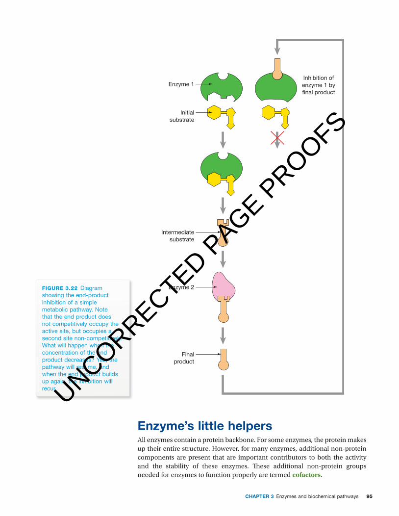
Th e presence of an allosteric binding site on an enzyme enables regu-lation of metabolic pathways to occur through a process termed end-product inhibition. Figure 3.22 shows an example of end-product inhibition where the end product of a metabolic pathway binds non-competitively to the fi rst enzyme in the pathway, stopping the action of the enzyme. Regulation of this type means that products will not continue to be produced regardless of the need for them.
UNCORRECTED PAGE P
ROOFS
95CHaPter 3 Enzymes and biochemical pathways
c03EnzymesAndBiochemicalPathways 95 24 October 2016 10:44 AM
Enzyme 1
Enzyme 2
Initialsubstrate
Inhibition ofenzyme 1 by�nal product
Intermediatesubstrate
Finalproduct
figuRE 3.22 Diagram showing the end-product inhibition of a simple metabolic pathway. Note that the end product does not competitively occupy the active site, but occupies a second site non-competitively. What will happen when the concentration of the end product decreases? Yes, the pathway will resume, and when the end product builds up again, the inhibition will recur.
enzyme’s little helpersAll enzymes contain a protein backbone. For some enzymes, the protein makes up their entire structure. However, for many enzymes, additional non-protein components are present that are important contributors to both the activity and the stability of these enzymes. Th ese additional non-protein groups needed for enzymes to function properly are termed cofactors.
UNCORRECTED PAGE P
ROOFS

Nature of biology 296
c03EnzymesAndBiochemicalPathways 96 24 October 2016 10:44 AM
Cofactors include both inorganic and organic molecules:1. Inorganic cofactors include metal ions such as Mg2+, Cu2+, Mn2+, or clus-
ters of several ions. Examples of enzymes with a metal (inorganic) cofactor are shown in table 3.4.
TABLE 3.4 Examples of enzymes with the requirement for metal ion cofactors.
enzyme ion
catalase Fe2+ or Fe3+
cytochrome oxidase Cu2+
glucose-6-phosphatase Mg2+
DNA polymerase Zn2+
2. Organic cofactors are sometimes divided into coenzymes and prosthetic groups, with prosthetic groups generally regarded as being those organic molecules that are bound tightly to particular enzymes.• Prosthetic groups: Some enzymes have a tightly bound heme group
that is identifi ed as a prosthetic group. Th ese heme-containing enzymes include catalase and cytochrome c, and the heme molecule is a perma-nent part of these enzymes. Catalase is an oligomeric enzyme with four polypeptide chains and each chain has an associated heme molecule.
• Coenzymes: Among the coenzymes are a number of key coenzymes that play a critical role in transferring functional groups in enzyme-catalysed reactions. Th ese key coenzymes include NAD, NADP and ATP.
Let’s look at NAD, which can exist in two forms:Nicotinamide adenine dinucleotide (NAD) is found in all living cells. It is
present in a loaded form as NADH and is a source of electrons and a helper for any enzyme that catalyses a reaction in which its substrate is reduced. When NAD unloads its electrons, it is oxidised and the enzyme substrate is reduced. NAD is also present in cells in an unloaded form in which NAD is a receiver of electrons and is a helper for any enzyme that catalyses a reaction in which its substrate is oxidised. When NAD loads up electrons, it is reduced and the substrate is oxidised. As NAD transitions between its loaded and unloaded forms, it switches from being a helper for the group of enzymes that catalyse reduction reactions to being a helper for those enzymes catalysing oxidation reactions.
Figure 3.23 shows the loaded and unloaded (‘empty’) forms of the NAD coenzyme.
So, NAD is a versatile molecule involved in a large number of redox reactions, sometime donating electrons and sometimes accepting them. It never rests . . . whatever form it is in, either loaded or unloaded, NAD can serve as a coenzyme for groups of enzymes involved in metabolic pathways. A similar story applies to the coenzyme NADP, which plays a comparable role in plants.
Let’s now look at ATP as a coenzyme that transports chemical energy within cells for use in metabolic pathways. Th is coenzyme exists in the energy-rich form of ATP (adenosine triphosphate), which can provide energy to drive anabolic reactions when one of its phosphate groups is hydrolysed and it is converted to ADP (adenosine diphosphate).
So, ATP is consumed in cells by energy-requiring (endergonic) processes, but ATP is regenerated by energy-releasing (exergonic) processes. Th rough these processes, ATP transfers energy between various metabolic pathways occurring in cells. Th ere are many ATP-dependent enzymes in cells and these include ATP-dependent DNA ligases, ATP-dependent restriction enzymes, ATP-dependent deoxyribonucleases, and ATP-dependent phosphofructokinases.
unit 3 atP as the energy currency for cellsSummary screen and practice questions
AOs 1
Topic 5
concept 7
unit 3
see moreATP is the energy currency of cells
AOs 1
Topic 5
concept 7
unit 3 the role of NaDH and NaDSummary screen and practice questions
AOs 1
Topic 5
concept 8
N
NAD+ + H+ + 2e− NADH
+
H NH2
O
Rib
Unloaded form Loaded form
Reduction
Oxidation
ADP
N
H H NH2
O
RibADP
figuRE 3.23 Diagram showing the two forms of nicotinamide adenine dinucleotide (NAD) — the loaded form, NADH, and the empty or unloaded form, NAD.
UNCORRECTED PAGE P
ROOFS
97CHAPTER 3 Enzymes and biochemical pathways
c03EnzymesAndBiochemicalPathways 97 26 October 2016 10:05 AM
BIOLOGIST AT WORK
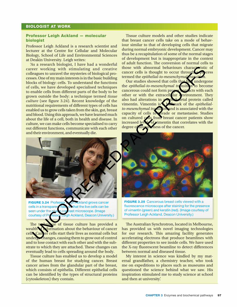
Professor Leigh Ackland — molecular biologistProfessor Leigh Ackland is a research scientist and lecturer at the Centre for Cellular and Molecular Biology, School of Life and Environmental Sciences at Deakin University. Leigh writes:
‘As a research biologist, I have had a wonderful career working with stimulating and creative colleagues to unravel the mysteries of biological pro-cesses. One of my main interests is in the basic building blocks of biology: cells. To understand the functions of cells, we have developed specialised techniques to enable cells from di� erent parts of the body to be grown outside the body: a technique termed tissue culture (see figure 3.24). Recent knowledge of the nutritional requirements of di� erent types of cells has enabled us to grow cells taken from the skin, gut, breast and blood. Using this approach, we have learned much about the life of a cell, both in health and disease. In culture, we can make cells become specialised to carry out di� erent functions, communicate with each other and their environment, and eventually die.
FIGURE 3.24 Professor Leigh Ackland grows cancer cells in a transparent � ask so that the live cells can be seen under the phase contrast microscope. (Image courtesy of Professor Leigh Ackland, Deacon University.)
� e technique of tissue culture has provided a wealth of information about the behaviour of cancer cells. Cancer cells start their lives as normal cells but undergo changes, causing them to grow out of control and to lose contact with each other and with the sub-strate to which they are attached. � ese changes can eventually lead to cells spreading around the body.
Tissue culture has enabled us to develop a model of the human breast for studying cancer. Breast cancer arises from the glandular part of the breast, which consists of epithelia. Di� erent epithelial cells can be identi� ed by the types of structural proteins (cytoskeleton) they contain.
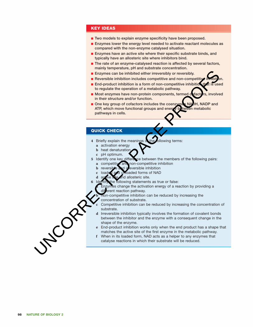
Tissue culture models and other studies indicate that breast cancer cells take on a mode of behav-iour similar to that of developing cells that migrate during normal embryonic development. Cancer may thus be a recapitulation of some of the normal stages of development but is inappropriate in the context of adult function. � e conversion of normal cells to those with abnormal behaviours characteristic of cancer cells is thought to occur through a process termed the epithelial-to-mesenchymal transition.
Our studies showed that cells that had undergone the epithelial-to-mesenchymal transition to become cancerous could not form proper contacts with each other or with the extracellular environment. � ey also had alterations in a cytoskeletal protein called vimentin. Vimentin is a hallmark of the epithelial-to-mesenchymal transition and is associated with the capacity of cells to invade or metastasise. Studies on cultured cells from breast cancer patients show increased levels of vimentin that correlates with the degree of invasiveness of the cancer.
FIGURE 3.25 Cancerous breast cells viewed with a � uorescence microscope after staining for the presence of vimentin (green) and keratin (red). (Image courtesy of Professor Leigh Ackland, Deacon University.)
� e Australian Synchrotron, located in Melbourne, has provided us with novel imaging technologies for our research. � is amazing facility generates accelerating electrons that produce beamlines with di� erent properties to see inside cells. We have used the X-ray � uorescent beamline to detect di� erences between normal and diseased tissue.
My interest in science was kindled by my mat-ernal grandfather, a chemistry teacher, who took me on expeditions to places such as museums and questioned the science behind what we saw. His inspiration stimulated me to study science at school and then at university’.
UNCORRECTED PAGE P
ROOFS
Nature of biology 298
c03EnzymesAndBiochemicalPathways 98 24 October 2016 10:44 AM
kEy idEAs
■ Two models to explain enzyme specificity have been proposed. ■ Enzymes lower the energy level needed to activate reactant molecules as compared with the non-enzyme catalysed situation.
■ Enzymes have an active site where their specific substrate binds, and typically have an allosteric site where inhibitors bind.
■ The rate of an enzyme-catalysed reaction is affected by several factors, mainly temperature, pH and substrate concentration.
■ Enzymes can be inhibited either irreversibly or reversibly. ■ Reversible inhibition includes competitive and non-competitive inhibition. ■ End-product inhibition is a form of non-competitive inhibition that is used to regulate the operation of a metabolic pathway.
■ Most enzymes have non-protein components, termed cofactors, involved in their structure and/or function.
■ One key group of cofactors includes the coenzymes NADH, NADP and ATP, which move functional groups and energy between metabolic pathways in cells.
Quick chEck
4 Briefly explain the meanings of the following terms:a activation energyb heat denaturationc pH optimum.
5 Identify one key difference between the members of the following pairs:a competitive and non-competitive inhibitionb reversible and irreversible inhibitionc loaded and unloaded forms of NADd active site and allosteric site.
6 Identify the following statements as true or false:a Enzymes change the activation energy of a reaction by providing a
different reaction pathway.b Non-competitive inhibition can be reduced by increasing the
concentration of substrate.c Competitive inhibition can be reduced by increasing the concentration of
substrate.d Irreversible inhibition typically involves the formation of covalent bonds
between the inhibitor and the enzyme with a consequent change in the shape of the enzyme.
e End-product inhibition works only when the end product has a shape that matches the active site of the first enzyme in the metabolic pathway.
f When in its loaded form, NAD acts as a helper to any enzymes that catalyse reactions in which their substrate will be reduced.
UNCORRECTED PAGE P
ROOFS

99CHaPter 3 Enzymes and biochemical pathways
BIOCHALLENGE
c03EnzymesAndBiochemicalPathways 99 24 October 2016 10:44 AM
1 The graph immediately below (fi gure 3.26) shows the behaviour of enzyme X under fi ve different conditions.a What variable is plotted: i on the vertical axis of this graph ii on the horizontal axis?b How many experimental results are shown on this graph?c What condition was kept constant throughout the
course of one experiment?
A dependent variable is the variable being tested in a scientifi c experiment and its value depends on the independent variable. As scientists change the independent variable, they observe and record the change in the dependent variable.
d In these experiments, what is: i the dependent variable ii the independent variable?e What conclusion(s) about enzyme X can be drawn
from these results?
Enzyme Y was isolated from extremophile microbes that live in hot volcanic waters.
f Would you predict that enzyme Y would show similar behaviour to enzyme X when subjected to the same experimental conditions? Explain.
Examine the data shown in these graphical results and answer the following questions:
g At what temperature did enzyme X lose about half its activity after just a few minutes?
h After one hour at 60 °C, what percentage activity of enzyme X remained?
i At 65 °C, about how long did it take for enzyme X to lose about 80% of its maximum activity level?
j What happened to enzyme X during the fi rst 10 minutes of its exposure at 70 °C?
k Explain why the behaviour of enzyme X differs at 50 °C compared with its behaviour at 70 °C.
2 Do an internet search using the term ‘metabolic pathways’ and ask for images. You can expect to see some complex images that show the numerous metabolic pathways that operate in living organisms. Remember that the purpose of this activity is simply to give you a sense of the complexity of metabolic pathways and to alert you to the key role of enzymes in virtually every step in these pathways.a Examine several of these images to see some detail of
metabolism. Is the scope of cellular metabolism what you expected?
b One site states that these metabolic pathways are ‘daunting in their majesty beauty’. Do you agree?
c Many of the images may show the various substrates and their products in metabolic pathways, but omit the enzymes involved in the various steps.
Can you locate a metabolic map that also gives the enzymes involved?
00
20
40
% m
axim
um a
ctiv
ity
60
80
100
10Time (min)
20 30 40 50 60
50 °C
55 °C
60 °C
65 °C70 °C
figuRE 3.26
UNCORRECTED PAGE P
ROOFS

100 Nature of biology 2
Chapter review
c03EnzymesAndBiochemicalPathways 100 24 October 2016 10:44 AM
unit 3 structure and regulation of biochemical pathways Practice questions
AOs 1
Topic 5
Key wordsactivation energyactive siteallosteric siteanabolismcatabolismcoenzymecofactorscouplingendergonic
end-product inhibitionenzyme inhibitorenzyme-substrate
(E-S) complexexergonicgulono-lactone oxidase
(GULO)induced fi t modelinorganic cofactors
irreversible inhibitionloaded formlock-and-key modelmetabolismmonomeric enzymesoligomersorganic cofactorsproductproenzymes
prosthetic groupreversible inhibitionscurvysimilar shapesubstratesubstrate binding sitesystematic nametrivial nameunloaded form
Questions 1 Making connections between concepts ➜ Use at least eight of the key words from this chapter to construct a
concept map relating to enzymes. 2 Applying knowledge and understanding ➜ Examine fi gure 3.27. Compound A is a type of protein.
a What is the general name given to the type of protein represented by compound A?b What are the general names given to each of the parts labelled B, C, D and E?c Name the type of reaction shown in this diagram — is it catabolic or anabolic? Explain.d Is this reaction more likely to be endergonic or exergonic? Explain.
A B C D A E
+
figuRE 3.27
3 Applying knowledge and understanding to make predictions ➜ Th e graph below shows the eff ect of temperature on the activity of an enzyme from three diff erent organisms.
Rat
e o
f rea
ctio
n
Temperature (°C)5 37 93
a What predictions, if any, might be made about the organisms that were the sources of
these enzymes? Give a brief rationale for any predictions that you make.
b Note that the shapes of the curves are diff erent. Suggest a reasonable explanation for these diff erences.
4 Drawing conclusions ➜ Amino acids in a polypeptide chain are numbered from 1 to n, starting from the amino terminal; for example, the third amino acid might be Val 3 and the tenth in the sequence might be Leu 10, and so on . . .
Th e active site of the enzyme trypsin, a digestive enzyme, consists of three amino acid as follows: Asp 102, His 57 and Ser 195, that is, these three amino acids are the 57th, the 102nd and the 195th amino acids in the primary structure of this enzyme.a Identify two events that could occur at the active
site of an enzyme.b What stabilises a substrate in the active site of an
enzyme?
UNCORRECTED PAGE P
ROOFS
101CHaPter 3 Enzymes and biochemical pathways
c03EnzymesAndBiochemicalPathways 101 24 October 2016 10:44 AM
One student suggested that the three amino acids at the active site of trypsin had been misnumbered. Th e student said that there is quite a distance separating them so that they cannot be close together at the active site.c Do you agree with the student? Give a clear
explanation for the decision you have made. 5 Demonstrating knowledge and communication ➜
Identify a key diff erence between the members of the following pairs:a ‘lock-and-key’ model and ‘induced fi t’ modelb a prosthetic group and a coenzymec loaded NAD and unloaded NADd competitive inhibition and end-product inhibitione catabolism and anabolismf pepsinogen and pepsin.
6 Interpreting diagrammatic information ➜
Inhibitor
ReactionE
S
E
Active site isdistorted
Substratecan’t bind
No reaction
I
I
S
S
E
figuRE 3.28
Examine fi gure 3.28 above. What story does this diagram tell? 7 Demonstrating understanding and
communication ➜ Consider the following fi gure, which shows the total amount of product formed during an enzyme-catalysed reaction.
Time
Tota
l am
oun
t o
f p
rod
uct
form
ed Explain the following features of this graph:
a the initial steep slopeb the slowdown in the rate of product formationc the fl attening of the curve to form a plateau.
8 Developing logical explanations ➜ Consider each of the following observations and formulate an explanation for the observation.a Salivary amylase enzyme breaks down starch. Th e
process begins when saliva mixes with food in the mouth, but, when it reaches the stomach, the amylase enzyme no longer functions.
b Th e jelly-like white of a raw hen’s egg consists principally of the protein albumin, but, after it is cooked, it forms a white solid.
c When an inhibitor binds to an enzyme at a site other than the active site, this inhibition cannot be overcome by increasing the concentration of substrate molecules.
d Enzyme-catalysed reactions operate at much faster rates than non-catalysed reactions.
9 Demonstrating knowledge and understanding ➜ Protein X is an enzyme.
Disul�de bonds(help hold shapeof protein)
Chemicalpresent
Chemical removed
Denaturedprotein
Protein X
a What is the general shape of protein X (as shown in the left-hand image)?
b When treated with a particular chemical, the shape of protein X was radically changed, as shown in the middle image. What specifi c eff ects has the chemical had on protein X?
c Describe what happened when the chemical was removed.
d What labels would you put on the action of the chemical on protein X?
e How, if at all, would the ability of protein X to catalyse its specifi c reaction be aff ected when it was in the ‘long snake form’?
UNCORRECTED PAGE P
ROOFS

UNCORRECTED PAGE P
ROOFS