chapter fungal deterioration of cultural heritage objects
TRANSCRIPT

1
Chapter
Fungal Deterioration of Cultural Heritage ObjectsŽeljko Savković, Miloš Stupar, Nikola Unković, Aleksandar Knežević, Jelena Vukojević and Milica Ljaljević Grbić
Abstract
Significant percent of world cultural heritage artifacts is threatened by fungal infestation. Fungi can deteriorate different substrates via various physical and chemical mechanisms. Hyphal growth and penetration into the substrate can cause symptoms like discoloration, biopitting, cracking, exfoliation and patina formation. On the other hand, chemical mechanisms include acid secretion, release of extracel-lular enzymes, pigment production, oxidation/reduction reactions and secondary mycogenic minerals formation. These processes can lead to serious, both esthetic and structural, alterations which may be irreversible and could permanently impair artworks. Proper isolation and identification of autochthonous isolates, as well as employment of different microscopic techniques and in vitro biodegradation tests are pivotal in understanding complex biodeterioration mechanisms caused by microorganisms, including fungal deteriogens. Biodeterioration and biodegradation studies require multidisciplinary approach and close collaboration of microbiolo-gists, chemists, geologists and different personnel responsible for the safeguarding of cultural heritage monuments and artifacts, especially restorers and conservators.
Keywords: alterations, biodegradation, cultural heritage, fungi, multidisciplinary research
1. Introduction
Ars longa, vita brevis – states the ancient Roman proverb, emphasizing that human need for artistic expression is as old as the civilization itself. Unfortunately, extant artworks are only a fragment of humanity’s creations throughout history. Along with artistic creation, there is a need for protection of the artwork from external, frequently damaging influences. Since works of art are an essential part of the cultural heritage legacy of every nation, they ought to be protected for future generations. Biodeterioration is defined as any undesired alteration of the property of the material which is caused by living organisms and cultural heritage objects are frequently prone to this process [1]. Mentioned alterations can be induced by both macroorganisms (plants and animals) and microorganisms (bacteria, algae and fungi). Inadequate storage and irregular maintenance of artifacts in archives, muse-ums and depots oftentimes favorize microbial, especially fungal, proliferation [2]. Since fungi are ubiquitous organisms, with pronounced metabolic activities, they

Biodegradation
2
are capable of colonizing various types of microenvironments therefore constantly causing problems in cultural heritage collections around the world [3].
Fungal propagules - spores and mycelial fragments, are always present in the air, their concentrations being dependent on environmental factors [4, 5]. Namely, during their life cycle, fungi produce various types of sexual and asexual spores which are actively or passively released into the surrounding environment and dispersed by air currents to available substrates [3]. The successful colonization of available substrates requires propagules to be viable in addition to favorable growth conditions [4, 6]. It is known that due to their metabolic activities, numerous fungal species could cause both esthetic and physical damage to a variety of substrates, including stone, paint, paper, wood, textile and other materials of which cultural heritage artworks are made. Therefore, the application of adequate microscopic techniques, proper species identification and physiological characterization of autochthonous isolates are very important to appropriately assess potential threats to cultural heritage artworks, especially on those stored in inadequate conditions [3]. Consequently, biodeterioration and biodegradation studies require a multidis-ciplinary approach and a close collaboration of scientists (microbiologists, chem-ists, geologists etc.) and the specialists responsible for the safeguarding of cultural heritage objects, such as restorers and conservators. Therefore, this work addresses general mechanisms of biodeterioration caused by fungi and their role in the deterioration of different materials which constitute cultural heritage artworks.
2. Biodeterioration mechanisms
Fungi present on artworks can affect them in two ways – mechanically and chemically. The aforementioned processes, more often than not, are taking place simultaneously. Depending on the substrate’s nature, exogenic and endogenic factors, the effect of one process can prove more prominent than the other [7, 8]. Notably, depending on its location, fungal colonizers can affect the substrate in two ways – from the surface to its interior and vice versa [7].
2.1 Physical processes
Physical processes are taking place under the influence of hyphal apical growth or by the formation of fruiting bodies on the surface and/or the inner layers of the colonized material. If the fungal growth is superficial, it results in the formation of spreading mycelium which covers the substrate and changes the original appear-ance, hence the esthetic value of the artifact [7]. Inner fungal growth might lead to further damage of the artworks and, especially if paintings are concerned, to the detachment of painted layers (exfoliation). Melanized micromycetes are well known inducers of mechanical deterioration, especially of stone substrates, since melanin provides mechanical rigidness to fungal structures, enhances the turgor pressure and facilitates hyphal penetration [8, 9]. In order to study mechani-cal deterioration, the application of different microscopic techniques is pivotal, especially in situ optical microscopy and scanning electron microscopy (Figure 1). The multimicroscopic approach is essential to ensure detailed information, not only about the deterioration status, but also to elucidate alterations that affect works of art, and to detect potential biodeterioration “culprits” [10].
2.2 Chemical processes
Mechanisms of chemical biodeterioration are much more complex and promi-nent than physical ones. Fungi can chemically alter the substrate via assimilation
3
Fungal Deterioration of Cultural Heritage ObjectsDOI: http://dx.doi.org/10.5772/intechopen.98620
and dissimilation processes [11]. In case of the former, fungi utilize nutrients from the substrate by secreting various enzymes which catalyze the macromolecules’ degradation. In contrast, dissimilation represents the production of various extra-cellular metabolites such as organic acids and pigments. These substances modify
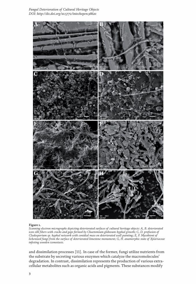
Figure 1. Scanning electron micrographs depicting deteriorated surfaces of cultural heritage objects: A, B. deteriorated icon silk fibers with cracks and gaps formed by Chaetomium globosum hyphal growth; C, D. profusion of Cladosporium sp. hyphal network with conidial mass on deteriorated wall painting; E, F. Mycobiont of lichenized fungi from the surface of deteriorated limestone monument; G, H. anamorphic state of Xylariaceae infesting wooden iconostasis.
Biodegradation
4
or damage the colonized substrate. Since hyphae have high surface to volume ratio, these metabolites can quickly diffuse between the cells as well as from the cell into the substrate [7, 8]. Nowadays, various microbiological, biochemical and petrographical tests are employed to study chemical biodeterioration. In vitro tests provide rapid, cost effective estimation of fungal degradation capacity, which helps in evaluating a potential risk to cultural heritage artifacts [3].
Acid production and acidolysis are the most studied biodeterioration mecha-nisms, particularly on inorganic materials [8, 12]. Due to their metabolic activities, fungi produce organic acids such as gluconic, citric, oxalic, malic, succinic, itaconic etc. [13]. Once the spore germination occurs, organic acids are produced by respira-tion in mitochondria as intermendiary products of the citric acid cycle. If the fungi grow on nutrient enriched substrates, these acids are formed in excess and excreted as secondary metabolites [6]. The secreted acids then react with different sub-stances via cation solubilization and chelation reactions. The reaction of acids with different metals (i.e. K, Fe and Mn) results in the formation of organic salts and complex compounds [7]. It should be mentioned that many organic acids, especially the oxalic, are able to chelate different metals in the process called complexolysis. The oxalic acid is able to form complexes with diverse metals (Ca, Mg, Fe, Cu and others), consequently leading to secondary mycogenic minerals formation, calcium oxalate being the most well-known [14]. The aforementioned crystals are present in patinas on stone, frescoes, oil paintings, glass, wood and other materials [8]. It is ascertained that most of the fungi have the ability, in greater or lesser extent, to pro-duce oxalic acid, and subsequently precipitate oxalates [3]. Furthermore, CO2, as a product of respiration, in the conditions of increased humidity is transformed into carbonic acid, which then solubilizes calcium carbonate and magnesium carbonate present in limestone, mortar and gypsum. As a result, water soluble bicarbonates are formed. Additionally, increased H+ concentration favorizes the colonization of acidofilic fungi, which further facilitates the biodeterioration process [7, 8].
Enzymes. Fungi are able to digest organic matter, altering and weakening those materials, by the action of extracellular hydrolytic enzymes, such as lignocellu-lases, proteases, lipases, pectinases, chitinases, etc. [15, 16]. Enzymes that convert large, complex and often water-insoluble compounds (cellulose, hemicellulose, lignin, proteins and lipids) into low-molecular-weight soluble compounds, play an important role in the biodeterioration and biodegradation processes [7]. Although filamentous fungi primarily use simple sugars as a carbon source, they can be producers of lignocellulolytic enzymes to depolymerize wood or cellulose material for nutritional purposes [17]. Cellulolytic enzyme complex, which is responsible for degradation of cellulose to glucose monomers, comprises of: endoglucanase (hydrolyzes β-1,4-glycosidic bonds within cellulose fibers), exoglucanase (hydro-lyzes β-glycosidic bonds and remove cellobiose units from the free ends of chains) and β-glucosidase (hydrolyzes cellobiose and cellodextrin to glucose) [18, 19]. Hemicellulases hydrolyze hemicellulose, which is made up of hexoses (mannose, glucose, galactose) and pentoses (xylose, arabinose) to monomeric sugars and acetic acid. The complex of enzymes that hydrolyze hemicellulose consists of at least eight enzymes: endo-1,4-β-D xylanase, exo-1,4-β-D xylocuronidase, α-L arabinofuranosidase, endo-1,4-β- D mananase, β-mannosidase, acetyl-acid ester-ase, α-glucuronidase and α-galactosidase [19]. Lignin degradation, characteristic for white-rot fungi, is catalyzed by nonspecific polyphenol oxidases: manganese oxidizing peroxidases, lignin peroxidases, and laccase. This process involves break-ing inter-monomer bonds, demethylation, hydroxylation, side chain modification, and aromatic ring cleavage [20, 21]. Some fungal species inhabit art objects that are substrates rich in fibrillar proteins (wool, parchment, leather, silk, etc.). Proteolytic enzymes (proteases) degrade various protein fibers such as collagen (wool), fibroin

5
Fungal Deterioration of Cultural Heritage ObjectsDOI: http://dx.doi.org/10.5772/intechopen.98620
(silk) and keratin (parchment) [11]. Lipases catalyze the hydrolysis of triacylglyc-erols to glycerol and fatty acids. These enzymes might take part in the degradation of widely used painting constituents, linseed and shellac, derived primarily from unsaturated oleic, linoleic and linolenic acids [22].
Pigment production. Micromycetes produce pigments which vary in chemical composition and color and are species specific [7, 8]. They are present in hyphae, conidia, or are secreted into the substrate whilst their production is determined by the availability of nutrients and minerals, UV radiation, pH, temperature and other environmental factors [6]. Pigment secretion on/into the substrate leads to the appearance of different, frequently irreversible, colorations leading to the observable changes on cultural heritage objects. This diminishes the aesthetic value of the artwork and accelerates biodeterioration process [7, 11, 23]. Many fungi produce dark colored pigments – melanins, which are responsible for characteristic brownish color of mycelia and reproductive structures. These water insoluble, very stable and resistant, molecules are formed by oxidative polymerization of phenolic compounds (ortho-dihydroxy phenols) [24]. Dark colored melanins are character-istic for dematiaceous fungi (representatives of the former family Deamtiaceae) and are present in cell walls in both granular of amorphic form, while their amount increases with aging [6, 25]. On the other hand, the secretion of various water soluble exopigments into the substrate can also cause different aesthetic damage to artworks, especially on those made from organic materials. A vast number of different pigments are identified and grouped in three main families: the derivatives of toluquinone, of naphthoquinone and of beta-methyl-quinone [8].
3. Stone artifacts
A significant percentage of world cultural heritage objects is represented by stone artworks, such as architectural monuments, statues and tombstones, to name just a few [26]. Stone is considered an extreme environment for microbial growth and proliferation, mostly due to the intensive oscillations of diurnal and seasonal microclimatic parameters and low nutrient and water content [27]. Stone surfaces directly exposed to sunlight could achieve temperatures above 60°C, while being simultaneously susceptible to freezing–thawing cycles [28, 29]. Regardless, certain groups of microorganisms, the so called lithobionts, are able to colonize such environments. Primary colonizers are photoautothrophic organisms – cyanobac-teria, algae and lichens, while hemolithotrophic and hemoorganotrophic bacteria and fungi are considered secondary. The latter are oligotrophic or poikilotrophic organisms which adapted to survive and grow in harsh or variable environmental conditions [29].
Microbial dwellers of stone surfaces frequently form biofilm, highly structurised microbial consortium embedded in a mutual extracellular matrix. Biofilm forma-tion starts with unspecific, reversible interactions, followed by stable interactions which are initiated as a consequence of the formation of specific molecules and structures (lipopolysaccharides, membrane proteins, flagellae). After initial adhe-sion, extracellular polymeric substances are produced and excreted, enhancing the adhesion and cohesion of cells [8]. Evolutionary advantages of biofilm are to provide protection, resistance to physical and chemical stressors, metabolic cooper-ation and mutually regulated gene expression [30]. All groups of microbial dwellers of stone are characterized with a high phenotypic plasticity, which is reflected in polymorphism – the change of growth or sporulation forms with regard to external conditions. Therefore, micromycetes, which colonize this environment, are able to form different somatic and reproductive structures - sclerotia, chlamidospores,
Biodegradation
6
conidial clusters, perithecia and pycnidia [29, 31]. Moreover, stone interior is a specific microenvironment for the growth of certain microorganisms such are endolithic fungi. The microclimatic conditions in this environment are a little more favorable – water retention is higher and solar radiation and air current intensities are lower [27].
Among fungal colonizers of stone, micorcolonial fungi constitute a specific ecological group characterized by its slow growth and formation of irregular shaped cells, often packed in aggregates. They rarely form specialized reproductive struc-tures and in turn, by active growth, form thick, pigmented cell walls which enable transition to the state of dormancy during prolonged, unfavorable environmental conditions. The ability to secrete extracellular polymeric substances and thick cell walls, enable water retention, nutrient absorption, desiccation reduction and cellular adhesion/cohesion. These organisms are able to survive for long periods without metabolic activities and their metabolic rates are low even during optimal environmental conditions [27].
Dematiaceous fungi are considered the most important agents of stone dete-rioration. All representatives intensively produce dark colored melanins which provide protection from excessive environmental radiation (UV radiation, x- and γ-rays) and chemical stressors. This group encompasses microcolonial fungi and black yeasts, species of genera: Acrodictys, Aureobasidium, Capnobotryella, Coniosporium, Exophiala, Hormonema, Hortaea, Knuffia, Lichenothelia, Monodictys, Phaeococcus, Phaeococcomyces, Phaeosclera, Sarcinomyces and Trimmatostroma, which are very important stone colonizers in arid and semiarid environments [27, 32]. Additionally, deatiaceous fungi include cosmopolitan filamentous species of genera Alternaria, Cladosporium, Ulocladium, Epicoccum etc. which are main colonizers of stone in more favorable conditions of temperate and humid environments [32]. They are especially important deteriogens of restored stone artifacts [27].
Fungi can deteriorate stone via physical and chemical mechanisms. Physical mechanisms include hyphal penetration of the rock surface which causes its frag-mentation, while chemical ones include secretion of acidic metabolites and pig-ments and oxidation of mineral forming cations. Although many microorganisms are able to produce acids, fungi are considered as the most potent ones in nature that degrade rocks and minerals. The production of various acidic metabolites leads to the biocorrosion - dissolution of the mineral substrate, resulting in the forma-tion of various secondary mycogenic minerals [26]. In vitro acid production and formation of calcium oxalate and calcium carbonate minerals have been reported by autochthonous isolates from limestone monuments such as ancient Roman stela and Portuguese king tomb [26, 33]. Synthesis and excretion of extracellular pigments mostly affect the stone aesthetically, although studies concerning pigment produc-tion on stone monuments are generally scarce [26]. Due to the mentioned processes, symptoms such as biopitting, biogenic patina and colorations can occur [11]. The growth of dematiaceous fungi results in the presence of dark stains while micro-colonial fungi are the main culprits of biopitting phenomena on limestone and marble artworks, [32, 34]. Sanctuary of Delos in Greece is an example of mentioned biopitting phenomena [35].
Lichenized fungi are important colonizers of stone substrates. These organisms have a high tolerance to variations of environmental factors, especially tempera-ture, insolation and water availability, which is responsible for their cosmopolitan distribution and ability to colonize extreme environments [36]. These organisms are poikilohydric, i.e. they are capable of enduring cycles of desiccation and rehy-dration due to their ability of lowering their metabolic rate and enter cryptobiosis under conditions of low water availability [37]. Endolithic lichens are of special importance to stone deterioration, since they are capable of the deepest penetration

7
Fungal Deterioration of Cultural Heritage ObjectsDOI: http://dx.doi.org/10.5772/intechopen.98620
into the stone compared to other microorganisms [38]. Apart from the fact that hyphae of the mycobiont can penetrate the rock surface (Figure 1E and F), perithe-cia formation by some endolithic species can penetrate the surface from the inside out, which leads to biopitting [27]. Additionally, lichen growth could cause exfolia-tions, encrustations and disaggregation of the stone surface [11, 39]. Conversely, lichen morphology and its adhesive capability aren’t always in correlation with its capacity to alter the substrate and physiological differences between the species are considered to be more significant [40, 41]. In fact, synthesis of different chemical deterioration agents is done by the mycobiont. Apart from carboxylic, lichens have the ability to produce lichen acids, semisoluble polyphenolic compounds which are able to form complexes with metal cations. The capability of lichens to absorb and maintain water enhances the duration of chemical reactions and therefore facilitates deterioration process [39, 41]. Lastly, some authors have reported the presence of orange-brownish patinas (scialbatura) on stone monuments made from limestone and marble. These colorations mainly consist of calcium carbonate and calcium oxalate minerals, sometimes intermixed with fragmented lichen thalli [42, 43]. Although this symptom is associated with deterioration, it is hypothesized that it may have a protective role to the monuments [27].
4. Wall paintings
Wall painting, as the pictorial technique, encompasses all painting techniques aimed at beautifying wall surfaces. There is no universally accepted definition of fresco, as well as consensus on what techniques can be included in this type of wall painting, however, the term al fresco generally refers to paintings made on a fresh lime mortar with mineral pigments mixed with water [44]. On the contrary, in fresco a secco or al secco technique painting is done on a dry plaster with paints pre-pared by mixing mineral pigments with various organic binders [32, 45]. Although hallmarked as an extreme type of habitat, painted layer and lime mortar are also considered to be very suitable and bioreceptive substrates for fungal growth. This is due to the mineral composition and porous nature of lime mortar, and the fact that organic and inorganic components of the painted layer represent a suitable niche for the development of a wide range of heterotrophic microorganisms [46].
Fungal infestation of wall paintings can occur from several sources including contaminated indoor air as the main, but also soil and plants of immediate vicin-ity, visitors, contaminated conservation tools, and indoor hotspots as secondary sources [7]. Whether a certain fungus will be able to colonize the painted layer or mortar depends on the ecological and physiological requirements of a given spe-cies. If the requirements are met the process is further controlled by three main factors: nutrient availability, relative humidity, and temperature [32]. The origin of nutrients in fresco painting is related to (1) additives (chaff, wheat paste, barley flakes, animal hair, hemp and flax fibers, egg whites, oils, fats) mixed with min-eral and complex fillers of chopped straw and lime mortar; (2) additives used in the preparation of mortar (liquid resins, tar, polymer latex, emulsions, bitumen, milk, olive and linseed oil, lard, animal blood); (3) binders of plant and animal origin mixed with mineral pigments; (4) casein, paraloid mixtures, fixatives and consolidants based on polymer components (cellulose acetates, polyvinyl acetate, polymethyl acrylate, etc.) used in restoration works [32, 45, 47–50]. These organic components determine the richness of the fresco mycobiota. Since the composi-tion of the painted layer and mortar is predominated by inorganic components, its mycobiota differs greatly from the fungal communities established on other painted works of art [45]. Furthermore, heterogeneously pigmented zones of the painted

Biodegradation
8
layer can be considered as selective substrates that condition the development of a specific mycobiota [51]. Using culture-dependent methods, the most commonly documented fungi on painted layer and mortar of wall paintings are Ascomycota of genera Acremonium, Alternaria, Arthrinium, Aspergillus, Aureobasidium, Beauveria, Botrytis, Chaetomium, Chrysosporium, Cladosporium, Curvularia, Dreschlera, Engyodontium, Epicoccum, Eurotium, Exophiala, Fusarium, Geomyces, Gliomastix, Phoma, Penicillium, Scopulariopsis, Sepedonium, Sporotrichum, Stachybotrys, Stemphylium, Trichoderma, Trichotecium, Ulocladium and Verticillium [7, 45, 52–54]. Contamination by fungi from phyla Basidiomycota is rare (e.g. Coprinus spp.), while Zygomycota of genera Mucor and Rhizopus are isolated frequently but are considered only surface contaminants [45, 55].
Species of the genera Aspergillus, Aureobasidium, Alternaria, Cladosporium and Penicillium, are frequently listed as the most common wall paintings contaminants, as well as the primary fresco deterioration agents in temperate climates [56]. Many Cladosporium species are recognized as the main biological agents in the process of biodeterioration of wall paintings since they are able to not only induce brown discolorations, but also penetrate through the entire painted layer all the way to the mortar support [57] (Figure 1C and D). Many species of the genus Penicillium are known to develop and intensely sporulate in a period of only few days to a few weeks on periodically moist fresco paintings [58]. Furthermore, in addition to dominant members of fungal community, species from less represented genera can also significantly contribute to the damage of wall paintings. For example, isola-tion of Phoma species from the surface of painted layer indicates that given wall paintings are in an active process of decay [59]. Chaetomium, Aureobasidium and Epicoccum species, due to strong proteolytic activity, degrade protein binders of the painted layer, which results in the lifting and separation of the painted layer from the support. Likewise, it has been recently contemplated that species of the genera Mucor and Rhizopus might be more involved in process of biodeterioration of wall paintings than originally considered, as it was shown in in vitro experiments that they are able to degrade protein binders and epoxy resins [60].
Mechanical (1) and chemical (2) activity of fungi directly results in damages to structural and esthetic integrity of fresco paintings. (1) Hyphal penetration, together with formation of fruiting bodies and various modifications of mycelium, increases internal pressure thereby forming new cracks in the painted layer and mortar, as well as expanding the existing ones. Damages caused by mechanical activity are considered by some to be of greater importance compared to changes induced by environmental factors and fungal chemical activity [61]. Furthermore, aside from mechanical activity, damages as the result of change in substrate properties can also incur due to utilizing fresco components as a source of nutrients for fungal growth (2′1) and/or due to secretion and interaction of fungal metabolites with organic and inorganic components of the painted layer and mortar (2′2) [7, 62]. (2′1) Extracellular enzymes break down complex organic components into simpler molecules enabling their absorption and easier penetration of hyphae into the substrate which results in cracking and peeling of the painted layer and mortar. The main enzymes involved in this process are β-glucosidase, phosphatase, lipase, arylsulfatase, esterase, protease and endo-N-acetyl-PD-glucosamidase [63, 64]. (2′2) Excreted organic acids chelate metal ions present in mineral pigments and mortar, resulting in the formation of mineral salts and complex compounds that increase pressure in pores, which leads to cracking, peeling, and loss of fragments of the painted layer and mortar [65, 66]. Additionally, salts stimulate formation of surface irregularities that serve as suitable sites for the settlement of heterotrophic microorganisms, thereby increasing the bioreceptivity of fresco painting [67]. In these circumstances, there is an uncontrolled biofilm development and acceleration of chemical dissimilation activity through
9
Fungal Deterioration of Cultural Heritage ObjectsDOI: http://dx.doi.org/10.5772/intechopen.98620
oxidation, reduction and transformation of metal ions in pigments, primarily Fe and Mn, but also As, Pb, Cu, Zn and Hg, resulting in the alterations to the original color of the painted layer [53, 68]. Aside from organic acids, very stable and persistent fungal pigments (melanins, mycosporins, quinones, hydroxyanthraquinones and carot-enoids) secreted onto the surface induce changes in the original coloration, which is process that depends on the chemical composition of the pigment, environmental conditions and interactions with substrate components [7, 32].
5. Canvas oil paintings
One of the best-known pictorial technique today, oil painting on canvas, emerged in the Middle Ages and has since been one of the most important art expressions, constituting outstanding works of art with important historic and cultural value [69]. Structurally speaking, these works of art are composed of the pictorial layer between the protective covering varnish and the ground (or prepara-tory layer) spread on a linen canvas. Compared to the other forms of artwork, oil paintings on canvas possibly provide the widest range of microhabitats and nutri-ents that may be exploited by a large variety of microbial species [70]. Materials that constitute the painting, i.e. the cellulose of the canvas support, organic adhe-sives (i.e. various animal, fish and plant glues) used in sizing the support, natural varnishes, and the oils used in binding the pigments (linseed, turpentine and other oils) are all composed of organic molecules of high nutritional content that are all easily degraded [32, 63]. These organic molecules encompass sugars, gums, and other polysaccharides, proteins and waxes, but also less chemically defined mix-tures of biomolecules, such as egg yolk, bile, and urine, as well [45]. Organic glue pastes used to coat the back of paintings with linen canvas, i.e. “re-lining”, may also represent a rich nutrient source [71]. Furthermore, dirt, dust and other environ-mental contaminants (volatile hydrocarbons released from machinery, respiration and cigarette smoke) deposited on the surface of the oil paintings provide nutrients as well [63].
Given the wide range of organic molecules that are present in oil paintings, many different microorganisms may grow provided inadequate storage and favorable environmental conditions, primarily high relative humidity and temperature, are met [45]. These specific environmental conditions may start and/or accelerate the micro-bial growth, which otherwise would persist on the obverse and the reverse side of the painting in a dormant metabolic state [70]. Among multitudes of different microor-ganisms, fungi are notorious for their ability to inhabit and decay paintings due to their enormous metabolic activity and ability to grow at low aw values [32]. However, to the best of our knowledge, despite being one of the most numerous objects exhibited and stored in museums and warehouses worldwide, relatively fewer number of studies to date have been engaged in describing the fungal communities dwelling on canvas oil paintings compared to the other forms of art. Using culture-dependent methods, the most commonly documented fungi on painted surface, canvas and wooden frame are Ascomycota of genera Alternaria, Aspergillus, Aureobasidium, Botrytis, Cladosporium, Drechslera, Epicoccum, Fusarium, Penicillium, Ulocladium, Scopulariopsis, Stemphylium, Trichoderma and Wardomyces, with occasional observations of teleomorphs from Chaetomium, Emericella and Eurotium genera [32, 63, 69, 70, 72, 73]. Review of the above referenced literature data has highlighted fungi that were most frequently isolated from the infested oil paintings: Alternaria alternata, Aspergillus flavus, A. niger, A. versicolor, Aureobasidium pullulans, Chaetomium globosum, Cladosporium cladospo-rioides, Eurotium chevalieri, and Penicillium chrysogenum. Aside from the Ascomycota, the only other documented species are Zygomycota of Cunninghamella, Mucor and

Biodegradation
10
Rhizopus genera, but they are in most cases regarded as transients only, i.e. part of the surface dust deposits, and not partaking in the complex process of biodeteriora-tion [32, 74]. Air mycobiota of the depots and exhibition rooms, where paintings are stored and exhibited, respectively, was shown to correlate well with fungal com-munities observed on the surface of the paintings indicating airborne origin of the infestations. Furthermore, higher density and diversity of the fungal communities on the obverse of the painting is a direct result of the higher amount of airborne fungal propagules deposited on the painted surface due to gravitational settling of propagules from the air [22]. It has to be noted, however, that there are discrepan-cies between results of the studies utilizing culture-dependent and -independent methods. Results of the limited number of studies on non-culturable fraction of the oil paintings fungal communities showed discrepancies in numbers, with unculturable or not viable part of the community being more dense and prevalent, as well as in composition: Ascomycota of genera Alternaria, Ascoidea, Aspergillus, Blastomyces, Candida, Chaetomium, Coccidioides, Diplodia, Eutypa, Exophiala, Fusarium, Gaeumannomyces, Histoplasma, Marssonina, Microsporum, Neofusicoccum, Paracoccidioides, Parastagonospora, Penicillium, Penicilliopsis, Pestalotiopsis, Pichia, Rasamsonia, Lodderomyces, Neurospora, Sordaria, Talaromyces, Thermothelomyces, Thielavia, Trichoderma, Tuber, and Verticillium were dominant, followed by Mucoromycota of genera Phycomyces and Lobosporangium and Basidiomycota of genus Puccinia [63, 75, 76].
Fungal induced deterioration of canvas oil paintings can occur on both the obverse and the reverse side. It usually starts on the reverse side as canvas components are more readily degraded than those found on the obverse side. In addition, support polymers and the glue sizing in the canvas act as supplementary substrates for fungal growth [22]. Canvas was shown to be one of the most sus-ceptible painting materials (only surpassed by linseed oil), with the susceptibil-ity depending on the percentage content of cellulose, lignin, and other organic components [74, 77]. The higher the percentage of cellulose and lignin, the more resistant it will be to fungal attack [78]. Due to their ability to produce cellulolytic enzymes responsible for cellulose fibers dissolution, fungi of the genus Alternaria, Aspergillus, Cladosporium and Ulocladium are considered to be the main agents of canvas deterioration [63, 69, 76].
On the other hand, the degree of deterioration on the obverse side depends on the oil paints and their mode of application. Varnish, added to provide protec-tion against environmental attacks, is the least susceptible painting material to fungal attack, while many pigments are known to possess antifungal properties [74]. Fungal communities are found to be less dense and diverse in pictorial lay-ers containing pigments with heavy metals (e.g. Pb, Cu and Hg), compared to those found in pictorial layers without such compounds [75]. On the obverse side, hydrolytic activities that the fungi undertake to sustain growth results in the detachment of the paint layer from the support, with further increase in the loss of material happening due to excretion of destructive metabolites, i.e. organic or inorganic acids and the additional production of extracellular enzymes: lipases, esterases, endo-N-acetyl-glucosaminidases and proteases [22]. Such activities lead to formation of structural impairments usually manifested as exfoliation of paint layers, cracking, peeling, formation of paint blisters, detachment of the paint layer from the support, deformations and loss of strength of support. Strongly linked to these damages are the esthetic impairments (preceding the structural damages or forming as a resulting consequence) manifesting as the change of the original coloration due to pigment alterations, biofilm formation on the painted surface or staining as the result of pigment excretion by fungi [75]. Fungi of Aspergillus and Penicillium genera degrade glues and oil binders, and dissolve paints, contributing

11
Fungal Deterioration of Cultural Heritage ObjectsDOI: http://dx.doi.org/10.5772/intechopen.98620
to chromatic alterations of the painted surfaces and detachment of the support, while Aureobasidium pullulans is considered by many to be the main biological agent of paint deterioration [1, 45, 69].
6. Wood and paper
Wood as a material has been widely used as a structural element for many types of constructions or for ritual, religious and decorative purpose. Historically, human usage of wood is embedded in wood cultural heritage reflecting past and present human life, culture, ideals, symbols and values. Wood cultural heritage objects can be classified as: moveable (musical instruments, frames, furniture, sculptures, iconic altar etc.), immovable (temples, churches, chapels, royal palaces, pagodas, wooden bridges etc.) and underwater (shipwrecks, foundation piles, wooden cargo or contents which were partially or totally underwater, periodically or continuously for at least 100 years), according to UNESCO [79]. Lignocellulose is the major com-ponent of wood biomass and consists of three types of polymers, cellulose (40–55%), hemicelluloses (24–40%) and lignin (18–35%) that are strongly intermeshed and chemically bonded by non-covalent bonds and by covalent crosslinkages [19]. Organic nature and optimal water content make the wooden substrate suitable for microbial attack [80]. However, microbial deterioration of these materials occurs only under poor conservation conditions: high humidity level, soil contact, poor ventilation, and rare maintenance [81]. Even though deterioration of wood cultural heritage is a process conducted by all groups of microorganisms, fungi have the most significant potential to affect this type of historic artworks [82]. Biodegradation and biodeterioration of wood materials is predominantly dependent on its moisture content, requiring a minimum of 20% of water. Despite dry wooden objects are considered to be resistant to fungal degradation due to low moisture content, occasional wetting, leaks and flooding can increase humidity, enabling conditions for fungal growth. The mechanism of biodeterioration implies the devel-opment of fungi on the surface (Figure 1G and H) or between internal structures, the production of extracellular enzymes, the structural change of basic biopoly-mers, which ultimately results in visible changes of the object [81]. Generally, fungi that attack wooden material can be distinguished as white-rot, brown-rot and soft rot fungi. White rot fungi are the only organisms that can completely depolymerize and degrade all lignin components as well as cellulose and hemicellulose. The largest number, of about 1500 species, belongs to the Basidiomycota and a smaller number belongs to Ascomycota. Most commonly found species are from genera Bjerkandera, Donkioporia, Fomes, Irpex, Phanerochaete, Pholiota, Pleurotus and Trametes. Brown rot fungi decompose cellulose and hemicellulose while lignin degradation is lim-ited to the process of demethylation of methoxyl groups, partial oxidation and depolymerization in a non-enzymatic catalytic cycle of the Fenton type where the free radical reaction is initiated by hydroxyl radicals (OH•). Only 6% of the total number of species that have been confirmed to be able to decompose wood mass belong to this group, and almost all representatives inhabit coniferous wood (species from the genera Antrodia, Aspergillus, Coniophora, Coriolellus, Fusarium, Gloeophyllum, Merulis, Paxillus, Poria, Postia, Serpula, etc.). Soft rot fungi decom-pose cellulose and hemicellulose, while the process of lignin modification is limited to demethylation. It is typical for this group of fungi to attack wood mass with high levels of humidity and low lignin content, forming a microscopic cavities inside the wood, sometimes leading to discoloration and occurrence of cracking pattern similar to brown rot (species from the genera Alternaria, Chaetomium, Daldinia, Humicola, Stemphylium, Xylaria, etc.) [79, 82, 83]. Soft-rot decay has been described
Biodegradation
12
frequently from construction timbers, ancient Egyptian wooden coffins, wooden structures of Buddhist temples, waterlogged archeological wooden material, which can be related to more tolerant growth conditions. In comparison to soft-rot spe-cies, other two groups of wood decay fungi have a relatively narrow spectrum of growth conditions (preferring moisture content between 35% and 50%) but were documented in wooden churches, historic timbers and various historic buildings [79]. Objects of wood cultural heritage in the outdoors have been more disposed to deterioration process than those in the indoor as they are exposed to the relative humidity of 70% or higher, which is stimulatory for fungal growth. Additionally, the physical contact with moisture-absorbing surface can also provoke fungal decay [79]. On the other hand, fungal degradation of waterlogged and buried wood is much slower than for that found in dry environment, but once excavated decay can occur rapidly [82]. Finally, it should also be emphasized that in some cases fungal decay can be observed in extreme conditions such as in wood with 17.4% of mois-ture content [79].
Additionally, paper, which is mostly produced by mechanical and chemical processing of cellulose fibers, originating from wood, is the most important mate-rial on which cultural achievements in the whole world are recorded and preserved. Since it is created as a product of the wood industry and consists of 90–99% of cel-lulose fibers, in the ecological sense, paper is considered to be a cellulose substrate. Books, documents, writings, old maps, photographs, etc. are objects made of paper that are most often kept in libraries, archives and museums. Apart from paper, cotton and linen are fabrics which main components are cellulose fibers. Also, paraments, defined as hangings or ornaments used for decorations of Christian churches’ interiors are often tailored of cotton and linen. In that sense, it should be emphasized that, art objects made of cellulose fibers can be colonized by cellulolytic fungi. These fungi can degrade cellulose fibers via process off cellulolysis, defined as an enzymatic hydrolysis of cellulose polymer into glucose units. In that sense, the fungi capable for production of cellulolytic enzymes are frequently isolated from paper, especially from old books or documents kept in libraries, archives and museum depots. Among the frequently encountered species on the paper sub-strates are the members of genera Chaetomium, Penicillium, Aspergillus, Eurotium, and Trichoderma [84, 85]. Some authors reported the presence of Fusarium sp., Humicola sp., Paecilomyces variotti and Trichoderma viride on deteriorated art photographs which were part of the collection of the Museum of Contemporary Art (Belgrade, Serbia) [86]. Due to their ability to degrade cellulose fibers, these fungi are referred to soft rot fungi [87]. Ascospores and conidia of different cel-lulolytic microfungi ubiquitously present in the environment worldwide could easily be deposited on papers and other cellulolytic materials (books), and when optimal conditions are met, they could germinate, elongate and proliferate and consequently lead to fast book decay. Also, fungi can deteriorate the paper-based materials mechanically via hyphal penetration or through production and excretion of pigments and organic acids [88]. A specific and irreversible phenomenon in the form of brown to red spots on paper material has been described in the literature [89]. Since the color of these spots resembles the color of fox fur, the phenomenon is called “foxing”. The origin of this phenomenon on paper documents is explained by two theories - abiotic and biotic. According to the abiotic theory, “foxing” is a consequence of natural chemical processes, most often oxidation, which takes place on paper material, as well as a consequence of the deposition of certain compounds on the paper surface [90]. According to the biotic theory, “foxing” is caused by microorganisms, especially fungi that produce organic acids which deteriorate the paper, permanently damaging it [89]. Isolation of a large number of fungal species from the parts of the paper on which the symptoms of “foxing” are
13
Fungal Deterioration of Cultural Heritage ObjectsDOI: http://dx.doi.org/10.5772/intechopen.98620
observed, speaks in favor of biotic theory. The famous piece of art affected with foxing symptoms is self-portrait of Leonardo da Vinci’s drawn in red chalk on paper and deposited in Royal Library in Turin. As a main culprit responsible for “foxing” spots on this famous piece of art, certain authors reported the fungus Eurotium halophilicum, which spores are documented near the foxing spots via SEM analy-ses, along with oxalates of fungal origin [91]. Additionally, they pointed out that tonophilic fungi can germinate on paper materials and also can metabolize organic acids, oligosaccharides and proteins, which react chemically with the material at a low water activity, forming brown products and, via oxidative reactions, leading to foxing spots.
7. Textile
Textile is defined as elastic material produced by spinning of natural or syn-thetic raw fibers and which are in final form composed of interlocked network of threads or yarns. Apart from synthetic fibers, materials used for textile production could be of plant or animal origin. Cotton, linen, hemp and jute are widely used fabrics of plant origin and hence they are composed of cellulose fibers. Many old-fashioned and vintage attires and garments, worn by our ancestors were woven from these fabrics and deposited in museum depots and exhibition rooms, or still worn during traditional festivities. In that sense, those fibers could be easily attacked by cellulolytic fungi, and mechanisms of biodeterioration are similar to those of the fungi capable of degrading paper-based materials. On the other hand, animal fibers include wool and silk [92]. Wool is a textile fiber obtained from various hairy mammals, but mostly from sheep, and main constituent of wool is a protein keratin. When compared with textile fibers of plant origin, wool is more resistant to fungal attack due to its specific cross-linked structure with disulphide bonds [93]. However, fungi capable for keratinolysis can attack wool fibers and cause wool degradation. Pioneer research by some authors demonstrated that fungi are the main “culprits” responsible for wool degradation, in much higher degree then bacteria, and members of genera Aspergillus, Chaetomium, Fusarium, Microsporum, Penicillium, Rhizopus and Trichophyton are among the most frequent wool colonizers [94]. In an in vitro study, other investigators showed that fungi do not grow directly on the wool fibers but rather between the fibers, and reported that proteolytic fungi Cladosporium cladosporioides and Penicillium corylophilum have the most intensive growth when inoculated on wool and also the highest impact on degradation and aging of wool fibers [93]. Although wool is very resilient to microbial attack, the silk is considered to be a natural fiber most resistant to the biodeterioration [92]. Silks are defined as fibrous proteins spun into fibers through activity of spiders and insects. The main producer of commercial silk is a domestic silkworm Bombyx mori. Chemically, raw silks are composed of highly crystalline polypeptide fibers, fibroin, linked to one another by a gum-like protein, sericin [95]. The amino acid composition of silk polypeptides results in a very stable β-pleated crystal structure, making fibroin totally insoluble in aqueous solvents and hence very resistant to enzymatic hydrolysis [96]. In that sense, scientific reports regarding the microbial deterioration of silk material are scarce. Still, some authors reported for the first time the mechanical deterioration of Japanese silk from Serbian museum collections caused by proteolytic fungus Chaetomium globosum. Scanning electron microscopy analysis of the analyzed scroll indicated that C. globosum hyphae are capable of the mechanical deterioration of silk, causing cracks and gaps in fibroin fibers and consequently lead to visible impairment of silk artifact itself (Figure 1A and B) [97].

Biodegradation
14
Author details
Željko Savković*, Miloš Stupar, Nikola Unković, Aleksandar Knežević, Jelena Vukojević and Milica Ljaljević GrbićFaculty of Biology, Institute of Botany and Botanical Garden “Jevremovac”, University of Belgrade, Belgrade, Serbia
*Address all correspondence to: [email protected]
8. Conclusion
Specific morphology and physiology of fungi enables them to colonize multifari-ous substrates, including cultural heritage artifacts. Due to their pronounced meta-bolic capacity, fungal deteriogens are able to significantly influence both aesthetical appearance and integrity of monuments, sculptures, murals, paintings, textile and documentary heritage. Nowadays, conversance of fungal biology is becoming cru-cial in proper assessment of contamination and colonization of artworks but also in their adequate storage and protection. Since mycology as a science gains more and more application in the conservation and restauration procedures, the investiga-tions in this scientific filed become essential in cultural heritage safeguard.
© 2021 The Author(s). Licensee IntechOpen. This chapter is distributed under the terms of the Creative Commons Attribution License (http://creativecommons.org/licenses/by/3.0), which permits unrestricted use, distribution, and reproduction in any medium, provided the original work is properly cited.
15
Fungal Deterioration of Cultural Heritage ObjectsDOI: http://dx.doi.org/10.5772/intechopen.98620
References
[1] Sterflinger K, Piñar G. Microbial deterioration of cultural heritage and works of art - tilting at windmills? Applied Microbiology and Biotechnology. 2013;97:9637-9646. DOI: 10.1007/s00253-013-5283-1
[2] di Carlo, E., Barresi, G., Palla, F. Biodeterioration. In: Palla, F., Barresi, G., editors. Biotechnology and conservation of cultural heritage. Springer, Berlin; 2017. p. 49-66.
[3] Savković Ž, Stupar M, Unković N, Ivanović Ž, Blagojević J, Vukojević J. In vitro biodegradation potential of airborne Aspergilli and Penicillia. The Science of Nature. 2019;106(3-4):8. DOI:10.1007/s00114-019-1603-3
[4] Kasprzyk, I. Aeromycology - Main research fields of interest during the last 25 years. Annals of Agricultural and Environmental Medicine. 2008; 15:1-7.
[5] Savković Ž, Stupar M, Unković N, Ivanović Ž, Blagojević J, Popović S, Vukojević J., Ljaljević Grbić M. Diversity and seasonal dynamics of culturable airborne fungi in a cultural heritage conservation facility. International Biodeterioration & Biodegradation. 2021;157:105163. DOI: 10.1016/j.ibiod.2020.105163
[6] Florian MLE. Fungal facts: solving fungal problems in heritage collections. London: Archetype; 2002. 146 p.
[7] Garg KL, Kamal KJ, Mishra AK. Role of fungi in the deterioration of wall paintings. The Science of the Total Environment. 1995;167:255-271. DOI: 10.1016/0048-9697(95)04587-Q
[8] Pinna D, Salvadori O. Processes of biodeterioration: general mechanisms. In: Caneva G, Nugari MP, Nugari MP, Salvadori O, editors Plant biology for cultural heritage: biodeterioration and
conservation. Los Angeles: Getty Conservation Institute; 2008. p. 15-34.
[9] Diakumaku E, Gorbushina AA, Krumbein WE, Panina L, Soukharjevski S. Black fungi in marble and limestones — an aesthetical, chemical and physical problem for the conservation of monuments. Science of The Total Environment. 1995;167 (1-3):295-304. DOI: 10.1016/ 0048-9697(95)04590-W
[10] Unković N, Ljaljević Grbić M, Stupar M, Savković Ž, Jelikić A, Stanojević D. Vukojević J. Fungal-induced deterioration of mural paintings: in situ and mock-model microscopy analyses. Microscopy and Microanalysis. 2016;22(2):410-421. DOI: 10.1017/S1431927616000544
[11] Caneva G, Maggi O, Nugari MP, Pietrini AM, Piervittori V, Ricci S, Roccardi A. The biological aerosol as a factor of biodeterioration. In: Mandrioli P, Caneva G, Sabbioni C, editors. Cultural heritage and aerobiology. Methods and Measurement Techniques for Biodeterioration Monitoring. Dordrecht: Springer Science+Business Media; 2003. p. 3-29.
[12] Warscheid Th, Braams J. Biodeterioration of stone: a review. International Biodeterioration & Biodegradation. 2000;46(4):343-368. DOI: 10.1016/S0964-8305(00)00109-8
[13] Gomez-Alarcon G, de la Torre MA. The effect of filamentous fungi on stone monuments - the Spanish experience. In: Singh J, Singh J, editors, Building mycology, management of decay and health in buildings. London: E & FN Spon; 1994. p. 295-309.
[14] Gadd GM. Fungal production of citric and oxalic acid: importance in metal speciation, physiology and
Biodegradation
16
biogeochemical processes. Advances in microbial physiology. 1999;41:47-92. DOI: 10.1016/S0065-2911(08)60165-4
[15] Knežević A, Stajić M, Jovanović VM, Kovačević V, Ćilerdžić J, Milovanović I, Vukojević J. Induction of wheat straw delignification by Trametes species. Scientific Reports. 2016;6:26529. DOI: 10.1038/srep26529
[16] Sabatini L, Sisti M, Campana R. Evaluation of fungal community involved in the bioderioration process of wooden artworks and canvases in Montefeltro area (Marche, Italy). Microbiological Research. 2018;207:203-210. DOI: 10.1016/j.micres.2017. 12.003
[17] Ćilerdžić J, Stajić M, Duletić- Laušević S, Vukojević J, Knežević A. Potential of Trametes hirsuta to produce ligninolytic enzymes during degradation of agricultural residues. Bioresources. 2011;6(3):2885-2895. DOI: 10.15376/biores.6.3.2885-2895
[18] Ferreira JA, Mahboubi A, Lennartsson PR, Taherzadeh MJ. Waste biorefineries using filamentous Ascomycetes fungi: Present status and future prospects. Bioresource Technology. 2016;215:334-345. DOI: 10.1016/j.biortech.2016.03.018
[19] Sánchez C. Lignocellulosic residues: Biodegradation and bioconversion by fungi. Biotechnology Advances. 2009;27:185-194. DOI: 10.1016/j.biotechadv.2008.11.001
[20] Souza SCMM, Melo IS, Oliveira PR. Ligninolytic enzyme production by Ganoderma spp. Enzyme and Microbial Technology. 2005;37:324-329. DOI: 10.1016/j.enzmictec.2004.12.007
[21] Knežević A, Stajić M, Milovanović I, Vukojević J. Degradation of beech wood and wheat straw by Trametes gibbosa. Wood Science and Technology.
2017;51:1227-1247. DOI: 10.1007/s00226-017-0921-x
[22] López-Miras M, Piñar G, Romero-Noguera J, Bolivar-Galiano FC, Ettenauer J, Sterflinger K, Martin-Sanchez I. Microbial communities adhering to the obverse and reverse sides of an oil painting on canvas: identification and evaluation of their biodegradative potential. Aerobiologia. 2013;29:301-314. DOI: 10.1007/s10453-012-9281-z1
[23] Savković ŽD, Stupar MČ, Ljaljević Grbić MV, Vukojević JB. Comparison of anti-Aspergillus activity of Origanum vulgare L. essential oil and commercial biocide based on silver ions and hydrogen peroxide. Acta Botanica Croatica. 2016;75(1):121-128. DOI: 10.1515/botcro-2016-0011
[24] Jacobson ES. Pathogenic roles for fungal melanins. Clinical Microbiology Reviews. 2000;13(4):708-717. DOI: 10.1128/CMR.13.4.708
[25] Butler MJ, Day AW. Fungal melanins: a review. Canadian Journal of Microbiology. 1998;44(12):1115-1136. DOI: 10.1139/w98-119
[26] Savković Ž, Unković N, Stupar M, Franković M, Jovanović M, Erić S, Šarić K, Stanković S, Dimkić I, Vukojević J, Ljaljević Grbić M. Diversity and biodeteriorative potential of fungal dwellers on ancient stone stela. International Biodeterioration & Biodegradation. 2016;115:212-223. DOI: 10.1016/j.ibiod.2016.08.027
[27] Pinna D, Salvadori O. Stone and related materials. In: Caneva G, Nugari MP, Nugari MP, Salvadori O, editors. Plant biology for cultural heritage: biodeterioration and conservation. Los Angeles: Getty Conservation Institute; 2008. p. 128-144.

17
Fungal Deterioration of Cultural Heritage ObjectsDOI: http://dx.doi.org/10.5772/intechopen.98620
[28] Jenkins KA, Smith BJ. Daytime rock surface temperature variability and its implications for mechanical rock weathering: Tenerife, Canary Islands. CATENA. 1990;17(4-5):449-459. DOI: 10.1016/0341-8162(90)90045-F
[29] Gorbushina AA, Krumbein WE. The poikilotrophic micro-organism and its environment. In: Seckbach J, editor. Enigmatic microorganisms and life in extreme environments. Cellular origin and life in extreme habitats. Vol. 1. Dordrecht: Springer; 1999. p. 175-185.
[30] Ramage G, Rajendran R, Sherry L, Williams C. Fungal biofilm resistance. International Journal of Microbiology. 2012;2012:1-14. DOI: 10.1155/2012/ 528521
[31] Cooke RC, Rayner ADM. Ecology of saprotrophic fungi. London; New York: Longman; 1984. 415 p.
[32] Sterflinger K. Fungi: Their role in deterioration of cultural heritage. Fungal Biology Reviews. 2010;24:47-55. DOI: 10.1016/j.fbr.2010.03.003
[33] Trovão J, Gil F, Catarino L, Soares F, Tiago I, Portugal A. Analysis of fungal deterioration phenomena in the first Portuguese King tomb using a multi-analytical approach. International Biodeterioration & Biodegradation. 2020;149:104933. DOI: 10.1016/j.ibiod.2020.104933
[34] Gorbushina AA, Krumbein WE, Hamman CH, Panina L, Soukharjevski S, Wollenzien U. Role of black fungi in color change and biodeterioration of antique marbles. Geomicrobiology Journal. 1993;11 (3-4):205-221. DOI: 10.1080/01490 459309377952
[35] Sterflinger K, Krumbein WE. Dematiaceous fungi as a major agent for biopitting on Mediterranean marbles and limestones. Geomicrobiology
Journal. 1997;14(3):219-230. DOI:10.1080/01490459709378045
[36] Rikkinen J. What’s behind the pretty colours? a study on the photobiology of lichens. Helsinki: Finnish Bryological Society; 1995. 239 p.
[37] Piervittori R, Nimis P, Tretiach M. Lichens. In: Caneva G, Nugari MP, Nugari MP, Salvadori O, editors. Plant biology for cultural heritage: biodeterioration and conservation. Los Angeles: Getty Conservation Institute; 2008. p. 77-81.
[38] Salvadori O. Characterisation of endolithic communities of stone monuments and natural outcrops. In: Ciferri O, Tiano P, Mastromei G, editors. Of Microbes and Art. Boston, MA: Springer; 2000 p. 89-101.
[39] Chen J, Blume H-P, Beyer L. Weathering of rocks induced by lichen colonization — a review. CATENA. 2000;39(2):121-146. DOI: 10.1016/S0341-8162(99)00085-5
[40] Adamo P, Marchetiello A, Violante P. The weathering of mafic rocks by lichens. The Lichenologist. 1993;25(03):285. DOI: 10.1017/S0024282993000349
[41] Adamo P. Weathering of rocks and neogenesis of minerals associated with lichen activity. Applied Clay Science. 2000;16(5-6):229-256. DOI: 10.1016/S0169-1317(99)00056-3
[42] Del Monte M, Sabbioni C, Zappia G. The origin of calcium oxalates on historical buildings, monuments and natural outcrops. Science of The Total Environment. 1987;67(1):17-39. DOI: 10.1016/0048-9697(87)90063-5
[43] Unković N, Erić S, Šarić K, Stupar M, Savković Ž, Stanković S, Stanojević O, Dimkić I, Vukojević J, Ljaljević Grbić M. Biogenesis of secondary mycogenic minerals related

Biodegradation
18
to wall paintings deterioration process. Micron. 2017;100:1-9. DOI: 10.1016/j.micron.2017.04.004
[44] Stanojević D. Kreč kao istorijski materijal. In: Zbornik radova Seminara i radionice “Kreč kao istorijski materijal”; 25-26 August 2014; Sopoćani. Srbija: Republički zavod za zaštitu spomenika kulture – Beograd; 2014. p. 3-13.
[45] Ciferri O. Microbial Degradation of Paintings. Applied and Environmental Microbiology. 1999; 65: 879-885. DOI: 10.1128/AEM.65.3.879-885.1999
[46] Milanesi C, Baldi F, Borin S, Vignani R, Ciampolini F, Faleri C, Cresti M. Biodeterioration of a fresco by biofilm forming bacteria. International Biodeterioration and Biodegradation. 2006;57:168-173. DOI: 10.1016/j.ibiod.2006.02.005
[47] Heyn C, Petersen K, Krumbein WE. Investigation of microbial degradation of synthetic polymers used in the conservation and restoration of art objects. In: Bousher A, Chandra M, Edyvean R, editors. Biodeterioration and Biodegradation. UK: IchemE; 1995. p. 73-79.
[48] Warscheid T. The evaluation of biodeterioration processes on cultural objects and approaches for their effective control. In: Koestler RJ, Koestler VH, Charola AE, Nieto-Fernandez FE, editors. Art, Biology, and Conservation: Biodeterioration of Works of Art. USA: The Metropolitan Museum of Art; 2003. p. 14-27.
[49] Miličić Lj. Aditivi krečnih maltera. In: Zbornik radova Seminara i radionice “Kreč kao istorijski materijal”; 25-26 August 2014; Sopoćani. Srbija: Republički zavod za zaštitu spomenika kulture – Beograd; 2014. p. 89-92.
[50] Stanojlović M. Kreč kao polazni materijal za slikanje al fresco. In: Zbornik radova Seminara i radionice “Kreč kao istorijski materijal”; 25-26 August 2014; Sopoćani. Srbija: Republički zavod za zaštitu spomenika kulture – Beograd; 2014. p. 3-13
[51] Giustetto R, Gonella D, Bianciotto V, Lumini E, Voyron S, Costa E, Diana E. Transfiguring biodegradation of frescoes in the Beata Vergine del Pilone Sanctuary (Italy): Microbial analysis and minero-chemical aspects. International Biodeterioration & Biodegradation. 2015;98:6-18. DOI: 10.1016/j.ibiod.2014.10.020
[52] Gorbushina AA, Petersen K. Distribution of microorganisms on ancient wall paintings as related to associated faunal elements. International Biodeterioration & Biodegradation. 2000;46:277-284. DOI: 10.1016/S0964-8305(00)00103-7
[53] Gorbushina AA, Heyrman J, Dornieden T, Gonzalez-Delvalle M, Krumbein WE, Laiz L, Petersen K, Sáiz-Jiménez C, Swings J. Bacterial and fungal diversity and biodeterioration problems in mural painting environments of St. Martins church (Greene-Kreiensen, Germany). International Biodeterioration & Biodegradation. 2004;53:13-24. DOI: 10.1016/j.ibiod.2003.07.003
[54] Saarela M, Alakomi HL, Siuhko ML, Maunuksela L, Raaska L, Mattila-Sandholm T. Heterotrophic microorganisms in air and biofilm samples from roman catacomb, with special emphasis on actinobacteria and fungi. International Biodeterioration & Biodegradation. 2004;54:27-37. DOI: 10.1016/j.ibiod.2003.12.003
[55] Ripka K. Identification of microorganisms on stone and mural paintings using molecular methods [thesis]. Austria: Faculty of Natural Sciences; 2005.
19
Fungal Deterioration of Cultural Heritage ObjectsDOI: http://dx.doi.org/10.5772/intechopen.98620
[56] Pepe O, Palomba S, Sannino L, Blaiotta G, Ventorino V, Moschetti G, Villani F. Characterization in the archaeological excavation site of heterotrophic bacteria and fungi of deteriorated wall painting of Herculaneum in Italy. Journal of Environmental Biology. 2011; 32:241-250.
[57] Ruga L, Orlandi F, Romano B, Fornaciari M. The assessment of fungal bioaerosols in the crypt of St. Peter in Perugia (Italy). International Biodeterioration & Biodegradation. 2015;98:121-130. DOI: 10.1016/j.ibiod.2014.12.010
[58] Nevalainen A, Täubel M, Hyvärinen A. Indoor fungi: companions and contaminants. Indoor Air. 2015;25:125-156. DOI: 10.1111/ina.12182
[59] Karbowska-Berent J. Microbiodeterioration of mural paintings: a review. In: Koestler RJ, Koestler VH, Charola AE, Nieto-Fernandez FE, editors. Art, Biology, and Conservation: Biodeterioration of Works of Art. USA: The Metropolitan Museum of Art. 2003; p. 266-301.
[60] Pangallo D, Bučková M, Kraková L, Puškárová A, Šaková N, Grivalský T, Chovanová K, Zemánková M. Biodeterioration of epoxy resin: a microbial survey through culture-independent and culture-dependent approaches. Environmental Microbiology. 2014;17:462-479. DOI: 10.1111/1462-2920.12523
[61] Dornieden T, Gorbushina AA, Krumbein WE. Biodecay of cultural heritage a space/time-related ecological situation - an evaluation of a series of studies. International Biodeterioration & Biodegradation. 2000;46:261-270. DOI: 10.1016/S0964-8305(00) 00107-4
[62] Sayer JA, Gadd GM. Binding of cobalt and zinc by organic acids and culture filtrates of Aspergillus niger grown in the absence or presence of insolubile cobalt or zinc phosphate. Mycological Research. 2001;105:1261-1267. DOI: 10.1016/S0953-7562(08) 61998-X
[63] del Mar López-Miras M, Martín-Sánchez I, Yebra-Rodríguez Á, Romero-Noguera J, Bolívar-Galiano F, Ettenauer J, Sterflinger K, Piñar G. Contribution of the microbial communities detected on an oil painting on canvas to its biodeterioration. PloS ONE. 2013;8:e80198. DOI: 10.1371/journal.pone.0080198
[64] Rosado T, Candeias A, Caldeira AT, Mirão J, Gil M. Evaluation of mural paintings biodeterioration by oxalate formation. In: Rogerio-Candelera MA, Lazzari M, Cano E, editors. Science and Technology for the Conservation of Cultural Heritage. UK: Taylor & Francis Group; 2013. p. 147-150. DOI: 10.1201/b15577-35
[65] Sterflinger K. Fungi as geologic agents. Geomicrobiology Journal. 2000;17:97-124. DOI: https://doi.org/10.1080/01490450050023791
[66] Piñar G, Ripka K, Weber J, Sterflinger K. The micro-biota of a sub-surface monument the medieval chapel of St. Virgil (Vienna, Austria). International Biodeterioration & Biodegradation. 2009;63:851-859. DOI: 10.1016/j.ibiod.2009.02.004
[67] Roldán M, Clavero E, Hernãndez-Mariné M. Aerophytic biofilms in dim habitats. In: Sáiz-Jiménez C, editor. Molecular Biology and Cultural Heritage. The Netherlands: Swets & Zeitlinger; 2003. p. 163-169. DOI: 10.1201/978020374 6578-21
[68] Urzì C, Realini M. Color changes of Noto's calcareous sandstone as related to

Biodegradation
20
its colonization by microorganisms. International Biodeterioration & Biodegradation. 1998;42:45-54. DOI: 10.1016/S0964-8305(98)00045-6
[69] Salvador C, Bordalo R, Silva M, Rosado T, Candeias A, Caldeira AT. On the conservation of easel paintings: evaluation of microbial contamination and artists materials. Applied Physics A. 2017;123:80. DOI: 10.1007/s00339-016-0704-5
[70] Caselli E, Pancaldi S, Baldisserotto C, Petrucci F, Impallaria A, Volpe L, D’Accolti M, Soffritti I, Coccagna M., Sassu G, Bevilacqua F, Volta A, Bisi M, Lanzoni L, Mazzacane S. Characterization of biodegradation in a 17th century easel painting and potential for a biological approach. PLoS ONE. 2018;13:e0207630. DOI: 10.1371/journal.pone.0207630
[71] Capodicasa S, Fedi S, Porcelli AM, Zannoni D. The microbial community dwelling on a biodeteriorated 16th century painting. 2010;64:727-733. DOI: 10.1016/j.ibiod.2010.08.006
[72] Inoue M, Koyano M. Fungal contamination of oil paintings in Japan. International Biodeterioration. 1991;28:23-35. DOI: 10.1016/0265-3036(91)90031-L
[73] Vukojević J, Ljaljević Grbić M. Moulds on paintings in Serbian fine art museums. African Journal of Microbiology Research. 2010;4:1453-1456. DOI: 10.5897/AJMR.9000517
[74] Paner CM. Chemical control of fungi infesting easel oil paintings at the University of Santo Tomas, Museum of Arts and Sciences. Manila: University of Santo Tomas Graduate School; 2009.
[75] Santos A, Cerrada A, García S, San Andrés M, Abrusci C, Marquina D. Application of Molecular Techniques to the Elucidation of the Microbial
Community Structure of Antique Paintings. Microbial Ecology. 2009;58:692-702. DOI: 10.1007/s00248-009-9564-2
[76] Piñar G, Poyntner C, Lopandic K, Tafer H, Sterflinger K. Rapid diagnosis of biological colonization in cultural artefacts using the MinION nanopore sequencing technology. International Biodeterioration & Biodegradation. 2020;148:104908. DOI: 10.1016/j.ibiod.2020.104908
[77] Rivera LEC, Ramos AP, Sánachez JIC, Serrano MED. Origin and Control Strategies of Biofilms in the Cultural Heritage. In: Kırmusaoğlu S, editor. Antimicrobials, Antibiotic Resistance, Antibiofilm Strategies and Activity Methods. London: IntechOpen; 2018. p. 51. DOI: 10.5772/intechopen.79617.ch4
[78] Poyatos F, Morales F, Nicholson AW, Giordano A. Physiology of biodeterioration on canvas paintings. Journal of Cellular Physiology. 2018;233:2741-2751. DOI: 10.1002/jcp.26088
[79] Kim YS, Singh AP. Wood as cultural heritage material and its deterioration by biotic and abiotic agents, In: Kim YS, Funada R, Singh AP, editors. Secondary Xylem Biology: Origins, Functions, and Applications. London, UK: Academic Press; 2016;233-257. Chapter 12. DOI: 10.1016/B978-0-12-802185-9.00012-7
[80] Goodell B, Winandy JE, Morrell JJ. Fungal degradation of wood: Emerging data, new insights and changing perceptions. Coatings. 2020;10(12):1210. DOI:10.3390/coatings10121210
[81] Tiano P. Biodegradation of cultural heritage: Decay mechanisms and control methods, 9th ARIADNE Workshop "Historic Material and their Diagnostic", ARCCHIP, Prague, 22 to 28 April 2002.

21
Fungal Deterioration of Cultural Heritage ObjectsDOI: http://dx.doi.org/10.5772/intechopen.98620
Available at: http://www.arcchip.cz/w09/w09_tiano.pdf
[82] Pyzik A, Ciuchcinski K, Dziurzynski M, Dziewit L. The bad and the good - Microorganisms in cultural heritage environments - An update on biodeterioration and biotreatment approaches. Materials. 2021;14:177. DOI:10.3390/ma14010177
[83] Knežević A, Milovanović I, Stajić M, Lončar N, Brčeski I, Vukojević J, Ćilerdžić J. Lignin degradation by selected fungal species. Bioresource Technology. 2013;138:117-123. DOI: https:10.1016/j.biortech.2013.03.182
[84] Szczepanowska H, Cavaliere AR. Fungal deterioration of 18th and 19th century documents: a case study of the tilghman family collection, wye house, easton. Maryland. International Biodeterioration & Biodegradation. 2000;46(3):245-249. DOI: 10.1016/S0964-8305(00)00061-5
[85] Corte AM, Ferroni A, Salvo VS. Isolation of fungal species from test samples and maps damaged by foxing, and correlation between these species and the environment. International Biodeterioration & Biodegradation. 2003;51(3):167-173. DOI: 10.1016/S0964-8305(02)00137-3
[86] Ljaljević Grbić M, Stupar M, Vukojević J, Maricić I, Bungur N. Molds in museum environments: Biodeterioration of art photographs and wooden sculptures. Archives of Biological Sciences. 2013;65(3):955-962. DOI: 10.2298/ABS1303955G
[87] Erickson KEL, Blanchette RA, & Ander P. Microbial and enzymatic degradation of wood and wood components. Berlin: Springer – Verlag. 1990: p. 407.
[88] Micheluz A, Manente S, Tigini V, Prigione V, Pinzari F, Ravagnan G, et al. The extreme environment of a library: Xerophilic fungi inhabiting indoor
niches. International Biodeterioration & Biodegradation. 2015;99:1-7. DOI: 10.1016/j.ibiod.2014.12.012
[89] Arai H. Foxing caused by Fungi: twenty-five years of study. International Biodeterioration & Biodegradation. 2000;46(3):181-188. DOI: 10.1016/S0964-8305(00)00063-9
[90] Cain E, Miller BA. Photographic, spectral and chromatographic searchesi nto the nature of foxing. Milwaukee: Preprints American Institute for Conservation, 10th Annual Meeting. 1982: pp. 54-52.
[91] Piñar G, Tafer H, Sterflinger K, Pinzari F. Amid the possible causes of a very famous foxing: molecular and microscopic insight into L eonardo da V inci’s self-portrait. Environmental Microbiology Reports. 2015;7(6):849-859. DOI: 10.1111/1758-2229.12313
[92] Szostak-Kotowa J. Biodeterioration of textiles. International Biodeterioration & Biodegradation. 2004;53(3):165-170. DOI: 10.1016/S0964-8305(03)00090-8
[93] Kavkler K, Demšar A. Impact of fungi on contemporary and accelerated aged wool fibres. Polymer Degradation and Stability. 2012;97(5):786-792. DOI: 10.1016/j.polymdegradstab. 2012.02.002
[94] Agarwal PN, Puvathingal JM. Microbiological deterioration of woollen materials. Textile Research Journal 1969;39-38. DOI: 10.1177/0040517569 03900107
[95] Seves A, Romanò M, Maifreni T, Sora S, Ciferri O. The microbial degradation of silk: a laboratory investigation. International Biodeterioration & Biodegradation. 1998;42(4):203-211. DOI: 10.1016/S0964-8305(98)00050-X
[96] Otterburn MS. The chemistry and reactivity of silk. In: Asquith RS, editor.
Biodegradation
22
The Chemistry of Natural Fibres. New York, London: Plenum Press; 1997. p. 53-79.
[97] Ljaljević Grbić M, Unković N, Stupar M, Vukojević J, Nedeljković T. Implementation of ATP biolumenescence method in the study of the fungal deterioration of textile artefacts. Fibres & Textiles in Eastern Europe. 2014;22(6):132-136.