chapter 13oprfhsanatomy.weebly.com/uploads/5/7/0/8/5708724/chapter_13_ppt...overview of breathing...
TRANSCRIPT

Chapter 13
The Respiratory System
https://www.youtube.com/watch?v=hc1YtXc_84A
https://www.youtube.com/watch?v=9fxm85Fy4sQ
http://ed.ted.com/lessons/what-do-the-lungs-do-emma-bryce

Primary Function of Breathing
Gas Exchange (simple diffusion)
– Oxygen from the air enters the blood to be carried to all body cells
– Carbon dioxide from the blood enters the lungs to be removed
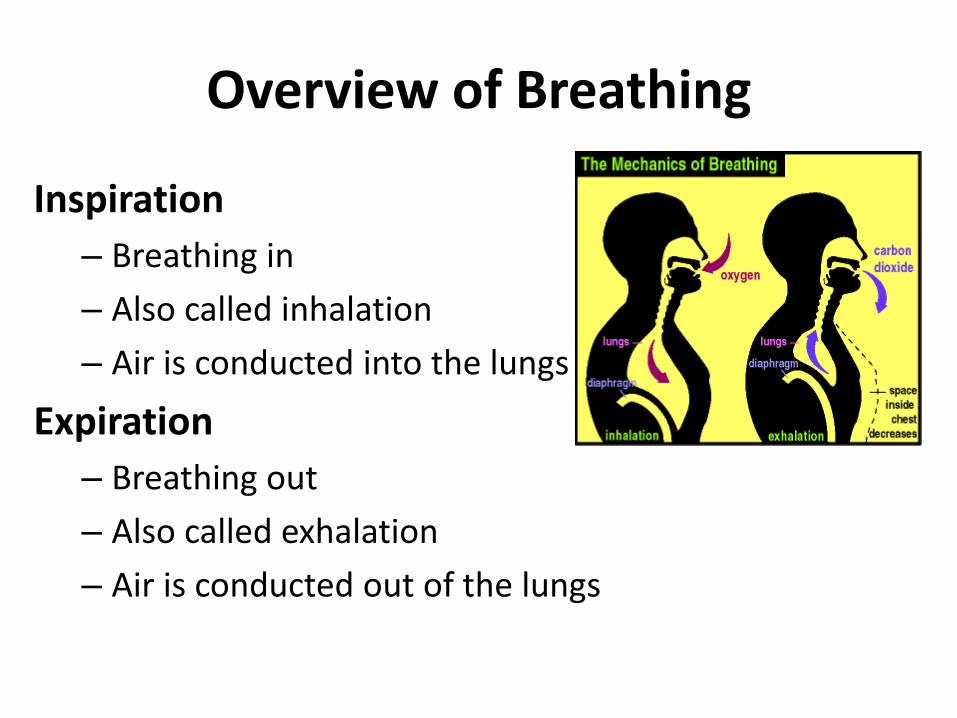
Overview of Breathing
Inspiration
– Breathing in
– Also called inhalation
– Air is conducted into the lungs
Expiration
– Breathing out
– Also called exhalation
– Air is conducted out of the lungs

Four Respiratory Events
Pulmonary Ventilation (breathing)
• the entry and exit of air into and out of lungs
External Respiration
• Gas exchange between air and blood
site = lungs
Internal Respiration
• Gas exchange between blood and tissue fluid
site = tissues
Transport of gases
• To and from the lungs and the tissues

Why O2? Cellular Respiration
• Production of ATP in the cells
– ATP = energy supply for the cells
• Requires oxygen
• Releases carbon dioxide
• Cellular respiration requires the four respiratory events

Overview Upper
• Nose
• Nasal Cavity
• Pharynx
• Larynx
Lower
• Trachea
• Bronchi
• Lungs

The Nasal Cavity
Nasal Conchae – bony ridges
• Increase surface area
Olfactory Epithelium – high in the nasal cavity
• Odor receptors on the cilia of cells
The Nasal Septum
• Bony separation between the nasal cavities

The Nasal Cavity…
• cleansing of air by coarse nostril hair, cilia, and mucus
• Cilia inside nasal cavity beat mucus into the throat for swallowing
• Cilia in trachea and bronchi beat mucus upward into the pharynx
• Lysozymes in mucus-kills bacteria

The Nasal Cavity…
• mucous membrane warms and moistens inhaled the air
• Cools air during expiration
• Moisture deposited on trachea
• Nose may drip – condensation

Mucociliary Escalator • What tissue type?


The Pharynx
• Commonly referred to as the “throat”
• Passageway and connection between the nasal and oral cavities
• Three parts:
– Nasopharynx
– Oropharynx – common path
for food and air
– Laryngopharynx -Leads to the larynx
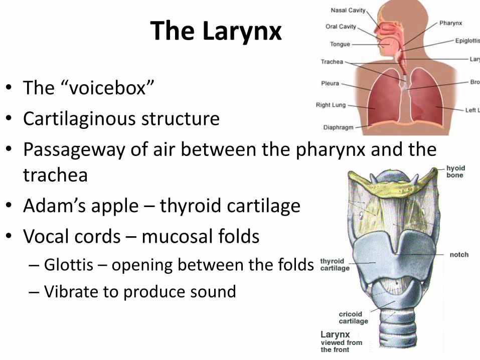
The Larynx
• The “voicebox”
• Cartilaginous structure
• Passageway of air between the pharynx and the trachea
• Adam’s apple – thyroid cartilage
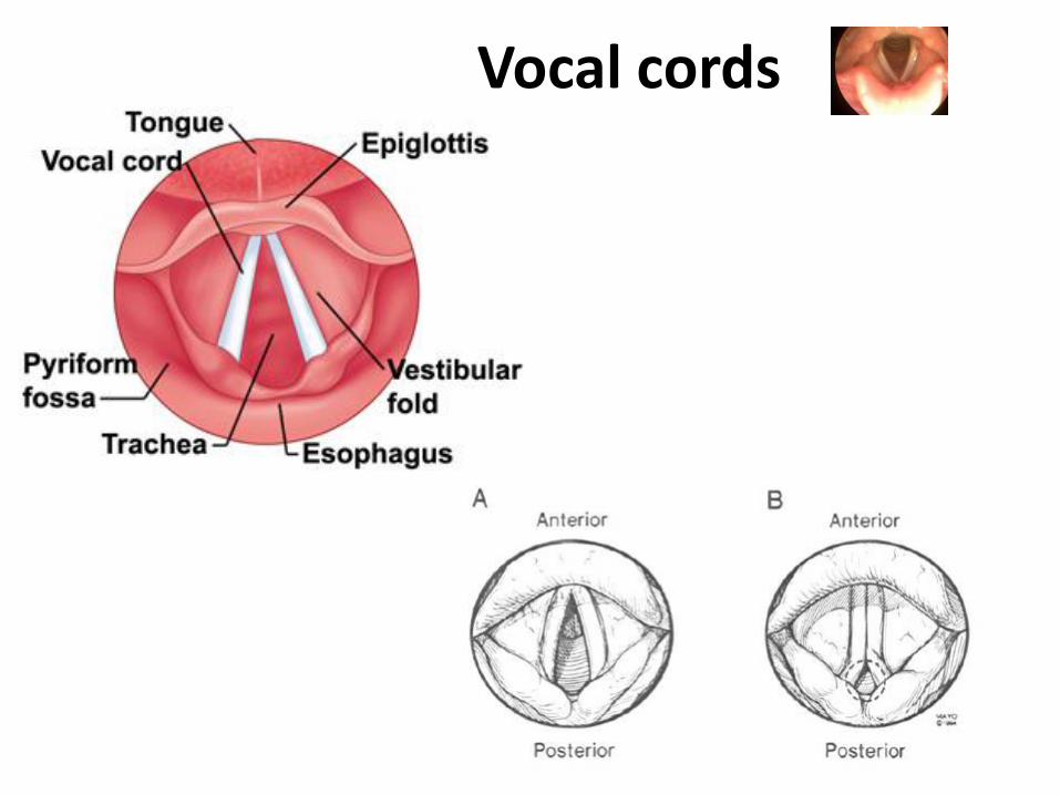
• Vocal cords – mucosal folds
– Glottis – opening between the folds
– Vibrate to produce sound

Swallowing of food
• The larynx moves up against the epiglottis
• Epiglottis – a flap of elastic cartillage closes
• Food is prevented from entering the larynx through the glottis

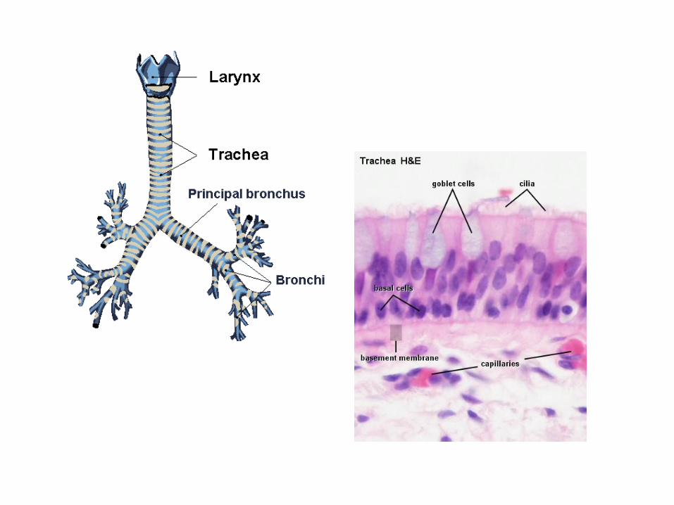
The Trachea
• Commonly known as the “windpipe”
• Flexible tube – connects larynx to the primary bronchi
• Ventral (anterior) to the esophagus
• Is held open via a C-shaped cartilage
• Is lined by pseudostratified ciliated columnar epithelium

Tracheostomy
• Insertion of a breathing tube via an incision in the trachea

The Bronchial Tree
• The trachea divides into R and L primary bronchi
• The primary bronchi branch into secondary bronchi
• The secondary bronchi branch into tertiary bronchi
• Brochioles – the smallest conducting airways
• Contraction of smooth muscle during Asthma attack

Alveoli
• Pockets (sacs) connected to each bronchiole
• Fill up with air during inhalation
• Simple squamous epithelium
• Site of gas exchange
• Surrounded by pulmonary
capillaries

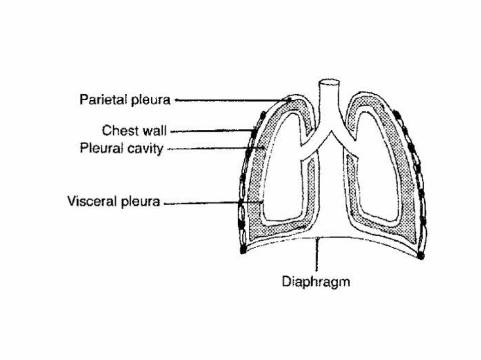
The Lungs • Paired organs inside the ribcage
• Each lung is inside its own pleural cavity
• Each lung is surrounded by visceral and parietal pleura
• Apex – superior narrow portion
• Base – inferior broad portion
– Lies on top of the diaphragm
• Divided into lobules made of alveoli

Location of the Lungs
• Thoracic cavity (sealed)
– Ribs join–Sternum anteriorly & Vertebrae posteriorly
– Intercostal muscles – between ribs
– Diaphragm – thin muscle, forms the floor
• Pleurae
– Visceral – on the lung surface
– Parietal – attached to the thoracic wall
– Pleural fluid in between
– Intrapleural pressure – between the pleura
• Lower than the atmospheric pressure
• Keeps lungs inflated
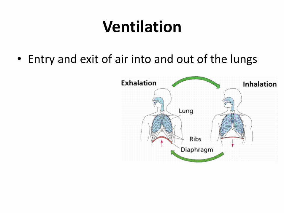
Ventilation
• Entry and exit of air into and out of the lungs
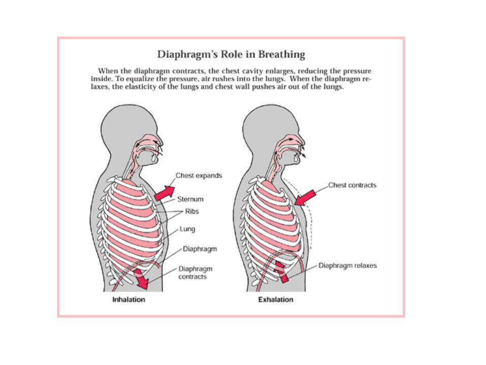

Inspiration
• Active phase of ventilation
• Diaphragm and intercostals contract
– Diaphragm flattens
– Ribs move up and out
• Lungs volume increases
• Air pressure in alveoli decreases (intrapulmonary pressure)
– Partial vacuum
– Alveolar P < atmospheric P
– Inward passive airflow until pressures equalize

Expiration
• Usually a passive phase
– No muscle effort required
• Diaphragm and external intercostal muscles relax
– Dome shaped diaphragm
– Rib cage down and in
• Lungs recoil
– P intra-alveolar (intrapulm.) > P atmosphere
– Prevention of collapse due to surfactant on alveoli
– Surfactant lowers surface tension

Maximum Inspiratory Effort • Exercise, Strenuous
activities
• Accessory Muscles of respiration required:
– Erector spinae (back), abdominals
– Pectoralis minor (chest)
– Scalene and sternocleidomastoid (neck)
• Maximum lung expansion

Forced Expiration
• Blowing into a musical instrument
• Using Internal intercostals, abdominals

Spirometer
Determines if a medical problems is preventing lungs from:
• Filling with air
• Releasing air
Spirometer records the volume of air exchanged during:
• Normal and deep breathing
Spirogram is created


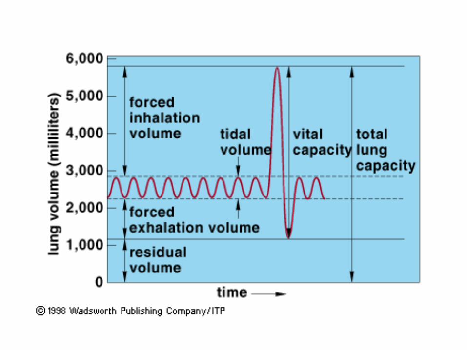
Respiratory Volumes
• Tidal Volume – amount of air that moves in and out when we are relaxed
– 500ml
• Vital Capacity - amount of air that moves in and out during deep breathing
Inspiratory Reserve V + Expiratory Reserve V.
– Inspiratory Reserve Volume
• 2900ml above the tidal volume – forced inspiration
– Expiratory Reserve Volume
• 4900 ml

Dead Air Space • Inhaled air that never reaches the alveoli
• Remains in the nasal cavities, trachea, bronchi, and bronchioles
• About 30% of the tidal volume
• Can reduce this by breathing slowly and deeply

Residual Volume
• Air that is not exhaled and remains in the alveoli
• About 1000ml
• Has a large amount of CO2
• Is not used for gas exchange
• Increase in residual volume in Emphysema
• Vital capacity is reduced
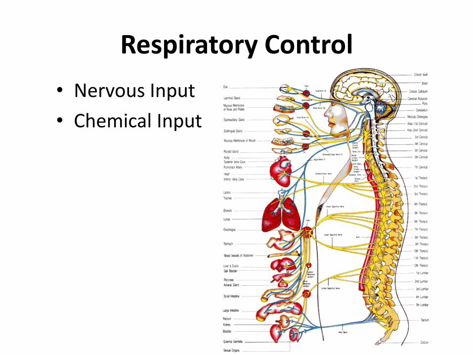
Respiratory Control
• Nervous Input
• Chemical Input

Normal Ventilation
• Normal adult breathing rate:
– 12 – 20 ventilations/minute
• Controlled by the primary respiratory center
– Located in medulla oblongata

Primary Respiratory Center
• Automatic motor input to the diaphram
– The phrenic nerve

Normal Breathing Rhythm
• Pons in conjunction with medulla oblongata

Other Nervous Input
• Additional input from the higher Centers of the brain can cause changes in breathing
– cerebral cortex
– limbic system
– hypothalamus
– emotions
• Conscious control of respiration:
– Temporary holding of breath

Protective Mechanisms
• Hering-Breuer Reflex
– Limits extent of inspiration
– Prevents over-expansion of lungs
– Vagus control
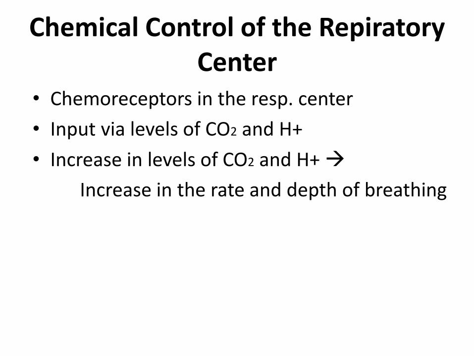
Chemical Control of the Repiratory Center
• Chemoreceptors in the resp. center
• Input via levels of CO2 and H+
• Increase in levels of CO2 and H+
Increase in the rate and depth of breathing

Influence of Oxygen levels
• No direct influence on the primary resp. center
• Low O2 levels sensed by chemoreceptors in:
– Carotid bodies
– Aortic bodies
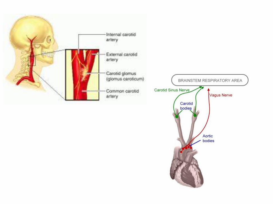

Control via Carotid and Aortic Bodies
• Low blood oxygen concentration
increase in the rate of respiration

Pulmonary Circulation
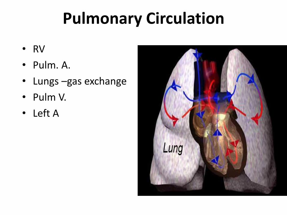
Pulmonary Circulation
• RV
• Pulm. A.
• Lungs –gas exchange
• Pulm V.
• Left A

Gas Exchange in the Lungs
• Capillaries surround alveoli
• O2 diffuses into the capillaries
• CO2 diffuses out of the capillaries
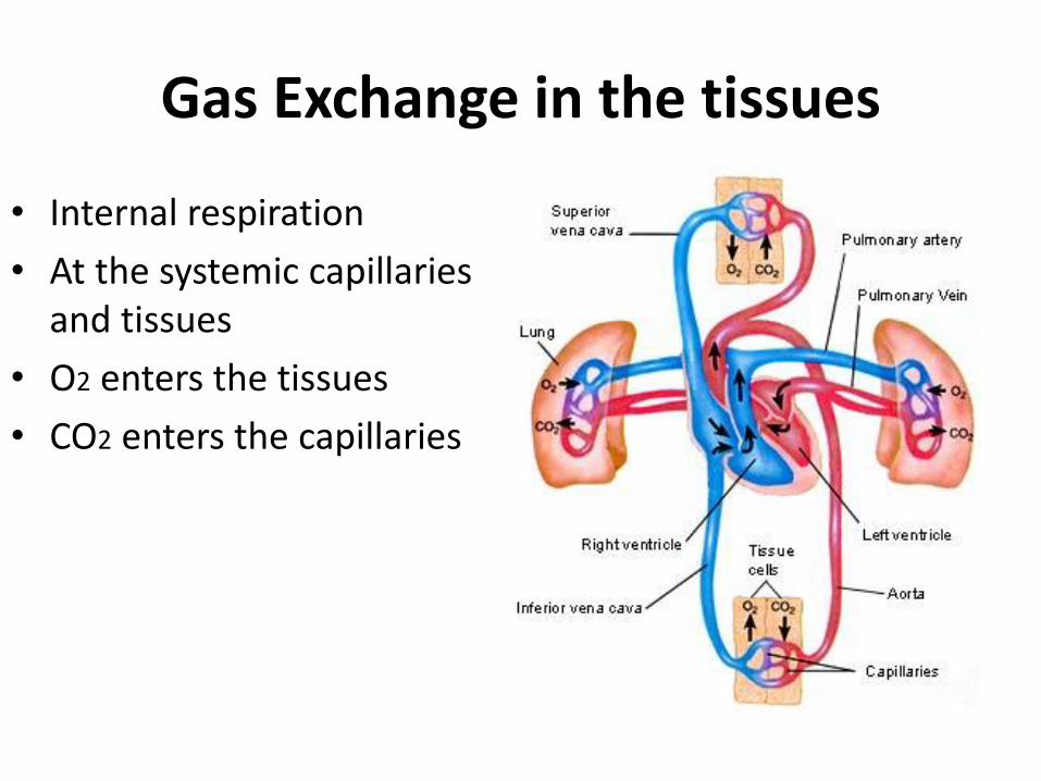
Gas Exchange in the tissues
• Internal respiration
• At the systemic capillaries and tissues
• O2 enters the tissues
• CO2 enters the capillaries

Gas transport
• O2 transport via:
– Fe in Hb
– plasma (2-3% only)
• CO2 transport:
– Plasma + cytoplasm of RBCs (10%)
– Globin of Hb (30%)
– CO2 + H2O Carbonic acid (H2CO3) (H+) + (HCO3-)
• majority
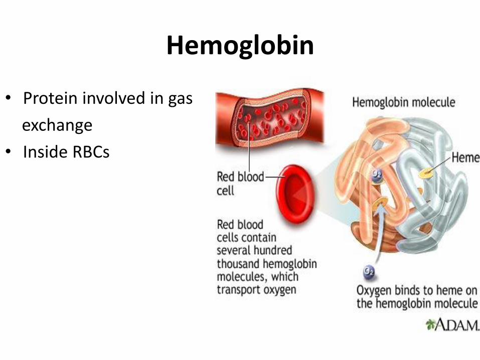
Hemoglobin
• Protein involved in gas
exchange
• Inside RBCs

Homeostasis
• Regulation of blood pH via respiratory control!