chapter05 121204002529-phpapp01
TRANSCRIPT

Chapter 5Chapter 5Chapter 5Chapter 5
The Human Body
National EMS Education National EMS Education Standard Competencies Standard Competencies (1 of 3)(1 of 3)
National EMS Education National EMS Education Standard Competencies Standard Competencies (1 of 3)(1 of 3)
Preparatory
Applies fundamental knowledge of the emergency medical services (EMS) system, safety/well-being of the emergency medical technician (EMT), medical/legal and ethical issues to the provision of emergency care.
National EMS Education National EMS Education Standard Competencies Standard Competencies (2 of 3)(2 of 3)
National EMS Education National EMS Education Standard Competencies Standard Competencies (2 of 3)(2 of 3)
Anatomy and Physiology
Applies fundamental knowledge of the anatomy and function of all human systems to the practice of EMS.
National EMS Education National EMS Education Standard Competencies Standard Competencies (3 of 3)(3 of 3)
National EMS Education National EMS Education Standard Competencies Standard Competencies (3 of 3)(3 of 3)
Pathophysiology
Applies fundamental knowledge of the pathophysiology of respiration and perfusion to patient assessment and management.
IntroductionIntroduction
• A working knowledge of anatomy is important.
• Knowledge of anatomy helps to communicate correct information:– To professionals, who know medical terms
– To others, who may not understand medical terms
Topographic AnatomyTopographic Anatomy
• Superficial landmarks – Serve as guides to structures that lie beneath
them
• Topographic anatomy applies to a body in the anatomic position.– Patient stands facing you, arms at side, palms
forward.
Planes of the Body (1 of 2)Planes of the Body (1 of 2)
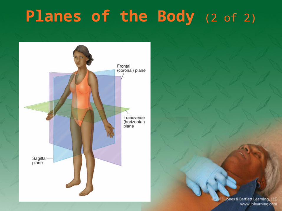
• Imaginary straight lines that divide the body
• Three main areas– Coronal plane: front/back
– Transverse (axial) plane: top/bottom
– Sagittal (lateral) plane: left/right
Planes of the Body (2 of 2)Planes of the Body (2 of 2)
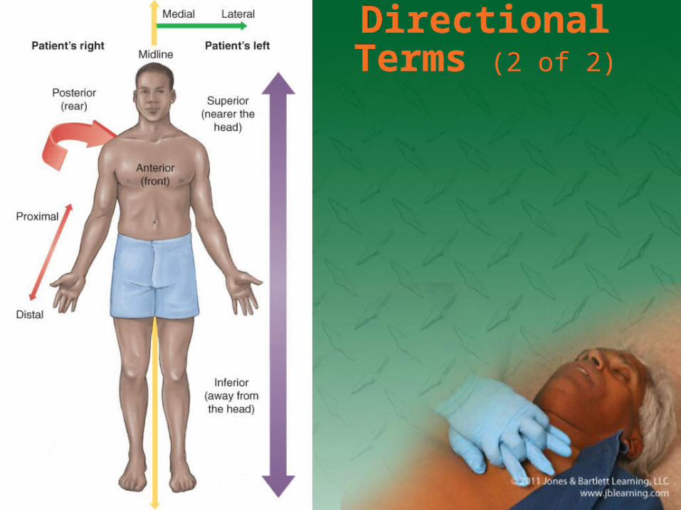
Directional Terms (1 of 2)Directional Terms (1 of 2)
• Important when discussing injury location or pain radiation. Examples include:– Anterior (ventral)
– Posterior (dorsal)
– Right, left (patient’s right or left)
– Superior (closest to head)
– Inferior (closest to feet)
Directional Terms (2 of 2)
Directional Terms (2 of 2)
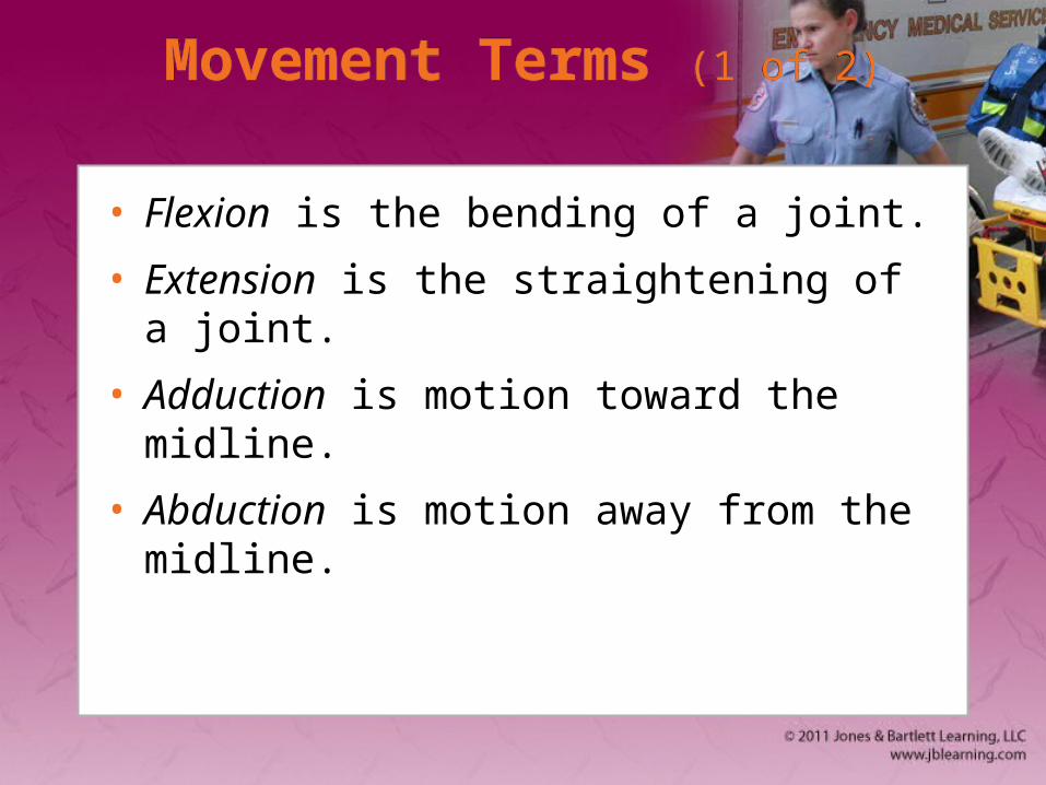
Movement Terms (1 of 2)Movement Terms (1 of 2)
• Flexion is the bending of a joint.
• Extension is the straightening of a joint.
• Adduction is motion toward the midline.
• Abduction is motion away from the midline.
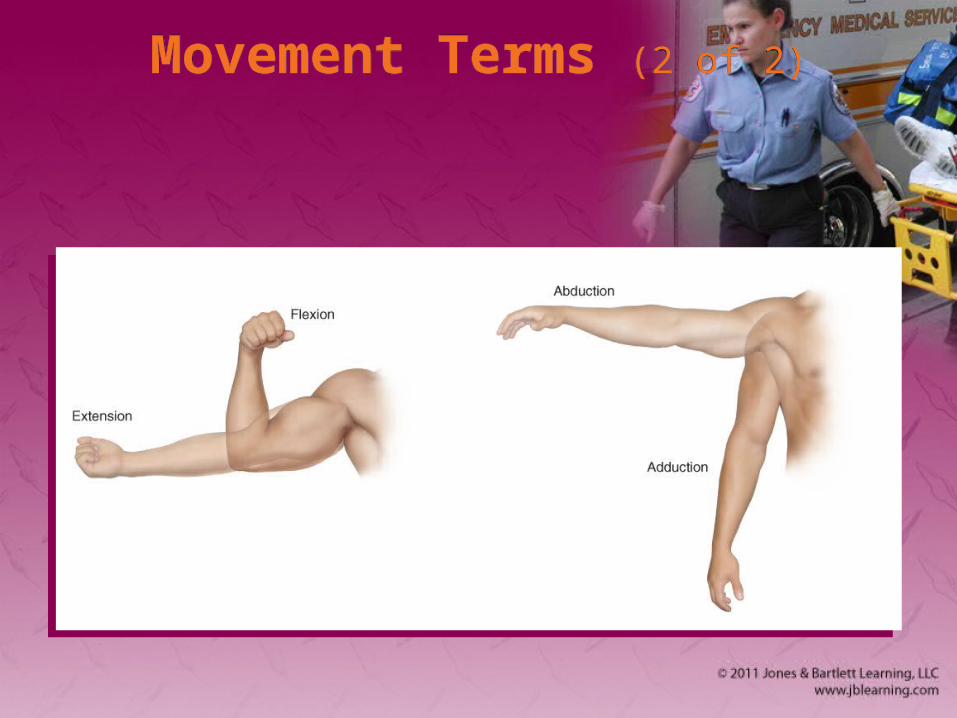
Movement Terms (2 of 2)Movement Terms (2 of 2)
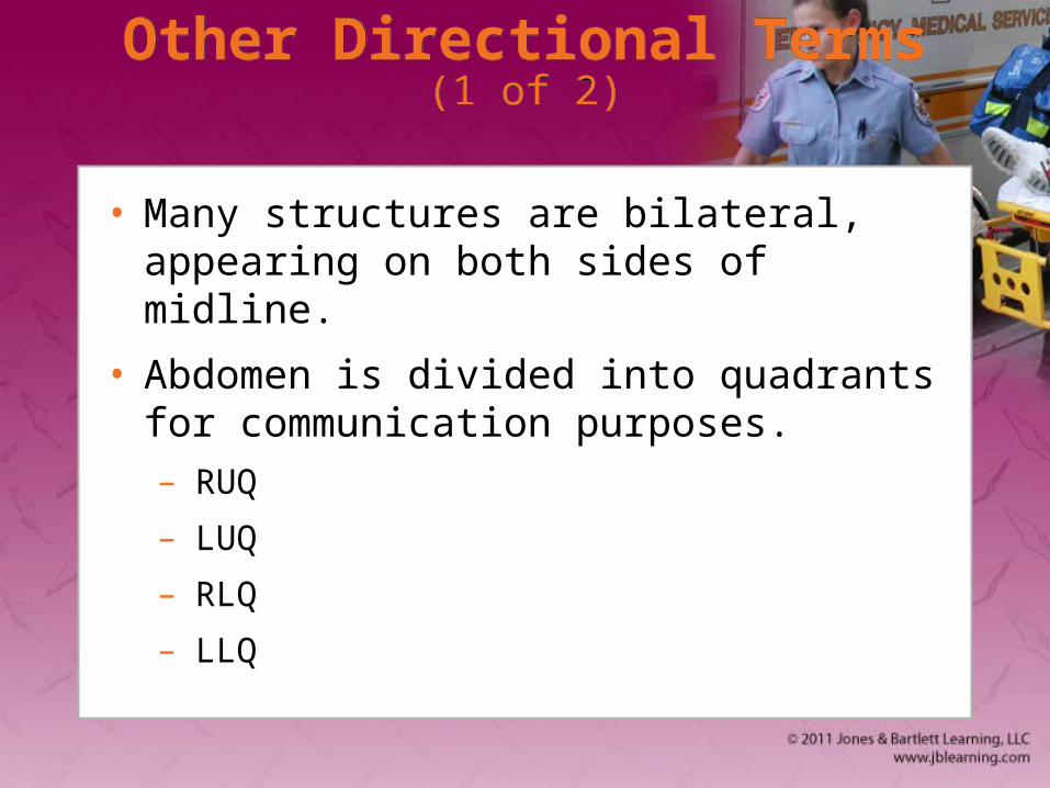
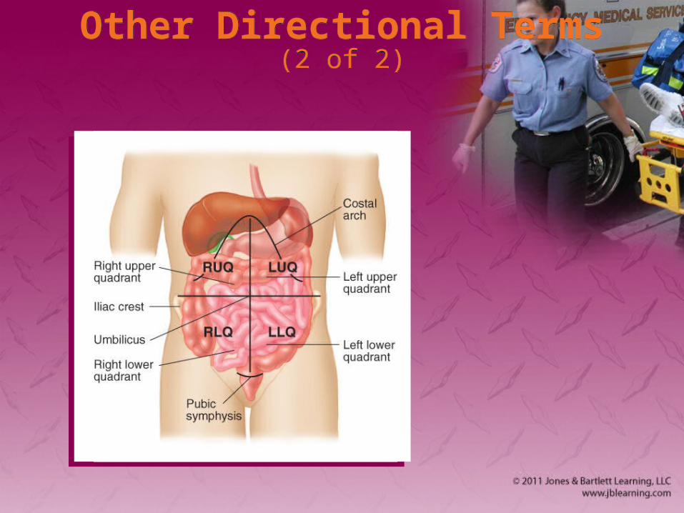
Other Directional Terms (1 of 2)Other Directional Terms (1 of 2)
• Many structures are bilateral, appearing on both sides of midline.
• Abdomen is divided into quadrants for communication purposes.– RUQ
– LUQ
– RLQ
– LLQ
Other Directional Terms (2 of 2)Other Directional Terms (2 of 2)
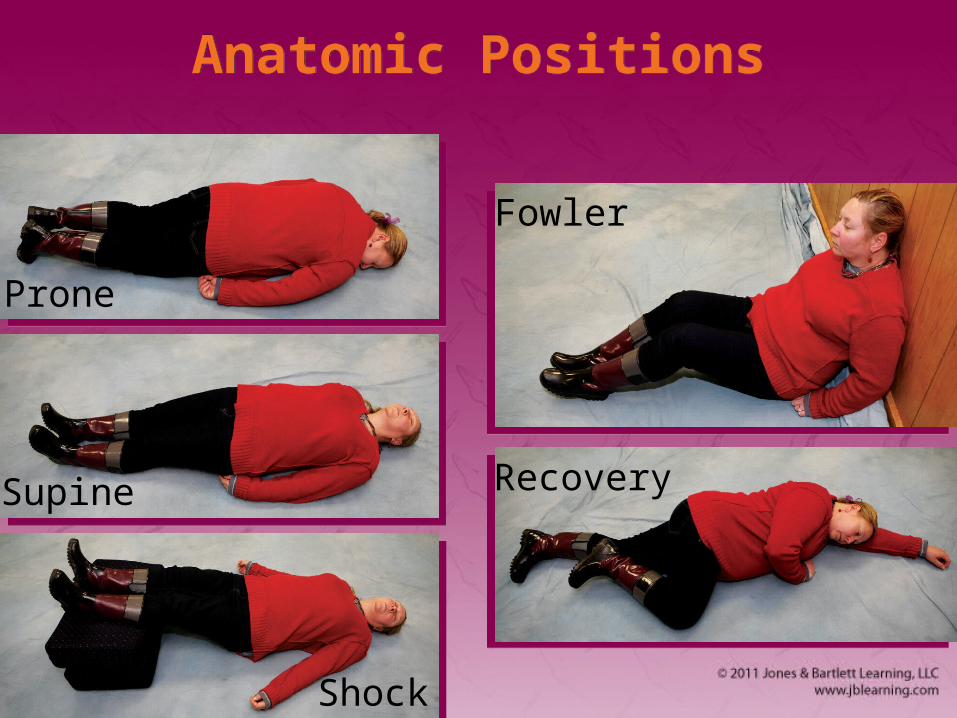
Anatomic PositionsAnatomic Positions
Prone
Supine
Shock
Fowler
Recovery
The Skeletal System: Anatomy The Skeletal System: Anatomy
• Skeleton gives us our recognizable human form.
• Protects vital internal organs
• Contains – Bones
– Ligaments
– Tendons
– Cartilage
The Axial Skeleton (1 of 4)The Axial Skeleton (1 of 4)
• Foundation on which the arms and legs are hung. Includes:– Skull
– Spinal column
– Thorax
The Axial Skeleton (2 of 4)The Axial Skeleton (2 of 4)
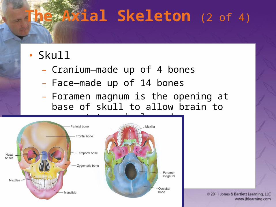
• Skull– Cranium—made up of 4 bones
– Face—made up of 14 bones
– Foramen magnum is the opening at base of skull to allow brain to connect to spinal cord.
The Axial Skeleton (3 of 4)The Axial Skeleton (3 of 4)
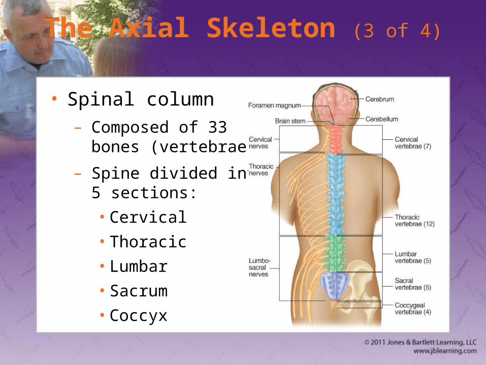
• Spinal column– Composed of 33
bones (vertebrae)
– Spine divided into 5 sections:
• Cervical
• Thoracic
• Lumbar
• Sacrum
• Coccyx
The Axial Skeleton (4 of 4)The Axial Skeleton (4 of 4)
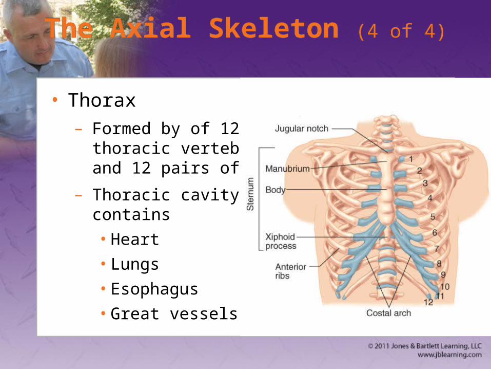
• Thorax– Formed by of 12
thoracic vertebrae and 12 pairs of ribs
– Thoracic cavity contains
• Heart
• Lungs
• Esophagus
• Great vessels
The Appendicular SkeletonThe Appendicular Skeleton
• Arms, legs, their connection points, and pelvis
• Includes:– Upper extremity
– Pelvis
– Lower extremity
The Upper Extremity (1 of 4)The Upper Extremity (1 of 4)
• Upper extremity extends from shoulder girdle to fingertips– Composed of arms, forearms, hands, fingers

The Upper Extremity (2 of 4)The Upper Extremity (2 of 4)
– Shoulder girdle
• Three bones come together, allowing arm to be moved:
– Clavicle, scapula, humerus
The Upper Extremity (3 of 4)The Upper Extremity (3 of 4)
– Arm
• The humerus is the supporting bone of the arm.
• The forearm consists of the radius and ulna.– Radius on lateral side of forearm
– Ulna on medial side of forearm
The Upper Extremity (4 of 4)The Upper Extremity (4 of 4)
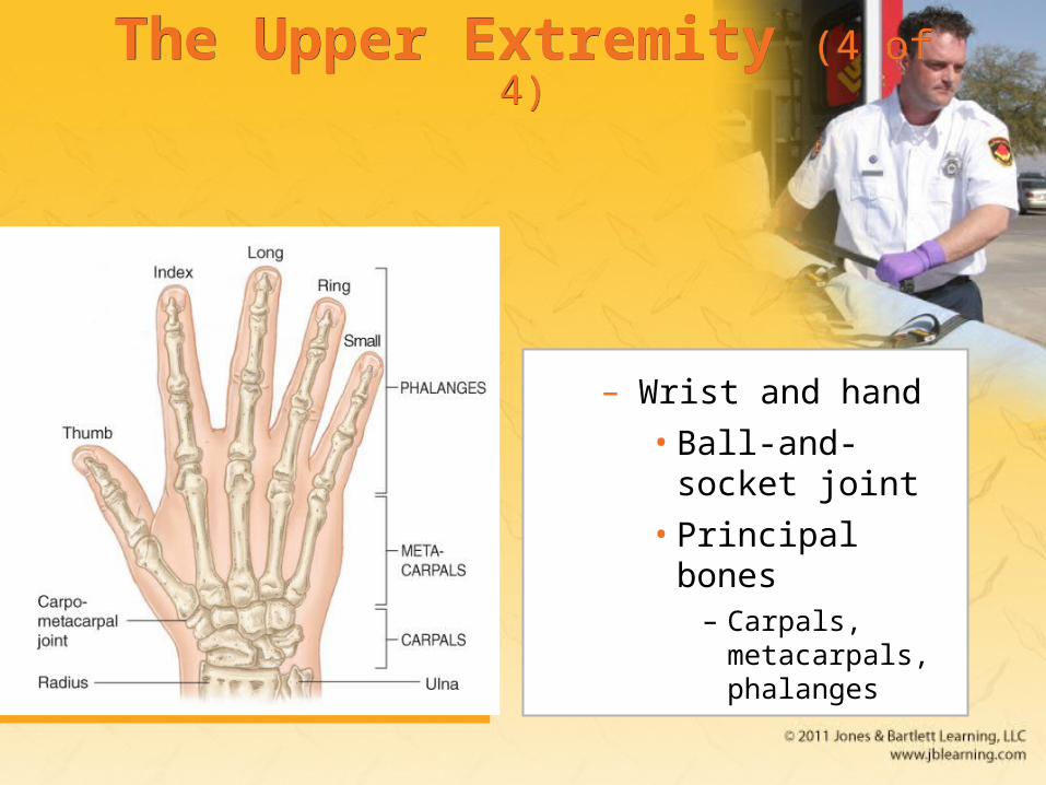
– Wrist and hand
• Ball-and-socket joint
• Principal bones– Carpals,
metacarpals, phalanges
The Pelvis (1 of 2)The Pelvis (1 of 2)
• Closed bony ring consisting of three bones– Sacrum
– Two pelvic bones
• Each pelvic bone is formed by fusion of ilium, ischium, and pubis.
The Pelvis (2 of 2)The Pelvis (2 of 2)
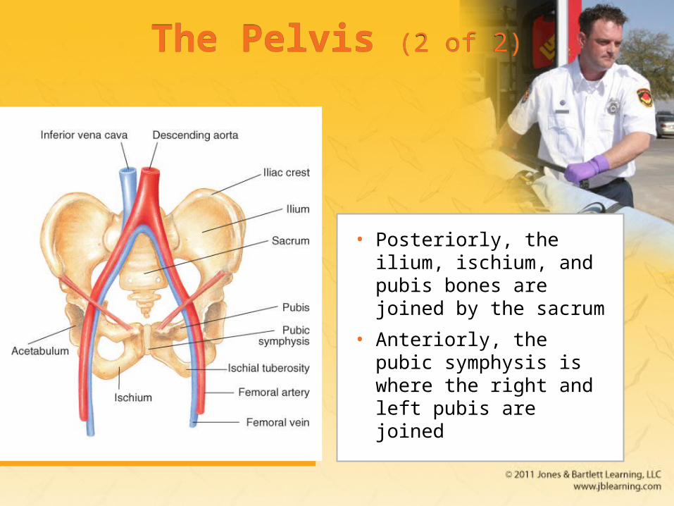
• Posteriorly, the ilium, ischium, and pubis bones are joined by the sacrum
• Anteriorly, the pubic symphysis is where the right and left pubis are joined

The Lower Extremity (1 of 4)The Lower Extremity (1 of 4)
• Main parts are thigh, leg, foot.
• Upper leg: femur (thigh bone) – Longest bone in body, femur connects into
acetabulum (pelvic girdle) by ball-and-socket joint.
– Greater and lesser trochanter are where major muscles of thigh connect to femur.
The Lower Extremity (2 of 4)The Lower Extremity (2 of 4)
• Knee connects upper leg to lower leg– Kneecap (patella)
• Lower Leg– Tibia (shin bone)
• Anterior of leg
– Fibula
• Lateral side of leg
The Lower Extremity
(3 of 4)
The Lower Extremity
(3 of 4)
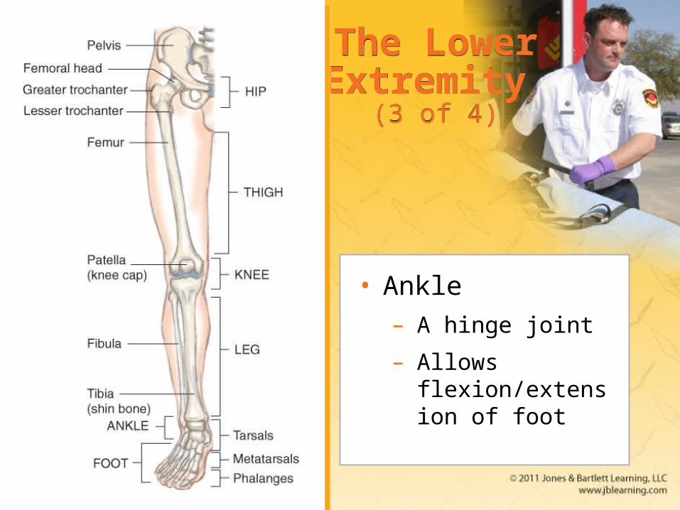
• Ankle– A hinge joint
– Allows flexion/extension of foot
The Lower Extremity (4 of 4)The Lower Extremity (4 of 4)
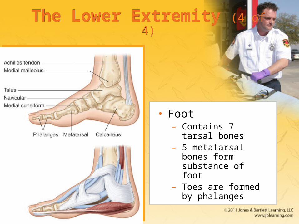
• Foot– Contains 7 tarsal
bones– 5 metatarsal bones
form substance of foot
– Toes are formed by phalanges
Joints (1 of 2)Joints (1 of 2)
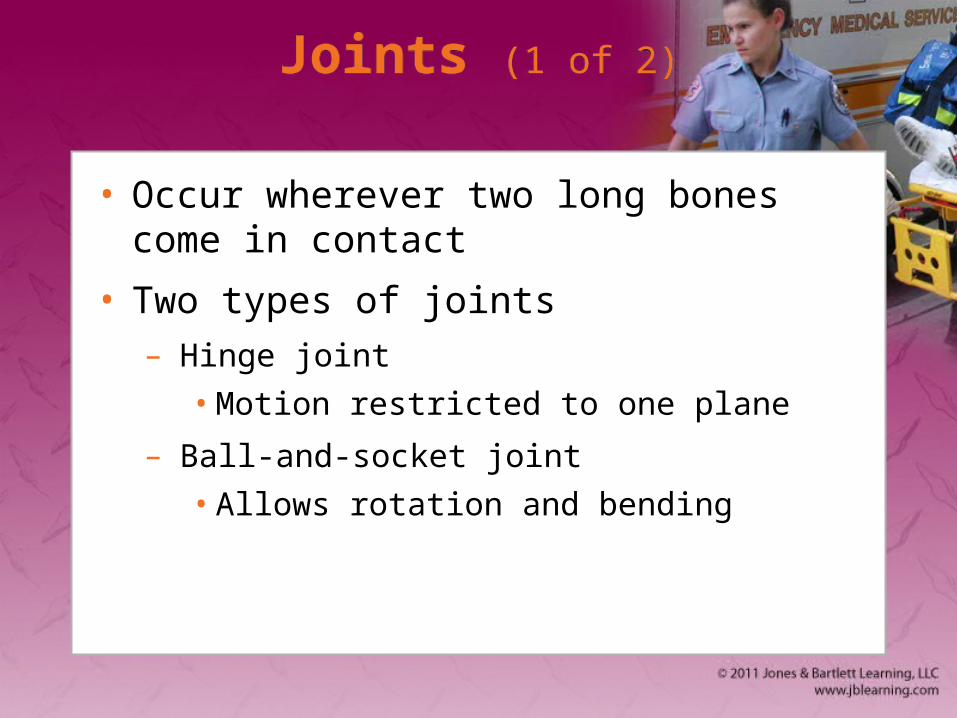
• Occur wherever two long bones come in contact
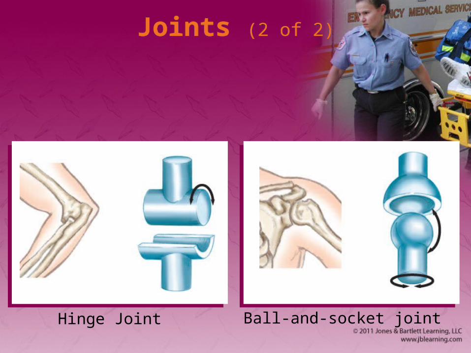
• Two types of joints– Hinge joint
• Motion restricted to one plane
– Ball-and-socket joint
• Allows rotation and bending
Joints (2 of 2)Joints (2 of 2)
Hinge Joint Ball-and-socket joint
The Skeletal System: Physiology
The Skeletal System: Physiology
• The skeletal system:– Gives body shape
– Provides protection of fragile organs
– Allows for movement
– Stores calcium
– Helps create blood cells
The Musculoskeletal System: Anatomy (1 of 4)
The Musculoskeletal System: Anatomy (1 of 4)
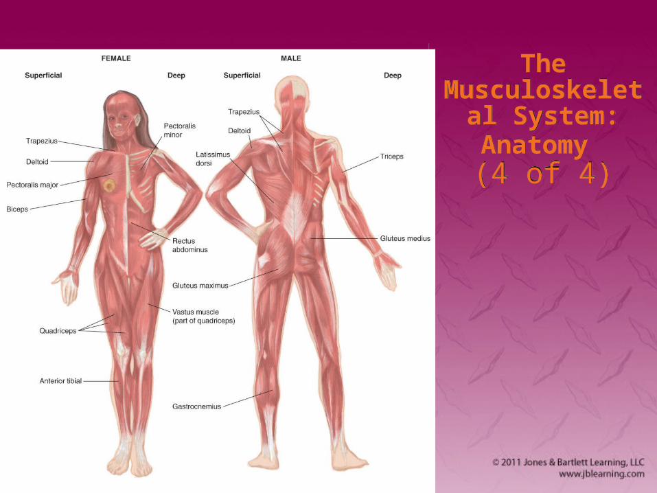
• Musculoskeletal system provides:– Form
– Upright posture
– Movement
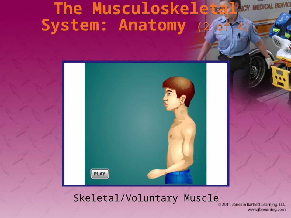
• More than 600 muscles attach to bone.– Called skeletal (or voluntary) muscles
The Musculoskeletal System: Anatomy (2 of 4)
The Musculoskeletal System: Anatomy (2 of 4)
Skeletal/Voluntary Muscle
The Musculoskeletal System: Anatomy (3 of 4)
The Musculoskeletal System: Anatomy (3 of 4)
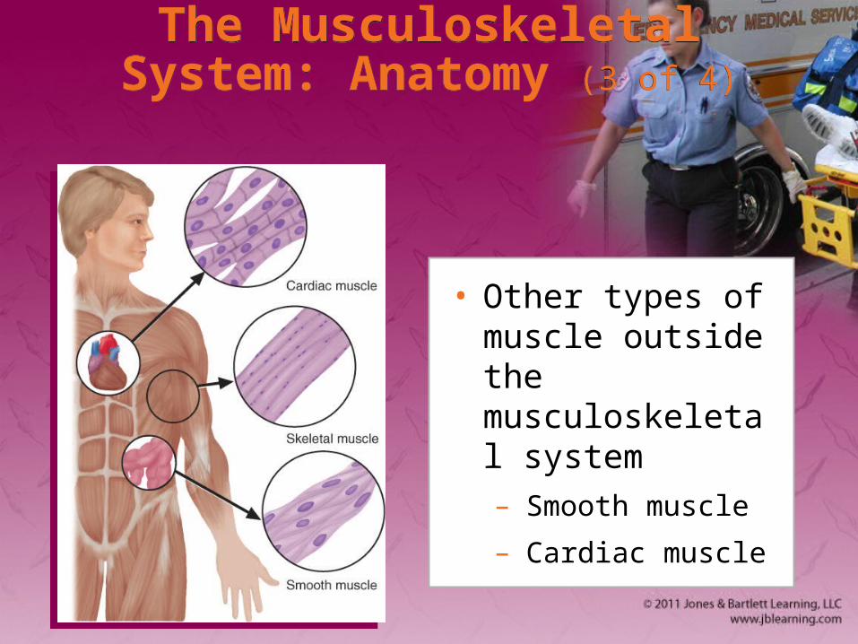
• Other types of muscle outside the musculoskeletal system– Smooth muscle
– Cardiac muscle
The Musculoskeletal
System:Anatomy (4 of 4)
The Musculoskeletal
System:Anatomy (4 of 4)
The Musculoskeletal System: Physiology
The Musculoskeletal System: Physiology
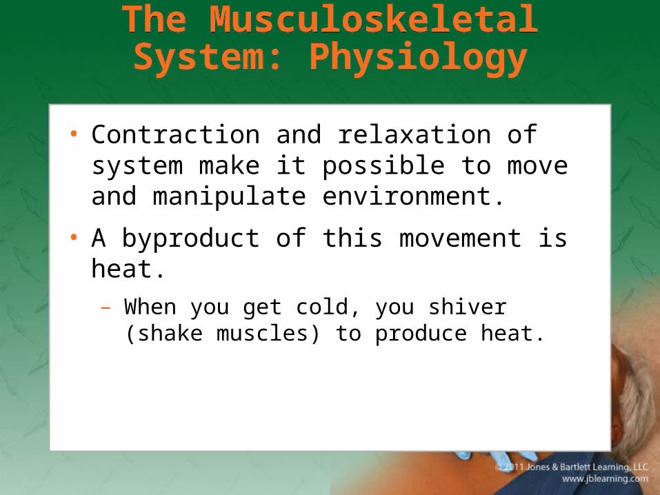
• Contraction and relaxation of system make it possible to move and manipulate environment.
• A byproduct of this movement is heat.– When you get cold, you shiver (shake muscles)
to produce heat.
The Respiratory System: Anatomy
The Respiratory System: Anatomy
• Structures of the body that contribute to respiration (the process of breathing)
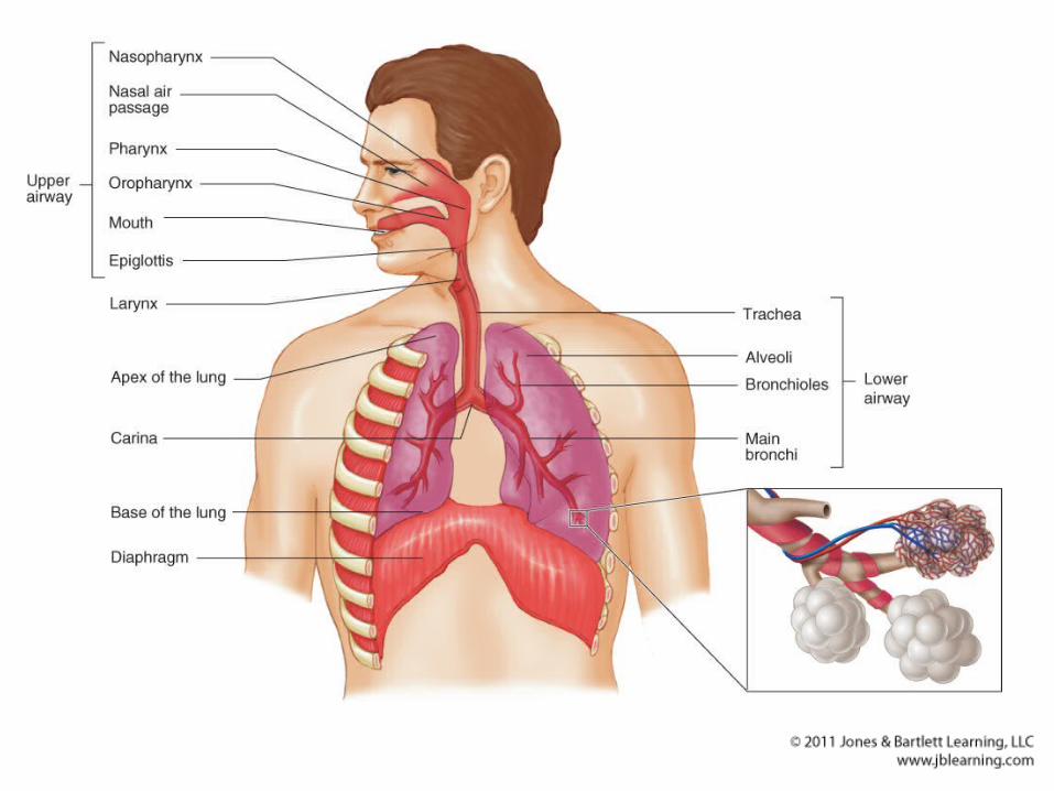

Upper Airway (1 of 3)Upper Airway (1 of 3)
• Includes:– Nose
– Mouth
– Tongue
– Jaw
– Oral cavity
Upper Airway (2 of 3)Upper Airway (2 of 3)
• Upper airway includes (cont’d)– Pharynx
• Nasopharynx
• Oropharynx
• Laryngopharynx– Larynx is anterior
– Esophagus is posterior
Upper Airway (3 of 3)Upper Airway (3 of 3)
• Upper airway (cont’d)– Epiglottis
• Prevents food and liquid from entering trachea
Lower AirwayLower Airway
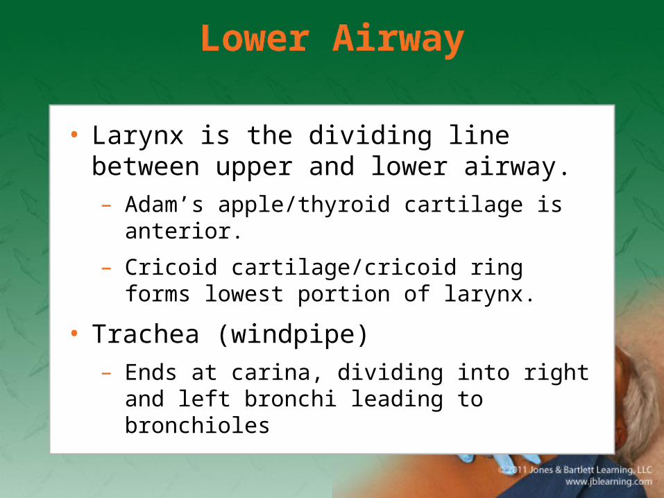
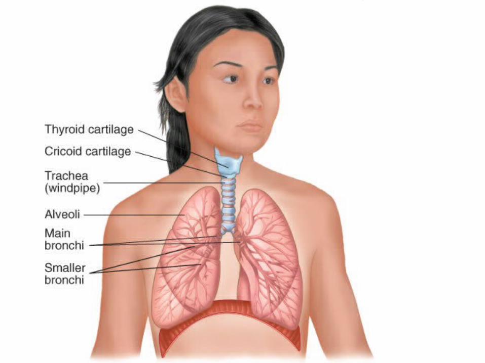
• Larynx is the dividing line between upper and lower airway.– Adam’s apple/thyroid cartilage is anterior.
– Cricoid cartilage/cricoid ring forms lowest portion of larynx.
• Trachea (windpipe)– Ends at carina, dividing into right and left
bronchi leading to bronchioles
Lungs (1 of 2)Lungs (1 of 2)
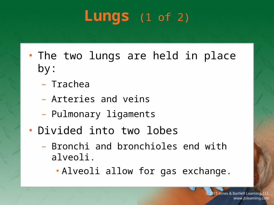
• The two lungs are held in place by:– Trachea
– Arteries and veins
– Pulmonary ligaments
• Divided into two lobes– Bronchi and bronchioles end with alveoli.
• Alveoli allow for gas exchange.
Lungs (2 of 2)Lungs (2 of 2)
• Lungs are covered by smooth, glistening tissue called pleura
Muscles of Breathing (1 of 2)Muscles of Breathing (1 of 2)
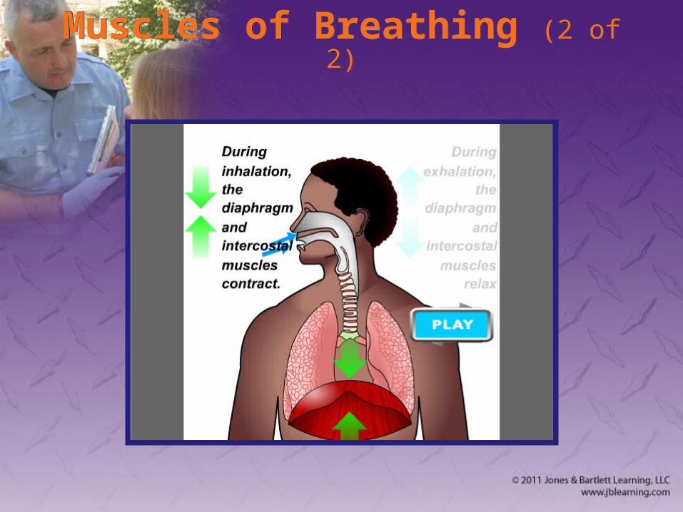
• Diaphragm is primary muscle.
• Also involved are:– Intercostal muscles
– Abdominal muscles
– Pectoral muscles
Muscles of Breathing (2 of 2)Muscles of Breathing (2 of 2)
The Respiratory System: Physiology (1 of 7)
The Respiratory System: Physiology (1 of 7)
• Function is to provide body with oxygen and eliminate carbon dioxide.
• Ventilation and respiration are two separate, interdependent functions of the respiratory system.
The Respiratory System: Physiology (2 of 7)
The Respiratory System: Physiology (2 of 7)
• Respiration is the exchange of oxygen and carbon dioxide in alveoli and tissue.– Brain stem controls breathing.
– Hypoxic drive is backup system.
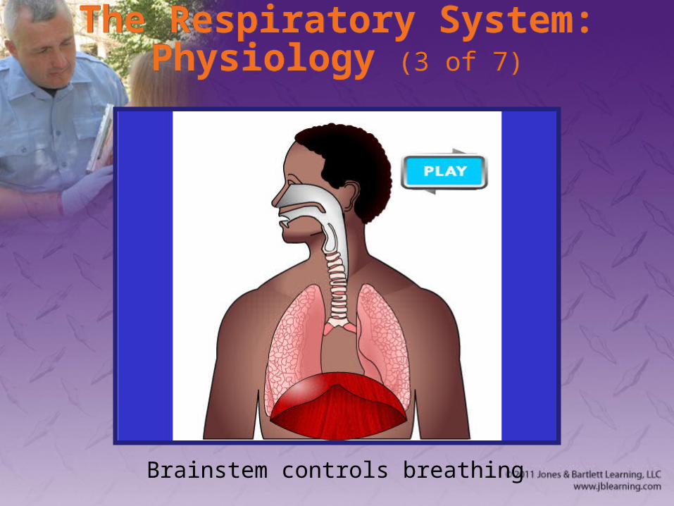
The Respiratory System: Physiology (3 of 7)
The Respiratory System: Physiology (3 of 7)
Brainstem controls breathing
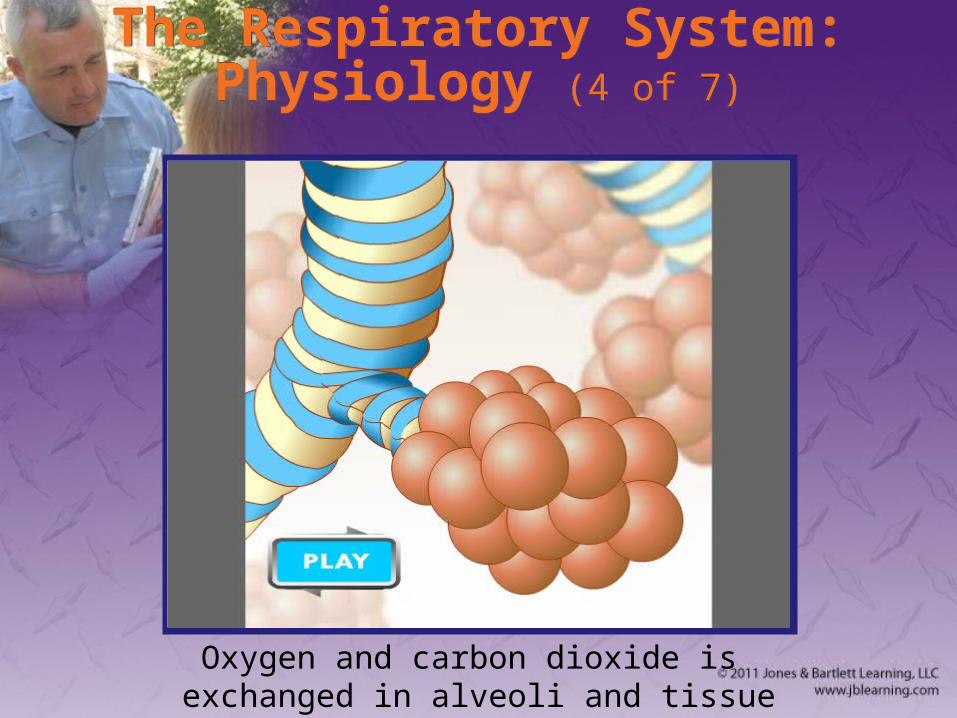
The Respiratory System: Physiology (4 of 7)
The Respiratory System: Physiology (4 of 7)
Oxygen and carbon dioxide is exchanged in alveoli and tissue
The Respiratory System: Physiology (5 of 7)
The Respiratory System: Physiology (5 of 7)
• Respiration (cont’d)– Medulla initiates ventilation cycles.
• Dorsal respiratory group (DRG)– Initiates inspiration
• Ventral respiratory group (VRG)– Provides forced inspiration or expiration when
needed
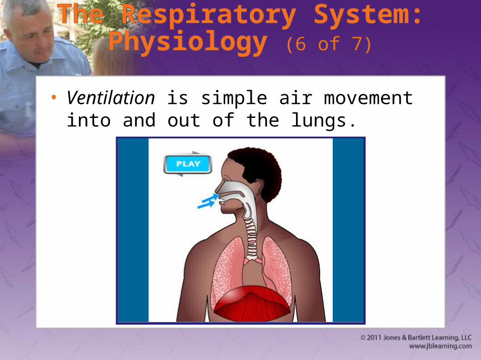
The Respiratory System: Physiology (6 of 7)
The Respiratory System: Physiology (6 of 7)
• Ventilation is simple air movement into and out of the lungs.
The Respiratory System: Physiology (7 of 7)
The Respiratory System: Physiology (7 of 7)
• You provide ventilation when you administer oxygen.
• Tidal volume is amount of air moved into or out of lungs in a single breath.
Characteristics of Normal Breathing (1 of 2)
Characteristics of Normal Breathing (1 of 2)
• Normal rate and depth (tidal volume)
• Regular rhythm or pattern of inhalation and exhalation
• Good audible breath sounds on both sides of chest
Characteristics of Normal Breathing (2 of 2)
Characteristics of Normal Breathing (2 of 2)
• Regular rise and fall movement on both sides of the chest
• Movement of the abdomen
Inadequate Breathing Patterns in Adults
Inadequate Breathing Patterns in Adults
• Labored breathing
• Muscle retractions
• Pale, cyanotic, cool, damp skin
• Tripod position
• Agonal gasps (gasping breaths)
The Circulatory System: Anatomy (1 of 2)
The Circulatory System: Anatomy (1 of 2)
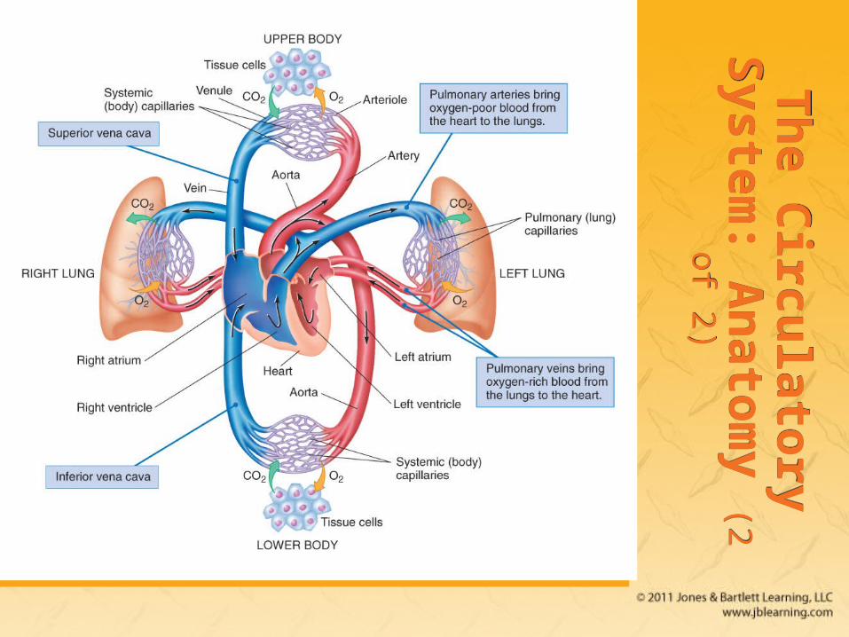
• Complex arrangement of connected tubes– Arteries, arterioles, capillaries, venules, veins
• Two circuits– Systemic circulation—body
– Pulmonary circulation—lungs
Th
e Circu
latory S
ystem:
An
atom
y (2 of 2)T
he C
irculato
ry System
: A
nato
my (2 of 2)
The Heart (1 of 7)The Heart (1 of 7)
• Hollow muscular organ the size of an adult’s clenched fist
• Made of specialized cardiac muscle (myocardium)
• Works as two paired pumps– Septum divides right and left sides.
The Heart (2 of 7)The Heart (2 of 7)
• Each side is divided into:– Atrium (upper chamber)
– Ventricle (lower chamber)
The Heart (3 of 7)The Heart (3 of 7)
• Circulation– Heart receives its blood from aorta.
– Right side receives blood from veins.
– Left side receives blood from lungs.
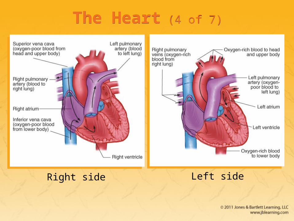
Right side Left side
The Heart (4 of 7)The Heart (4 of 7)
The Heart (5 of 7)The Heart (5 of 7)
• Circulation (cont’d)
The Heart (6 of 7)The Heart (6 of 7)
• Normal resting heart rate (HR) is 60 to 100 beats/min.
• Stroke volume (SV)– Amount of blood moved by one beat
• Cardiac output (CO)– Amount of blood moved in 1 minute
– HR × SV = CO
The Heart (7 of 7)The Heart (7 of 7)
• In 1 minute, body’s entire blood volume (5 to 6 L) is circulated through all the vessels.
• Electrical conduction network– Causes smooth, coordinated contractions
– Contractions produce pumping action
Arteries (1 of 4)Arteries (1 of 4)
• Arteries carry blood from heart to all body tissues.– Branch into arterioles
– Arterioles branch into capillaries
• Pulse is created by blood pumping out of left ventricle into major arteries.
Arteries (2 of 4)Arteries (2 of 4)
• Major arteries – Aorta (heart)
– Pulmonary (right ventricle)
– Carotid (neck)
– Femoral (thigh)
– Posterior tibial (lower leg)
– Dorsalis pedis (foot)
Arteries (3 of 4)Arteries (3 of 4)
• Major arteries (cont’d)– Brachial (upper arm)
– Radial (lower arm)
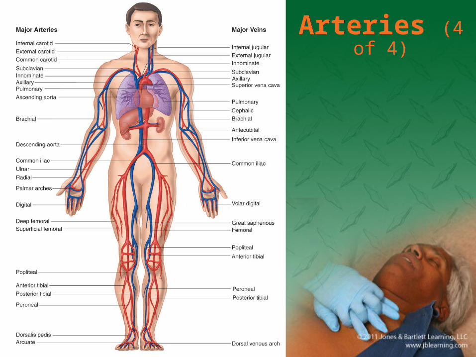
Arteries (4 of 4)Arteries (4 of 4)
CapillariesCapillaries
• Connect arterioles to venules
• Fine end divisions of arterial system
• Allow contact between blood and cells
• Billions of capillaries in body
VeinsVeins
• Return oxygen-depleted blood to the heart
• Superior vena cava carries blood returning from head, neck, shoulders, upper extremities.
• Inferior vena cava carries blood from abdomen, pelvis, lower extremities.
• Join at right atrium
SpleenSpleen
• Solid organ located under rib cage
• Filters blood
• Is particularly susceptible to injury from blunt trauma– Can lead to severe internal bleeding
Blood CompositionBlood Composition
• Plasma
• Red blood cells (erythrocytes)
• White blood cells (leukocytes)
• Platelets
The Circulatory System: Physiology (1 of 2)
The Circulatory System: Physiology (1 of 2)
• Blood pressure is pressure blood exerts against walls of arteries.
• When left ventricle of heart contracts, it pumps blood from ventricle into aorta.– Called systole
The Circulatory System: Physiology (2 of 2)
The Circulatory System: Physiology (2 of 2)
• When muscle of ventricle relaxes, ventricle fills with blood.– Called diastole
• Blood pressure readings – Systolic blood pressure (high point of wave)
– Diastolic blood pressure (low point of wave)
Normal Circulation in Adults (1 of 2)
Normal Circulation in Adults (1 of 2)
• Automatically adjusted and controlled
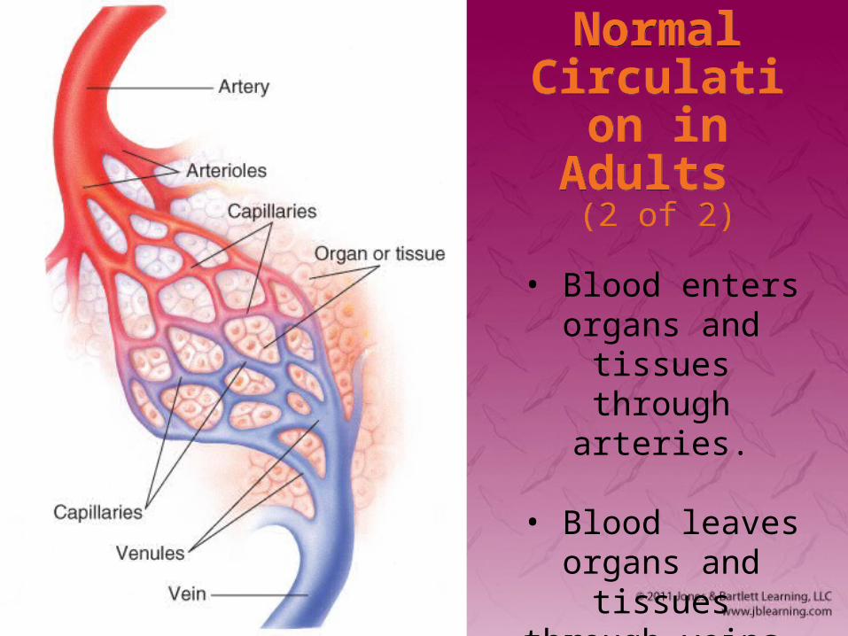
• Perfusion is circulation of blood in organ or tissue in adequate amounts to meet the needs of cells.
Normal Circulation in Adults
(2 of 2)
Normal Circulation in Adults
(2 of 2)
• Blood enters organs and tissues
through arteries.
• Blood leaves organs and tissues
through veins.
Inadequate Circulation in Adults
Inadequate Circulation in Adults
• The system can adjust to small blood loss.– Vessels constrict.
– Heart pumps more rapidly.
• With a large loss, adjustment fails, and patient goes into shock.
The Function of BloodThe Function of Blood
• Fighting infection
• Transporting oxygen
• Transporting carbon dioxide
• Controlling pH
• Transporting wastes and nutrients
• Clotting (coagulation)
Nervous System Control of the Cardiovascular System (1 of 2)
Nervous System Control of the Cardiovascular System (1 of 2)
• Sympathetic nervous system is responsible for fight-or-flight response.– Sends commands to adrenal glands
– Epinephrine and norepinephrine are secreted to stimulate heart and blood vessels.
Nervous System Control of the Cardiovascular System (2 of 2)
Nervous System Control of the Cardiovascular System (2 of 2)
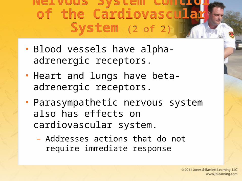
• Blood vessels have alpha-adrenergic receptors.
• Heart and lungs have beta-adrenergic receptors.
• Parasympathetic nervous system also has effects on cardiovascular system.– Addresses actions that do not require
immediate response
The Nervous System: Anatomy and Physiology (1 of 2)
The Nervous System: Anatomy and Physiology (1 of 2)
• The nervous system is perhaps the most complex organ in body
• Consists of:– Brain
– Spinal cord
The Nervous System: Anatomy and Physiology (2 of 2)
The Nervous System: Anatomy and Physiology (2 of 2)
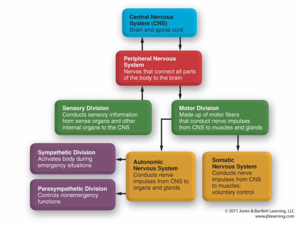
• Divided into two main portions:– Central nervous system (CNS)
– Peripheral nervous system
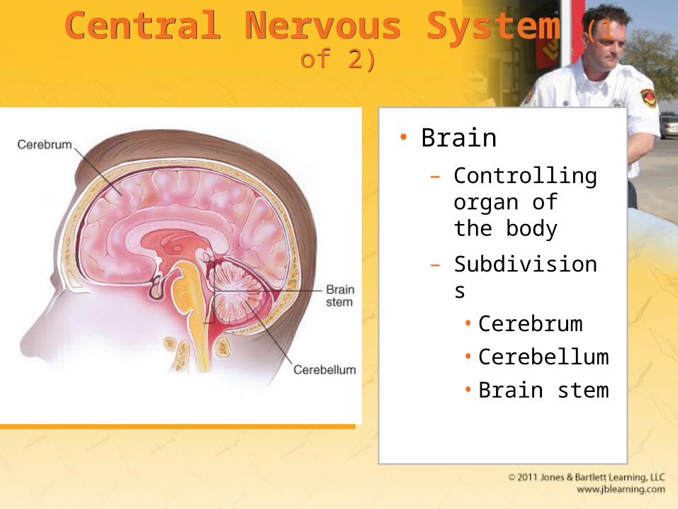
Central Nervous System (1 of 2)Central Nervous System (1 of 2)
• Brain– Controlling
organ of the body
– Subdivisions
• Cerebrum
• Cerebellum
• Brain stem
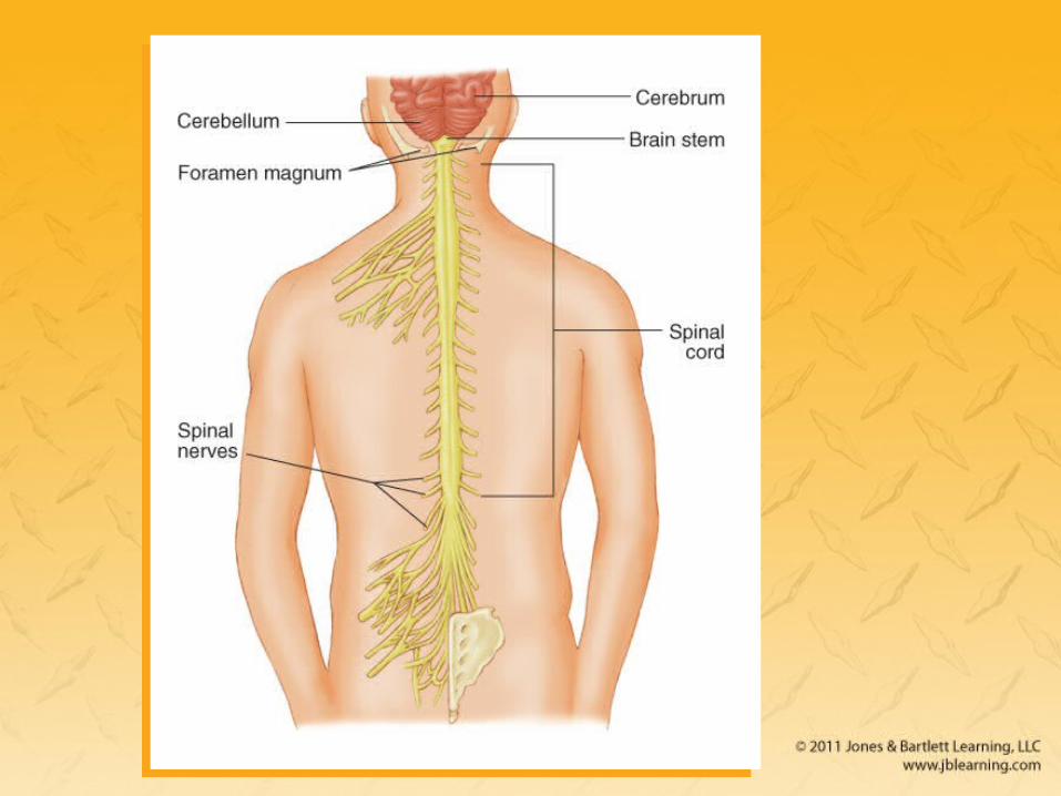
Central Nervous System (2 of 2)Central Nervous System (2 of 2)
• Spinal cord– Continuation of the brain
– Transmits messages between brain and body
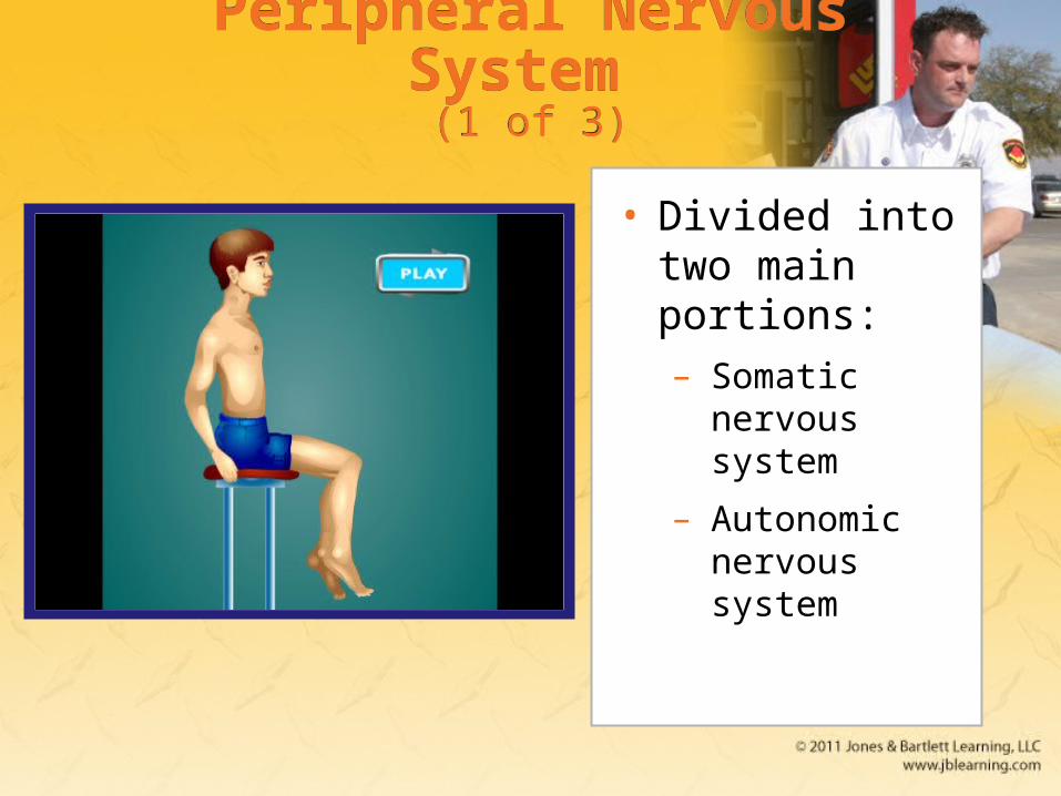
Peripheral Nervous System (1 of 3)
Peripheral Nervous System (1 of 3)
• Divided into two main portions:– Somatic
nervous system
– Autonomic nervous system
Peripheral Nervous System (2 of 3)
Peripheral Nervous System (2 of 3)
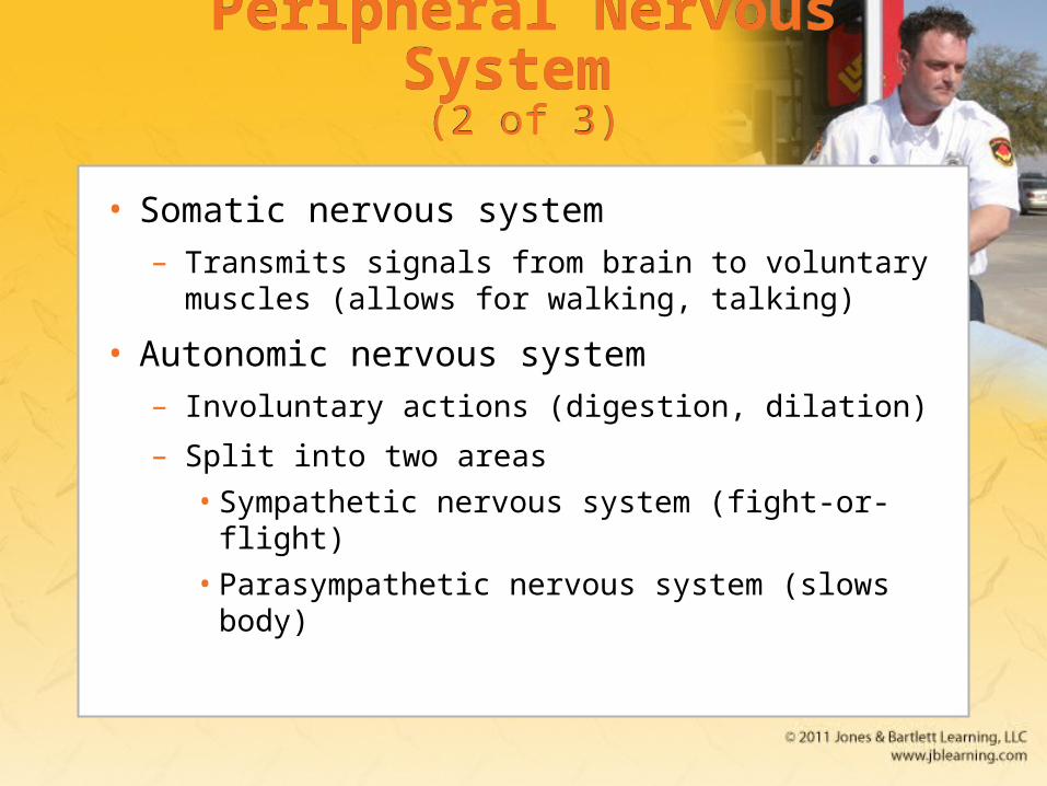
• Somatic nervous system– Transmits signals from brain to voluntary
muscles (allows for walking, talking)
• Autonomic nervous system– Involuntary actions (digestion, dilation)
– Split into two areas
• Sympathetic nervous system (fight-or-flight)
• Parasympathetic nervous system (slows body)
Peripheral Nervous System (3 of 3)
Peripheral Nervous System (3 of 3)
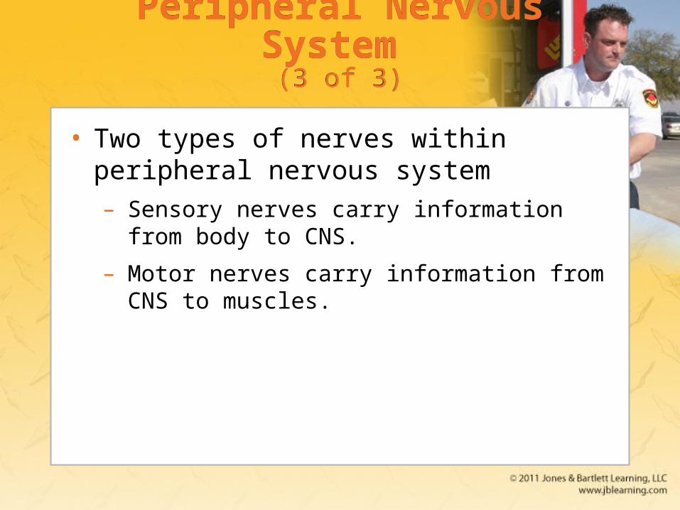
• Two types of nerves within peripheral nervous system– Sensory nerves carry information from body to
CNS.
– Motor nerves carry information from CNS to muscles.
The Integumentary System (Skin): Anatomy (1 of 2)
The Integumentary System (Skin): Anatomy (1 of 2)
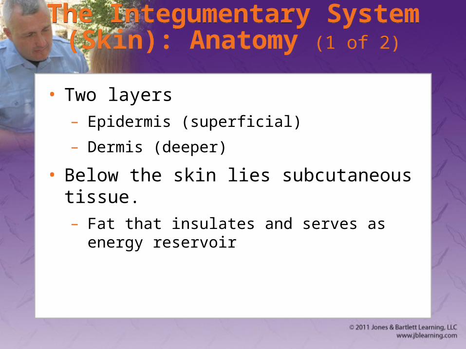
• Two layers– Epidermis (superficial)
– Dermis (deeper)
• Below the skin lies subcutaneous tissue. – Fat that insulates and serves as energy
reservoir
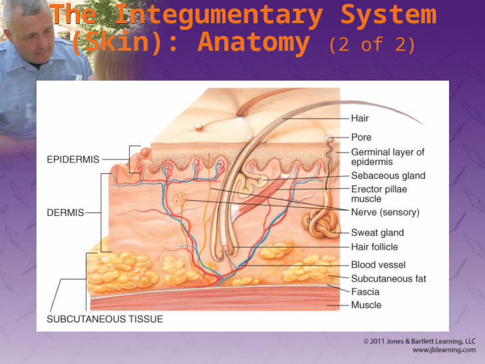
The Integumentary System (Skin): Anatomy (2 of 2)
The Integumentary System (Skin): Anatomy (2 of 2)
The Integumentary System (Skin): Physiology
The Integumentary System (Skin): Physiology
• Skin is the largest single organ
• Three major functions– Protect the body in the environment
– Regulate body temperature
– Transmit information from environment to brain
The Digestive System: Anatomy (1 of 4)
The Digestive System: Anatomy (1 of 4)
• Function of system is digestion.
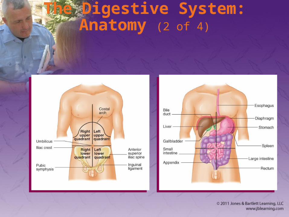
• Abdomen is second major body cavity.– Contains major organs of digestion and
excretion
– Quadrants are easiest way to identify areas
• RUQ/LUQ
• RLQ/LLQ
The Digestive System: Anatomy (2 of 4)
The Digestive System: Anatomy (2 of 4)
The Digestive System: Anatomy (3 of 4)
The Digestive System: Anatomy (3 of 4)
• Mouth– Lips, cheeks, gums, teeth, tongue
– Salivary glands
• Oropharynx
• Esophagus
• Stomach
• Pancreas
The Digestive System: Anatomy (4 of 4)
The Digestive System: Anatomy (4 of 4)
• Liver
• Small intestine
• Large intestine
• Appendix
• Rectum
The Digestive System: Physiology
The Digestive System: Physiology
• Enzymes are added to food.– By salivary glands, stomach, liver, pancreas,
small intestine
• Enzymes convert food into basic sugars, fatty acids, amino acids.– Further processed by liver
– Circulated via blood throughout body
The Endocrine System: Anatomy and Physiology (1 of 2)
The Endocrine System: Anatomy and Physiology (1 of 2)
• Complex message and control system
• Integrates many body functions
• Hormones are released directly into bloodstream.– Examples: epinephrine, norepinephrine, insulin
The Endocrine System: Anatomy and Physiology (2 of 2)
The Endocrine System: Anatomy and Physiology (2 of 2)
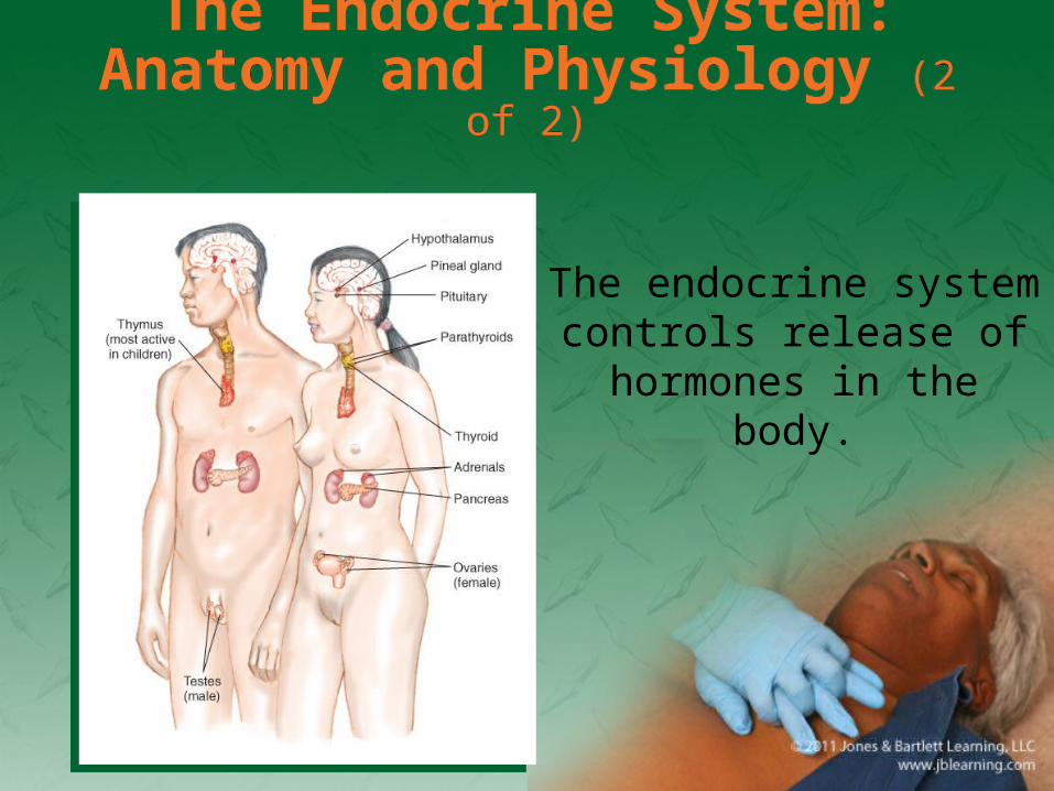
The endocrine system controls release of
hormones in the body.
The Urinary System: Anatomy and Physiology (1 of 2)
The Urinary System: Anatomy and Physiology (1 of 2)
• Controls fluid balance in the body
• Filters and eliminates wastes
• Controls pH balance
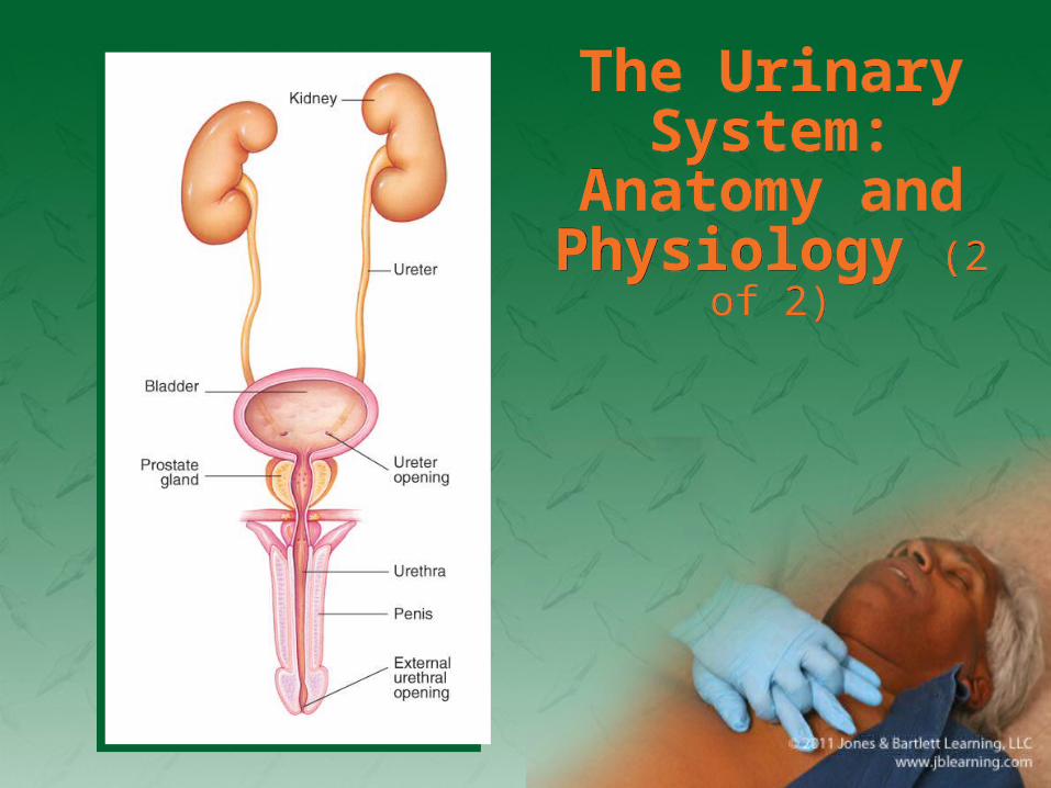
The Urinary System:
Anatomy and Physiology (2 of 2)
The Urinary System:
Anatomy and Physiology (2 of 2)
The Genital System: Anatomy and Physiology (1 of 2)
The Genital System: Anatomy and Physiology (1 of 2)
• Controls reproductive processes
• Male system consists of– Testicles
– Epididymis
– Vasa Deferentia
– Penis
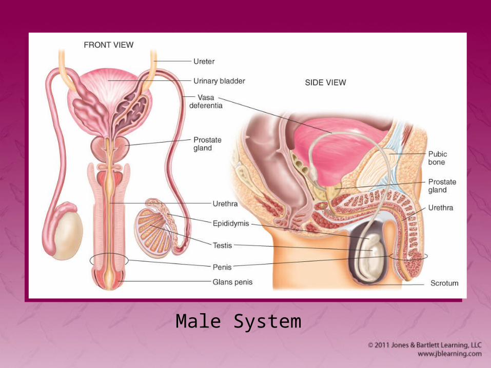
Male System
The Genital System: Anatomy and Physiology (2 of 2)
The Genital System: Anatomy and Physiology (2 of 2)
• Female system consists of– Ovaries
– Fallopian tubes
– Uterus
– Cervix
– Vagina
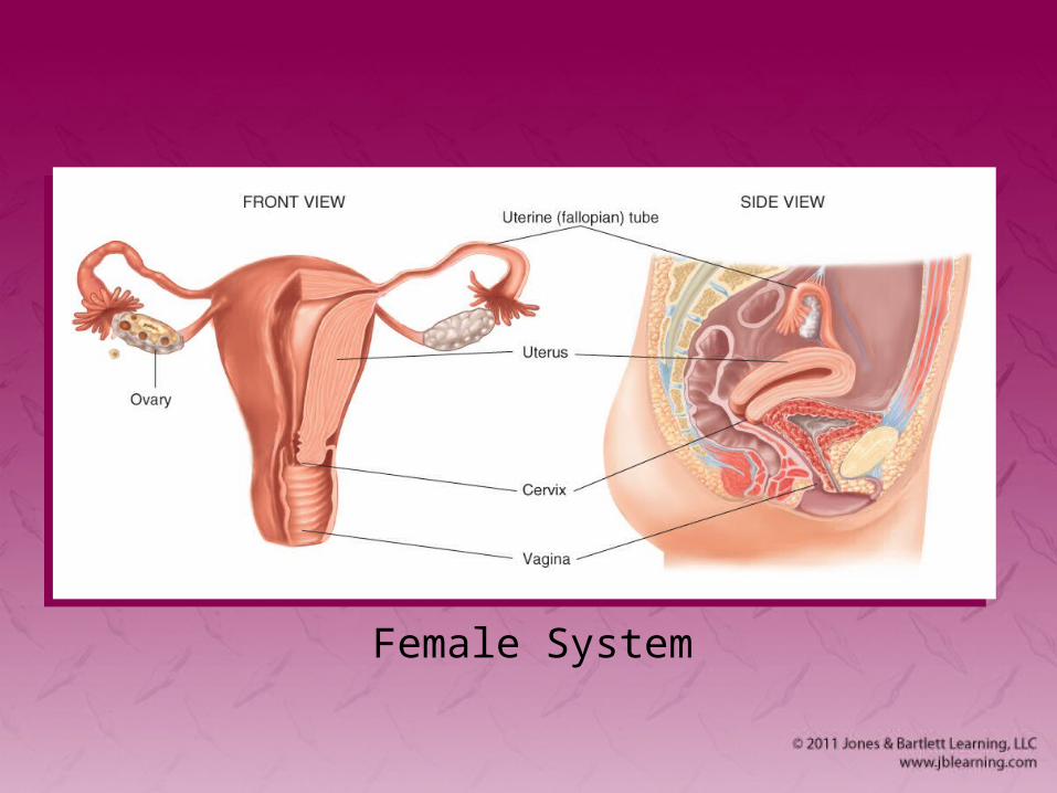
Female System
Life Support Chain (1 of 3)Life Support Chain (1 of 3)
• All cells in body require oxygen, nutrients, and removal of waste.
• Circulatory system is the carrier of these supplies and wastes.
• If interference occurs, cells become damaged and die.
Life Support Chain (2 of 3)Life Support Chain (2 of 3)
• Adenosine triphosphate (ATP) – Involved in energy metabolism
– Used to store energy
• Aerobic metabolism uses oxygen.
• Cells switch to anaerobic metabolism when oxygen is limited.– Lactic acid is damaging waste product.
Life Support Chain (3 of 3)Life Support Chain (3 of 3)
• Movement of oxygen, waste, nutrients occurs by diffusion.
• pH is critical to diffusion.– Measure of acidity or alkalinity
• Body spends large amount of energy to maintain normal pH.
PathophysiologyPathophysiology
• The study of functional changes that occur when body reacts to disease
• Respiratory compromise can lead to:– Shock
– Alteration of cellular metabolism
Summary Summary (1 of 9)(1 of 9)Summary Summary (1 of 9)(1 of 9)
• Understand human anatomy and physiology so you can assess the patient’s condition and communicate with others.
• Know superficial landmarks of the body and what lies underneath the skin.
Summary Summary (2 of 9)(2 of 9)Summary Summary (2 of 9)(2 of 9)
• Bones, ligaments, tendons, and cartilage give the body its recognizable human form.
• The skeletal system provides protection for organs, allows for movement, and gives the body its shape.
Summary Summary (3 of 9)(3 of 9)Summary Summary (3 of 9)(3 of 9)
• The contraction and relaxation of the musculoskeletal system gives the body its ability to move.
• The respiratory system includes the nose, mouth, throat, larynx, trachea, bronchi, and bronchioles.

Summary Summary (4 of 9)(4 of 9)Summary Summary (4 of 9)(4 of 9)
• The function of the respiratory system is to provide the body with oxygen and eliminate carbon dioxide.
• The circulatory system is a complex arrangement of connected tubes, including arteries, arterioles, capillaries, venules, and veins.
Summary Summary (5 of 9)(5 of 9)Summary Summary (5 of 9)(5 of 9)
• The nervous system is the most complex organ system within the human body. It consists of the brain, spinal cord, and nerves.
• The skin is divided into two parts: the superficial epidermis and the deeper dermis.
Summary Summary (6 of 9)(6 of 9)Summary Summary (6 of 9)(6 of 9)
• The skin is the largest single organ in the body.
• The skin serves three major functions: to protect the body in the environment, to regulate the temperature of the body, and to transmit information from the environment to the brain.
Summary Summary (7 of 9)(7 of 9)Summary Summary (7 of 9)(7 of 9)
• The digestive system is composed of the gastrointestinal tract (stomach and intestines), mouth, salivary glands, pharynx, esophagus, liver, gallbladder, pancreas, rectum, and anus.
Summary Summary (8 of 9)(8 of 9)Summary Summary (8 of 9)(8 of 9)
• The endocrine system is a complex message and control system that integrates many body functions.
• The urinary system controls the discharge of certain waste materials filtered from the blood by the kidneys.
Summary Summary (9 of 9)(9 of 9)Summary Summary (9 of 9)(9 of 9)
• The genital system controls the reproductive processes.
• Pathophysiology is the study of how the body reacts to diseases.
ReviewReview
1. The __________ lies in the retroperitoneal space.A. liver
B. spleen
C. kidneys
D. stomach
ReviewReview
Answer: C
Rationale: The kidneys lie in the retroperitoneal space—the space behind the abdominal cavity. The spleen, liver, and stomach are all located within the anterior (true) abdomen.
ReviewReview
1. The __________ lies in the retroperitoneal space.
A. LiverRationale: The liver lies immediately beneath the diaphragm in the anterior abdomen.
B. SpleenRationale: The spleen lies under the rib cage in left upper quadrant of the abdominal cavity.
C. KidneysRationale: Correct answer.
D. StomachRationale: The stomach lies in the left upper quadrant of the abdominal cavity.
ReviewReview
2. The cartilaginous tip of the sternum is called the:A. costal arch.
B. manubrium.
C. angle of Louis.
D. xiphoid process.
ReviewReview
Answer: D
Rationale: The xiphoid process projects from the lower part of the sternum. It is made of cartilage, and, relative to other parts of the sternum (eg, manubrium, angle of Louis), is soft to palpation.
ReviewReview
2. The cartilaginous tip of the sternum is called the:A. costal arch.
Rationale: This is the bridge of cartilage that connects the ends of the 6th through 10th ribs to lower sternum.
B. manubrium.Rationale: This is the upper section of the sternum, one of three parts.
C. angle of Louis.Rationale: This is at the level where the second rib is attached to the sternum.
D. xiphoid process.Rationale: Correct answer.
ReviewReview
3. A person with bilateral femur fractures has:A. fractured one of his or her femurs.
B. fractured both of his or her femurs.
C. one femur fractured in two places.
D. fractured the lateral aspect of the femur.
ReviewReview
Answer: B
Rationale: The term bilateral refers to both sides of the body with reference to the midline. Therefore, bilateral femur fractures would indicate that both femurs are fractured.
ReviewReview
3. A person with bilateral femur fractures hasA. fractured one of his or her femurs.
Rationale: Bilateral means two.
B. fractured both of his or her femurs.Rationale: Correct answer
C. one femur fractured in two places.Rationale: A bilateral fracture is one fracture that occurs in two bones.
D. fractured the lateral aspect of the femur.Rationale: This means that the outside portion of the femur is broken.
ReviewReview
4. The MOST prominent landmark on the anterior surface of the neck is the:A. mastoid process.
B. cricoid cartilage.
C. thyroid cartilage.
D. cricothyroid membrane.
ReviewReview
Answer: C
Rationale: The thyroid cartilage, commonly referred to as the “Adam's Apple,” is the most prominent landmark on the anterior (front) surface of the neck. The cricoid cartilage is located directly inferior to (below) the thyroid cartilage; it is a less prominent landmark.
Review (1 of 2)Review (1 of 2)
4. The MOST prominent landmark on the anterior surface of the neck is the:
A. mastoid process.Rationale: This is the prominent boney mass at the base of the skull.
B. cricoid cartilage.Rationale: This is the firm ridge of cartilage inferior (below) to the thyroid cartilage.
Review (2 of 2)Review (2 of 2)
4. The MOST prominent landmark on the anterior surface of the neck is the:
C. thyroid cartilage.Rationale: Correct answer
D. cricothyroid membrane.Rationale: This is the thin sheet of connective tissue that joins the thyroid cartilage and the cricoid cartilage.
ReviewReview
5. Insulin is produced in the:A. liver.
B. pancreas.
C. thyroid gland.
D. adrenal glands.
ReviewReview
Answer: B
Rationale: The pancreas is a solid organ that produces both insulin and digestive juices. Insulin is produced in the islets of Langerhans, which are a part of the pancreas.
ReviewReview
5. Insulin is produced in the:A. liver.
Rationale: This is where poisonous bi-products of digestion are rendered harmless.
B. pancreas.Rationale: Correct answer
C. thyroid gland.Rationale: This is found in the neck over the larynx and regulates the body’s metabolism.
D. adrenal glands.Rationale: These are located in the kidneys and regulate salt levels, sugar levels, and sexual function.
ReviewReview
6. The medial aspect of a bone is that part of a bone that lies:A. nearer to the feet.
B. nearer to the back.
C. closer to the midline of the body.
D. away from the midline of the body.
ReviewReview
Answer: C
Rationale: The term medial means toward the midline of the body, while lateral means away from the midline of the body. A part of the body that is nearer to the back is said to be posterior; if it is nearer to the feet, it is said to be inferior.
ReviewReview
6. The medial aspect of a bone is that part of a bone that lies:
A. nearer to the feet.Rationale: This is inferior.
B. nearer to the back.Rationale: This is posterior.
C. closer to the midline of the body.Rationale: Correct answer
D. away from the midline of the body.Rationale: This is lateral.

ReviewReview
7. The normal resting adult heart rate is:A. 50 to 70 beats/min.
B. 60 to 100 beats/min.
C. 80 to 110 beats/min.
D. 110 to 120 beats/min.
ReviewReview
Answer: B
Rationale: The normal resting heart rate for an adult is 60 to 100 beats/min. Bradycardia exists when the adult heart rate is less than 60 beats/min, and tachycardia exists when it is greater than 100 beats/min.
ReviewReview
7. The normal resting adult heart rate is:A. 50 to 70 beats/min.
Rationale: Less than 60 beats/min is bradycardia.
B. 60 to 100 beats/min.Rationale: Correct answer
C. 80 to 110 beats/min.Rationale: Normal is more than 100 beats/min.
D. 110 to 120 beats/min.Rationale: More than 100 beats/min is tachycardia.
ReviewReview
8. The left atrium of the heart receives ___________ blood from the ___________.A. oxygenated, lungs
B. deoxygenated, body
C. oxygenated, body
D. deoxygenated, lungs
ReviewReview
Answer: A
Rationale: The left atrium receives oxygenated blood from the lungs via the pulmonary veins. The right atrium receives deoxygenated blood from the body via the vena cavae.
Review (1 of 2)Review (1 of 2)
8. The left atrium of the heart receives ___________ blood from the ___________.
A. oxygenated, lungsRationale: Correct answer
B. deoxygenated, bodyRationale: The right atrium of the heart receives deoxygenated blood from the body.
Review (2 of 2)Review (2 of 2)
8. The left atrium of the heart receives ___________ blood from the ___________.
C. oxygenated, bodyRationale: No part of the heart receives oxygenated blood from the body. It only receives oxygenated blood from the lungs.
D. deoxygenated, lungsRationale: The right atrium and right ventricle are the only parts of the heart that receive deoxygenated blood from the body.
ReviewReview
9. The largest part of the brain is the:A. cerebrum.
B. brain stem.
C. cerebellum.
D. foramen magnum.

ReviewReview
Answer: A
Rationale: The three major parts of the brain are the cerebrum, the brain stem, and the cerebellum. The largest part of the brain is the cerebrum, which is sometimes called the “grey matter,” The cerebellum—sometimes called the “athletes brain”—is the smallest part of the brain. The brain stem is responsible for vital functions such as heart rate, breathing, and blood pressure. The foramen magnum is the large opening at the base of the skull through which the spinal cord passes.
ReviewReview
9. The largest part of the brain is the:A. cerebrum.
Rationale: Correct answer
B. brain stem.Rationale: The bottom portion of the brain is responsible for vital functions, heart rate, breathing, and blood pressure.
C. cerebellum.Rationale: This is the smallest part of brain. It is sometimes called the athlete’s brain.
D. foramen magnum.Rationale: This is the large opening at the base of the skull through which the spinal cord passes.
ReviewReview
10. Which of the following statements about red blood cells is FALSE?A. They contain iron.
B. They carry oxygen.
C. They help to fight infection.
D. They give color to the blood.

ReviewReview
Answer: C
Rationale: The hemoglobin molecules in red blood cells contain iron, give color to the blood, and carry oxygen. White blood cells play a role in helping the body to fight infection.
ReviewReview
10. Which of the following statements about red blood cells is FALSE?A. They contain iron.
Rationale: This is true. Hemoglobin found in red blood cells carries iron.
B. They carry oxygen.Rationale: This is true. Hemoglobin found in red blood cells carries oxygen.
C. They help to fight infection.Rationale: Correct answer
D. They give color to the blood.Rationale: This is true. Hemoglobin found in red blood cells gives blood color.
CreditsCredits
• Chapter opener: © National Cancer Institute/Photodisc/Getty Images
• Background slide images: © Jones & Bartlett Learning. Courtesy of MIEMSS.