characterisation of macaque testicular leucocyte populations and … · r. de rose et al. / journal...
TRANSCRIPT

CT
RWa
b
ARRA
KMTMTIF
1
oitr
3
0h
Journal of Reproductive Immunology 100 (2013) 146– 156
Contents lists available at ScienceDirect
Journal of Reproductive Immunology
jo ur n al homep age : w ww.elsev ier .com/ locate / j repr imm
haracterisation of macaque testicular leucocyte populations and-lymphocyte immunity
obert De Rosea,1, Caroline S. Fernandeza,1, Mark P. Hedgerb, Stephen J. Kenta,endy R. Winnall a,∗
Department of Microbiology and Immunology, University of Melbourne, Parkville, VIC AustraliaCentre for Reproduction and Development, Monash Institute of Medical Research, Monash University, Clayton, VIC, Australia
a r t i c l e i n f o
rticle history:eceived 3 May 2013eceived in revised form 29 August 2013ccepted 3 September 2013
eywords:acaque
estisacrophage
-lymphocytemmune privilegelow cytometry
a b s t r a c t
The rodent testis is well established as a site of immune privilege where both innate andacquired immune responses are suppressed. Immune cells and responses within human ornon-human primate testes, by contrast, are poorly characterised. This study used multi-colour flow cytometry to characterise the leukocytes in testicular cells isolated from 12young adult pigtail macaques (Macaca nemestrina) by collagenase dispersal, and to measurethe cytokine responses of macaque testicular T-lymphocytes to mitogens. B-lymphocytesand granulocytes were present in very low numbers (0.24% and 3.3% of leukocytes respec-tively), indicating minimal blood contamination. A median of 30.8% of the recoveredtesticular leukocytes were CD3+ lymphocytes, with CD4 and CD8 T-lymphocyte propor-tions similar to those in the blood. The proportion of naïve T-lymphocytes in the testiswas low, with significantly higher frequencies of central memory cells, compared with theblood. A median of 42.7% of the testicular leukocytes were CD163+ macrophages, while4.5% were CD14+CD163− monocyte-like macrophages. Small populations of myeloid andplasmacytoid dendritic cells, NK cells and NKT cells were also detected. Following mitogenstimulation, 19.7% of blood T-lymphocytes produced IFN� and/or TNF, whereas signifi-
cantly fewer (4.4%) of the testicular T-lymphocytes responded to stimulation. Our resultscharacterise the immune cells within the adult macaque testis and identify a suppressionof T-lymphocyte responses. This study provides a baseline to examine the immunology ofthe primate testis and suggests that testicular immune privilege could also be present inprimates.. Introduction
The rodent testis is well established as a regionf immune privilege, where both innate and acquired
mmune responses are suppressed. This may serve to pro-ect the immunogenic germ cells from acquired immuneesponses and from the deleterious effects of inflammation∗ Corresponding author at: University of Melbourne, Parkville, VIC010, Australia. Tel.: +61 3 8344 9938; fax: +61 3 8344 3846.
E-mail address: [email protected] (W.R. Winnall).1 Denotes equal contributions of first authors.
165-0378/$ – see front matter © 2013 Elsevier Ireland Ltd. All rights reserved.ttp://dx.doi.org/10.1016/j.jri.2013.09.003
© 2013 Elsevier Ireland Ltd. All rights reserved.
(Meinhardt and Hedger, 2011). Currently, there are fewstudies that have addressed the presence or absence ofimmune privilege in the testes of humans or non-humanprimates. Impeding this area of research is the relativelypoor characterisation of the leucocyte populations of theprimate testis.
The testicular leukocytes reside in the testicular inter-stitium, and share this compartment with the Leydigcells, connective tissue cells such as fibroblasts and peri-
cytes, and vascular endothelial cells. Leukocytes are neverfound in the seminiferous tubules under normal conditions(Hedger, 1997). In all species studied to date, the majority oftesticular leukocytes are macrophages (Hedger, 2002). The
ductive Immunology 100 (2013) 146– 156 147
Table 1Antibodies.
Antigena Fluorophore Clone Titre Cataloguenumberb
CD3 Alexa fluor 488 SP34.2 1/200 557705CD3 Pacific blue SP34.2 1/100 558124CD3 APC-Cy7 SP34.2 1/100 557757CD4 Alexa fluor 700 L200 1/100 560836CD8 PerCP SK1 1/70 347314CD8 APC-H7 SK1 1/100 641400CD8 PE-Cy7 SK1 1/1000 335787CD8 APC SK1 1/800 340584CD11c BV711 3.9 1/20 301629c
CD14 PE-Cy7 M5E2 1/66 557742CD14 APC-H7 MP�9 1/100 560270CD16 APC-H7 3G8 1/40 560195CD19 PE HIB19 1/20 555413CD20 PE-Cy7 L27 1/67 335793CD25 Alexa fluor 488 BC96 1/50 53-0259-42d
CD28 PerCP-Cy5.5 L293 1/25 337181CD45 V450 D058-1283 1/100 561291CD49d – L25 N/A 340976CD66 PE B6.2 1/200 551478CD68 FITC Y1/82A 1/50 11-0689-41d
CD95 FITC DX2 1/12.5 556640CD123 APC 7G3 1/125 560087CD159a
(NKG2A)APC Z199 1/66 A60797
CD161 BV605 HP-3G10 1/40 339915c
CD163 PE GHI/61 1/25 560933Lin1 FITC e 1/40 340546HLA-DR PerCP-Cy5.5 L243 1/20 552764V�7.2 PE 3C10 1/40 351705TNF PE-Cy7 mAb11 1/100 557647IFN� APC B27 1/800 554702
a All were raised to human antigens except non-human primate CD45.b All were from BD Biosciences except where indicated.c Purchased from BioLegend.d
R. De Rose et al. / Journal of Repro
macrophage population of the rat testis is the best charac-terised and consists primarily of resident macrophages thatare CD163+. It also includes monocyte-like macrophagesthat lack CD163, but express other macrophage mark-ers such as CD68 (i.e. CD68+CD163−) (Wang et al., 1994;Winnall and Hedger, 2013). CD68+ macrophages have beendetected in the human and macaque testis (Pöllänen andNiemi, 1987; Frungieri et al., 2002; Shehu-Xhilaga et al.,2007), but the macrophage populations of these speciesare otherwise poorly characterised. However, data fromthe human testis suggest that both CD163+ and CD163−
macrophages are present (Frungieri et al., 2002). These tes-ticular macrophages play roles in the response to infection,the regulation of spermatogenesis and steroidogenesis, andare proposed to contribute to immunosuppression in thetestis (Hales, 2002; Hedger, 2002; Winnall and Hedger,2013).
In rat and human testes, T-lymphocytes make up anestimated 10–20% of the leukocytes (El-Demiry et al.,1985; Pöllänen and Niemi, 1987) with CD8+ T-lymphocytespresent as the predominant population (Ritchie et al.,1984; Pöllänen and Niemi, 1987; Wang et al., 1994;Tompkins et al., 1998; Jacobo et al., 2009). Smaller pop-ulations of dendritic cells, mast cells, eosinophils andnatural killer (NK) cells have also been detected in thetestes of rodents and some other species (Derrick et al.,1993; Itoh et al., 1995; Anton et al., 1998; Fijak et al.,2005; Fijak and Meinhardt, 2006; Rival et al., 2006),but these remain largely uncharacterised in the pri-mate testis. Neutrophils and B-lymphocytes, which arecommonly found in large numbers in the circulation,have not been detected in rodent testes under normal,homeostatic conditions (Hedger, 1997; Tompkins et al.,1998).
The mechanisms behind immune privilege are incom-pletely defined, but studies in rodents have proposedthat anatomical sequestration of germ cells by the Sertolicell tight junctions, potential immunosuppressive prop-erties of steroid hormones, unique MHC compositioninside seminiferous tubules of rodents and humans, aswell as the actions of testicular macrophages, contributeto tolerance in the male gonads (Pöllänen and Niemi,1987; Fiszer et al., 1997; Hutter and Dohr, 1998; Livaand Voskuhl, 2001; Cheng and Mruk, 2002; Hedger andHales, 2006; Winnall et al., 2011). At least in rodents,the responses of the T-lymphocyte population of thetestis to foreign antigens are believed to be inhibitedor altered, leading to a prolonged tolerance (Head andBillingham, 1985; Dai et al., 2005; Nasr et al., 2005; Winnallet al., 2011). Local production of immunosuppressivefactors such as IL-10, TGF�, activin A and the pres-ence of immunosuppressive lyso-glycerophosphocholines,which regulate T-lymphocyte activation and survival, aresuspected to promote this diminished response of testic-ular T-lymphocytes (Pollanen et al., 1993; Foulds et al.,2008; Winnall et al., 2011; Bistoni et al., 2012; Hedgerand Winnall, 2012). The response of primate testis T-
lymphocytes to activation has not been characterised. Thepresent study aims to characterise the leukocytes presentin the macaque testis and their response to activation bymitogens.Purchased from eBioscience.e Lin1 FITC comprises CD3, CD14, CD16, CD19, CD20 and CD56, which
are clones SK7, 3G8, SJ325C1, L27, M�P9, and NCAM16.2, respectively.
2. Materials and methods
2.1. Animals
Testes were sourced from 12 healthy young adultmale pigtail macaques (Macaca nemestrina) that wereinvolved in an unrelated study. These animals were unin-fected controls and no procedures were performed thatcould have affected the testes. Animals were euthanisedusing ketamine sedation (1 mg/kg) and pentobarbitone(0.5 ml/kg) and orchidectomy was performed immediatelyat autopsy. Concurrent blood samples were collected fromanimals in heparinised collection tubes. Animals werehoused in the Australian Animal Health Laboratory and theCSIRO Animal Health animal ethics committees approvedall studies.
2.2. Reagents and antibodies
All antibodies and other reagents were purchased from
BD Biosciences (San Jose, CA, USA) and raised againsthuman antigens unless otherwise indicated. Antibodiesare described in Table 1 and were all mouse monoclon-als raised against human antigens, with the exception of
1 ductive I
alislBDb
2
rtutttfBtbclaccmignwbnewdgc
2
fBafwaalrcLfSloiic
48 R. De Rose et al. / Journal of Repro
nti-non-human primate CD45. Mouse CD1d-tetramer PEoaded with an �-galactosylceramide analogue was maden-house (peripheral blood isolated from macaques wastained with CD1d tetramer loaded with an �-GalCer ana-ogue, PBS-44, [kindly provided by Professor Paul Savage,righam Young University]). A LIVE/DEAD Fixable Aquaead Cell Stain Kit was purchased from Invitrogen (Carls-ad, CA, USA).
.3. Isolation of testicular cells
Testes were decapsulated and major blood vesselsemoved manually. Approximately half of each testis, distalo the more vascular rete testis, was recovered. Testic-lar tissue was roughly chopped using scissors (untilubules were approximate 5-mm pieces) and incubated in aotal of 10 ml PBS with 0.07 mg/ml collagenase (Worthing-on Biochemicals) and 1.4 �g/ml DNase (Roche) at 37 ◦Cor 60 min. After collagenase digestion, 40 ml PBS/0.5%SA/2 mM EDTA was added, tissue was briefly mixed bywo inversions and tubules were allowed to settle for 2 minefore removal of the PBS containing suspended cells. Theell preparation contained few aggregates. Cells were pel-eted and resuspended in PBS/0.5% BSA/2 mM EDTA forntibody incubations or activation with mitogens. Cellounts were performed using a CELL DYN Emerald cellounter (Abbott Diagnostics, Lake Forest, IL, USA). Lighticroscopy observations indicated considerable contam-
nation of the testicular interstitial cell preparations witherm cells, mostly spermatids released from broken semi-iferous tubules, at approximately 20% of the total. No cellsith the characteristic morphology of Sertoli cells could
e seen using light microscopy. To confirm that collage-ase treatment did not affect the leucocyte surface antigenxpression we tested macaque PBMC samples with andithout collagenase treatment. Flow cytometry analysisemonstrated that the surface expression of the major anti-ens used in this study was not significantly changed byollagenase treatment.
.4. Freezing and thawing testicular cells and PBMC
Fresh blood, stored in heparin-coated vials was care-ully layered over 95% Ficoll-Paque PLUSTM (GE Healthcare,uckinghamshire, England)/5% water (v/v) and centrifugedt 1000 × g for 25 min at room temperature. The inter-ace was recovered using a transfer pipette, washedith RPMI media and centrifuged at 500 × g for 5 min
t room temperature. Cell counts were performed using CELL DYN Emerald cell counter. For freezing, testicu-ar cell-enriched preparations or PBMC were pelleted andesuspended in a solution of 90% FCS (Bovogen Biologi-als, VIC, Australia)/10% (v/v) DMSO (Sigma–Aldrich, St.ouis, MO, USA) at a concentration of 5 × 10⁶ cells/ml androzen in a “Mr Frosty” freezing container (ThermoFishercientific, Waltham, MA, USA) at −80 ◦C for 12 h beforeong-term storage in liquid nitrogen. Frozen testicular cells
r PBMC were rapidly thawed in a 37 ◦C water-bath thenncubated for 10 min in 3 ml RPMI/10%FCS 66 U/ml DNasen a 37 ◦C water-bath, followed by washing in PBS. Thawedells were used for the dendritic cell staining only; freshlymmunology 100 (2013) 146– 156
isolated blood and testicular interstitial cells were used forall other experiments.
2.5. Phenotypic analysis of testicular and bloodleukocytes
Whole blood (100 �l/samples), thawed PBMC(5 × 10⁶ cells in 100 �l, for dendritic cell staining only)or testicular interstitial cells in PBS/0.5% BSA/2 mMEDTA (5 × 10⁶ cells in 200 �l) were incubated with 0.5 �lLIVE/DEAD Aqua to stain dead cells for 30 min, thenantibody cocktails were added for a 20-min incubation.Testicular cells were washed in PBS/0.5% BSA/2 mM andfixed in Stabilising Fixative (BD). Blood samples were fur-ther incubated 1× FACSTM Lysing Solution (BD) for 10 minto lyse red blood cells, followed by washing in PBS/0.5%BSA/2 mM EDTA and fixing with Stabilising Fixative. Allincubations were performed at room temperature in thedark after mixing by gentle vortex and centrifugation wasat 500 × g for 5 min.
2.6. Intracellular cytokine staining assay
IFN� and TNF production by blood and testicularcells was measured by an intracellular cytokine-stainingassay similar to previously described assays (De Roseet al., 2007) Peripheral whole blood (100 �l) or 100 �lof 5 × 10⁶ testicular interstitial cells in RMPI/10% FCSwere mixed with anti-CD28 and anti-CD49d at 1 �g/�leach, brefeldin A (10 �g/�l) and stimulated with eitherPBS (unstimulated controls) or a mix of StaphylococcusEnterotoxin-B (1 �g/�l, Sigma–Aldrich), phytohaemagglu-tinin (100 �g/�l, Sigma–Aldrich) and phorbol myristateacetate (30 �g/�l, Sigma–Aldrich). Cell activation was per-formed at 37 ◦C for 6 h in the same media, followed bystorage at 4 ◦C overnight. Cells were then incubated with0.5 �l LIVE-DEAD Aqua dead cell stain for 30 min, followedby anti-CD3 APC-Cy7, CD4 AF700, CD8 PerCP and CD45V450 for 20 min. Testicular cells were fixed by incuba-tion in 1% formaldehyde/PBS while blood was treated with1× FACsTM Lysing Solution (BD) for 10 min followed bywashing in PBS/0.5% BSA/2 mM EDTA. All cells were per-meabilised using 1× FACsTM Permeabilising Solution (BD)for 10 min, washed in PBS/0.5% BSA/2 mM EDTA, then incu-bated with anti-IFN� APC and TNF PE-Cy7 for 60 min. Cellswere washed then fixed in Stabilising Fixative (BD). Allincubations were performed at room temperature in thedark after mixing by gentle vortex. PBS controls were per-formed for every sample.
2.7. Flow cytometry
Acquisition was performed on an LSR Fortessa orLSRII flow cytometer (BD) according to the manufac-turer’s instructions and data were analysed using FlowJoVersion 9.6 (TreeStar, Ashland, OR, USA). Fluorochromecompensation correction was performed using the auto-
compensation function in DIVA 6.1, using CalibrateTMbeads (BD) for FITC, PE, PerCP APC, CompBead PLUS anti-mouse Ig � capture beads (BD) for other fluorochromes andArC beads (Invitrogen) for LIVE/DEAD Aqua. Testicular or

ductive I
R. De Rose et al. / Journal of Reproblood populations were gated according to scatter proper-ties or CD45 expression, and aggregates and dead cells wereexcluded. Specific populations were gated within testicu-lar interstitial cells compared with blood by calculating themedian percentage of the population among all live CD45+
cells (aggregates excluded). Alternatively, in Figs. 2 and 5,medians of specific T-lymphocyte populations were calcu-lated as a percentage of live CD45+CD3+ cells, and in Fig. 3C,as a percentage of live CD45+CD163+ cells.
2.8. Statistical analyses
Two testicular cell isolation experiments were per-formed on different days with 6 animals in each. Thesesamples were analysed by staining with different antibodycocktails, making the sample size n = 6 animals for eachexperiment, with the exception of dendritic cell stainingon thawed samples, done at n = 3 (statistical analyses werenot used for these). Statistical analyses were performedusing SPSS version 18 software (IBM, Armonk, NY, USA).Wilcoxon Ranked Sign Tests were used to compare testisand animal-matched blood data in Figs. 2C, D and 5C. A pvalue ≤0.05 was considered to indicate a significant differ-ence.
3. Results
3.1. Macaque testicular interstitial-enriched cellpreparations contain low numbers of contaminatingblood B-lymphocytes and granulocytes
To characterise the macaque testicular leucocyte pop-ulation, testicular interstitial cell-enriched preparationswere isolated by collagenase digestion of fresh testiculartissue and phenotyped using antibodies. Leukocytes, gatedas CD45+, were present at a median of 4.44% (interquar-tile range (IQR) 2.05%) of the testicular cell preparation,once aggregates and dead cells were excluded (Fig. 1A).There were very few CD20+ B-lymphocytes present inthe interstitial cell preparations (0.235% IQR 0.105% ofCD45+ leukocytes) compared with the animal-matchedblood samples (8.28% IQR 4.02%; Fig. 1B and C). From thesedata, it was calculated that an average of ≤3.7% of the tes-ticular leukocytes were likely to be contaminating bloodcells from the testicular blood vessels.
3.2. T-lymphocyte populations in the macaque testicularinterstitium
Testicular T-lymphocytes were distinguished from theother testicular interstitial cells according to CD45+ andCD3+ co-expression (Fig. 2A). The proportions of CD4+ andCD8+ T-lymphocytes in the testis were not significantly dif-ferent from those in blood (Fig. 2A, B, middle panels, and C).Expression of CD28 and CD95 wad used to differentiatingpopulations of naïve, central memory and effector memoryT-lymphocytes similar to previous studies (Pitcher et al.,
2002). There were significantly fewer naïve T-lymphocytesin the testes (2.2% IQR 2.29% of CD45+CD3+ leukocytes)than the blood (27.8% IQR 16.7%, Wilcoxon Ranked SignTest, p = 0.028, Fig. 2A, B (right side panels) and D). Whilemmunology 100 (2013) 146– 156 149
effector memory T-lymphocytes were present at similarlevels, the central memory proportion was larger in thetestis (74.5% IQR 13.9% of CD45+CD3+ leukocytes) thanthe blood (41.3% IQR 15.4%, Wilcoxon Ranked Sign Test,p = 0.046). A median of 1.08% (range 0.62–1.14%) of thetesticular T-lymphocytes expressed the activation markerCD25 compared with 2.57% (range 2.05–4.57%) in the bloodsamples (data not shown). Also detected was a small pop-ulation of leukocytes (3.28%, IQR 2.07%) displaying thecharacteristic large side scatter properties of granulocytes,consistent with low levels of blood contamination (Fig. 2A).This population would likely contain neutrophils from theblood, but may also contain eosinophils, as found in otherspecies (Anton et al., 1998).
3.3. Mononuclear phagocyte and dendritic cellpopulations in the macaque testicular interstitium
CD3 and CD163 staining of testicular leukocytes indi-cated that a median of 30.8% (IQR 11.3%) of testicular CD45+
leukocytes were CD3+ T-lymphocytes, 42.7% (IQR 16.75%)were CD163+ resident macrophages and the remainderwere mostly a CD3−CD163− population (26.7% IQR 5.73%)that could include NK cells, dendritic cells and mono-cytes (Fig. 3A and B). In rats, the mononuclear phagocytepopulation of the testis has been characterised based onexpression of either or both CD163 and CD68 (Wang et al.,1994; Winnall and Hedger, 2013). CD14 was used to furthercharacterise the mononuclear phagocytes, as the avail-able anti-human CD68 antibody did not cross-react withmacaque cells. Two populations could be discerned fromthe testicular cell preparation: CD14+CD163+ cells, whichwere most likely resident testicular macrophages, andCD14+CD163−, which may be monocytes or monocyte-likemacrophages (Fig. 3A). The CD163− population was calcu-lated to be a median of 4.5% (IQR 5%) of testicular CD45+
leukocytes. Similar proportions of these populations werefound in each of 6 animals (Fig. 3A and C).
Small populations of dendritic cells were detected inthe testis, identified as lymphocyte- and monocyte-lineagenegative (Lin−), HLA-DR+ and either CD11c+ (myeloid) orCD123+ (plasmacytoid; Fig. 3D). Overall, dendritic cellswere calculated to be a median of 0.22% (range 0.18–0.42%)of the recovered CD45+ testicular leukocytes, whereas0.09% (range 0.07–0.11%) of macaque blood CD45+ leuko-cytes were classified as dendritic cells using the samegating strategy (Fig. 3D). Factoring in the low level of bloodcontamination in the testes sample (≤3.7%), >98% of thedendritic cells identified in the testicular cell preparationswere predicted to be of testicular origin.
3.4. Minor lymphocyte populations in the macaquetesticular interstitium
Natural killer cells were detected in the CD3− popula-tion of testicular leukocytes by their surface expressionof NKG2A, which marks 97% of NK cells in macaques
(Mavilio et al., 2005). NK cells were a median of 5.27%of the testicular CD45+ leukocytes compared with 12.6%of blood leukocytes (Fig. 4A and B). Whereas only 1.98%of the NK cells in the blood were CD8−, 35.3% of the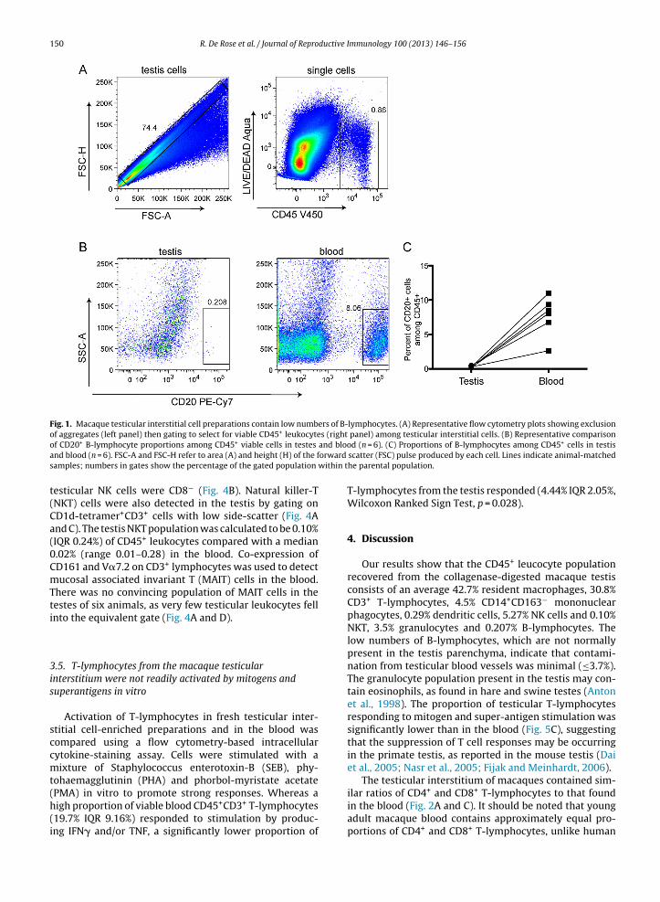
150 R. De Rose et al. / Journal of Reproductive Immunology 100 (2013) 146– 156
Fig. 1. Macaque testicular interstitial cell preparations contain low numbers of B-lymphocytes. (A) Representative flow cytometry plots showing exclusionof aggregates (left panel) then gating to select for viable CD45+ leukocytes (right panel) among testicular interstitial cells. (B) Representative comparisono and blooa orward
s within t
t(Ca(0CmTti
3is
sccmt(h(i
f CD20+ B-lymphocyte proportions among CD45+ viable cells in testes
nd blood (n = 6). FSC-A and FSC-H refer to area (A) and height (H) of the famples; numbers in gates show the percentage of the gated population
esticular NK cells were CD8− (Fig. 4B). Natural killer-TNKT) cells were also detected in the testis by gating onD1d-tetramer+CD3+ cells with low side-scatter (Fig. 4And C). The testis NKT population was calculated to be 0.10%IQR 0.24%) of CD45+ leukocytes compared with a median.02% (range 0.01–0.28) in the blood. Co-expression ofD161 and V�7.2 on CD3+ lymphocytes was used to detectucosal associated invariant T (MAIT) cells in the blood.
here was no convincing population of MAIT cells in theestes of six animals, as very few testicular leukocytes fellnto the equivalent gate (Fig. 4A and D).
.5. T-lymphocytes from the macaque testicularnterstitium were not readily activated by mitogens anduperantigens in vitro
Activation of T-lymphocytes in fresh testicular inter-titial cell-enriched preparations and in the blood wasompared using a flow cytometry-based intracellularytokine-staining assay. Cells were stimulated with aixture of Staphylococcus enterotoxin-B (SEB), phy-
ohaemagglutinin (PHA) and phorbol-myristate acetate
PMA) in vitro to promote strong responses. Whereas aigh proportion of viable blood CD45+CD3+ T-lymphocytes19.7% IQR 9.16%) responded to stimulation by produc-ng IFN� and/or TNF, a significantly lower proportion ofd (n = 6). (C) Proportions of B-lymphocytes among CD45+ cells in testisscatter (FSC) pulse produced by each cell. Lines indicate animal-matchedhe parental population.
T-lymphocytes from the testis responded (4.44% IQR 2.05%,Wilcoxon Ranked Sign Test, p = 0.028).
4. Discussion
Our results show that the CD45+ leucocyte populationrecovered from the collagenase-digested macaque testisconsists of an average 42.7% resident macrophages, 30.8%CD3+ T-lymphocytes, 4.5% CD14+CD163− mononuclearphagocytes, 0.29% dendritic cells, 5.27% NK cells and 0.10%NKT, 3.5% granulocytes and 0.207% B-lymphocytes. Thelow numbers of B-lymphocytes, which are not normallypresent in the testis parenchyma, indicate that contami-nation from testicular blood vessels was minimal (≤3.7%).The granulocyte population present in the testis may con-tain eosinophils, as found in hare and swine testes (Antonet al., 1998). The proportion of testicular T-lymphocytesresponding to mitogen and super-antigen stimulation wassignificantly lower than in the blood (Fig. 5C), suggestingthat the suppression of T cell responses may be occurringin the primate testis, as reported in the mouse testis (Daiet al., 2005; Nasr et al., 2005; Fijak and Meinhardt, 2006).
The testicular interstitium of macaques contained sim-
ilar ratios of CD4+ and CD8+ T-lymphocytes to that foundin the blood (Fig. 2A and C). It should be noted that youngadult macaque blood contains approximately equal pro-portions of CD4+ and CD8+ T-lymphocytes, unlike human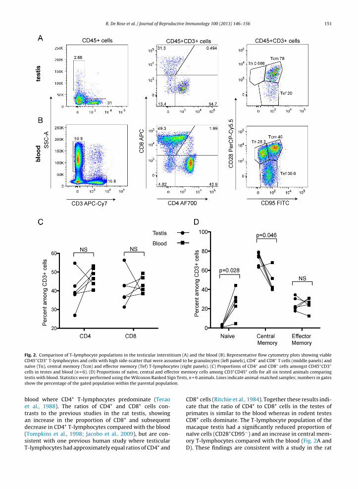
R. De Rose et al. / Journal of Reproductive Immunology 100 (2013) 146– 156 151
Fig. 2. Comparison of T-lymphocyte populations in the testicular interstitium (A) and the blood (B). Representative flow cytometry plots showing viableCD45+CD3+ T-lymphocytes and cells with high side-scatter that were assumed to be granulocytes (left panels), CD4+ and CD8+ T cells (middle panels) andnaïve (Tn), central memory (Tcm) and effector memory (Tef) T-lymphocytes (right panels). (C) Proportions of CD4+ and CD8+ cells amongst CD45+CD3+
ctor megn Testsion.
cells in testes and blood (n = 6). (D) Proportions of naïve, central and effetestis with blood. Statistics were performed using the Wilcoxon Ranked Sishow the percentage of the gated population within the parental populat
blood where CD4+ T-lymphocytes predominate (Teraoet al., 1988). The ratios of CD4+ and CD8+ cells con-trasts to the previous studies in the rat testis, showingan increase in the proportion of CD8+ and subsequent
decrease in CD4+ T-lymphocytes compared with the blood(Tompkins et al., 1998; Jacobo et al., 2009), but are con-sistent with one previous human study where testicularT-lymphocytes had approximately equal ratios of CD4+ andmory cells among CD3+CD45+ cells for all six tested animals comparing, n = 6 animals. Lines indicate animal-matched samples; numbers in gates
CD8+ cells (Ritchie et al., 1984). Together these results indi-cate that the ratio of CD4+ to CD8+ cells in the testes ofprimates is similar to the blood whereas in rodent testesCD8+ cells dominate. The T-lymphocyte population of the
macaque testis had a significantly reduced proportion ofnaïve cells (CD28+CD95−) and an increase in central mem-ory T-lymphocytes compared with the blood (Fig. 2A andD). These findings are consistent with a study in the rat
152 R. De Rose et al. / Journal of Reproductive Immunology 100 (2013) 146– 156
Fig. 3. Mononuclear phagocyte (A)–(C) and dendritic cell (D) populations in the testicular interstitium compared with the blood. (A) Representativeflow cytometry plot showing CD163+ macrophages and CD3+ T-lymphocytes gated from CD45+ testicular leukocytes (left panel). Cells with high sidescatter (likely granulocytes) were excluded from CD45+ leukocytes to avoid CD14+ neutrophils (not shown), then mononuclear phagocytes were gated asCD14+CD163− and CD14+CD163+ populations (right panel). (B) Proportions of T-lymphocytes (CD3+) resident macrophages (CD163+) and CD3−CD163−
cells among CD45+ cells for all six animals. (C) Proportions of CD14+CD163− and CD14+CD163− cells among CD45+ with low side scatter for the same sixanimals. (D) Representative flow cytometry plots showing gating of dendritic cell populations (Lin1−HLA-DR+ cells) from live testicular single cells (panel1 asmacytc o calculs ion.
sieo(
rmcelmttTo
) compared with the blood (panel 3). Dendritic cells were classified as plells that were either CD123+ or CD11c+ were counted as dendritic cells thow the percentage of the gated population within the parental populat
howing only 19% of testicular T-lymphocytes as being pos-tive for CD45RA, indicating a naïve phenotype (Tompkinst al., 1998) and being consistent with low proportionsf naïve T-lymphocytes found in other peripheral tissuesWeninger et al., 2002).
Regulatory T-lymphocytes (Tregs) play an importantole in peripheral tolerance and the prevention of autoim-unity (Chatila, 2009). Since the testicular environment
ontains high levels of TGF� (Le Magueresse-Battistonit al., 1995), which can stimulate Treg production, and lowevels of T-lymphocyte activation, at least in rodents, it
ight be expected that numerous Tregs would be found in
his region. In one previous study, approximately 2% of tes-icular interstitial cells of the rat were CD4+CD25+Foxp3+regs (Jacobo et al., 2009). The present study found thatnly 1% of testicular lymphocytes were CD25+ in the
oid (CD123+) or myeloid (CD11c+) in panels 2 and 4. Only Lin1−HLA-DR+
ate the proportion of dendritic cells among leukocytes. Numbers in gates
macaque testis (data not shown), suggesting that levels ofCD25+Foxp3+ T cells are likely to be low. Further study ofTregs in the macaque testes is warranted.
Stimulation of macaque testicular T-lymphocytes witha combination of three mitogens, including the super-antigen SEB, resulted in very little production of TNF orIFN� compared with stimulation of blood T-lymphocytes.The exact mechanisms behind this immunosuppressionare unknown. We propose that the failure of the T-lymphocytes to be activated in our cultures may be dueeither to an intrinsically suppressed state as a resultof originating in the testis, or alternatively that testic-
ular cells present in the culture (and in vivo) exert animmunosuppressive effect in the assay. The current viewof testicular immunosuppression is that local factors affectthe responses of T-lymphocytes (Hedger and Hales, 2006).
R. De Rose et al. / Journal of Reproductive Immunology 100 (2013) 146– 156 153
Fig. 4. Minor lymphocyte populations in the testicular interstitium compared with the blood. Lymphocytes/smaller cells were selected based on scatterproperties and then aggregates were excluded prior to this gating (not shown). (A) Representative flow cytometry plot showing gating strategy for detectingCD3− NK and CD3+ NKT cells and MAIT cells among testicular cells (left panels) and blood (right panels). (B) NKG2A+ NK cells were divided into CD8+ andCD8− NK cells. (C) NKT cells were selected as CD3+CD1d tetramer+. (D) A V�7.2+CD161+ MAIT cell population could be found in the blood, but not in thetestis. Numbers in gates show the percentage of the gated population within the parental population.

154 R. De Rose et al. / Journal of Reproductive Immunology 100 (2013) 146– 156
Fig. 5. T-lymphocytes from the macaque testicular interstitium were not readily activated by mitogens in vitro. (A) Representative flow cytometry plotsshowing gating on viable CD45+ cells (left panel), gating on cells with the low forward and side scatter characteristics of lymphocytes (middle panel) thenCD3+ cells (right panel). (B) Representative flow cytometry plots showing cytokine production by CD3+ testicular T-lymphocytes (upper row) comparedwith blood (lower row). Unstimulated controls (PBS) are shown on the left side compared with mitogen-stimulated samples on the right. (C) Proportionso ts showS imals. Sc percent
AsStcda2ttbfusst
f T-lymphocytes producing IFN� and/or TNF in all six animals tested. Plotatistics were performed using the Wilcoxon Ranked Sign Tests, n = 6 anell. Lines indicate animal-matched samples; numbers in gates show the
lthough factors produced by Sertoli cells can be immuno-uppressive (Fallarino et al., 2009; Bistoni et al., 2012), fewertoli cells were present in our cultures. However ampleesticular macrophages and Leydig cells were present thatould be affecting T-lymphocyte activation, likely by pro-uction of soluble factors that inhibit activation (Bornnd Wekerle, 1982; Hedger et al., 1990; Winnall et al.,011). Previous studies showed that T-lymphocytes ofhe mouse testis entered into apoptosis upon transplan-ation of pancreatic islet allografts or are more likely toecome Tregs (Dai et al., 2005; Nasr et al., 2005). Theate of mitogen-exposed T-lymphocytes in our cultures is
nknown and will be interesting to determine in futuretudies. Importantly, these results are the first to demon-trate a suppression of immune responses in macaqueestis cultures and suggest that immune privilege, whichbackground-subtracted proportions (as percentage) of responding cells.SC-A refers to area (A) of the side scatter (SSC) pulse produced by each
age of the gated population within the parental population.
has so far only been demonstrated in the rodent testis, mayalso extend to the primate testis.
The rat testis contains three populations of mononu-clear phagocytes: CD68−CD163+ cells, which are consid-ered tissue-resident macrophages, CD68+CD163−, whichare predicted to be “newly arrived”, monocyte-likemacrophages, and a double-positive population thatmay be in the process of differentiating into residentmacrophages (Wang et al., 1994; Winnall et al., 2011). Thepresent study indicates that CD163+ cells are the majorityof the recovered testicular leukocytes in the macaque, atan average of 42.7% (Fig. 3A and B). Analysis of CD14 versus
CD168 staining revealed two potential populations ofmononuclear phagocytes: a predominating CD14+CD163hipopulation that is likely to be the resident macrophagepopulation, and CD14+CD163− that is likely to be “newly

ductive I
R. De Rose et al. / Journal of Reproarrived” monocyte-like macrophages (Fig. 3A and C). Theseresults confirm that the macaque testis has at least twopopulations of macrophages that are CD163+ and CD163−
cells, similar to the rat and human testis (Frungieri et al.,2002).
We detected a population of NK cells in the testis at5.27% of leukocytes (17% of lymphocytes), compared with12.6% of blood lymphocytes (Fig. 4B). The macaque tes-ticular NK population was similar in proportion to the rattesticular NK population at 25% of lymphocytes (Tompkinset al., 1998). Interestingly, the macaque testicular NK cellswere more likely to be CD8− than blood NK cells, indicat-ing that they are less prone to activation (Srour et al., 1990;Vargas-Inchaustegui et al., 2011). We also detected NKTcells in the testis that were present at 5-fold higher lev-els than in the blood, indicating that the small amount ofblood contamination (3.7%) in our testicular cell prepara-tions could not have been the source of all of these cells.Mucosal associated invariant T (MAIT) cells are unique Tcells found in mucosal regions that are the portals of entryfor many pathogens (Gold and Lewinsohn, 2011). SmallMAIT cell populations were detected in macaque bloodsamples, but no discernable population could be seen in thetesticular interstitial cell samples. These results are not sur-prising, as the interstitium of the testis is neither a mucosalregion nor a common portal of pathogen entry.
Dendritic cells were detected in the testis, with bothmyeloid and plasmacytoid phenotypes. Myeloid dendriticcells, which are CD11c+, are more commonly found inperipheral tissues (i.e. non-mucosal tissues) and have highspontaneous rates of migration out from the circulationinto peripheral tissues (De La Rosa et al., 2003). Plasma-cytoid dendritic cells (CD123+), on the other hand, areprimarily found in the blood and peripheral lymphoidtissues, with their migration more tightly controlled byspecific integrins and chemokines (De La Rosa et al., 2003).Interestingly, the testicular cell preparations contained rel-atively large proportions of plasmacytoid dendritic cellscompared with myeloid, indicating that specific signals,as yet uncharacterised, may be stimulating their migrationinto the testis.
Our results provide a baseline for future analyses ofthe immunomodulatory environment of the primate testis.Further, they allow us to probe the effects of local and sys-temic infections, such as simian immunodeficiency virus,on immune cell populations and immune responses in theprimate testis in detail.
Acknowledgements
We thank Sinthujan Jegaskanda, Thakshila Amarasena,Sheilajen Alcantara, Sarah Lloyd, Matthew Parsons andGayle Davey for expert technical assistance, providingreagents and/or for assistance with manuscript prepara-tion. This work was supported by NHMRC fellowships(1013221, 1041832, 1020269 and 1011578), the NHMRC
programme grant 510448 and the Victorian Government’sOperational Infrastructure Support Programme (to MonashInstitute of Medical Research).mmunology 100 (2013) 146– 156 155
References
Anton, F., et al., 1998. A comparative study of mast cells and eosinophilleukocytes in the mammalian testis. Zentralbl. Veterinarmed. A 45,209–218.
Bistoni, G., et al., 2012. Prolongation of skin allograft survival in rats by thetransplantation of microencapsulated xenogeneic neonatal porcinesertoli cells. Biomaterials 33, 5333–5340.
Born, W., Wekerle, H., 1982. Leydig cells nonspecifically suppress lympho-proliferation in vitro: implications for the testis as an immunologicallyprivileged site. Am. J. Reprod. Immunol. 2, 291–295.
Chatila, T.A., 2009. Regulatory T cells: key players in toleranceand autoimmunity. Endocrinol. Metab. Clin. North Am. 38,265–272.
Cheng, C.Y., Mruk, D.D., 2002. Cell junction dynamics in the testis: sertoli-germ cell interactions and male contraceptive development. Physiol.Rev. 82, 825–874.
Dai, Z., et al., 2005. Impaired recall of CD8 memory T cells in immunolog-ically privileged tissue. J. Immunol. 174, 1165–1170.
De La Rosa, G., et al., 2003. Migration of human blood dendriticcells across endothelial cell monolayers: adhesion molecules andchemokines involved in subset-specific transmigration. J. Leukoc. Biol.73, 639–649.
De Rose, R., et al., 2007. Comparative efficacy of subtype AE simian-human immunodeficiency virus priming and boosting vaccines inpigtail macaques. J. Virol. 81, 292–300.
Derrick, E.K., et al., 1993. The tissue distribution of factor XIIIa positivecells. Histopathology 22, 157–162.
El-Demiry, M.I., et al., 1985. Lymphocyte sub-populations in the malegenital tract. Br. J. Urol. 57, 769–774.
Fallarino, F., et al., 2009. Therapy of experimental type 1 diabetes by iso-lated sertoli cell xenografts alone. J. Exp. Med. 206, 2511–2526.
Fijak, M., Meinhardt, A., 2006. The testis in immune privilege. Immunol.Rev. 213, 66–81.
Fijak, M., et al., 2005. Identification of immunodominant autoantigens inrat autoimmune orchitis. J. Pathol. 207, 127–138.
Fiszer, D., et al., 1997. Analysis of HLA class Ib gene expression in malegametogenic cells. Eur. J. Immunol. 27, 1691–1695.
Foulds, L.M., et al., 2008. Molecular identification of lyso-glycerophosphocholines as endogenous immunosuppressivesin bovine and rat gonadal fluids. Biol. Reprod. 79, 525–536.
Frungieri, M.B., et al., 2002. Number, distribution pattern, and identifica-tion of macrophages in the testes of infertile men. Fertil. Steril. 78,298–306.
Gold, M.C., Lewinsohn, D.M., 2011. Mucosal associated invariant tcells and the immune response to infection. Microbes Infect. 13,742–748.
Hales, D.B., 2002. Testicular macrophage modulation of leydig cellsteroidogenesis. J. Reprod. Immunol. 57, 13–18.
Head, J.R., Billingham, R.E., 1985. Immune privilege in the testis. II. Evalu-ation of potential local factors. Transplantation 40, 269–275.
Hedger, M.P., 1997. Testicular leukocytes: what are they doing? Rev.Reprod. 2, 38–47.
Hedger, M.P., 2002. Macrophages and the immune responsiveness of thetestis. J. Reprod. Immunol. 57, 19–34.
Hedger, M.P., Hales, D.B., 2006. Immunophysiology of the male repro-ductive tract. In: Neill, J.D. (Ed.), Knobil and Neill’s Physiology ofReproduction. , 3rd ed. Elsevier, Amsterdam.
Hedger, M.P., Winnall, W.R., 2012. Regulation of activin and inhibinin the adult testis and the evidence for functional roles in sper-matogenesis and immunoregulation. Mol. Cell. Endocrinol. 359,30–42.
Hedger, M.P., et al., 1990. Intragonadal regulation of immune system func-tions. Reprod. Fertil. Dev. 2, 263–280.
Hutter, H., Dohr, G., 1998. HLA expression on immature and mature humangerm cells. J. Reprod. Immunol. 38, 101–122.
Itoh, M., et al., 1995. Phenotypical heterogeneity of testicularmacrophages/dendritic cells in normal adult mice: an immuno-histochemical study. J. Reprod. Immunol. 28, 217–232.
Jacobo, P., et al., 2009. Differential changes in CD4+ and CD8+ effectorand regulatory T lymphocyte subsets in the testis of rats undergoingautoimmune orchitis. J. Reprod. Immunol. 81, 44–54.
Le Magueresse-Battistoni, B., et al., 1995. Expression of mRNAs for trans-forming growth factor-beta receptors in the rat testis. Endocrinology
136, 2788–2791.Liva, S.M., Voskuhl, R.R., 2001. Testosterone acts directly on CD4+T lymphocytes to increase IL-10 production. J. Immunol. 167,2060–2067.

1 ductive I
M
M
N
P
P
P
R
R
S
56 R. De Rose et al. / Journal of Repro
avilio, D., et al., 2005. Identification of NKG2a and NKp80 as specificnatural killer cell markers in rhesus and pigtailed monkeys. Blood 106,1718–1725.
einhardt, A., Hedger, M.P., 2011. Immunological, paracrine andendocrine aspects of testicular immune privilege. Mol. Cell.Endocrinol. 335, 60–68.
asr, I.W., et al., 2005. Testicular immune privilege promotes trans-plantation tolerance by altering the balance between memory andregulatory t cells. J. Immunol. 174, 6161–6168.
itcher, C.J., et al., 2002. Development and homeostasis of T cell memoryin rhesus macaque. J. Immunol. 168, 29–43.
öllänen, P., Niemi, M., 1987. Immunohistochemical identification ofmacrophages, lymphoid cells and HLA antigens in the human testis.Int. J. Androl. 10, 37–42.
ollanen, P., et al., 1993. Role of transforming growth factor beta in testic-ular immunosuppression. J. Reprod. Immunol. 24, 123–137.
itchie, A.W.S., et al., 1984. Intra-epithelial lymphocytes in the normalepididymis. A mechanism for tolerance to sperm auto-antigens? Br. J.Urol. 56, 79–83.
ival, C., et al., 2006. Identification of a dendritic cell population in nor-
mal testis and in chronically inflamed testis of rats with autoimmuneorchitis. Cell Tissue Res. 324, 311–318.hehu-Xhilaga, M., et al., 2007. The testis and epididymis are productivelyinfected by SIV and SHIV in juvenile macaques during the post-acutestage of infection. Retrovirology 4, 7.
mmunology 100 (2013) 146– 156
Srour, E.F., et al., 1990. Cytolytic activity of human natural killer cellsubpopulations isolated by four-color immunofluorescence flow cyto-metric cell sorting. Cytometry 11, 442–446.
Terao, K., et al., 1988. Development of lymphocyte subsets in pigtailedmacaques. Hum. Immunol. 21, 33–48.
Tompkins, A.B., et al., 1998. Characterization of lymphocytes in the adultrat testis by flow cytometry: effects of activin and transforminggrowth factor beta on lymphocyte subsets in vitro. Biol. Reprod. 58,943–951.
Vargas-Inchaustegui, D.A., et al., 2011. A CD8alpha(−) subpopula-tion of macaque circulatory natural killer cells can mediate bothantibody-dependent and antibody-independent cytotoxic activities.Immunology 134, 326–3240.
Wang, J., et al., 1994. Leukocyte populations of the adult rat testis follow-ing removal of the leydig cells by treatment with ethane dimethanesulfonate and subcutaneous testosterone implants. Biol. Reprod. 51,551–561.
Weninger, W., et al., 2002. Migration and differentiation of CD8+ T cells.Immunol. Rev. 186, 221–233.
Winnall, W.R., Hedger, M.P., 2013. Phenotypic and functional heterogene-
ity of the testicular macrophage population: a new regulatory model.J. Reprod. Immunol. 97, 147–158.Winnall, W.R., et al., 2011. Rat resident testicular macrophages havean alternatively activated phenotype and constitutively produceinterleukin-10 in vitro. J. Leukoc. Biol. 90, 133–143.