characteristics and analysis of electropherotypes of avian reovirus field isolates
TRANSCRIPT

Veterinary Microbiology, 23 (1990) 273-281 273 Elsevier Science Publishers B.V., Amsterdam - - Printed in The Netherlands
Characteristics and analysis of electropherotypes of avian reovirus field isolates
M.R. Rekik, A. Silim and M.A.S.Y. Elazhary Universitd de Montreal, Facultk de Mkdecine V~t~rinaire, C.P. 5000, St-Hyacinthe, Que. J2S 7C6
(Canada)
ABSTRACT
Rekik, M.R., Silim, A. and Elazhary, M.A.S.Y., 1990. Characteristics and analysis of electrophero- types of avian reovirus field isolates. Vet. Microbiol., 23:273-281.
Genomic segments of 10 selected isolates of avian reoviruses recovered from the intestine of birds affected with malabsorption syndrome or runting/stunting syndrome were separated by polyacrylam- ide gel electrophoresis. Different electropherotypes were observed and analysed, depending on the period of recovery and particular geographic locations. The analysis showed great variability in the dsRNA profiles of the isolates and higher mobility of the segments Lb St, Sz, $3 and $4. There was no correlation between electropherotype and geographic origin of the isolate. The analysis also showed the emergence of electropherotypically distinct strains since the introduction of modified live reovirus vaccines.
I N T R O D U C T I O N
Avian reovirus has been suspected as the aetiologic agent of several disease conditions, among which malabsorption syndrome is the most important (Van der Heide et al., 1981; Page et al., 1982). Although it is considered to be the cause of avian arthritis (Olson and Solomon, 1968; Jones et al., 1975) it is also known to be present in clinically normal chickens (Mustaffa-Babjee and Spradbrow, 1971; Robertson et al., 1984 ). The virus is endemic in most poul- try farms and has become of significant economic importance to have led to the development of several vaccines. In Quebec, even with the use of killed and live virus vaccines in broiler breeders, malabsorption syndrome contin- ues to be a problem.
Avian reovirus has a double stranded (ds)RNA genome, consisting of 10 segments, that can be separated by polyacrylamide gel electrophoresis (PAGE) (Spandidos and Graham, 1976). Examination of electropherotypes of a number of field isolates of avian reovirus from birds with tenosynovitis and respiratory symptoms demonstrated that the virus exhibits a great poly-
0378-1135/90/$03.50 © 1990 Elsevier Science Publishers B.V.

274 M.R. REKIK ET AL.
morphism in genome migration patterns, and that no correlation can be made between electropherotypes and serotypes or disease expression (Gouvea and Schnitzer, 1982b). This study was undertaken to characterize 10 virus iso- lates by genetic analysis of their dsRNA migration patterns and to assess the molecular epidemiology of this virus.
MATERIALS AND METHODS
Virus isolates The 10 avian reovirus isolates selected were recovered in 1982 and 1985
from commercial broiler chickens with leg problems, reduced weight gain and poor feed conversion (Table 1 ). The chickens, varying from 5 to 42 days of age, were collected from different broiler farms in Quebec. Reovirus strain S1133 was obtained from a Salsbury modified live reovirus vaccine "VA- Chick. Vac." and used as control. All virus isolates were passaged at least four times in chicken embryo liver (CEL) cell culture and plaque purified twice. The isolated virus wa~ adapted for propagation in Yero cells after 6 to 8 passages.
Cell culture Vero cells (ATCC) were grown in medium 199 containing 5% fetal bovine
serum (FBS), 100 i.u. penicillin, 100/tg streptomycin and 25 U fungizone
TABLE1
Origin of the avian reoviruses isolated from broiler chickens with malabsorption syndrome
Virus Year Age Outbreak Clinical signs isolate isolated (days) location
704 1982 28 St-Hyacinthe Leg weakness 902 1982 14 St-Hyacinthe Leg weakness
1048 1982 34 St-Foy Diarrhoea Leg weakness
1103 1982 42 St-Hyacinthe Diarrhoea Bad feathering
1405 1985 42 St-Hyacinthe Poor growth Leg weakness
684 1985 18 St-Felix Bad feathering Leg weakness
6207 1985 10 St-Dominique Poor growth Mortality
5037 1985 21 St-Dominique Poor growth Bad feathering
644 1985 5 St-Felix Diarrhoea Mortality
615 1985 10 St-Germain Rickets Pale shanks

ELECTROPHEROTYPES OF AVIAN REOVIRUS FIELD ISOLATES 275
per ml. The cells were maintained in the same medium but with 2% FBS. Cell cultures from CEL were prepared from SPF chicken embryos (SPAFAS), as described by Springer et al. ( 1983 ).
Virus purification Virus stocks were prepared in 850 cm 2 roller bottles (Falcon) of Vero cell
cultures and harvested by freezing and thawing, when max imum CPE was observed. The harvested culture fluids were centrifuged at 10 000 X g for 30 min to separate med ium from cell debris. The cellular material was resus- pended in 5 ml of NMT buffer consisting of 0.15 M NaC1, 0.015 M MgC12 and 0.01 M Tris pH 7.4. The suspension was mixed with an equal volume of trifluoro-trichloroethane (Freon) and homogenized in a commercial blender at med ium speed for 5 min. The mixture was centrifuged at 2000Xg for 5 minutes and the supernatant was collected. The Freon phase containing cell debris was resuspended in an equal volume of NMT buffer, homogenized, centrifuged and the top layer collected. The latter procedure was repeated once more and the supernatants were pooled and recombined with the initial culture fluid.
The resulting virus suspension was pelleted by centrifugation through a 40% sucrose cushion, at 100 00 X g for 90 min at 4 °C using an SW 28 rotor (Beck- man Instrument) . The pellet was resuspended in NMT buffer and sonicated. The suspension was layered onto a three-step CsCl gradient (20, 30 and 40% w/w), prepared in the same buffer and centrifuged for 3 h at 150 000Xg. The virus band, corresponding to a density of 1.34 to 1.36 g/ml, was collected and pooled separately. The purified virus was pelleted by centrifugation for 90 min at 100 000Xg, resuspended in 1 ml NMT buffer and stored at - 7 0 ° C pending use.
Extraction of dsRNA The extraction of dsRNA from the purified reovirus, was performed by a
modified method described by Kang et al. ( 1986 ). In brief, sodium dodecyl sulphate (SDS) was added to the virus to a final concentration of 1% and incubated overnight at 37 ° C. The mixture was treated with proteinase K at a final concentration of 200/zg/ml of virus and incubated again at 37 °C for 30 min. To extract the dsRNA, an equal volume of phenol saturated NMT buffer was added and the mixture was homogenized and centrifuged at 500 Xg for 5 min. The upper phase was collected and set aside and 0.5 ml of NMT buffer was added to the remaining phenol phase, and the extraction procedure was repeated as described above. The pooled upper phase was treated further with an equal volume of phenol /chloroform solution and centrifuged at 500Xg for 5 min. The upper phase was collected, and treated with an equal volume of chloroform and centrifuged once more at 5 0 0 × g for 5 min. The upper aqueous layer was collected and mixed with three volumes of cold absolute ethanol. The genomic dsRNA was precipitated overnight at - 2 0 ° C , centri-

276 M.R. REKIK ET AL.
fuged at 12 000Xg for 30 min at 4°C and dried under vacuum. The dried RNA preparation was stored at - 70 ° C until analysed by electrophoresis.
Polyacrylamide gel electrophoresis Extracted dsRNA segments were separated in 7.5% polyacrylamide gel, 1
m m thick with a 4% stacking gel. Electrophoresis was carried out at 100 V for 20 h at room temperature, using a discontinuous buffer system originally de- scribed by Laemmli (1970), except that SDS was omit ted in all solutions. The gel was stained using a Bio-Rad silver staining kit and the genome elec- tropherotypes were analysed.
RESULTS
Genome profile analysis of $1133 and field isolates Electrophoresis of standard avian reovirus S 1133 strain showed a genome
composed of 10 dsRNA segments (Fig. 1 ). The segments were divided into three size classes: three large (L~, L2 and L3) , four medium (M~, M> M 3 and S~ ) and three small ($2, $3 and $4). The 4th segment of the medium class was called S~, although it was placed in the medium (M) class.
Analysis of migration patterns of field isolates showed considerable electro- phoretic variation in RNA segments between isolates (Fig. 2). Differences were found in at least five of the genome segments: L~, S1, $2, $3 and $4. When
Fig. 1. Genome profile of the avian reovirus S1133 strain analysed by PAGE. The sample was electropboresed from top to bottom and dsRNA segments were separated from largest to smallest.
ELECTROPHEROTYPES OF AVIAN REOVIRUS FIELD ISOLATES 277
A El G D E I i Q H I
L1
L3
M1 ~ . ~
S2 ~ . . . . $3 ~ ~ S4
m
d
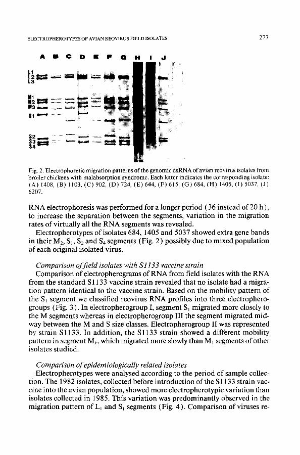
Fig. 2. Electrophoretic migration patterns of the genomic dsRNA of avian reovirus isolates from broiler chickens with malabsorption syndrome. Each letter indicates the corresponding isolate: (A) 1408, (B) 1103, (C) 902, (D) 724, (E) 644, (F) 615, (G) 684, (H) 1405, (I) 5037, (J) 6207.
RNA electrophoresis was performed for a longer period (36 instead of 20 h), to increase the separation between the segments, variation in the migration rates of virtually all the RNA segments was revealed.
Electropherotypes of isolates 684, 1405 and 5037 showed extra gene bands in their Mz, S~, $2 and $4 segments (Fig. 2 ) possibly due to mixed population of each original isolated virus.
Comparison of field isolates with $1133 vaccine strain Comparison of electropherograms of RNA from field isolates with the RNA
from the standard S 1133 vaccine strain revealed that no isolate had a migra- tion pattern identical to the vaccine strain. Based on the mobility pattern of the S~ segment we classified reovirus RNA profiles into three electrophero- groups (Fig. 3 ). In electropherogroup I, segment $1 migrated more closely to the M segments whereas in electropherogroup III the segment migrated mid- way between the M and S size classes. Electropherogroup II was represented by strain S1133. In addition, the S1133 strain showed a different mobility pattern in segment M~, which migrated more slowly than M~ segments of other isolates studied.
Comparison of epidemiologically related isolates Electropherotypes were analysed according to the period of sample collec-
tion. The 1982 isolates, collected before introduction of the S 1133 strain vac- cine into the avian population, showed more electropherotypic variation than isolates collected in 1985. This variation was predominantly observed in the migration pattern of L1 and S~ segments (Fig. 4). Comparison of viruses re-

278 M . R . R E K I K ET AL.
1 11 ! i l
m m 3
w
3 . . . . ~
1
S 2 3 ...... --, 4 ~
Fig. 3. Representative electropherotypes of each of the three electropherogroups identified ac- cording to the migration rate of segment S~.
N
'1 U 1 i . . . . M 2 ~ 3 ~
$
Fig. 4. Comparison of the different reovirus electropherotypes identified, with the vaccine strain
S1133.
covered in the same period from the same geographic area (Table 1 ) such as isolates 724, 902 and 1103 revealed an extensive variability in their genome migration profiles. On the contrary, isolates 6207 and 1405 recovered from different geographic locations demonst ra ted similar electropherotypes.

ELECTROPHEROTYPES OF AVIAN REOVIRUS FIELD ISOLATES 279
DISCUSSION
The characterization of individual reovirus isolates from different out- breaks by analysis of their dsRNA migration patterns is important for epide- miological investigations. The reports on electrophoretic analysis of avian reovirus studied to date were based on the isolates collected from outbreaks of tenosynovitis and arthritis (Gouvea and Schnitzer, 1982a; Wood and Thornton, 1986). The present report describes the analysis of dsRNA seg- ments from reoviruses isolated from outbreaks of malabsorption syndrome.
The results confirm an earlier finding of the slow migration of the avian reovirus S1 segment (Gouvea and Schnitzer, 1982b) compared to its mam- malian counterpart (Shatkin et al., 1968; Ramig et al., 1977). In addition, some electropherotypes analysed in this study, showed a slower mobility of the S1 segment than previously reported (Spandidos and Graham, 1976; Gouvea and Schnitzer, 1982b). Segment $1 of mammalian reovirus codes for two proteins (Jacobs and Samuel, 1985 ), whereas the larger $1 gene of avian reovirus appears to code for only one low molecular weight protein (Schnitzer, 1985 ). This supports the fact that great differences exist between S~ segments of each species and suggests that this avian reovirus segment should be grouped into the medium class instead of the slow class.
The great variability in the RNA profiles of isolates analysed in this study confirms the genetic polymorphism of avian reoviruses previously reported (Gouvea et al., 1982b). Whether a large genetic polymorphism exists among avian reoviruses is not known. According to Ramig and Fields ( 1983 ) changes observed in the genome ofreoviruses may be due to the high mutation rate or to genetic reassortment of the segmented RNA viruses.
The three electrophoretic patterns showing extra gene bands (Fig. 2 ) may have been due to simultaneous infection of birds by reoviruses of two differ- ent electropherotypes, which were prevalent at the same time. Although each isolate was plaque-purified twice, an unsuccessful plaque purification could have occurred leading to a mixed stock virus population. Mixed infection is a prerequisite for genomic reassortment.
The shift in the molecular weight of S~ segments detected in 1985, as op- posed to the 1982 isolates, and the absence of S 1133-like electropherotypes, are probably the consequence of genome evolution under selective immune pressure exerted on the sigma c protein encoded by this gene. A similar selec- tion pressure might have influenced the mobility of the L1 gene segment. This immune pressure might also have caused the emergence of new strains such as isolate 615 which is serologically distinct from the S1133 vaccine strain (Rekik et al., 1987 ). It would be interesting to study the changes occuring in the segments in relation to the immunologic response of the host.
The great variation observed in the migration of $2 and $4 genes could be attributed to the heterogeneity of the avian population analysed (different

280 M.R. REKIK ET AL.
age and origin ) since these two genes might be associated with the initiation and maintenance of persistent infection in vivo (Huang et al., 197 ). The sig- nificance of the particular migration level observed in segment M~ of the vac- cine strain was not easy to assess since its function and the protein that it codes for have not yet been determined. The only possible association that could be made is with the lack of virulence of the vaccine strain.
The ability to characterize individual virus isolates by genetic analysis of their dsRNA migration patterns is of potential importance in epidemiological investigations. However, it is necessary to analyse a large number of virus strain to obtain more detailed epidemiological information. Despite this, the method is still invaluable for the differentiation of avian reovirus strains and detection of mixed infection, or for the identification of new emerging viruses.
REFERENCES
Gouvea, V.S. and Schnitzer, T.J., 1982a. Polymorphism of the genomic RNAs among the avian reoviruses. J. Gen. Virol., 61: 87-91.
Gouvea, V.S. and Schnitzer, T.J., 1982b. Polymorphism of the migration of double-stranded RNA genome segments of avian reoviruses. J. Virol., 43: 465-471.
Huang, D.D., Nugent, M.A., Rosenberger, J.K. and Schnitzer, T.J., 1987. Association of avian reovirus M and S genes with viral behavior in vivo. I. Viral persistence. Avian Dis., 31: 438- 445.
Jacobs, B.L. and Samuel, D.E., 1985. Biosynthesis of reovirus-specified polypeptides: The reo- virus sl mRNA encodes two primary translation products. Virology, 143: 63-74.
Jones, R.C., Jordan, F.T.W. and Lioupis, S., 1975. Characteristics of reovirus isolated from ruptured gastrocnemius tendons of chickens. Vet. Rec., 96:153-154.
Kang, S.Y., Nagaraja, K.V. and Newman, J.A., 1986. Electropherotypic analysis of rotaviruses isolated from turkeys. Avian Dis., 30: 794-800.
Laemmli, U.K., 1970. Cleavage of structural proteins during the assembly of the head of bac- teriophage T4. Nature (Lond.), 277: 680-685.
Mustaffa-Babjee, A. and Spradbrow, P.B., 1971. The isolation of an avian reovirus. Aust. Vet. J., 47: 284.
Olson, N.O. and Solomon, D.P., 1968. A natural outbreak of synovitis caused by the viral ar- thritis agent. Avian Dis., 12:311-316.
Page, R.K., Fletcher, O.J., Rowland, G.N., Gaudry, D. and Villegas, P., 1982. Malabsorption syndrome in broiler chickens. Avian Dis., 26:618-624.
Ramig, R.F. and Fields, B.N., 1983. Genetics of reovirus. In: W. Joklik (Editor), The Reoviri- dae, Plenum, New York, pp. 197-228.
Ramig, R.F., Cross, R.S. and Fields, B.N., 1977. Genome RNAs and polypeptides of reovirus serotypes, 1, 2 and 3. J. Virol., 22: 726-733.
Rekik, M.R., Silim, A. and Bernier, G., 1987. Experimental infection of broiler chickens using different serotypes of reovirus from malabsorption syndrome cases. In: Proc. 36th Western Poultry Disease Conference, Davis, CA, pp. 146-148.
Robertson, M.D., Wilcox, G.E. and Kibenge, F.S.B., 1984. Prevalence of reoviruses in com- mercial chickens. Aust. Vet. J., 61 : 319-322.
Schnitzer, T.J., 1985. Protein coding assignment of the S genes of the avian reovirus S 133. Vi- rology, 141: 167-170.

ELECTROPHEROTYPES OF AVIAN REOVIRUS FIELD ISOLATES 281
Shatkin, A.J., Sipe, J.D. and Loh, P.C., 1968. Separation of 10 reovirus genome segments by polyacrylamide gel electrophoresis. J. Virol., 2: 986.
Spandidos, D.A. and Graham, A.F., 1976. Physical and chemical characterisation of avian reo- virus. J. Virol., 19: 968-976.
Springer, W.T., Olson, N.O., Kerr, K.M. and Fabacher, C.J., 1983. Responses of specific-path- ogen-free chicks to concomitant infections of reovirus (WVU-2937) and infectious bursal disease virus. Avian Dis., 27:911-917.
Van der Heide, L., Liitticken, D. and Horzinek, M., 1981. Isolation of avian reovirus as a pos- sible etiologic agent of esteoporosis (brittle bone disease: femoral head necrosis) in broiler chickens. Avian Dis., 25: 847-856.
Wood, G.W. and Thornton, D.H., 1986. Application of polyacrylamide gel electrophoresis of genome fragment to control of reovirus products. Dev. Biol. Standard, 64:213-218.