characteristics of bronchoalveolar lavage fluid in patients with sulfur mustard gas–induced asthma...
TRANSCRIPT
Characteristics of Bronchoalveolar Lavage Fluid inPatients with Sulfur Mustard Gas–induced Asthma
or Chronic Bronchitis
Ali Emad, MD, FAMA, Gholam Reza Rezaian, MD
PURPOSE: To examine the pattern of immunoglobulins andcellular constituents in bronchoalveolar lavage fluid obtained frompatients with sulfur mustard gas–induced asthma or chronic bron-chitis as compared with healthy control subjects.SUBJECTS AND METHODS: We studied two groups of non-smoking veterans with either bronchial asthma (n 5 21) orchronic bronchitis (n 5 28) believed to have been caused bysulfur mustard gas exposure and a third group of healthy, non-smoking, non–sulfur mustard gas exposed controls (n 5 17).Bronchoalveolar lavage was performed in all three groups. Thecellular constituents, albumin content, and immunoglobulinconcentrations were determined.RESULTS: The three groups did not differ in age or in the serumalbumin and immunoglobulin concentrations. The volume ofbronchoalveolar lavage fluid recovered was approximately 10%less in the patients with asthma and chronic bronchitis (P50.008).
The proportions of lymphocytes among the bronchoalveolar la-vage cells were similar in all three groups, whereas the proportionof eosinophils was greater in lavage fluid from the asthmatic sub-jects than in either the healthy control subjects or the patients withchronic bronchitis (P 5 0.0001). Both the total number of therecovered cells per milliliter of lavage fluid and the proportion ofneutrophils were significantly greater in bronchoalveolar lavagefrom patients with chronic bronchitis than in healthy subjects or inthe patients with asthma (all P ,0.001).
CONCLUSION: The bronchoalveolar lavage cellular constitu-ents of patients with sulfur mustard gas–induced asthma andchronic bronchitis are similar to those that have been observedpreviously in patients with asthma and chronic bronchitis fromother common causes. Am J Med. 1999;106:625– 628. q1999by Excerpta Medica, Inc.
Sulfur mustard gas is a lipophilic alkylating agentthat was recently used in the Iran–Iraq war (1,2).Acutely, it can damage the upper and lower respira-
tory tracts. Chronically, it can lead to the development ofairway hyperreactivity (3,4) and chronic bronchitis, asmanifest by chronic cough and sputum production (5–7). This study was carried out to define the characteristicsof the bronchoalveolar lavage fluid in a group of sulfurmustard gas– exposed patients who had developed bron-chial asthma or chronic bronchitis.
PATIENTS AND METHODS
In an earlier series of 197 veterans whose mustard gasexposure was toxicologically confirmed, there were 21patients with bronchial asthma and 116 patients withmild to severe bronchitis (5). All of these patients hadsevere respiratory symptoms immediately after their ini-tial mustard gas exposure in 1986 and were hospitalizedat that time (5). The presence of bronchiectasis, airwaystricture, or fibrosis was excluded by high-resolution
computerized tomographic scan of the chest. All 21 pa-tients with asthma and 28 of the 80 patients with mild tomoderate bronchitis volunteered to participate in thisstudy. (The 36 patients with severe bronchitis were notconsidered for participation, because they were thoughtto be at very high risk of complications from bronchoal-veolar lavage.) The control group consisted of 17 healthy,non–sulfur mustard gas exposed veterans. All subjectswere lifetime nonsmokers, and none had a previous his-tory of asthma or allergic disorders. Patients with a familyhistory of asthma or other allergic respiratory disorderswere excluded. Patients with proven cardiovascular dis-eases and those with exposure to other environmental orpharmacologic agents known to cause extrinsic allergicalveolitis, or with evidence of recent infection or exacer-bation of their disease, were excluded. All subjects weremen. Patients in the asthma group met at least two of thefollowing criteria: diurnal variability in peak expiratoryflow (PEF) rate .20% (8,9); reversibility of the forcedexpiratory volume in 1 second (FEV1) (10); or typicalhistory of attacks of dyspnea, wheezing, or both, and noc-turnal cough either spontaneously or triggered by irri-tants, respiratory infections, or exercise. Patients in thechronic bronchitis group had a history of cough and spu-tum production for at least 3 months per year during thelast 2 years and an irreversible obstructive pattern ob-served at spirometric testing (11).
All subjects signed informed written consent and had acomplete history and physical examination. A chest
From the Department of Internal Medicine (AE), Section of PulmonaryDiseases, and Department of Internal Medicine (GRR), Shiraz Univer-sity of Medical Sciences, Shiraz, Iran.
Requests for reprints should be addressed to Ali Emad, MD, FAMA,PO Box 71345-1674, Shiraz University of Medical Sciences, Shiraz, Iran.
Manuscript submitted March 13, 1998, and accepted in revised formNovember 10, 1998.
q1999 by Excerpta Medica, Inc. 0002-9343/99/$–see front matter 625All rights reserved. PII S0002-9343(99)00127-8
roentgenogram and electrocardiogram were obtained.Peripheral blood was examined for total and differentialleukocyte counts. The albumin, IgG, and total IgA con-centrations in serum were measured by immunoneph-elometry (12). The IgM concentration in the serum wasdetermined by immunoradiometric assay (13).
Spirometry and single breath diffusing capacity forcarbon monoxide were performed according to interna-tional standards (14). Spirometric data are expressed aspercentages of predicted values.
Bronchoalveolar LavageBronchoalveolar lavage was performed in all subjects us-ing a flexible fiberoptic bronchoscope (Olympus BF1T,Tokyo, Japan). No patient was allowed to receive sys-temic or inhaled corticosteroids for at least 1 month be-fore bronchoalveolar lavage. The upper respiratory tractwas anesthesized with 2% lidocaine. Atropine (0.75 mgintramuscularly) was given before the procedure. Supple-mental oxygen was given throughout the procedure, andthe oxygen saturation was monitored by continuouspulse oximetry. The bronchoscope was wedged for lavagein the middle lobe segmental bronchus, and four 60-mLaliquots of sterile physiologic saline solution, warmed to378C, were infused. The fluid was immediately recoveredby gentle suction after each instillation. The first aliquot,consisting of a bronchial sample, was discarded, whereasthe others were pooled for study. The recovered lavagefluid was passed through a monolayer surgical gauze toeliminate mucus and was centrifuged at 4003 g for 10minutes. The cell pellet was washed once in Hanks9 bal-anced salt solution (without calcium and magnesium). AMay-Grunwall-Giemsa stained smear was used to iden-tify differential profiles after cytospin preparation. Totalcell counts were determined with a hemocytometer. Thedifferential cell count for lymphocytes, neutrophils, mac-rophages, and eosinophils was made under light micros-copy by counting approximately 300 cells in randomfields. The results are expressed as cells 3 103/mL. Un-concentrated supernatant was frozen at 2708C beforeprotein concentrations were measured.
The concentrations of the albumin, IgG, total IgA, and
IgM were measured in bronchoalveolar lavage fluids su-pernatant by immunoradiometric assay (13). Proteinconcentrations were normalized to the albumin concen-tration.
Statistical AnalysisContinuous variables are presented as means 6 SD. Dif-ferences among groups were ascertained using Kruskal-Wallis analysis of variance; intergroup comparisons weremade using the Mann-Whitney test. A P value of ,0.05was considered statistically significant. Statistical analyseswere performed using Statview II software (Statview Inc.,Berkeley, California).
RESULTS
The age and the results of pulmonary function testing inthe three groups are shown in Table 1. There were nodifferences in total cell count, absolute leukocyte count,or serum protein concentrations among the patientgroups and controls (data not shown).
Cell Content of Bronchoalveolar Lavage FluidThe mean volume of recovered bronchoalveolar lavagefluid was 8% to 11% less in patients with asthma orchronic bronchitis than in controls (P ,0.01, Table 2).The mean number of recovered cells per milliliter ofbronchoalveolar lavage fluid was greater in patients withchronic bronchitis than either the patients with asthma orthe control group. Compared with controls, the propor-tions of macrophages were significantly lower in patientswith asthma and chronic bronchitis (P 5 0.001). Theproportions of lymphocytes were similar in all threegroups. The proportion of eosinophils was substantiallygreater in patients with asthma than in controls (P 50.001) or patients with chronic bronchitis (P 5 0.0001).In contrast, the proportion of neutrophils was signifi-cantly greater in patients with chronic bronchitis thaneither the controls (P 5 0.0001) or the patients withasthma (P 5 0.0001).
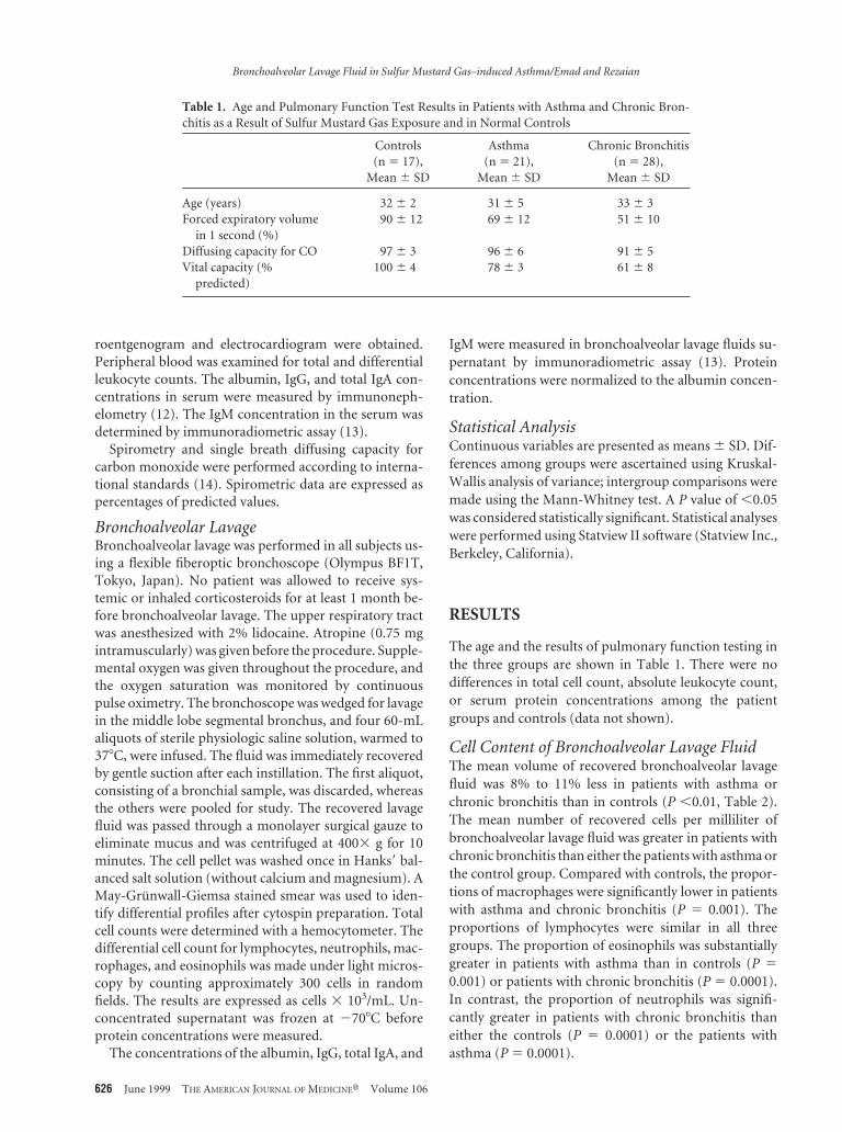
Table 1. Age and Pulmonary Function Test Results in Patients with Asthma and Chronic Bron-chitis as a Result of Sulfur Mustard Gas Exposure and in Normal Controls
Controls(n 5 17),
Mean 6 SD
Asthma(n 5 21),
Mean 6 SD
Chronic Bronchitis(n 5 28),
Mean 6 SD
Age (years) 32 6 2 31 6 5 33 6 3Forced expiratory volume
in 1 second (%)90 6 12 69 6 12 51 6 10
Diffusing capacity for CO 97 6 3 96 6 6 91 6 5Vital capacity (%
predicted)100 6 4 78 6 3 61 6 8
Bronchoalveolar Lavage Fluid in Sulfur Mustard Gas–induced Asthma/Emad and Rezaian
626 June 1999 THE AMERICAN JOURNAL OF MEDICINEt Volume 106

Serum and Bronchoalveolar Lavage FluidAlbumin and ImmunoglobulinsSerum albumin and immunoglobulin concentrations ofpatients with asthma or chronic bronchitis were not sig-nificantly different from those of the control subjects. Inall three groups, analysis of bronchoalveolar lavage fluidshowed that IgG was the predominant immunoglobulin(Table 3). There were, however, no significant differencesin concentrations of bronchoalveolar lavage fluid albu-min or any of the immunoglobulins among the threegroups.
DISCUSSION
Bronchial asthma and hyperreactivity of the airways maybe a late complication of sulfur mustard gas exposure (5).The epithelial lining of the airways is particularly sensitiveto this gas, which is a lipophilic agent with severe vesicantproperties. Inhalational exposure is thought to causeDNA alkylation, death, and shedding of bronchial epithe-lial cells (3), but the pathogenesis of the delayed appear-ance of asthma, chronic bronchitis, pulmonary fibrosis,or “reactive airway dysfunction syndrome” (RADS) is notknown (15).
Bronchoalveolar lavage enables the inferential study ofthe inflammatory and immune process in the lower re-spiratory tract (16,17) and can be used safely in patientswith asthma and chronic bronchitis (18). The cellularconstituents in bronchoalveolar lavage fluid that we ob-served among veterans who developed asthma after sul-fur mustard gas exposure resemble those found in thelungs and airways of subjects with “ordinary” allergicasthma, in that the proportion of eosinophils was signif-icantly greater than in either the control group or theveterans with chronic bronchitis. Eosinophils recruitedto allergen-exposed airways, presumably in response tothe release of chemokines, are thought to mediate manyof the characteristic changes of asthma (19 –22). Thefinding that eosinophils are present in bronchoalveolarlavage fluid from patients with sulfur mustard gas–in-duced asthma supports the still controversial view thatthese cells may play a role in initiating or maintainingasthma after an acute toxic exposure (15).
Chronic bronchitis is the most common chronic pul-monary sequela of sulfur mustard gas exposure, occur-ring in .50% of those exposed, and often associated withmarked physical disability (5). The total number of cellsrecovered in bronchoalveolar lavage of exposed veterans
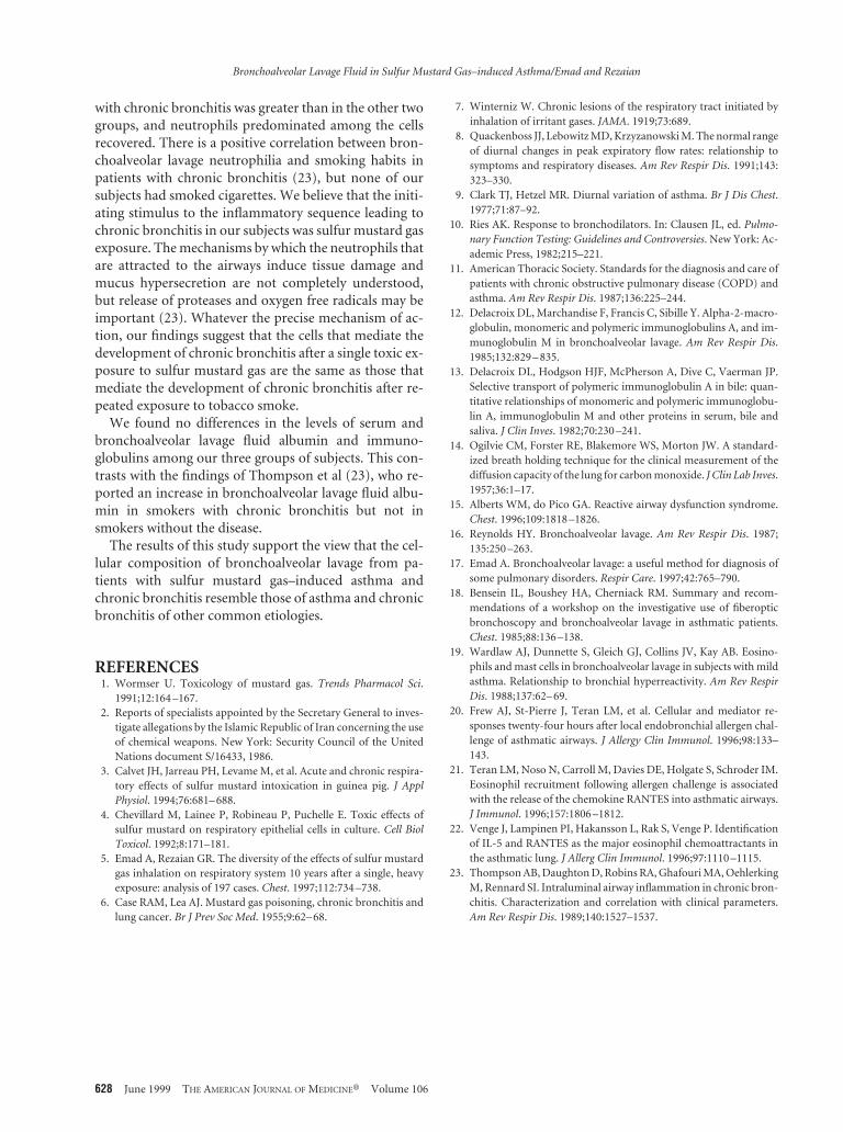
Table 2. Comparison of Bronchoalveolar Lavage Fluid Cellular Constituents in Patients withAsthma and Chronic Bronchitis as a Result of Sulfur Mustard Gas Exposure and in NormalControls
Controls(n 5 17),
Mean 6 SD
Asthma(n 5 21),
Mean 6 SD
Chronic Bronchitis(n 5 28),
Mean 6 SD P Value
Fluid recovered (mL) 161.3 6 12 144.5 6 19 149.2 6 15 0.0001Leukocytes (3103/mL) 85 6 9 80 6 8 104 6 18 0.008Macrophages (%) 86 6 3 81 6 5 74 6 7 0.0001Macrophages (3103/mL) 73 6 8 65 6 9 78 6 18 0.006Lymphocytes (%) 12 6 3 11 6 3 11 6 4 0.59Lymphocytes (3103/mL) 10 6 3 9 6 2 11 6 3 0.06Neutrophils (%) 2 6 1 3 6 2 14 6 5 0.0001Neutrophils (3103/mL) 2 6 1 3 6 1 15 6 5 0.0001Eosinophils (%) 0.6 6 0.7 5 6 4 0.6 6 0.8 0.0001Eosinophils (3103/mL) 0.5 6 0.6 4 6 3 0.6 6 0.8 0.0001
Table 3. Bronchoalveolar Lavage Fluid Albumin and Immunoglobulin Concentrations in Patientswith Asthma and Chronic Bronchitis as a Result of Sulfur Mustard Gas Exposure and in NormalControls
Controls(n 5 17),
Mean 6 SD
Asthma(n 5 21),
Mean 6 SD
Chronic Bronchitis(n 5 28),
Mean 6 SD P Value
Albumin (mg/mL) 166 6 23 159 6 16 161 6 16 0.21IgG (mg/dL) 9 6 2 9 6 2 11 6 3 0.20IgM (mg/dL) 0.02 6 0.02 0.01 6 0.02 0.04 6 0.08 0.78IgA (mg/dL) 1.9 6 0.5 1.9 6 0.7 2.0 6 0.5 0.72
Bronchoalveolar Lavage Fluid in Sulfur Mustard Gas–induced Asthma/Emad and Rezaian
June 1999 THE AMERICAN JOURNAL OF MEDICINEt Volume 106 627
with chronic bronchitis was greater than in the other twogroups, and neutrophils predominated among the cellsrecovered. There is a positive correlation between bron-choalveolar lavage neutrophilia and smoking habits inpatients with chronic bronchitis (23), but none of oursubjects had smoked cigarettes. We believe that the initi-ating stimulus to the inflammatory sequence leading tochronic bronchitis in our subjects was sulfur mustard gasexposure. The mechanisms by which the neutrophils thatare attracted to the airways induce tissue damage andmucus hypersecretion are not completely understood,but release of proteases and oxygen free radicals may beimportant (23). Whatever the precise mechanism of ac-tion, our findings suggest that the cells that mediate thedevelopment of chronic bronchitis after a single toxic ex-posure to sulfur mustard gas are the same as those thatmediate the development of chronic bronchitis after re-peated exposure to tobacco smoke.
We found no differences in the levels of serum andbronchoalveolar lavage fluid albumin and immuno-globulins among our three groups of subjects. This con-trasts with the findings of Thompson et al (23), who re-ported an increase in bronchoalveolar lavage fluid albu-min in smokers with chronic bronchitis but not insmokers without the disease.
The results of this study support the view that the cel-lular composition of bronchoalveolar lavage from pa-tients with sulfur mustard gas–induced asthma andchronic bronchitis resemble those of asthma and chronicbronchitis of other common etiologies.
REFERENCES1. Wormser U. Toxicology of mustard gas. Trends Pharmacol Sci.
1991;12:164 –167.2. Reports of specialists appointed by the Secretary General to inves-
tigate allegations by the Islamic Republic of Iran concerning the useof chemical weapons. New York: Security Council of the UnitedNations document S/16433, 1986.
3. Calvet JH, Jarreau PH, Levame M, et al. Acute and chronic respira-tory effects of sulfur mustard intoxication in guinea pig. J ApplPhysiol. 1994;76:681– 688.
4. Chevillard M, Lainee P, Robineau P, Puchelle E. Toxic effects ofsulfur mustard on respiratory epithelial cells in culture. Cell BiolToxicol. 1992;8:171–181.
5. Emad A, Rezaian GR. The diversity of the effects of sulfur mustardgas inhalation on respiratory system 10 years after a single, heavyexposure: analysis of 197 cases. Chest. 1997;112:734 –738.
6. Case RAM, Lea AJ. Mustard gas poisoning, chronic bronchitis andlung cancer. Br J Prev Soc Med. 1955;9:62– 68.
7. Winterniz W. Chronic lesions of the respiratory tract initiated byinhalation of irritant gases. JAMA. 1919;73:689.
8. Quackenboss JJ, Lebowitz MD, Krzyzanowski M. The normal rangeof diurnal changes in peak expiratory flow rates: relationship tosymptoms and respiratory diseases. Am Rev Respir Dis. 1991;143:323–330.
9. Clark TJ, Hetzel MR. Diurnal variation of asthma. Br J Dis Chest.1977;71:87–92.
10. Ries AK. Response to bronchodilators. In: Clausen JL, ed. Pulmo-nary Function Testing: Guidelines and Controversies. New York: Ac-ademic Press, 1982;215–221.
11. American Thoracic Society. Standards for the diagnosis and care ofpatients with chronic obstructive pulmonary disease (COPD) andasthma. Am Rev Respir Dis. 1987;136:225–244.
12. Delacroix DL, Marchandise F, Francis C, Sibille Y. Alpha-2-macro-globulin, monomeric and polymeric immunoglobulins A, and im-munoglobulin M in bronchoalveolar lavage. Am Rev Respir Dis.1985;132:829 – 835.
13. Delacroix DL, Hodgson HJF, McPherson A, Dive C, Vaerman JP.Selective transport of polymeric immunoglobulin A in bile: quan-titative relationships of monomeric and polymeric immunoglobu-lin A, immunoglobulin M and other proteins in serum, bile andsaliva. J Clin Inves. 1982;70:230 –241.
14. Ogilvie CM, Forster RE, Blakemore WS, Morton JW. A standard-ized breath holding technique for the clinical measurement of thediffusion capacity of the lung for carbon monoxide. J Clin Lab Inves.1957;36:1–17.
15. Alberts WM, do Pico GA. Reactive airway dysfunction syndrome.Chest. 1996;109:1818 –1826.
16. Reynolds HY. Bronchoalveolar lavage. Am Rev Respir Dis. 1987;135:250 –263.
17. Emad A. Bronchoalveolar lavage: a useful method for diagnosis ofsome pulmonary disorders. Respir Care. 1997;42:765–790.
18. Bensein IL, Boushey HA, Cherniack RM. Summary and recom-mendations of a workshop on the investigative use of fiberopticbronchoscopy and bronchoalveolar lavage in asthmatic patients.Chest. 1985;88:136 –138.
19. Wardlaw AJ, Dunnette S, Gleich GJ, Collins JV, Kay AB. Eosino-phils and mast cells in bronchoalveolar lavage in subjects with mildasthma. Relationship to bronchial hyperreactivity. Am Rev RespirDis. 1988;137:62– 69.
20. Frew AJ, St-Pierre J, Teran LM, et al. Cellular and mediator re-sponses twenty-four hours after local endobronchial allergen chal-lenge of asthmatic airways. J Allergy Clin Immunol. 1996;98:133–143.
21. Teran LM, Noso N, Carroll M, Davies DE, Holgate S, Schroder IM.Eosinophil recruitment following allergen challenge is associatedwith the release of the chemokine RANTES into asthmatic airways.J Immunol. 1996;157:1806 –1812.
22. Venge J, Lampinen PI, Hakansson L, Rak S, Venge P. Identificationof IL-5 and RANTES as the major eosinophil chemoattractants inthe asthmatic lung. J Allerg Clin Immunol. 1996;97:1110 –1115.
23. Thompson AB, Daughton D, Robins RA, Ghafouri MA, OehlerkingM, Rennard SI. Intraluminal airway inflammation in chronic bron-chitis. Characterization and correlation with clinical parameters.Am Rev Respir Dis. 1989;140:1527–1537.
Bronchoalveolar Lavage Fluid in Sulfur Mustard Gas–induced Asthma/Emad and Rezaian
628 June 1999 THE AMERICAN JOURNAL OF MEDICINEt Volume 106