characteristics of extremophylic fungi from chernobyl nuclear
TRANSCRIPT

Characteristics of Extremophylic Fungi from Chernobyl Nuclear Power
Plant
Т. Belozerskaya 1, K. Aslanidi
2, А. Ivanova
3, N. Gessler
1, A. Egorova
1, Yu. Karpenko
4, and S.
Olishevskaya4
1 A.N. Bach Institute of Biochemistry, Russian Academy of Sciences, Moscow, Russia, 2 Institute of Theoretical and Experimental Biophysics, Russian Academy of Sciences, Pushchino, Moscow Region,
Russia, 3 Soil Science Department, Moscow State University, Moscow; Russia, 4 Zabolotny Institute of Microbiology and Virology, National Academy of Sciences, Ukraine
Fungi isolated from Chernobyl Nuclear Power Plant (ChNPP) where radiation dose was from 3 to 5 orders higher than the
background radioactivity offered following properties: aggregation of hyphae; oligotrophy; high resistance to oxidative
stress due to adaptation to low (0,2%) carbohydrate content in the medium; resistance to an order higher H2O2
concentration than fungi from habitats with background radiation; ability to resume growth after severe oxidative stress
(10-2M H2O2); activation of antioxidant enzymes, and increased biosynthesis of melanins in Paecilomyces lilacinus
possessing hyaline mycelium. Biochemical characteristics point that adaptation mechanisms to oxidative stress are strain
specific. Ability to survive in hostile environment makes these micromycetes perspective models to study evolutionary
process of extremophylic fungi.
Keywords Chernobyl fungi; strain specific properties; aggregation of hyphae; oligotrophy; high resistance to oxidative
stress
1. Introduction
Of the major groups of soil microorganisms, fungi are of fundamental importance with primary roles in plant
pathogenesis, symbiosis and the cycling of important nutrient elements, e.g. carbon, nitrogen, phosphorus, sulphur and
many trace metals [1]. According to their trophic characteristics, the overwhelming majority of the saprophytic fungi
belong to the group of decomposers, which actively destroy various vegetable and technogenic substrates [2].
Microfungi (anamorphs, producing conidiospores) represent an extensive group of organisms in soil. They have been
shown to take up and translocate in the mycelium a waste array of naturally occurring as well as man-made
radionuclides [3]. Dead mycelia (cell walls) made into filters have been used to adsorb heavy metals and radionuclides
from industrial effluents [3].
Grassland soil saprotrophic fungi are capable of the transformation of radioactive particles with high specific activity
to a soluble form. Unfortunately these soluble elements might be accumulated into food webs [4]. Absorption of
radionuclides by fungi seems to be strain-specific [3]. Fungi are able to absorb not only 137
Cs, 121
Sr and 152
Eu, but also
such radioisotopes as 239
Pu and 241
Am [4, 5]. This suggests that fungi could be long-term retainers of radionuclides in
the environment [3].
To cope such environment these fungi worked out resistance mechanisms such as asexuality, synthesis of melanin-
like pigments, flexible morphology, and growth under limited nutrient content in the habitat.
Recent work from in and around the remains of the ChNPP demonstrates that soil microfungal communities have
been altered by the intense radiation fallout, leading to simpler community structure and a dominance of melanin-
containing (pigmented) fungal species [6]. During the last 15 years, about 2000 strains of 200 species of 98 genera of
fungi have been isolated around the Chernobyl Atomic Energy Station. Some of these microfungi show formerly
unknown adaptive features, such as directed growth of fungi to sources of ionizing radiation [7, 8].
The mechanisms whereby fungi arrive at sources of radionuclides in the environment and the processes that occur
during the decomposition/modification of those sources are ecologically important in regulating radionuclide movement
and for potential site remediation. Capacity to acquire radionuclides in micromycetes from Chernobyl appears to be an
interaction between the physical nature of the radioactive source, fungal species and, presumably, their enzymatic and
pigment potential.
It is well known that ionizing radiation induces the formation of reactive oxygen species (ROS), which provoke
oxidative stress in cells and can eventually cause their death [9, 10]. (Fig. 1). In cells with an aerobic type of
metabolism, hydrogen peroxide is the most long-lived transformation product of ROS. It is always present in such cells
at low concentrations, being involved in intra- and intercellular signal transduction [11]. In fungi, hydrogen peroxide is
a necessary component of the intracellular signaling system in such processes as cell differentiation and proliferation
[12, 13]. Hydrogen peroxide is of great interest for simulating the effect of radiation on living objects, since up to 90%
of the damage induced by ionizing radiation is due to water radiolysis products [9].
_______________________________________________________________________________________

Fig. 1. Scheme illustrating ROS formation
The aim of the present work was to reveal some morphological, physiological and biochemical peculiarities of
Chernobyl’s microfungi allowing for adaptation to the extreme environment hostile to most eukaryotes. The individual
parameters (characteristic values) of Chernobyl’s fungi were also studied under oxidative stress via H2O2 treatment.
2. Fungal strains under investigation
Experiments were carried out with four species of microscopic fungi isolated from the exclusion zone of the ChNPP,
which has a high level of radioactive contamination, and from habitats with a background level of radioactive
contamination (see Table 1). The strains differed the structure of their mycelium (either septate or nonseptate), the
presence of melanin pigments in the cell wall (dark- colored and hyaline mycelia), and in their growth rate (slow- and
fast-growing strains).
Table 1 Fungal isolates studied
Species,
№ strain
Isolates habitats Level of
radioactive
pollution, Bk/kg
Mycelial structure
Alternaria alternata (Fr.:Fr.) Keissler 1912
56 ChNPP territory. Wall of the 4th
block. Isolated
1998.
α=500 Bq/cm2;
β= 2x10⁴ Bq/cm
2;
γ=700 mR/h
Melanin-containing,
septate
60 Moscow region. Umbric Albeluvisols. Isolated
1989.
Background -“-
224 Moscow region. Umbric Albeluvisols
Anthropic. Isolated 1989.
Background -“-
Cladosporium cladosporioides (Fres.) de Vries 1952
4 ChNPP territory. Soil of Red Forest. Isolated
1986.
3,6 x 10⁵ Melanin-containing,
septate
5 ChNPP territory. Surface of carbon radioactive
debris from the reactor. Isolated 1993.
3,7 x 10⁹ -“-
396 Lvov region. Chernozems Anthropic. Isolated
1957.
Background * -“-
Mucor hiemalis Wehmer 1903
111 Moscow region. Umbric Albeluvisols
Anthropic. Isolated 1997.
Background hyaline, coenocytic
Paecilomyces lilacinus (Thom) Samson 1974
1941 ChNPP territory. Soil of Red Forest. Isolated
1994г.
5,9 х 105 hyaline, septate
1492 ChNPP territory. Soil of Red Forest. Isolated
1992.
2,7 х 105 -“-
744 ChNPP territory. Soil of Western Track.
Isolated 1992.
1,3 х 103 -“-
1786 ChNPP territory. Soil of Red Forest. Isolated
1993.
1,2 х 102 -“-
SM Smolensk region. Village Gnesdovo. Umbric
Albeluvisols. Isolated 2004.
Background -“-
SB Moscow. Serebryany Bor. Umbric
Albeluvisols. Isolated 2005.
-“- -“-
VR Moscow region. City Voskresensk. Umbric
Albeluvisols. Isolated 2009.
-“- -“-
O2 O2-• Н2O2 НO• 2Н2O
+е‾
+е‾
+е‾
+2Н+ +2Н
+
+е‾
+4 е-
+4 Н+
_______________________________________________________________________________________

T2 Tuva Republic, Tere-Hole-lake depression.
Calci-Mollic Cryosols. Isolated 2007.
-“- -“-
10 Novgorod region. Sappric Histosols. Isolated
2000.
-“- -“-
146 Lugansk region. Hutor Kartamush. Chernozem.
Isolated 2002.
-“- Copper
pollution
-“-
804 Donetsk region. Village Konstantinovka.
Chernozem. Isolated 2004.
-“-, Heavy metal
pollution
-“-
* - The soil contamination of the Russian Federation territory is about 0,22 Bk/sm2 by 137Cs and 0,15 Bk/sm2 by 90Sr. Soils are
considered to be unpolluted when the contamination level is 3,7 Bk/sm2 by 137Cs. γ-radiation exposure dose rate on the ground (EDR)
in Russia, except for contaminated areas, is within the natural background radiation - 9-17 µcR / h [14].
The second part of the work was devoted to investigation of P.lilacinus strains widely distributed all around the
world. The genus Paecilomyces is a cosmopolitan filamentous fungus, and the strain P.lilacinus has been isolated from
a wide range of habitats including cultivated and uncultivated soil as well as from soil with anthropogenic effects. In
Chernobyl there is a trend of change in dominance of fungal species with radiation level. At present P.lilacinus is one of
the indicators of high levels of radionuclide soil contamination (3.7 x 10⁶–3.7 x 10
8 Bq/kg) there [3].
All of the strains isolated from around ChNPP showed an aggregated growth of their hyphae (Fig 2)
Fig 2 Aggregated hyphae of Alternaria alternata 56 isolated from ChNPP, inner wall of contaminated structure. 1998. Radioactivity
level is shown in Table 1. 40 min growth on agar Chapek media.
The hyphal aggregation is typical of fungi growing under unfavorable conditions [4, 5]. This aggregated growth also
precedes their differentiation under the conditions of oxidative stress and, hence, like some other morphological
characteristics (such as the formation of sclerotia), may be considered as a mechanism of adaptation to high
concentrations of free radicals in the medium [4, 5]. In the fungi studied, the capacity for aggregated growth of hyphae
has probably been acquired due to their growth under extreme environmental conditions.
3. Olygotrophic growth
It has been shown previously that some species of Chernobyl habitat can grow as olygotrophs [16]. Olygotrophs are
microorganisms that can grow in the presence of low concentrations of nutrients, or even nutrients appear to be
nonexistent. The term olygotrophy is generally used to describe the strategy used by microorganisms to grow on low
concentration of organic carbon, more strictly referred to as olygocarbotrophy [15]. Oligotrophic microorganisms cause
a number of problems associated with biocontamination and biodeterioration, e.g. contaminate bottled drinking water
and colonize solid surfaces such as glass, metals and electronic components.
On investigation of growth peculiarities of P.lilacinus strains it was found that in all the strains tested radial growth
rates were higher under glucose level lower than 0,5% (Fig. 2). Strains of P.lilacinus from around Chernobyl showed
higher radial growth rates under lowest glucose concentrations (0,02%-0,2%) in comparison with the strains from
habitats with background radioactivity.
The data presented confirm previous investigations [6] and allow referring to P.lilacinus Chernobyl strains studied as
olygocarbotrophs for their ability to show maximal growth rate under low glucose content.
10 µm
_______________________________________________________________________________________

Fig. 3 The alteration of hyphal radial growth rate
Kr(i) under various glucose concentrations in the
medium relatively to radial growth rate Kr(0) in the
medium lacking glucose .
Future investigations will unravel how these fungi could provide life energy: through CO2 fixation, acceptance of
volatile compounds or using melanins as energy source [15-18].
4. Oxidative stress and fungal growth under various glucose concentrations
According to some authors the specific growth rate is an important physiological indicator of microbial resistance to
stresses such as starvation and hydrogen peroxide [19]. The data presented in this paper show that H2O2 increase in the
medium (10-9
- 10-1
M) influenced elongation rate of fungal hyphae (Fig. 4). The growth of fungi isolated from habitats
with a background level of radioactive contamination was stopped by 10–4
- 10–3
M H2O2, whereas the growth of fungi
that were isolated from habitats with high levels of radioactive contamination was arrested only by 10-2
- 10–1
M H2O2.
Adaptation slowdown reaction has been found as a result of the first 10–30 min H2O2 treatment most profound in
the ChNPP P.lilacinus 1941 strain. Under 10–3
M H2O2 growth of 1941 resumed with 20% loss in growth rate after such
a slowdown. Cessation of growth of the control strain 10 (Table 1) was observed under these conditions [20].
One of the important physiological criteria of resistance to oxidative stress is the ability to re-establish hyphal
growth after H2O2 (10-2
М, 90 min) treatment. All the fungi from Chernobyl habitat were able to regenerate after 10-2
М
H2O2 for 90 min. Cessation of growth in all the fungi from background habitats except for A. alternata 60 (40%
resumed growth) was observed [21].
Thus Chernobyl fungi were more resistant to oxidative stress. They could cope with severe oxidative stress: an order
higher H2O2 concentration than isolates from habitats with background radioactivity.
Among strains investigated, those isolated from “background” habitats (A. alternata 60, A. alternata 224, C.
cladosporioides 396, and P.lilacinus 10) [Table 1] exhibited three types of responses to H2O2 (Fig.4 A).
Type 1. An initial increase in the elongation rate of hyphae upon an increase of H2O2 concentration from 10-6
to 10-5
M, followed by a decrease of hyphal elongation and complete growth arrest at 10–2
–10–1
M (Fig.4 A, strain 2).
Fig. 4 Scheme illustrating different types of hyphal growth of the soil fungi isolated from habitats with background radiation (A)
and the strains isolated in the Chernobyl zone (B) under various H2O2 concentrations. Dependences of relative rates of leading hyphal
elongation (Vi/V0, %) for different species and strains of filamentous fungi on the H2O2 concentration (C, M), in an agar nutrient
medium are shown. The rates were normalized to the corresponding values of elongation rates, obtained in the hydrogen peroxide-
free medium (10-9 M H2O2 [22]. Designations: (1) M. hiemalis (2) A. alternata 60; (3) A. alternata224; (4) C. cladosporioides 396;
(5) P. lilacinus 10; (6) A. alternata 56; (7) C. cladosporioides 5; (8) C. cladosporioides 4; (9) P. lilacinus 1941.
A)
0
1
VI /V0
1 2 3 4 5
10 -9
10 -7
10-6
10 -5
10-4
10-3
10-2
10 -1
H2 O2 Concentration, M
B)
0
1
6 7 8 9
10 -7
10-6
10 -5
10-4
10-3
10-2
10-1
10 -9
H2O 2 Concentration, M
0,70
0,75
0,80
0,85
0,90
0,95
1,00
1,05
1,10
1,15
1,20
0 1 2 3 4 5
glucose, %
Kr(i)/Kr(0%)
from radioactiva contaminated habitats
from background habitats
Kr(i)/Kr(0)
_______________________________________________________________________________________
Type 2. A gradual decrease of the hyphal elongation rate as a result of increasing Н2О2 concentration. Growth was
completely inhibited at 10–3
–10–2
M of Н2О2 (Fig.4 A, strain 3).
Type 3. A constant rate of hyphal growth observed at 10-9
-10-4
M H2O2, a growth rate decrease at 10-3
M H2O2, and
growth arrest at 10-2
M H2O2 (Fig.4 A, strain 5).
Strains isolated from the radioactively contaminated zones (A. alternata 56, C. cladosporioides 4, 5, P. lilacinus
1941) [Table 1] showed only second and third types of response (Fig. 4 B). The broadest H2O2 concentration activity
range allowing hyphal growth (10-9
-10-1
M) was described in A. alternata 56 isolated from the wall of the 4th block of
the ChNPP (Table 1). In all the studied strains of filamentous fungi elongation rates of the leading hyphae determined
on H2O2-free media (10-9
M) [22], fell within a range from 0.5±0.2 to 5.5± 0.5 µm/min [14].
These different types of growth of microscopic fungi under varying H2O2 concentrations (especially A and B) were
basis for creation of a mathematical model of fungal growth in a gradient of H2O2 [23]. As it was mentioned earlier,
upon interaction with water, all types of radiation sources generate reactive oxygen species (ROS), primarily H2O2, as
the most stable ROS. These isolated radiation sources create local gradients of H2O2 in moist substrates that affect the
growth rates of microscopic fungus hyphae. Unfortunately only a few studies have been devoted to the directional
growth of hyphae towards these source of ionizing radiation (hot particles, or carbon-based radioactive graphite from
the 4th block ChNPP 1-100nm size) [7, 8].
Analysis of the model worked out revealed that H2O2 action on fungal growth apparently determines the
radioactivity level that enables overgrowth of the hot particles by fungi [23].
Upon comparative study of fungi from Chernobyl zone and fungi from habitats with background radiation the
question arises what fungal species are more stressed growing under various glucose concentrations? To answer the
question protein carbonyls of P.lilacinus strains from different habitats grown under various glucose concentrations
have been studied.
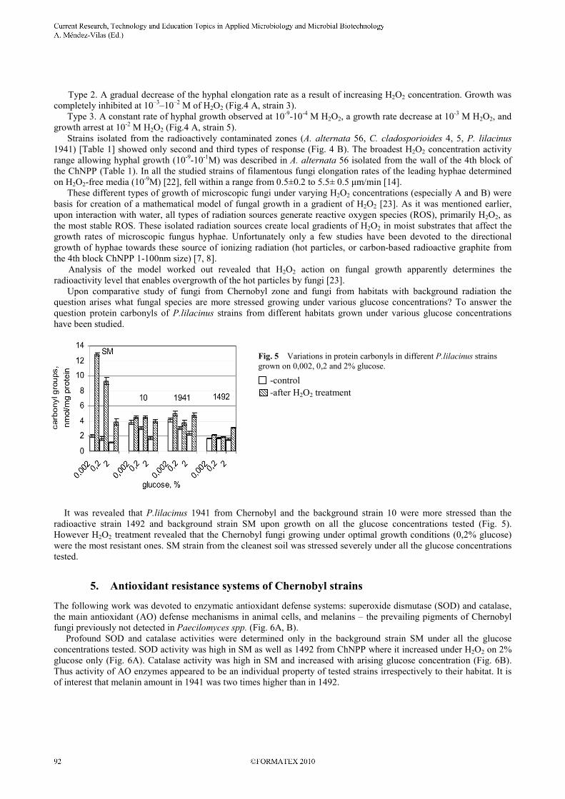
Fig. 5 Variations in protein carbonyls in different P.lilacinus strains
grown on 0,002, 0,2 and 2% glucose.
It was revealed that P.lilacinus 1941 from Chernobyl and the background strain 10 were more stressed than the
radioactive strain 1492 and background strain SM upon growth on all the glucose concentrations tested (Fig. 5).
However H2O2 treatment revealed that the Chernobyl fungi growing under optimal growth conditions (0,2% glucose)
were the most resistant ones. SM strain from the cleanest soil was stressed severely under all the glucose concentrations
tested.
5. Antioxidant resistance systems of Chernobyl strains
The following work was devoted to enzymatic antioxidant defense systems: superoxide dismutase (SOD) and catalase,
the main antioxidant (AO) defense mechanisms in animal cells, and melanins – the prevailing pigments of Chernobyl
fungi previously not detected in Paecilomyces spp. (Fig. 6A, B).
Profound SOD and catalase activities were determined only in the background strain SM under all the glucose
concentrations tested. SOD activity was high in SM as well as 1492 from ChNPP where it increased under H2O2 on 2%
glucose only (Fig. 6A). Catalase activity was high in SM and increased with arising glucose concentration (Fig. 6B).
Thus activity of AO enzymes appeared to be an individual property of tested strains irrespectively to their habitat. It is
of interest that melanin amount in 1941 was two times higher than in 1492.
-control
-after H2O2 treatment
_______________________________________________________________________________________

A)
1492SM
10 1941
0
200
400
600
800
1000
1200
1400
1600
0,00
2 0,2 2
0,00
2 0,2 2
0,00
2 0,2 2
0,00
2 0,2 2
glucose, %
SO
D a
ctiv
ity,
un
it/m
g p
rote
in
Fig. 6 Activity of antioxidant enzymes SOD (A) and catalase (B) in some P.lilacinus strains.
Melanin pigments are found in all biological kingdoms. Melanins are complex molecules with a variety of properties
able to absorb all types of electromagnetic radiation [25]. They are capable of both: energy transduction and shielding.
It has been proposed recently that melanins have functions analogue to other energy harvesting pigments such as
chlorophylls [17, 18]. Antioxidant properties of fungal melanins have been studied long ago. Antioxidant capacity of
fungal melanins is similar to AO activity of macrophages [26]. Modern investigations of Ascomycetes and Fungi
Imperfecti have demonstrated convincingly that melanins of these fungi mainly originate from 1,8-dihydroxynaftalene
[16, 27].
Melanins in P.lilacinus were detected by ESR method (Fig. 7). Electron spin resonance spectroscopy (ESR) of dried
mycelia of each P.lilacinus spp. showed the presence of stable free radical population, distinguishing of melanin [28].
This population varies among Paecillomyces strains. The highest numbers of stable free radicals were revealed in
Chernobyl strain 1941 (4,6х1016
spins/g) and lowest in the strain 10 (1,4х1016
spins/g). Dominance of melanin containing (pigmented) fungal species with higher radionuclide absorption capacity has been
observed in around Chernobyl lately [3]. Thus finding the melanins in P.lilacinus strains indicators of high radioactive
contamination at present confirms the pivotal role of these pigments in adaptation to extreme conditions of ChNPP.
Fig. 7 ESR spectra of standard DOPA-melanin (1) and of P.lilacinus 1941
melanin (2)
6. Conclusion
Fungi from around ChNPP show aggregation of hyphae, increased resistance to oxidative stress, certain growth
peculiarities under H2O2 treatment, increased growth rate under low glucose concentrations. Adaptation to low glucose
content in the medium (0.2%) is coupled with an increased resistance to oxidative stress. These fungi demonstrate
strain-specific mechanisms of antioxidant defense.
Acknowledgements The work was supported by Russian Foundation for Basic Research. Grant N 08-04-01833.
-control, -after H2O2 treatment
_______________________________________________________________________________________

References
[1] Gadd G, Ramsay L, Crawford J, Ritz K. Nutritional influence on fungal colony growth and biomass distribution in response to
toxic metals. FEMS Microbiol. Lett. 2001. 204:311–316.
[2] Moore D. Fungal morphogenesis. Cambridge University Press, USA; 1998.
[3] Dighton J, Tugay T, Zhdanova N. Fungi and ionizing radiation from radionuclides. FEMS Microbiol. Lett. 2008. 281:109-120.
[4] Zhdanova N, Redchitz, T, Zheltonozhsky V, Sadovnikov L, Gerzhabek M, Ollson S, Strebl F, Mück K. Accumulation of
radionuclides from radioactive substrata by some micromycetes. Journal of Environmental Radioactivity. 2003. 67: 119-130.
[5] Zhdanova N, Redchitz T, Zheltonozhsky V, Zheltonozhskaya M, Sadovnikov L. Ability of certain soil fungi to interact with rare
earth and transuranic elements 152Eu and 239Pu. Abstracts of the II Interdisciplinary Mycological Forum. Moscow 2010:60.
[6] Zhdanova N, Zakharchenko V, Haselwandter K. Radionuclides and fungal communities. In: Dighton J, White JF, Oudemans P,
eds. The Fungal Community: Its Organization in Ecosystems. CRC Press, Baton Rouge; 2005:759-768.
[7] Zhdanova N, Tugay T, Dighton J, Zheltonozhsky V, McDermott P. Ionizing radiation attracts soil fungi. Mycol. Res. 2004.
108:1089-1096.
[8] Tugay T, Zhdanova N, Zheltonozhsky V, Sadovnikov L, Dighton J. The influence of ionizing radiation on spore germination and
emergent hyphal growth response reactions of microfungi. Mycologia 2006. 94:521-526.
[9] Korystov Yu. Contributions of the direct and indirect effects of ionizing radiation to reproductive cell death, Radiat. Res. 1992.
129:228–234.
[10] Burlakova E, Mikhailov V, Mazurik V. System of redox homeostasis in the radiation-induced genome instability, Radiats. Biol.
Radioecol. 2001. 41:489–499.
[11] Gamalei I, and Klyubin N. Hydrogen peroxide as a signal molecule. Tsitologiya (in Russian). 1996. 38:1233–1247.
[12] Hansberg W, Aguirre J. Hyperoxidant states cause microbial cell differentiation by cell isolation from dioxygen, J. Theor. Biol.
1990. 142:287–293.
[13] Sidery, M. and Georgiou, Ch.D., Differentiation and hydrogen peroxide production in Sclerotium rolfsii are induced by the
oxidizing growth factors, light, and iron. Mycologia, 2000. 92:1033–1042.
[14] Ivanova A, Aslanidi K, Karpenko Yu, Belozerskaya T. The effect of hydrogen peroxide on the growth of microscopic mycelial
fungi isolated from habitats with different level of radioactive contamination. Microbiology 2005. 74:655-663.
[15] Wainwrite M. Olygotrophyc growth of fungi. In: Dighton J, White JF, Oudemans P, eds. The Fungal Community: Its
Organization in Ecosystems. CRC Press, Baton Rouge; 2005:643-658.
[16] Zhdanova N, Vasilevskaya A. Melanincontaining fungi in extreme conditions. (in Russian). Kiev:Naukova dumka 1988.
[17] Dadacheva K, Bryan R, Huang X, Moadel T, Schweitzer A, Alsen P, Nosanchuk J, Casadevall A. Ionizing radiation changes
the electronic properties of melanin and enhances the growth of melanized fungi. PLoS ONE 2007. 5:e457.
[18] Dadacheva K, Casadevall A. Ionizing radiation: how to cope, adapt, and exploit with the help of melanin. Curr. Opin.
Microbiol. 2008. 11:525-531.
[19] Ostrowski M, Cavicchioli R., Blaauw M, Gottschalk J. Specific growth rate plays a critical role in hydrogen peroxide resistance
of the marine oligotrophic ultramicrobacterium Sphingomonas alaskensis Strain RB2256. Appl. Environ. Microbiol. 2001.
67:1292–1299.
[20] Aslanidi K, Ivanova A, Gessler N, Egorova A, Belozerskaya T. A Comparative investigation of adaptation to oxidative stress
factors a strain of mycelial fungus Paecilomyces lilacinus from Chernobyl Atomic Energy Station and strains of the same
species from territories with basic level of radioactive pollution. Radiats. Biol. Radioekol. 2009. 49:425-431.
[21] Ivanova A, Gessler N, Egorova A, Belozerskaya T. Mechanisms of oxidative stress resistance of fungi from Chernobyl Atomic
Energy Station and fungi from soils with background radiation level. In “Organism resistance to unfavorable environment”
Siberian Institute of Plant Physiology and Biochemistry eds. Irkutsk. 2009:65-68
[22] Bruskov V, Malakhova L, Masalimov Kh, and Chernikov A. Heat-induced formation of reactive oxygen species and 8-
oxoguanine, a biomarker of damage to DNA. Nucleic Acids Res . 2002. 30:1354–1363.
[23] Aslanidi K, Tsyganov M, Ivanova A, Belozerskaya T, Ivanitskii G. Simulation of growth of colonies of filamentous fungi in a
hydrogen peroxide gradient. Dokl. Biochem. Biophys. 2007. 413:47–49.
[24] Zhdanova N, Tugay T, Dighton J, Zheltonozhsky V, McDermott P. Ionizing radiation attracts fungi. Mycol. Res. 2004.
108:1089–1096.
[25] Meredith P, Sarma T. The physical and chemical properties of eumelanin. Pigment Cell Res. 2006. 19:752-594.
[26] Jacobson E, Tinnel Sh. Antioxidant properties of fungal melanins J. Bacteriol. 1993. 175:7102-7104.
[27] Langfelder K, Streibel M, Jahn B, Haase G, Brakhage A, Biosynthesis of fungal melanins and their impotance for human
pathogenic fungi. Fungal Genetics and Biology. 2003; 38:143-158.
[28] Enochs W, Nilges M, Swartz H. A standardized test for the identification and characterization of melanins using electron
paramagnetic resonance (EPR) spectroscopy. Pigment Cell Res. 1993. 6:91-99.
_______________________________________________________________________________________